R E V I E W
Open Access
Extracellular matrix production
in vitro
in cartilage
tissue engineering
Jie-Lin Chen
1,2,3, Li Duan
1,2,3, Weimin Zhu
1,3, Jianyi Xiong
1,3and Daping Wang
1,3*Abstract
Cartilage tissue engineering is arising as a technique for the repair of cartilage lesions in clinical applications. However, fibrocartilage formation weakened the mechanical functions of the articular, which compromises the clinical outcomes. Due to the low proliferation ability, dedifferentiation property and low production of cartilage-specific extracellular matrix (ECM) of the chondrocytes, the cartilage synthesisin vitrohas been one of the major limitations for obtaining high-quality engineered cartilage constructs. This review discusses cells, biomaterial scaffolds and stimulating factors that can facilitate the cartilage-specific ECM production and accumulation in thein vitroculture system. Special emphasis has been put on the factors that affect the production of ECM macromolecules such as collagen type II and proteoglycans in the review, aiming at providing new strategies to improve the quality of tissue-engineered cartilage.
Keywords:Cartilage, Tissue engineering, Extracellular matrix, Collagen type II
Introduction
Articular cartilage lesions, once caused by trauma or pathology, are hard to heal by itself due to the poor cap-ability of chondrocytes to regenerate. The treatment for articular cartilage lesions is versatile and challenging, which develops from initial microfracture, mosaicplsty to the first (periosteum as the cover for the defects) and second (collagen type I/III as the cover for the defects) generation of autologous chondrocyte implantation (ACI) and gradually widely used matrix-assisted autologous chondrocyte implantation (MACI) [1,2]. The outcome of the treatment improves with the development of the tech-niques derived from cartilage tissue engineering. However, there are always some cases with fibrocartilage repair in-stead of hyaline cartilage regeneration, resulting in inferior mechanical functions of the cartilage [3].
Cartilage is a special avascular and aneural tissue with sparse chondrocytes embedding in the dense extracellular matrix (ECM) and the cartilage ECM macromolecules play a central role in cartilage functionality, primarily in
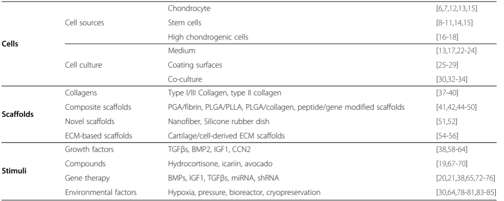
the establishment of the mechanical functions [4]. Colla-gen type II, proteoglycan and glycosaminoglycan (GAG), as the primary compositions in the ECM, have always been used as the criteria for the identification and evalu-ation of the chondrogenic capacity of cells and constructs obtained for cartilage tissue engineering. Parameters such as GAG content, cell mobility and water content usually can be reached up to the level of native tissue. However, the quantity of collagen type II (the major molecule for cartilage mechanical functions) is much lower from cells in vitrothan that from the native cartilage [5,6]. Therefore, an obvious obstacle currently is the inadequate production and accumulation of some chondrocyte-specific ECM macromolecules, especially collagen type II in cultured constructs comparing with native cartilage tissue, even though there has been a great progression in regeneration of the cartilage lesions using engineered constructs [5]. As collagen type II to a large extent provides the tissue with mechanical functions, strategies to promote its production and accumulation in engineered tissues need to be stressed. This review integrates cells, biomaterial scaffolds and stimuli that can facilitate ECM production and accumu-lation in cartilage tissue engineering (Table 1), with spe-cial focus on their effects on collagen type II expression and production. We endeavor to cover the above aspects that can improve the quality and function of engineered
* Correspondence:[email protected]
1Shenzhen Key Laboratory of Tissue Engineering, Shenzhen Second People’s
Hospital (The First Affiliated Hospital of Shenzhen University), Shenzhen 518035, Guangdong Province, China
3
Department of Orthopedics, Shenzhen Second People’s Hospital, Shenzhen 518035, Guangdong Province, China
Full list of author information is available at the end of the article
constructs, in the hope of promoting new technology to produce high-quality tissue engineered cartilage.
Cells
Cell sources
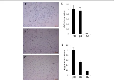
The cell sources employed in cartilage tissue engineering range from chondrocytes, stem cells to gene-modified cells. Several comparative studies revealed that chondro-cytes culturedin vitrocould produce more collagen type II than that of its stem cell counterparts as the collagen type II concentration was much higher in cultured chon-drocytes [7,8]. Shahin et al. used human chondrocytes for constructing tissue-engineered cartilage and the ob-tained collage type II concentration was around 8.5% of dry weight tissue. In contrast, in a similar study using human adipose-derived stem cells, the concentration of collagen type II was only 0.22% of dry weight tissue. However, adipose-derived stem cells showed inferior properties in chondrogenesis comparing with synovium-, bone marrow- and periosteum-derived stem cells in both human and rat [9,10]. Synovium-derived stem cells have been proved to be superior for the production of cartil-age matrix including collcartil-agen type II and have the great-est ability for chondrogenesis, among the other stem cell sources [1]. As for the comparison between chondro-cytes and synovium-derived stem cells, there was a re-search demonstrating synovium-derived stem cells and BMSCs produced more GAGs and collagen than chon-drocytes [11]. However, the chonchon-drocytes used in the research were P2 cells in monolayer culture (primary culture was termed P0 here), of which the collagen type II and aggrecan significantly decreased as shown in other studies and also in our experiments using a rabbit model (Additional file 1, Figure 1) [12,13]. Therefore, more in-vestigations need to be explored for the verification.
Nevertheless, a recent study showed that fibrocartilage-like tissue produced from synovium-derived stem cells was found in the superficial layer of cultured constructs [14]. While in another study comparing human bone marrow stromal cells (HBMSC) and articular chondro-cytes, cartilage-like tissue derived from chondrocytes was found in both superficial zone-like and middle zone-like structure. By contrast, relatively thicker fibrous capsules were shown in constructs from HBMSCs [15]. As superficial zone of the cartilage bears maximum load, it needs to be paid special attention to in cartilage tissue engineering. Chondrocyte and stem cells mentioned above have been summarized in Table 2.
Sorted single cell clone and clonal cell strains were also investigated for chondrogenic capacity. Fluorescence sorted chondrocytes with brighter CD49c and CD44 were detected to expression more collagen type II and higher levels of GAG than unsorted cells [16]. Magnetic-ally sorted stromal cells (STRO-1) from human bone marrow mononuclear populations have been used as seed cells with alginate-chitosan encapsulating and were proved to undergo chondrogenesis with increased colla-gen type II and proteoglycan in micromass culture and also in culture using a perfused bioreactor [17]. Induced pluripotent stem cells (iPSCs) were also used for screen-ing cells with high chondrogenic capacity. Fluorescence sorted mouse fibroblast-derived iPSCs with GFP driven by Col2 promoter/enhancer showed higher levels of col-lagen type II, aggrecan and GAGs [18]. Therefore, iden-tification of chondrocyte-specific markers and sorting of cells with relatively higher chondrogenic capacity would provide a new means for the seeding cells for cartilage tissue engineering.
[image:2.595.55.546.99.295.2]Gene-modified cells employed in cartilage tissue en-gineering have been paid more attention to nowadays Table 1 Summary of cells, scaffolds and stimuli
Cells
Cell sources
Chondrocyte [6,7,12,13,15]
Stem cells [8-11,14,15]
High chondrogenic cells [16-18]
Cell culture
Medium [13,17,22-24]
Coating surfaces [25-29]
Co-culture [30,32-34]
Scaffolds
Collagens Type I/III Collagen, type II collagen [37-40]
Composite scaffolds PGA/fibrin, PLGA/PLLA, PLGA/collagen, peptide/gene modified scaffolds [41,42,44-50]
Novel scaffolds Nanofiber, Silicone rubber dish [51,52]
ECM-based scaffolds Cartilage/cell-derived ECM scaffolds [54-56]
Stimuli
Growth factors TGFβs, BMP2, IGF1, CCN2 [38,58-64]
Compounds Hydrocortisone, icariin, avocado [19,67-70]
Gene therapy BMPs, IGF1, TGFβs, miRNA, shRNA [20,21,38,65,72-76]
with the rapid development of molecular biology and technology, including cells with single gene transduction and co-transduction of two genes or a gene with an shRNA [19-21]. All these methods aim at improving the chondrogenic capacity of cells and the quality of engi-neered constructs. As for the details of genes and gene-modified cells that can promote ECM synthesis and collagen type II expression, they will be elucidated in the later part ofgene therapy.
Cell culture
Dedifferentiation is the major cause of degeneration of chondrocytes in vitro and maintaining chondrocytes phenotype and features is always on focus. Likewise, for stem cells, chondrogenesis is the primary process to chondrocytes and differentiation of stem cells has been broadly investigated. Optimization of culture methods improves characteristics of chondrocytes and facilitates collagen type II and proteoglycan production in vitro. There are various ways for chondrocytes culture and chondrogenesis of stem cells, including using different media, coating proteins and co-culture systems. Different
A
B
C
D
[image:3.595.60.537.89.422.2]E
Figure 1Evaluation of GAGs, Col2a1 and aggrecan in passaged rabbit chondrocytes. (A-C)Toluidine blue staining showed production of GAGs from cells declined following passaging from P0(A), P1(B)to P2(C). Scale bar: 100μm.(D-E)Real time PCR showed the expression of Col2a1(D)and aggrecan(E)decreased with cell passaging.
Table 2 Summary of chondrocyte and stem cells in the part of cells
Cell type Species Passage Culture method Reference
Chondrocyte Human P2 Monolayer + 3D [7]
ADSC Human P4 Monolayer + 3D [8]
SDSC Human P2 Monolayer + 3D [11]
BMSC Human P2 Monolayer + 3D [11]
Chondrocyte Human P2 Monolayer [11]
Chondrocyte Rabbit P0-P4 Monolayer [12]
Chondrocyte Human P0-P4 Monolayer [13]
SDSC Human P4-P7 Monolayer + 3D [14]
Chondrocyte Human P0-P4 Monolayer + 3D [15]
BMSC Human P1-P4 Monolayer + 3D [15]
[image:3.595.56.289.555.707.2]scaffolds and growth factors are also involved, which will be discussed later.
A study specifically designed to optimize the chondro-genic medium using factorial design of experiments sug-gested standard chondrogenic medium with increased level of TGF-β1 and glucose and decreased level of dexa-methasone could promote collagen type II to collagen type I ratio (ColII/ColI) and better maintain the chondro-cyte properties [22]. TGF-β1 in serum free medium has been proved to increase collagen type II expression and better maintain human chondrocyte phenotype compar-ing with medium plus fetal calf serum (FCS) [23]. More-over, the redifferentiation of serum free medium cultured monolayer chondrocytes in a 3D scaffold was observed with the expression of collagen type II and proteoglycans; while it was not observed for chondrocytes and STRO-1 cells cultured in FCS supplemented medium [17,23,24]. One recent study showed chondrocytes cultured in medium supplemented with 20% platelet-rich plasma pro-duced thicker cartilage tissue than cells in medium with 20% FBS [25], suggesting platelet-rich plasma was more able to maintain chondrocyte propertiesin vitro.
Different coatings of cell culture surfaces influences chondrocyte culture in vitro. Cells cultured on collagen type I and II coating dishes expressed more collagen type II expression and produced more glycosaminogly-can (GAG) than uncoated control samples [26,27]. Though not significantly, higher collagen type II expres-sion was observed in chondrocytes on collagen type II coated dishes than collagen type I coated dishes. On the contrary, fibronectin was suggested to promote collagen type II degradation [28]. More specific research came from Shawnet al.,who investigated the effects of coating with zone-specific cartilage extracellular matrix molecules on cartilage formation [29]. Cells cultured in monolayer showed different outcomes with pellets culture. While hyaluronic acid (HA) and osteopontin coating increased Col2a1expression in monolayer culture, pellets culture of cells harvested from HA coated dishes showed decreased expression of collagen type II. On the contrary, tenascin C promoted collagen type II expression in pellet culture while inhibited its expression in monolayer culture. Cells on type II collagen coated dishes showed consistent in-creased expression of Col2a1 with both monolayer culture and pellet culture system [29]. As different coatings and different culture environments synergistically affect the cell behaviors, both need to be taken into considerations in an experimental design. Aside from protein coating, TiO2-coated coverslips were used for human MSCs cul-ture and proved to significantly enhance cell proliferation without weakening the chondrogenic capacity [30], which would also benefit the ECM production.
Cell co-culture systems have been used in many fields of biomedical sciences. As for cartilage tissue engineering,
co-culture of MSCs and chondrocytes provide us with a means to obtain promising cells. After a comparative study of chondrocytes, MSCs and co-culture of chondro-cytes and MSCs, the co-cultured cells were suggested to be the most prospective for cartilage tissue engineering [31]. Co-culture of primary chondrocytes with MSCs or embryonic stem cells or iPSCs increased collagen type II and Sox 9 expression and promoted cartilage matrix for-mation [3,32,33]. The optimal ratio of MSCs and chon-drocytes in co-cultures for cartilage engineering varied in different studies. Some study revealed that higher ratios of human MSCs to human articular chondrocytes was more beneficial to chondrogenesis as was shown by newly syn-thesized cartilaginous ECM and collagen type II gene up-regulation [34]. However, another study suggested 63:1 as the appropriate initial ratio of MSCs/chondrocytes for enough stable chondrocyte-like cells and the constructs from co-cultures were more cartilaginous than that from chondrocytes [35].
Development and optimization of cell culture methods aiming at elevating the chondrogenic capacity of seed cells will improve the cartilage-specific ECM production and finally benefit the cartilage tissue engineering with relatively higher quality of engineered constructs.
Scaffolds
There are various scaffolds used for cartilage tissue en-gineering as a 3D environment facilitates to maintain chondrocyte properties than monolayer culture. The characteristics of a scaffold involve mechanical strength, biocompatibility, biodegradability, porosity and toxicity [36]. The scaffold must be structurally stable, allow cells to infiltrate and growth factors to attach. All these char-acteristics of scaffolds provide cells with healthy micro-environment, beneficial cell-cell interaction and adequate nutrient exchangein vitro. Thus, the constructs with cells and ECM mimic the cartilage and are more close to the native cartilage tissue than monolayer cells when im-planted in the defects. Of so many scaffolds used, we will focus on clinically used scaffolds and those assist to pro-mote ECM production, especially collagen type II produc-tion here.
survival ability and increased the expression of collagen type II and also aggrecan [42]. Polyglycolic acid/fibrin (PGA/fibrin) scaffolds used for chondrocyte culture also enhanced the redifferentiation of ovine chondrocytes and the induction of collagen type II and aggrecan after culture. Biomechanical tests further supported this scaf-folds for chondrocyte culture with a high tensile strength of 3.6 N/mm2 [43]. Poly(L-lactic acid) (PLLA) and poly (lactic-co-glycolic) acid (PLGA), as materials that have been approved by the US Food and Drug Administration for human clinical uses [44], have been often used for the research of cartilage tissue engineering. Comparing with PLLA, collagen type II and Arg-Gly-Asp (RGD) peptide modified PLLA/PLGA (50:50), collagen type II modified PLLA/PLGA (50:50) exhibits higher collagen type II expression and shows superiority for chondro-genesis bothin vitro andin vivo[45]. Besides, based on type I collagen-PLGA scaffold, improved funnel-like collagen-PLGA hybrid scaffold was shown to be a stron-ger promoter for cartilage regeneration [46]. PLGA scaf-fold with hyaluronic acid incorporated substantially promoted collagen synthesis [47]. Moreover, as PLGA microspheres have been successfully used to immobilize DNA, siRNA and growth factors [48,49]. Sox9 expres-sion plasmid loaded PLGA microspheres have been in-vestigated for chondrogenesis. Human MSCs seeded on Sox9 gene and heparinized TGF-β3 coated dexameth-sone loaded PLGA microspheres could drastically in-crease collagen type II expression by 30 times compared with control [50]. Therefore, gene incorporation pro-vides a new and promising strategy to optimize the property of scaffolds for tissue engineering.
Comparing with the traditional scaffolds, more and more novel scaffolds have been under investigations, in-cluding peptide-modified scaffolds and mobile scaffolds. Mesenchymal stem cells cultured in Arg-Gly-Asp-Ser (RGDS) peptide incorporated PEG-based hydrogels had greater gene expression of collagen type II and produced significantly more GAGs comparing with control [51]. A newly designed synthetic link N peptide nanofiber was suggested to remarkably promote collagen type II and aggrecan production [52]. Since cell expansion would lead to contact inhibition and to avoid the dedifferenti-ation of chondrocytes through passaging, mobile scaffold was also examined for chondrocyte culture. Differently from traditional biomaterial scaffolds, the culture surface area of the high-extension silicone rubber dish could in-crease by 8 folds with a motorized device [53]. Thus, ad-equate cells could be obtained and extracellular matrix was simultaneously maintained, including collagen type II, which would drastically decrease through passaging.
ECM-based scaffold is an emerging approach in the field of cartilage tissue engineering in that it may be rela-tively easy to retain the natural growth factors in the
ECM [54]. Cartilage-derived and cell-derived ECM scaf-folds have been investigated and cartilage-like tissues have been observed [55-57]. However, the quality of formed cartilaginous tissue needs to be evaluated and compared. With improvement of the ECM-based scaf-fold, it would probably be a promising type of scaffolds for cartilage tissue engineering.
Stimuli
Growth factors and compounds
Various growth factors have been used for cartilage tis-sue engineering. Members in TGF-β family have been widely used to induce the chondrogenesis of MSCs and enhance ECM synthesis. TGF-β3 was proved to be more capable to induce chondrogenesis of MSCs and also in-crease the expression of collagen type II and aggrecan [41,58]. As for collagen type II production, the effect of TGF-β1 used in different culture systems was controver-sial. Depending on the design of the experiments,
TGF-β1 could either increase or decrease the expression of collagen type II [59-61]. BMP-2 and IGF-1 both impact chondrogenesis and promote collagen type II produc-tion. In a study which compared the effects of BMP-2, IGF-1 and their combination, BMP-2 treatment showed superior ability for collagen type II expression than IGF-1 [39]. Synergistical use of BMP-2 and BMP-7 gave better outcome of ECM production [62]. A more recent study showed standard chondrogenic medium plus TGF-β3, BMP-2 and IGF-1 demonstrated stronger capability for MSCs to produce collagen type II comparing with the other three groups including standard medium with
TGF-β3 alone, TGF-β3 + BMP-2 and TGF-β3 + IGF-1 [63]. An-other growth factor, connective tissue growth factor (CCN2) has also been proved to strongly enhance the car-tilaginous ECM production, including collagen type II and was suggested to reduce age-related changes of articular cartilage [64-67].
Gene therapy
Gene therapy has been grown rapidly and is widely used in biomedical sciences. Although gene therapy is in its infancy for cartilage engineering, it shows promising to contribute to the cartilage repair. Often, growth factors are been transfected into chondrocytes or MSCs to im-prove the properties of cells. IGF-1 and TGF-β1 have been successfully transfected into articular chondrocyte and BMSCs separately and both were proved to increase the expression of collagen type II and aggrecan [20,72]. Meanwhile, functional study further demonstrated IGF-I transfected implants accelerated the regeneration com-pared with lacZ implants [20]. Furthermore, one recent study investigating co-transduction of different gene combinations which involved any two of genes IGF-1, FGF-2, TGF-β1 and SOX-9 with adenovirus delivering system showed that cells co-transduced with IGF-1 and FGF-2 could significantly express higher aggrecan, colla-gen type II and other ECM proteins than other combi-nations [21].
In addition to delivering growth factors to cells to in-crease cartilage-specific gene expression, down regulation of some negative factors has also been proved to effect-ively promote collagen type II and aggrecan expression. MicroRNA-181b (miR-181b) has been identified to be a negative modulator for cartilage development and siRNA targeting miR-181b induced collagen type II expression and significantly reduced degeneration of cartilage [73]. MiR-145 was proved to target Sox9 and suppress the chondrogenesis of mesenchymal stem cells; while anti-miR-145 inhibitor could rescue the expression of Col2a1, aggrecan and relevant chondrogenic marker genes [74]. Besides, down regulation of some pro-inflammatory fac-tors that could induce cartilage destruction, such as IL-1, IL-6, TNF-α, would also help cartilage regeneration [68,75,76]. Therefore, inhibition of the negative modulator could be a promising therapeutic strategy for cartilage re-generation. Furthermore, targeting chondrocyte COL1A1 with siRNA could significantly improve chondrocyte phenotype, with increased ratio of COL2A1/COL1A1 and improved cartilage-like matrix formation [39].
Furthermore, the combination of delivering growth fac-tors and down-regulation of negative modulafac-tors has also been investigated recently. Co-transduction of TGF-β3 with lentiviral transduction system and shRNA targeting Col1a1 with adenoviral vectors demonstrated optimal effi-cacy for collagen type II expression and cartilage forma-tion [77]. As gene therapy is just arising in cartilage tissue engineering, much needs to be explored in this field.
Environmental factors
As cartilage resides in a microenvironment with reduced oxygen and various biomechanical stresses, creating a similar environment in vitro may provide a way to
maintain the characteristics of chondrocytes. Hypoxia with 5% or even 3% oxygen stimulated collagen type II expression and biosynthesis of hyaline-like cartilage matrix [39,78,79]. For dynamic environment, expression of collagen type II and aggrecan were induced after the stimulation of a uniaxial compressive load with 1kPa, 1Hz for 30 min [80]. Intermittent hydrostatic pressure benefit long-term chondrocyte culture and the cartil-aginous construct formation [81]. As for tensile strength, abnormal cyclic tensile strain inhibited collagen type II expression; while proper and cyclic tensile strain would promote collagen type II expression [65,82]. To provide an environment that could enhance nutrient transport and maintain dynamic state for cells, bioreactors have been used for cartilage tissue engineering. Combined with scaffolds as the support for cells, the system facili-tates chondrocyte proliferation and ECM accumulation comparing with static culture [83,84]. Improved bioreac-tor as wavy-wall bioreacbioreac-tor better increased the cartilage matrix than common dynamic culture using spinner flasks [84]. Perfusion bioreactor was also used to investi-gate chondrogenesis and chondrocyte growth. It was found that human articular chondrocytes within alginate beads only produced collage type II in a perfused envir-onment. Collage type II was not detected in the static culture condition by contrast [17]. However, one prob-lem caused by dynamic loading was less accumulation of matrix in the scaffold as part of the newly synthesized molecules would disperse into the medium [85]. Besides dynamic environment mentioned above, cryopreservation has a negative effect on the chondrogenic capacity of chondrocytes. Human chondrocytes showed significantly decreased collagen type II expression after cryopreserva-tion [86]. Although new methods and scaffolds are being under investigation for the preservation and revitalization of articular cartilage or chondrocytes [87-89], it is better to use freshly cultured chondrocytes for clinical applica-tion if it is possible.
Conclusions
tissue-engineered cartilage successful and lead the clin-ical approaches from ACI, MACI to the ideal tissue-engineered cartilage implantation (TEC).
Additional file
Additional file 1:Culture and characterization of rabbit chondrocyte.
Competing interest
The authors declare that they have no competing interests.
Authors’contributions
CJL conceived of the study, participated in the experiments and drafted the manuscript; DL participated in the design of the draft and revised it critically for important intellectual content; ZW participated in the experiments and interpreted the data obtained; XJ critically revised the draft for important intellectual content and obtained funding; WD supervised the study, revised the draft for important intellectual content, obtained funding and approval of the final version of the manuscript. All authors read and approved the final manuscript.
Acknowledgements
Our work was funded by Shenzhen Science and Technology Key Project (Project NO. 201001013) and Shenzhen Science and Technology International Cooperative Project (Project NO. 20130326210545).
Author details
1
Shenzhen Key Laboratory of Tissue Engineering, Shenzhen Second People’s Hospital (The First Affiliated Hospital of Shenzhen University), Shenzhen 518035, Guangdong Province, China.2School of Medicine, Sun Yat-sen University, Guangzhou 510182, Guangdong Province, China.3Department of
Orthopedics, Shenzhen Second People’s Hospital, Shenzhen 518035, Guangdong Province, China.
Received: 4 December 2013 Accepted: 31 March 2014 Published: 5 April 2014
References
1. Jiang YZ, Zhang SF, Qi YY, Wang LL, Ouyang HW:Cell transplantation for articular cartilage defects: principles of past, present, and future practice.
Cell Transplant2011,20:593–607.
2. Kon E, Filardo G, Di Martino A, Marcacci M:ACI and MACI.J Knee Surg2012, 25:17–22.
3. Chung C, Burdick JA:Engineering cartilage tissue.Adv Drug Deliv Rev2008, 60:243–262.
4. Huang AH, Farrell MJ, Mauck RL:Mechanics and mechanobiology of mesenchymal stem cell-based engineered cartilage.J Biomech2010, 43:128–136.
5. Mahmoudifar N, Doran PM:Chondrogenesis and cartilage tissue engineering: the longer road to technology development.Trends Biotechnol2012,30:166–176.
6. Dunkelman NS, Zimber MP, Lebaron RG, Pavelec R, Kwan M, Purchio AF: Cartilage production by rabbit articular chondrocytes on polyglycolic acid scaffolds in a closed bioreactor system.Biotechnol Bioeng1995, 46:299–305.
7. Shahin K, Doran PM:Strategies for enhancing the accumulation and retention of extracellular matrix in tissue-engineered cartilage cultured in bioreactors.PLoS One2011,6:e23119.
8. Mahmoudifar N, Doran PM:Chondrogenic differentiation of human adipose-derived stem cells in polyglycolic acid mesh scaffolds under dynamic culture conditions.Biomaterials2010,31:3858–3867. 9. Sakaguchi Y, Sekiya I, Yagishita K, Muneta T:Comparison of human stem
cells derived from various mesenchymal tissues: superiority of synovium as a cell source.Arthritis Rheum2005,52:2521–2529.
10. Yoshimura H, Muneta T, Nimura A, Yokoyama A, Koga H, Sekiya I: Comparison of rat mesenchymal stem cells derived from bone marrow, synovium, periosteum, adipose tissue, and muscle.Cell Tissue Res2007, 327:449–462.
11. Vinardell T, Sheehy EJ, Buckley CT, Kelly DJ:A comparison of the functionality and in vivo phenotypic stability of cartilaginous tissues engineered from different stem cell sources.Tissue Eng Part A2012, 18:1161–1170.
12. Benya PD, Padilla SR, Nimni ME:Independent regulation of collagen types by chondrocytes during the loss of differentiated function in culture.Cell 1978,15:1313–1321.
13. Hamada T, Sakai T, Hiraiwa H, Nakashima M, Ono Y, Mitsuyama H, Ishiguro N:Surface markers and gene expression to characterize the
differentiation of monolayer expanded human articular chondrocytes.
J Med Sci2013,75:101–111.
14. Ando W, Fujie H, Moriguchi Y, Nansai R, Shimomura K, Hart DA, Yoshikawa H, Nakamura N:Detection of abnormalities in the superficial zone of cartilage repaired using a tissue engineered construct derived from synovial stem cells.Eur Cell Mater2012,24:292–307.
15. Saha S, Kirkham J, Wood D, Curran S, Yang XB:Informing future cartilage repair strategies: a comparative study of three different human cell types for cartilage tissue engineering.Cell Tissue Res2013,352:495–507. 16. Grogan SP, Barbero A, Diaz-Romero J, Cleton-Jansen AM, Soeder S, Whiteside
R, Hogendoorn PC, Farhadi J, Aigner T, Martin I, Mainil-Varlet P:Identification of markers to characterize and sort human articular chondrocytes with enhanced in vitro chondrogenic capacity.Arthritis Rheum2007,56:586–595. 17. Forsey RW, Tare R, Oreffo RO, Chaudhuri JB:Perfusion bioreactor studies of
chondrocyte growth in alginate-chitosan capsules.Biotechnol Appl Biochem2012,59:142–152.
18. Diekman BO, Christoforou N, Willard VP, Sun H, Sanchez-Adams J, Leong KW, Guilak F:Cartilage tissue engineering using differentiated and purified induced pluripotent stem cells.Proc Natl Acad Sci U S A2012,109:19172–19177. 19. Henrotin YE, Deberg MA, Crielaard JM, Piccardi N, Msika P, Sanchez C: Avocado/soybean unsaponifiables prevent the inhibitory effect of osteoarthritic subchondral osteoblasts on aggrecan and type II collagen synthesis by chondrocytes.J Rheumatol2006,33:1668–1678.
20. Madry H, Kaul G, Cucchiarini M, Stein U, Zurakowski D, Remberger K, Menger MD, Kohn D, Trippel SB:Enhanced repair of articular cartilage defects in vivo by transplanted chondrocytes overexpressing insulin-like growth factor I (IGF-I).Gene Ther2005,12:1171–1179.
21. Garza-Veloz I, Romero-Diaz VJ, Martinez-Fierro ML, Marino-Martinez IA, Gonzalez-Rodriguez M, Martinez-Rodriguez HG, Espinoza-Juarez MA, Bernal-Garza D, Ortiz-Lopez R, Rojas-Martinez A:Analyses of chondrogenic induction of adipose mesenchymal stem cells by combined co-stimulation mediated by adenoviral gene transfer.Arthritis Res Ther2013,15:R80(81–13). 22. Enochson L, Brittberg M, Lindahl A:Optimization of a chondrogenic
medium through the use of factorial design of experiments.BioRes Open Access2012,1:306–313.
23. Giannoni P, Pagano A, Maggi E, Arbico R, Randazzo N, Grandizio M, Cancedda R, Dozin B:Autologous chondrocyte implantation (ACI) for aged patients: development of the proper cell expansion conditions for possible therapeutic applications.Osteoarthritis Cartilage2005,13:589–600. 24. Shao XX, Duncan NA, Lin L, Fu X, Zhang JY, Yu CL:Serum-free media for
articular chondrocytes in vitro expansion.Chin Med J2013,126:2523–2529. 25. Petrera M, De Croos JN, Iu J, Hurtig M, Kandel RA, Theodoropoulos JS:
Supplementation with platelet-rich plasma improves the in vitro formation of tissue-engineered cartilage with enhanced mechanical properties.
Arthroscopy2013,29:1685–1692.
26. Barbero A, Grogan SP, Mainil-Varlet P, Martin I:Expansion on specific substrates regulates the phenotype and differentiation capacity of human articular chondrocytes.Cell Biochem2006,98:1140–1149. 27. Rutgers M, Saris DB, Vonk LA, van Rijen MH, Akrum V, Langeveld D, van
Boxtel A, Dhert WJ, Creemers LB:Effect of collagen type I or type II on chondrogenesis by cultured human articular chondrocytes.Tissue Eng Part A2013,19:59–65.
28. Yasuda T, Poole AR:A fibronectin fragment induces type II collagen degradation by collagenase through an interleukin-1-mediated pathway.
Arthritis Rheum2002,46:138–148.
29. Grogan SP, Chen X, Sovani S, Taniguchi N, Colwell CW Jr, Lotz M, D’Lima D: Influence of cartilage extracellular matrix molecules on cell phenotype and neocartilage formation.Tissue Eng Part A2013,20:264–274. 30. Kaitainen S, Mahonen AJ, Lappalainen R, Kroger H, Lammi MJ, Qu C:TiO2
31. Meretoja VV, Dahlin RL, Wright S, Kasper FK, Mikos AG:The effect of hypoxia on the chondrogenic differentiation of co-cultured articular chondrocytes and mesenchymal stem cells in scaffolds.Biomaterials2013, 34:4266–4273.
32. Vats A, Bielby RC, Tolley N, Dickinson SC, Boccaccini AR, Hollander AP, Bishop AE, Polak JM:Chondrogenic differentiation of human embryonic stem cells: the effect of the micro-environment.Tissue Eng2006, 12:1687–1697.
33. Qu C, Puttonen KA, Lindeberg H, Ruponen M, Hovatta O, Koistinaho J, Lammi MJ:Chondrogenic differentiation of human pluripotent stem cells in chondrocyte co-culture.Int J Biochem Cell Biol2013,45:1802–1812. 34. Mo XT, Guo SC, Xie HQ, Deng L, Zhi W, Xiang Z, Li XQ, Yang ZM:Variations
in the ratios of co-cultured mesenchymal stem cells and chondrocytes regulate the expression of cartilaginous and osseous phenotype in alginate constructs.Bone2009,45:42–51.
35. Yang YH, Lee AJ, Barabino GA:Coculture-driven mesenchymal stem cell-differentiated articular chondrocyte-like cells support neocartilage development.Stem Cell Transl Med2012,1:843–854.
36. Mafi P, Hindocha S, Mafi R, Khan WS:Evaluation of biological protein-based collagen scaffolds in cartilage and musculoskeletal tissue engineering–a systematic review of the literature.Curr Stem Cell Res Ther 2012,7:302–309.
37. Iwasa J, Engebretsen L, Shima Y, Ochi M:Clinical application of scaffolds for cartilage tissue engineering.Knee Surg Sports Traumatol Arthrosc2009, 17:561–577.
38. Zheng MH, Willers C, Kirilak L, Yates P, Xu J, Wood D, Shimmin A: Matrix-induced autologous chondrocyte implantation (MACI): biological and histological assessment.Tissue Eng2007,13:737–746.
39. Legendre F, Ollitrault D, Hervieu M, Bauge C, Maneix L, Goux D, Chajra H, Mallein-Gerin F, Boumediene K, Galera P, Demoor M:Enhanced hyaline cartilage matrix synthesis in collagen sponge scaffolds by using siRNA to stabilize chondrocytes phenotype cultured with bone morphogenetic protein-2 under hypoxia.Tissue Eng Part C Methods2013,19:550–567. 40. Willers C, Chen J, Wood D, Xu J, Zheng MH:Autologous chondrocyte
implantation with collagen bioscaffold for the treatment of osteochondral defects in rabbits.Tissue Eng2005,11:1065–1076. 41. Chen WC, Yao CL, Wei YH, Chu IM:Evaluating osteochondral defect repair
potential of autologous rabbit bone marrow cells on type II collagen scaffold.Cytotechnology2011,63:13–23.
42. Lee CR, Grad S, Gorna K, Gogolewski S, Goessl A, Alini M: Fibrin-polyurethane composites for articular cartilage tissue engineering: a preliminary analysis.Tissue Eng2005,11:1562–1573.
43. Endres M, Neumann K, Zhou B, Freymann U, Pretzel D, Stoffel M, Kinne RW, Kaps C:An ovine in vitro model for chondrocyte-based scaffold-assisted cartilage grafts.J Orthop Trauma Res2012,7:1–14.
44. Nair LS, Laurencin CT:Biodegradable polymers as biomaterials.Prog Polym Sci2007,32:762–798.
45. Hsu SH, Chang SH, Yen HJ, Whu SW, Tsai CL, Chen DC:Evaluation of biodegradable polyesters modified by type II collagen and Arg-Gly-Asp as tissue engineering scaffolding materials for cartilage regeneration.
Artif Organs2006,30:42–55.
46. Lu H, Ko YG, Kawazoe N, Chen G:Culture of bovine articular chondrocytes in funnel-like collagen-PLGA hybrid sponges.Biomed Mater2011, 6:045011.
47. Yoo HS, Lee EA, Yoon JJ, Park TG:Hyaluronic acid modified biodegradable scaffolds for cartilage tissue engineering.Biomaterials2005,26:1925–1933. 48. Shen H, Hu X, Bei J, Wang S:The immobilization of basic fibroblast
growth factor on plasma-treated poly(lactide-co-glycolide).Biomaterials 2008,29:2388–2399.
49. Zhang XQ, Tang H, Hoshi R, De Laporte L, Qiu H, Xu X, Shea LD, Ameer GA: Sustained transgene expression via citric acid-based polyester elastomers.Biomaterials2009,30:2632–2641.
50. Park JS, Yang HN, Woo DG, Jeon SY, Park KH:SOX9 gene plus heparinized TGF-beta 3 coated dexamethasone loaded PLGA microspheres for inducement of chondrogenesis of hMSCs.Biomaterials2012,33:7151–7163. 51. Salinas CN, Cole BB, Kasko AM, Anseth KS:Chondrogenic differentiation
potential of human mesenchymal stem cells photoencapsulated within poly(ethylene glycol)-arginine-glycine-aspartic acid-serine thiol-methacrylate mixed-mode networks.Tissue Eng2007,13:1025–1034. 52. Ma K, Wu Y, Wang B, Yang S, Wei Y, Shao Z:Effect of a synthetic link N
peptide nanofiber scaffold on the matrix deposition of aggrecan and
type II collagen in rabbit notochordal cells.J Mater Sci Mater Med2013, 24:405–415.
53. Rosenzweig DH, Chicatun F, Nazhat SN, Quinn TM:Cartilaginous constructs using primary chondrocytes from continuous expansion culture seeded in dense collagen gels.Acta Biomater2013,9:9360–9369.
54. Benders KE, van Weeren PR, Badylak SF, Saris DB, Dhert WJ, Malda J: Extracellular matrix scaffolds for cartilage and bone regeneration.Trends Biotechnol2013,31:169–176.
55. Yang Q, Peng J, Guo Q, Huang J, Zhang L, Yao J, Yang F, Wang S, Xu W, Wang A, Lu S:A cartilage ECM-derived 3-D porous acellular matrix scaffold for in vivo cartilage tissue engineering with PKH26-labeled chondrogenic bone marrow-derived mesenchymal stem cells.
Biomaterials2008,29:2378–2387.
56. Jin CZ, Park SR, Choi BH, Park K, Min BH:In vivo cartilage tissue
engineering using a cell-derived extracellular matrix scaffold.Artif Organs 2007,31:183–192.
57. Schwarz S, Elsaesser AF, Koerber L, Goldberg-Bockhorn E, Seitz AM, Bermueller C, Durselen L, Ignatius A, Breiter R, Rotter N:Processed xenogenic cartilage as innovative biomatrix for cartilage tissue engineering: effects on chondrocyte differentiation and function.J Tissue Eng Regen Med2012, doi:10.1002/term.1650.
58. Gooch KJ, Kwon JH, Blunk T, Langer R, Freed LE, Vunjak-Novakovic G:Effects of mixing intensity on tissue-engineered cartilage.Biotechnol Bioeng2001, 72:402–407.
59. Chadjichristos C, Ghayor C, Herrouin JF, Ala-Kokko L, Suske G, Pujol JP, Galera P:Down-regulation of human type II collagen gene expression by transforming growth factor-beta 1 (TGF-beta 1) in articular chondrocytes involves SP3/SP1 ratio.J Biol Chem2002,277:43903–43917.
60. Galera P, Redini F, Vivien D, Bonaventure J, Penfornis H, Loyau G, Pujol JP: Effect of transforming growth factor-beta 1 (TGF-beta 1) on matrix synthesis by monolayer cultures of rabbit articular chondrocytes during the dedifferentiation process.Exp Cell Res1992,200:379–392.
61. Galera P, Vivien D, Pronost S, Bonaventure J, Redini F, Loyau G, Pujol JP: Transforming growth factor-beta 1 (TGF-beta 1) up-regulation of collagen type II in primary cultures of rabbit articular chondrocytes (RAC) involves increased mRNA levels without affecting mRNA stability and procollagen processing.J Cell Physiol1992,153:596–606.
62. Hicks DL, Sage AB, Shelton E, Schumacher BL, Sah RL, Watson D:Effect of bone morphogenetic proteins 2 and 7 on septal chondrocytes in alginate.Otolaryngol Head Neck Surg2007,136:373–379.
63. Abukawa H, Oriel BS, Leaf J, Vacanti JP, Kaban LB, Troulis MJ, Hartnick CJ: Growth factor directed chondrogenic differentiation of porcine bone marrow-derived progenitor cells.J Craniofac Surg2013,24:1026–1030. 64. Itoh S, Hattori T, Tomita N, Aoyama E, Yutani Y, Yamashiro T, Takigawa M:
CCN family member 2/connective tissue growth factor (CCN2/CTGF) Has anti-aging effects that protect articular cartilage from Age-related degenerative changes.PLoS One2013,8:e71156.
65. Furumatsu T, Matsumoto E, Kanazawa T, Fujii M, Lu Z, Kajiki R, Ozaki T:Tensile strain increases expression of CCN2 and COL2A1 by activating TGF-beta-Smad2/3 pathway in chondrocytic cells.J Biomech2013,46:1508–1515. 66. Tomita N, Hattori T, Itoh S, Aoyama E, Yao M, Yamashiro T, Takigawa M:
Cartilage-specific over-expression of CCN family member 2/connective tissue growth factor (CCN2/CTGF) stimulates insulin-like growth factor expression and bone growth.PLoS One2013,8:e59226.
67. Kubota S, Takigawa M:The role of CCN2 in cartilage and bone development.J Cell Commun Signal2011,5:209–217.
68. Wang J, Elewaut D, Hoffman I, Veys EM, Verbruggen G:Physiological levels of hydrocortisone maintain an optimal chondrocyte extracellular matrix metabolism.Ann Rheum Dis2004,63:61–66.
69. Zhang L, Zhang X, Li KF, Li DX, Xiao YM, Fan YJ, Zhang XD:Icariin promotes extracellular matrix synthesis and gene expression of chondrocytes in vitro.Phytother Res2012,26:1385–1392. 70. Li D, Yuan T, Zhang X, Xiao Y, Wang R, Fan Y:Icariin: a potential
promoting compound for cartilage tissue engineering.Osteoarthritis Cartilage2012,20:1647–1656.
71. Lee HS, Park SY, Park Y, Bae SH, Suh HJ:Yeast hydrolysate protects cartilage via stimulation of type II collagen synthesis and suppression of MMP-13 production.Phytother Res2013,27:1414–1418.
73. Song J, Lee M, Kim D, Han J, Chun CH, Jin EJ:MicroRNA-181b regulates articular chondrocytes differentiation and cartilage integrity.Biochem Biophys Res Commun2013,431:210–214.
74. Yang B, Guo H, Zhang Y, Chen L, Ying D, Dong S:MicroRNA-145 regulates chondrogenic differentiation of mesenchymal stem cells by targeting Sox9.PLoS One2011,6:e21679.
75. Legendre F, Dudhia J, Pujol JP, Bogdanowicz P:JAK/STAT but not ERK1/ ERK2 pathway mediates interleukin (IL)-6/soluble IL-6R down-regulation of Type II collagen, aggrecan core, and link protein transcription in articular chondrocytes. Association with a down-regulation of SOX9 expression.J Biol Chem2003,278:2903–2912.
76. Ryu B, Himaya SW, Napitupulu RJ, Eom TK, Kim SK:Sulfated chitooligosaccharide II (SCOS II) suppress collagen degradation in TNF-induced chondrosarcoma cells via NF-kappaB pathway.Carbohydr Res2012,350:55–61.
77. Zhang F, Yao Y, Su K, Fang Y, Citra F, Wang DA:Co-transduction of lentiviral and adenoviral vectors for co-delivery of growth factor and shRNA genes in mesenchymal stem cells-based chondrogenic system.
J Tissue Eng Regen Med2012, doi:10.1002/term.1656.
78. Hansen U, Schunke M, Domm C, Ioannidis N, Hassenpflug J, Gehrke T, Kurz B:Combination of reduced oxygen tension and intermittent hydrostatic pressure: a useful tool in articular cartilage tissue engineering.J Biomech 2001,34:941–949.
79. Scherer K, Schunke M, Sellckau R, Hassenpflug J, Kurz B:The influence of oxygen and hydrostatic pressure on articular chondrocytes and adherent bone marrow cells in vitro.Biorheology2004,41:323–333. 80. Waldman SD, Couto DC, Grynpas MD, Pilliar RM, Kandel RA:A single application of cyclic loading can accelerate matrix deposition and enhance the properties of tissue-engineered cartilage.Osteoarthritis Cartilage2006,14:323–330.
81. Hu JC, Athanasiou KA:The effects of intermittent hydrostatic pressure on self-assembled articular cartilage constructs.Tissue Eng2006,12:1337–1344. 82. Kanazawa T, Furumatsu T, Hachioji M, Oohashi T, Ninomiya Y, Ozaki T:
Mechanical stretch enhances COL2A1 expression on chromatin by inducing SOX9 nuclear translocalization in inner meniscus cells.J Orthop Res2012,30:468–474.
83. Freed LE, Marquis JC, Langer R, Vunjak-Novakovic G, Emmanual J: Composition of cell-polymer cartilage implants.Biotechnol Bioeng1994, 43:605–614.
84. Bueno EM, Bilgen B, Barabino GA:Wavy-walled bioreactor supports increased cell proliferation and matrix deposition in engineered cartilage constructs.Tissue Eng2005,11:1699–1709.
85. Lee CR, Grodzinsky AJ, Spector M:Biosynthetic response of passaged chondrocytes in a type II collagen scaffold to mechanical compression.
J Biomed Mater Res A2003,64:560–569.
86. Albrecht C, Tichy B, Nurnberger S, Zak L, Handl MJ, Marlovits S, Aldrian S: Influence of cryopreservation, cultivation time and patient’s age on gene expression in Hyalograft(R) C cartilage transplants.Int Orthop2013, 37:2297–2303.
87. Abazari A, Jomha NM, Elliott JA, McGann LE:Cryopreservation of articular cartilage.Cryobiology2013,66:201–209.
88. Lyu SR, Kuo YC, Ku HF, Hsieh WH:Cryopreserved chondrocytes in porous biomaterials with surface elastin and poly-L-lysine for cartilage regeneration.
Colloids Surf B Biointerfaces2013,103:304–309.
89. Xia Z, Duan X, Murray D, Triffitt JT, Price AJ:A method of isolating viable chondrocytes with proliferative capacity from cryopreserved human articular cartilage.Cell Tissue Bank2013,14:267–276.
doi:10.1186/1479-5876-12-88
Cite this article as:Chenet al.:Extracellular matrix productionin vitroin cartilage tissue engineering.Journal of Translational Medicine201412:88.
Submit your next manuscript to BioMed Central and take full advantage of:
• Convenient online submission
• Thorough peer review
• No space constraints or color figure charges
• Immediate publication on acceptance
• Inclusion in PubMed, CAS, Scopus and Google Scholar
• Research which is freely available for redistribution