Dissertation
“A STUDY ON THE ROLE OF ELECTIVE
LAPAROSCOPIC APPENDECTOMY FOR
CHRONIC RIGHT LOWER QUADRANT PAIN”
M.S. BRANCH - I GENERAL SURGERY
MADRAS MEDICAL COLLEGE
THE TAMILNADU
Dr. MGR MEDICAL UNIVERSITY
CHENNAI – TAMILNADU
CERTIFICATE
This is to certify that, the dissertation entitled “A STUDY
ON THE ROLE OF ELECTIVE LAPAROSCOPIC
APPENDECTOMY FOR CHRONIC RIGHT LOWER
QUADRANT PAIN” is the bonafide work done by Dr.KANNAN.G during his M.S. (General Surgery) course 2013-2016, done under my supervision and is submitted in partial fulfillment for the requirement of the M.S. ( BRANCH - I ) - General Surgery, April 2016 examination of The Tamilnadu Dr.M.G.R Medical University.
Prof. P. RAGUMANI, M.S., Prof. A. AFFEE ASMA,M.S.,
Professor & Director Professor of Surgery
Institute of General Surgery Institute of General Surgery
Madras Medical College Madras Medical College
Chennai - 3 Chennai - 3
Dr. R. VIMALA, M.D., THE DEAN
DECLARATION
I, declare that this dissertation titled “A STUDY ON THE ROLE OF ELECTIVE LAPAROSCOPIC APPENDECTOMY FOR CHRONIC RIGHT LOWER QUADRANT PAIN” represents a genuine work of mine. The contributions of any supervisors to the research are consistent with normal supervisory practice , and are acknowledged.
I also affirm that this bonafide work or part of this work was not submitted by me or any others for any award , degree or diploma to any other University board , either in India or abroad. This is submitted to The Tamil Nadu Dr. M.G.R Medical University, Chennai in partial fulfillment of the rules and regulations for the award of Master of Surgery Degree Branch- I ( General Surgery ).
Date:
ACKNOWLEDGEMENT
As I walk down the memory lane, I realize with a deep sense of humility that what I have done now would not have been possible, but for certain luminaries, who have enlightened my path to wisdom.
“Surgery is learnt by apprenticeship and not from textbooks, not even from one profusely illustrated ” - Ian Aird.
While I put these words together it is my special privilege and great pleasure to record my deep sense of gratitude to my revered Professor and guide Prof. A. Affee Asma. M.S., but for whose constant guidance, help and encouragement this research work would not have been made possible . The unflinching academic, moral and psychological support will remain ever fresh in my memory for years to come . Words cannot simply express my gratitude to them for imparting me the surgical skills I have acquired.
I would like to express my sincere thanks to Dr.S.umarani,M.S., Dr.A.anandi,M.S., Dr.S. Nedunchezhian, M.S., Dr.Selvakumar, M.S., Dr.Aravindh, M.S., Dr.Sampathkumar, M.S., Assistant Professors of Surgery, for all of them have given me invaluable advice , guided me and have been most kind and patient to me.
All along the way, I have been supported and encouraged by all my Associate Professors and Assistant Professors who helped me to reach where I am.
I also thank my fellow postgraduates , friends and colleagues who have extended their co - operation in my work.
I thank the Dean, MMC & RGGGH for permitting me to conduct this study.
I would be failing in my duty if I do not show my deep sense of gratitude to all the patients who have helped me to become a surgeon and especially those who consented to be part of this study.
ABSTRACT
BACKGROUND:
Chronic right lower quadrant abdominal pain(RLQ) is common clinical
entity and continues to remain a diagnostic and therapeutic problem. Although the
typical presentation of acute appendicitis is well known, there is a debate whether
the so called chronic or recurrent appendicitis may cause persistent or recurrent
pain in the RLQ. Chronic RLQ pain causes disability and distress and causes
significant cost to health services .
Laparoscopic is changing the view regarding exploration in patients with right
lower quadrant
AIMS AND OBJECTIVES:
1. To study the role of elective laparoscopic appendectomy for chronic or
recurrent right lower quadrant pain .
2. To stydy the relationship between clinical improvement and histopathological
METHODOLOGY:
An observational study was done in Institute of surgery, Rajiv Gandhi
government Hospital ,Chennai between jan 2014 to 2015. All 50 patients with
chronic or recurrent right lower quadrant pain for more than 3 months in whom
routine investigations ,radiological investigations didn’t reveal any pathology.
Those patients subjected to laparoscopic appendectomy, then patients followed
after 6 weeks and 3 months of interval.
The primary outcome measure was by pain scored by the patient ,secondary
outcome was relationship between clinical improvement and histopathological
findings of removed appendix.
RESULTS :
In our study .it shows that 40 (80%)had complete relief of RLQ pain after 6
weeks ,and at the end of 3 months 45(90%) patients had complete relief of pain but
still 5(10%) patients complained of RLQ abdominal pain.
In our study 26(52%) patients are female and 24(48%)patients are male, the
age group with maximum no of cases with chronic or recurrent RLQ pain was
The histopathological results for all 50 patients ,14 patients (28%) had
chronic appendicitis,12 patients (24%)had features of acute appendicitis and 24
patients (48%) had normal features.
In our study no post op complications or post op deaths were encountered..
CONCLUSION:
In patients with chronic right lower quadrant abdominal pain, elective laparoscopic
appendectomy could be an effective therapeutic procedure in properly selected
patients. Histopathological findings of removed appendix does not contribute in
establishing the diagnosis in these patients.
KEY WORDS: Chronic right lower quadrant pain (RLQ).Chronic appendicitis,
CONTENTS
S.No. TITLE Page No.
1 INTRODUCTION 1
2 AIMS AND OBJECTIVES 2
3 REVIEW OF LITERATURE 3
4 MATERIALS AND METHODS 61
5 DATA ANALYSIS AND RESULTS 63
6 DISCUSSION 71
7 CONCLUSION 75
8 BIBILIOGRAPHY
9 PROFORMA
10 MASTER CHART
1
INTRODUCTION
Chronic right lower quadrant abdominal pain can be diagnostic challenge . These patients are seen by a lot of different physicians and the surgeons are consulted at last after other modalities have failed to provide resolution of their symptomatology.
The recent advances in laparoscopic surgery provides precise visual assessment of intra abdominal conditions for diagnosis and prompt intervention . It has a significant diagnostic and therapeutic role in patients with chronic right lower quadrant abdominal pain. In case of diagnostic uncertainty, laparoscopy may help to avoid unnecessary laparotomy, aids in planning surgical treatment by providing accurate diagnosis.
2
AIMS & OBJECTIVES
1. To study the role of elective laparoscopic appendectomy for chronic or recurrent right lower quadrant pain .
2. To study the relationship between clinical improvement and histopathological findings of removed appendix.
3
REVIEW OF LITERATURE
HISTORICAL BACKGROUND
Aryateus of cappedocia in 3rd century A.D. is reputed to have described accurately about appendicular abscesses and cured patients by incision and drainage of the abscess through the abdominal wall.
Leonardo da Vinci (1492) clearly depicted the appendix organ in his anatomical drawings . he called it “ Orchid ”, literally
ear to denote the auricular appendage of the caecum .
Erasmus (1530), a great scholar was the first to record a case of appendicitis with an abscess formation.
Claudius Amyand (1735) performed the first reported appendectomy. He was the surgeon in St. George’s Hospital ,
4
Mestivier (1759) was the first to willfully open and drain the appendiceal abscess.
Hancock (1848) contributed to the greatest surgical advancement in treating appendiceal abscess by performing first deliberate laparotomy.
Willard Parker (1867) again practiced this same “ bold ” method of treatment.
Lawson Tait (1880) , a pioneer of abdominal surgery in great Britain , performed the first planned appendectomy on a girl with an appendiceal abscess She had recurrent pain in right iliac fossa. This milestone in history of appendicitis was not reported by Tait till 1890. Later John shepherd rediscovered Tait’s important contribution.
Samuel Fenwick (1884) in London had exhorted the surgeons to operate a perforated appendix as soon as the diagnosis was certain.
5
Reginald Fitz (1886) described acute and chronic appendicitis. He was a professor of medicine at Harvard varsity who gave a lucid and logical description of the clinical features and detailed description of the pathological changes of the disease ; he was also the first to use the term “ appendicitis ”.
Sands (1887) performed the closure of appendix perforation with suture.
Treves (1888) did the first interval appendectomy.
Charles McBurney (1889) described the pathological changes in appendicitis.
Rockey (1905) described the transverse skin incision done by Elliot in 1896. Murphy (1905) described the exact sequence of symptoms, pain then nausea, vomiting followed by fever with exaggerated local tenderness.
Semm (1982) is widely credited with performing the first successful laproscopic appendectomy.
6
negative laparatomy rate to assess the feasibility of decreasing the diagnostic error in scoring system that was made to aid in the diagnosis of acute appendicitis . It was concluded that the scoring system could have eliminated over 1/3rd of unnecessary laparotomies.
Alvarado et al. (1986) described a practical scoring system in establish the diagnosis of acute appendicitis. This includes,
Leucocytosis
Migration of pain from umbilical region to RIF
Shift to the left
Elevation of body temperature
Nausea
Rebound tenderness
Anorexia
Tenderness in the RIF.
Alvarado score is also called as “ MANTRELS ” score .
ANATOMY OF ABDOMEN
7
Thus , a considerable part of abdominal cavity is overlapped by thoracic cage above and by the bony pelvis below. Its dome – shaped roof is the thoracic diaphragm and its oblique floor is the pelvic inlet .
For the purpose of describing viscera’s locations within, the
abdomen is divided into nine regions by four planes.
The two horizontal planes namely
Transpyloric plane of addison
Transtubercular plane .
The two vertical planes are
Right lateral plane
Left lateral plane .
The Transpyloric plane of Addison is along the line passing through the tips of 9th costal cartilage on each side and passes through the lower border of vertebra L1 .
8
The vertical planes corresponds to the vertical lines passing through the mid – clavicular lines and mid – inguinal points on either side.
The nine regions marked out this way are arranged in three vertical zones namely median , right and left .
From above downwards the median regions are
Epigastric
Umbilical
9
From above downwards, right and left regions are
Hypochondriac
Lumbar
Iliac .
Right iliac fossa is the right lateral and lower most region. The anterior wall is formed by external oblique, internal oblique, transverse abdominus muscles and fascia transversalis. The psoas and quadratus lumborum muscles and thoraco lumbar fascia forms the posterior wall . Inferiorly bounded by the posterior part of the ileum and iliacus muscle. Lateral wall is formed by external oblique , internal oblique and transverse abdominis muscles and fascia transversalis.
10
APPENDIX
EMBRYOLOGY
At about 6th week of gestation a small diverticulum appears at the caudal limb of the midgut loop and later differentiates into caecum and appendix.
According to Borman, the increasing accumulation of meconium within the colon causes the increase in diameter of this section of the intestine , because of a mucosal fold the distal part cannot be filled completely with meconium so the growth is not stimulated there and the vermiform appendix borders against the caecum as a thin structure.
Post - partumly the caecum exhibits increased growth laterally resulting in the dislocation of the vermiform appendix along the medial direction.
11
Normal variations in the peritoneal fixation of appendix :
Intra - peritoneal
Intra - peritoneal retro - caecal with in paracaecal fossa
Extra - peritoneal retro - caecal , para - caecal fossa present
Extra - peritoneal retro - caecal , para - caecal fossa absent
Extra - peritoneal retro - caecal , lying anterior to the right
kidney associated with sub - hepatic caecum .
ANATOMY OF APPENDIX
12
The Taenia coli converges on the posteromedial area of the caecum, the site of the appendiceal base. The canal of the
appendix is small and opens into caecum by an orifice lying below and beyond the ileo - caecal opening , this being guarded by a mucosal fold called Gerlach valve.
The length of the appendix varies from 2 – 35 cms and average length is 9 cm in adults.
13
Para - colic or Para - caecal or 11 O’ clock position
Retro - caecal or Retro - colic or 12 O’ clock position
Splenic or 2 O’ clock position ( Pre - ileal and Post - ileal )
Promonteric or 3 O’ clock position
Pelvic or 4 O’ clock position
Mid inguinal or 6 O’ clock position ( Sub - caecal position )
Arterial Supply
The main appendicular artery is a branch of the lower division of ileo - colic artery, enters mesoappendix in a short distance from the base of appendix along with a branch of posterior caecal artery.
As the terminal part of artery lies on the wall of appendix , it gets thrombosed in appendicitis resulting in distal gangrene and necrosis.
14 Venous drainage
The veins in the appendix runs along the corresponding arteries and drains into the ileo - colic vein and into superior mesenteric vein, then to portal vein.
Lymphatic drainage :
Lymphatic vessels from the appendix pass to lymph nodes in the mesentery of the appendix , then to ileo - colic nodes.
Nerve Supply :
Parasympathetic nerve supply for the appendix are derived from the vagus nerves.
Sympathetic nerve supply is from the superior mesenteric ganglia and celiac ganglia.
The nerves are distributed in plexus around the ramification of the superior mesenteric artery.
15
The inner circular layer by the deeper interior muscle layers . Sub - mucosal layer has the lympho - epithelial tissue.
APPENDIX - Cross Section
16
CLASSIFICATION OF APPENDICITIS
Acute Appendicitis
Recurrent Appendicitis
i. Recurrent acute appendicitis ii. Recurrent sub - acute appendicitis
Chronic Appendicitis
CLASSIFICATION OF ACUTE APPENDICITIS
Acute appendicitis is again classified based on the gross and histopathological study of the inflamed appendix
Acute focal appendicitis
Acute suppurative appendicitis
Acute gangrenous appendicitis
Acute appendicitis with perforation
17 ACUTE APPENDICITIS :
The classical migratory pain is the single most reliable sign of acute appendicitis.
Typically in appendicitis, the symptoms includes abdominal pain in the umbilical region for several hours , associated with anorexia, nausea and vomiting and fever and malaise .
The pain is then localized to the McBurney’s point when inflammation reaches the parietal peritoneum from the visceral peritoneum layer .
Urine pregnancy test is a must in all women of child bearing age to rule out ectopic pregnancies.
The consequences of missing an ectopic pregnancy are of grave danger and potentially life threatening .
This typical progression of symptoms is not present in atypical histories like sub-acute and even in chronic appendicitis.
18
It might just include pain in the right lower quadrant as a starting symptom . so these patients requires radiological imaging using ultrasound or CT scanning .
Severe complications in a case of a ruptured appendix includes the painful inflammation of the inner abdominal wall lining and sepsis.
The classical findings of appendicitis in USG Abdomen :
Aperistaltic, non - compressible and dilated appendix with
an outer diameter more than 6mm .
Appendiceal layers appears distinct .
On axial section, target appearance is seen.
Appendicolith .
Peri - appendiceal collection of fluid.
Echogenic prominent pericaecal fat.
The technique used is known as Graded Compression and in this the linear probe is used over the site of maximum thickness.
19
USG - Appendix
Appendicitis with Appendicolith
White arrow - Appendicolith Black arrows - Posterior shadowing Arrow heads - Distended appendix
20
USG - Appendix
USG appendix showing dilated appendix.
The classical findings of Appendicitis in CT Abdomen :
Appendix fails to get filled with oral contrast medium
Diameter of the appendix greater than 6mm
Dilated bowel loops
Enhancement of wall of the appendix with an intravenous
21
Peri - appendiceal inflammation or inflammatory fatty
infiltration
Free fluid presence in cul - de - sac
Peri - caecal lymphadenopathy
Thickening of the caecal wall
Extra - luminal gas from the perforation
Abscess
Inflammatory (phlegmon) mass Air pockets
22
CLINICAL SIGNS
ACUTE APPENDICITIS :
Blumberg’s sign - rebound tenderness at McBurney’s
point in the right lower quadrant .
Rovsing’s sign - pain in the right lower quadrant when
left lower quadrant is palpated .
Obturator sign - pain on internal rotation of the hip
indicating pelvic appendicitis.
Iliopsoas sign - pain on extension of the hip indicating
the presence of retrocaecal appendicitis .
Ten Horn sign - pain caused by gentle traction of right
testicle
CHRONIC APPENDICITIS :
Bassler sign - sharp pain created by compressing appendix
23
HISTOLOGY
ACUTE APPENDICITIS :
In early acute appendicitis, there is congestion of the subserosal vessels, and perivascular neutrophilic infiltration in all layers of the wall .
24
The definitive diagnosis is based on the pathological findings of the removed appendix only.
The histological finding pathognomonic of appendicitis is neutrophils infiltrating the muscularis propria.
Although mucosal neutrophils and focal superficial ulceration are often seen during the pathological study, these are not used as specific markers of acute appendicitis.
CHRONIC APPENDICITIS:
In chronic inflammation of the appendix, there is lymphocytic and eosinophilic infiltration along with fibrosis, granulomatous reaction and foreign body giant – cell reaction (Rao et al. 1998 )
PAIN
Pain is derived from latin word ̏ poena ̋ , which means penalty or torment.
Pain is the most common of the gastrointestinal manifestations than any other symptoms that leads to surgical interventions.
25
Due to the complexity in the arrangement of innervations by parietal and visceral networks in the abdomen , it is a big deal to point out the precise site of pain and fortunately general patterns emerge .
The peritoneum includes a visceral layer with autonomic innervations and a parietal layer with somatic innervations.
During normal embryonic development of viscera in the abdomen, there is a procession of bilateral midline autonomic innervation.
So due to this when pain arises, it is usually perceived as a midline pain.
The pain from visceral layer is usually not well – localized, and is cramping or burning or gnawing type since they are of autonomic innervations.
And mostly accompanied by autonomic symptoms like sweating, nausea, vomiting, perspiration and pallor.
26
viscera is perceived so, because sensory input enters spinal cord from both sides.
The anterior and lateral abdominal walls are supplied by nerves of spinal segments from T7 to L1.
The posterior abdominal wall is innervated by spinal segments L2 to L5 and the pain fibers enters the spinal cord ipsilaterally , so it is perceived as originating from the sides and it is localized too.
On the other hand, somatic pain is localized and perceived as arising from one of the quadrants of the abdominal pain.
Epigastric pain - Foregut origin
Periumbilical pain - Midgut origin
Hypogastric pain - Hindgut origin
27
PAIN MAP OF ABDOMEN
Visceral stimulations that results in abdominal pain are
Stretching
Traction
Compression and torsion
Other factors are
Ischemia
28
Referred pain is the one in which pain is felt somewhere other the origin site and is a characteristic of abdominal pain.
Referred pain arises from deeper structures and is sharp, localized and kind of persisting at the distant site .
It occurs due to shared central pathways for the afferent neurons that are arising from different sites due to dermatomes.
Associated features are hyperalgesia of the skin along with increased muscle tone of the abdominal wall .
ACUTE ABDOMINAL PAIN :
Pain lasting for less than 8 hours .
CHRONIC ABDOMINAL PAIN :
Pain persisting more than 3 to 6 months .
It is divided into two types
1) Diagnosable 2) Undiagnosable
29
CAUSES OF RIGHT LOWER QUADRANT PAIN :
Chronic and recurrent appendicitis
Endometriosis
Adenomyosis
Uterine leiomyomas
Pelvic adhesive diseases
Infectious colitis
Meckel’s diverticulitis
Right ureteric colic
Right sided diverticula
Inflammatory bowel diseases
In addition to these diseases, in immunocompromised individuals neutropenic enterocolitis is considered in the differential diagnosis and tuberculosis of the abdomen is also considered.
CLINICAL EVALUATION :
Patients presenting with chronic right lower quadrant abdominal pain is evaluated by
1) History
30 3) Laboratory studies 4) Radiological studies 5) Empirical interventions
HISTORY :
Detailed history from the patient is obtained regarding ,
Site, intensity, character, duration of pain and aggravating, relieving factors.
Fever, nausea.
Appetite, satiety , diet, reflex emesis .
Stool pattern , consistency, completeness of evacuation .
Family history .
Medications and previous surgery.
Co – morbid illness.
PHYSICAL EXAMINATION :
General examination to assess the condition of the patient.
Abdominal findings like distension , location of pain .
Liver , spleen and ascites.
31
LABORATORY STUDIES :
CBC
TC
DC
ESR
Viral markers ( HIV , Hep B )
Urine analysis
Urine - culture and sensitivity
Stool for occult blood
Stool for parasites .
Further investigations can be added up based on the history and the findings of the physical examination of the patient .
RADIOLOGICAL STUDIES :
USG – Abdomen and pelvis mainly to rule out the non
intestinal causes of pain .
Abdominal CT for evaluation of extra – intestinal mass
32 EMPIRICAL INTERVENTIONS :
Empirical interventions are considered as a part of diagnostic evaluation .
It begins with educating the patients as well as their relatives regarding the differential diagnoses for the condition and the appropriate interventions.
Frequency of pain along with related events and responses like vomiting , fever must be documented.
After establishing the etiology for chronic right lower quadrant pain, careful follow- up of the patient is necessary.
It is to monitor the compliance of the patient with the method of treatment given and also in the restoration of normal activities of the patient.
33
DIAGNOSTIC LAPARASCOPY
Diagnostic laparascopy is a minimally invasive surgical procedure that allows the visual examination of intra – abdominal organs so as to detect pathology.
Diagnostic laparascopy was first introduced in 1901, german surgeon George Kelling performed a “ peritoneoscopy ” in a dog and these procedures were called as “ celioscopy ”.
A Swedish internist Hans Christian Jacobaeus performed the first diagnostic laparoscopy in humans in 1903 and he was the one who coined the term “ laparothorakoskopie ” in 1911 .
Betram Berheim , a surgeon from Johns Hopkins in 1911 performed laparoscopy with proctoscope in two of his patients with carcinoma of pancreas .
34
The diagnostic values of emergency laparoscopy have been established and been proved around 1950s and 1960s .
Philipe Mouret was the first to perform the laparoscopic cholecystectomy.
Diagnostic laparoscopy allows us to perform rapid and thorough inspection of the paracolic gutters and pelvic cavity which is not possible with open laparotomy
Diagnostic laparoscopy is better when compared to open laparotomy in the absence of adhesions where the whole peritoneal cavity can be visualized and it improves the diagnostic accuracy to avoid non – therapeutic laparotomy.
Diagnostic laparoscopy was mainly introduced as the final staging investigation in Gastro – intestinal cancer patients .
35
This can be useful in diagnosing
Acalculous cholecystitis ,
Perforated viscus ,
Acute appendicitis ,
Mesenteric ischemia ,
Other surgical emergencies in critically ill patients and
with equivocal abdominal examination.
CONTRA - INDICATIONS :
Hemodynamic instability ,
Mechanical or paralytic ileus ,
Generalized peritonitis ,
Uncorrected coagulopathy ,
Abdominal wall infection ,
Late pregnancy .
36
CHRONIC APPENDICITIS
Without the presence of an acute febrile illness, few patients complaints of right lower abdominal pain. On CT scan , some were found to have appendicoliths or enlarged appendiceal diameter by sonography.
There will be pathological and surgical evidences of chronic inflammation and also relief from symptoms after doing appendectomy, hence supporting the fact that appendicitis indeed shows a wide spectrum of inflammatory changes.
Chronic appendicitis could be the explanation in some patients with lower abdominal pain persisting for weeks to years , but they don’t present with typical features of acute appendicitis.
Diagnosis is more difficult because of typically normal radiological and lab studies, but confirmation is done by pathological studies only .
37 RECURRENT APPENDICITIS :
Recurrent appendicitis represents a pattern of symptoms with spontaneously resolving self - limiting right lower quadrant abdominal pain that lasts for few hours. Once the appendix becomes inflamed, further attacks occurs at gradually shortening intervals .
Diagnosis of recurrent appendicitis is mostly retrospective i.e after appendectomy .
After appendectomy , on pathological studies there are frequent signs of both acute and chronic inflammation , providing further more evidences for this clinical entity .
While in chronic appendicitis , the pain and tenderness is constant well-localised in the right lower quadrant with no other abdominal or pelvic diseases that could be identified.
38 CHRONIC PELVIC PAIN :
Chronic pelvic pain is defined as pain below umbilicus lasting for atleast 6 months or causing functional disability , that requires treatment .
As far as gynaecology is concerned, chronic pelvic pain could be due to
Endometriosis ,
Adenomyosis ,
Uterine leiomyomas ,
Adhesive diseases .
ENDOMETRIOSIS
Ectopic endometrial glands and stroma are found outside the uterus in Endometriosis . Endometriosis can cause large complex masses to be produced and are popularly known as ̏ chocolate cysts ̋ . Ovaries, pelvic peritoneal surfaces and uterosacral ligaments
are commonly involved.
39
Other possible sites of involvement are sigmoid colon, retroperitoneal space, rectovaginal septum, uterus, umbilicus, incisional scars, intraperitoneal organs and even in thoracic cavity
Endometriosis can also cause increase in serum Cancer antigen 125 (CA- 125). Endometriosis is treated by Operative laparoscopy with endometrial ablation using CO2 laser or electrocautery
and resection of deeply seated implants of endometrial glands .
PELVIC INFLAMMATORY DISEASE
It is an infection involving the uterus , fallopian tubes and ovaries of the upper female genital tract resulting in endometriosis, salphingitis, oophoritis. This involves adnexal pelvic organs resulting in peritonitis, tubo- ovarian abscesses and peri – hepatitis occasionally and this is known as Fitz - Hugh - Curtis syndrome.
Pain and tenderness is usually diffuse and confined to lower abdomen and the motion of the cervix can cause exquisite pain.
40
Fever and leukocytosis might be minimal or absent and because this pain occurs during the mid – point of the menstruation , it is termed as “mittelschmerzˮ
It is mostly an ascending sexually transmitted infection caused by Neisseria gonorrhoeae , Chlamydia trachomatis and even normal vaginal flora is involved . Intracellular diplococci could be demonstrated on the smear prepared from the purulent vaginal discharge of the patient with infection .
Differential diagnosis includes ,
Appendicitis ,
Cholecystitis ,
Pyelonephritis ,
Nephrolithiasis ,
Ovarian torsion ,
Inflammatory bowel disease .
41 INFECTIOUS COLITIS
Infectious colitis can be caused by
Campylobacter jejuni ,
Yersinia enterocolitica ,
Salmonella typhi ,
Clostridium difficile .
Y. enterocolitica infections are common in infants and younger age group, in the ileocaecal region typically, which produces a clinical picture similar to that of the appendicitis, Crohn’s
disease .
C. jejuni causes symptoms similar to that of non – specific IBDs which includes, bloody diarrhea, abdominal pain , fever , nausea and vomiting. It is treated with Ciprofloxacin .
42
S. typhi causes typhoid fever and the endotoxins produced by the bacterium is responsible for the symptoms . Intestinal perforations and toxic megacolon have been known to occur and massive lower GI bleeding and Gangrenous cholecystitis have been reported .
MECKEL’S DIVERTICULUM :
First reported in 1598 by Hildanus and later described in detail by John Meckel in 1809 . It is due to incomplete closure of omphalomesentric or vitello-intestinal duct and is located on the antimesentric border of ileum , proximal to the ileocaecal valve by 45 to 60 cm .
Meckel’s diverticulitis is the important differential diagnosis in patients presenting with right lower quadrant pain , which is indistinguishable from appendicitis clinically.
In children, diagnostic test with most accuracy is Scintigraphy with sodium 99mTC- pertechnetate, which is taken up by gastric tissue in the diverticulum preferentially.
43
In adults , the specificity and sensitivity of this test is increased by using pentagastrin and glucagon or cimetidine or other H2 receptor blockers , as they all result in higher radionuclide concentrations in the diverticulum’s wall .
The treatment is surgical resection of the diverticulum or the segment of the ileum with diverticulum ..
Segmental intestinal resection is necessary for patients with bleeding , as the bleeding site is mostly in the ileum adjacent to the diverticulum .
RIGHT URETERIC COLIC :
In right ureteric colic , loin pain radiates to the right iliac fossa and the genitalia
44 INVESTIGATIONS
1) Radiography :
A KUB film reveals kidneys , ureters and bladder . Branching of a renal calculus is consistent with the diagnosis .
An opacity maintaining its relative position in the urinary tract during respiration is most likely a calculi .
2) Contrast enhaced CT :
Spiral CT is the investigation preferred .
3) Excretion Urography :
Done to localize the exact anatomical location of the calculi.
4) Ultrasound :
Used in locating stones for treating with Extracorporeal shock wave lithotripsy ( ESWL ).
45 RIGHT - SIDED DIVERTICULA
Incidence of Right - sided diverticula is more common than left - sided diverticula in younger patients and are mostly asymptomatic.
As patients are young , presenting with right lower quadrant pain, they are mistaken for acute appendicitis and diagnosis is made only at the time of surgery.
For a minimally inflammed single large diverticulum, a diverticulectomy will suffice but ileocaecal resection is preferred.
INFLAMMATORY BOWEL DISEASE :
Ulcerative colitis ,
Crohn’s disease and
Indeterminate colitis .
Among the other factors ,
Mycobacterium paratuberculosis
Listeria monocytogenes
Paramyxovirus
46
Also have been attributed to the etiology for the inflammatory bowel disease.
In ulcerative colitis , inflammatory cells infiltrates mucosa and sub-mucosa and multiple pseudopolyps can be seen endoscopically and there is continuous involvement of rectum and colon unlike Crohn’s disease
In Crohn’s disease, inflammation is trans-mural and
predominantly sub-mucosal and there is rectal sparing or skip lesions and are seen endoscopically as deep serpiginous ulcers and “cobblestone” appearance.
The anti – saccharomyces cerevisiae antibody (ASCA) and perinuclear anticytoplasmic antibody (pANCA) are the serological markers used in differentiating crohn’s from ulcerative colitis .
CLINICAL PRESENTATION
Classical symptoms in Crohn’s disease are
Abdominal pain ,
Diarrhea ,
47
Anorexia ,
Fever ,
Recurrent aphthous ulcers .
DIAGNOSIS :
It is established by combining clinical, endoscopic, and radiologic features. The common radiological findings are skip lesions, longitudinal and transverse ulcers, cobblestone mucosa, haustral margin thickenings and irregular nodular defects.
CT aids in recognition of thickening of colon, intra- abdominal abscesses and adenopathies.
INDICATIONS FOR SURGERY
Intestinal obstruction
Intra-Abdominal abscess
Fulminant colitis
Toxic megacolon
Massive bleeding
48 SURGICAL PROCEDURES
Ileocecal resection
Total Proctocolectomy with End ileostomy
Total abdominal colectomy with ileorectal anastomosis
Segmental colon resection
TUBERCULOSIS :
Mostly symptoms starts with fever and attacks of abdominal pain and intermittent diarrhea.
Ileum is distended above the partial obstruction and due to stasis with super - added infections, it leads to steatorrhoea , anemia and loss of weight .
Differential diagnosis includes ,
Appendicular mass
Carcinoma caecum
Crohn’s disease
49
A small bowel enema or a barium meal follow-through will reveal the defect radiologically. It presents as mass in the right iliac fossa indistinguishable from ileo - cacecal tuberculosis.
Ileocaecal resection is the considered operative mode of treatment with Anti - tubercular therapy (ATT) .
IMMUNOSUPPRESSED PATIENTS :
In HIV – infected patients presenting with right lower quadrant pain , the differential diagnosis should be expanded when compared to general population . Opportunistic infections should be given importance as far as HIV – infected patients are concerned along with other possible causes of right lower quadrant pain.
Neutropenic enterocolitis ( typhilitis ) is also taken into consideration in differential diagnosis of HIV – infected patients with right lower quadrant pain.
MESENTERIC LYMPHADENITIS :
50
lymphadenitis. USG helps with the identification of enlarged ileal mesentry lymph nodes along with ileal wall thickening.
RARE DIFFERENTIAL DIAGNOSIS OF RLQ PAIN :
Sigmoid diverticulitis
Tabetic crisis
Tuberculosis of spine
Metastatic carcinoma in spine
Osteoporotic vertebral collapse
Multiple myeloma
Pre – herpetic pain of the right 10th and 11th dorsal
nerves
Abdominal crisis of porphyria
Abdominal crisis of diabetes mellitus
51
LAPAROSCOPIC APPENDECTOMY
Kurt Semm in 1982 performed the first reported laparoscopic appendectomy.
Under general anaesthesia , first patient is placed in supine position with left arm tucked and strapped . Oro – gastric or Naso – gastric tube and Foley’s catheter are placed under sterile
conditions .
52
Three ports are used in a standard laparoscopic appendectomy, one at umbilicus and a supra - pubic an a left lateral port.
53
VERESS NEEDLE
Open Hassan technique, use of Veress needle, use of an optical view Trocar under laparoscopic visualization are the important methods to gain a safe access into the peritoneal cavity.
HASSAN’ S TROCAR AND CANNULA
Great care must be taken to avoid injury to the epigastric vessels, underlying organs of the viscera and urinary bladder.
Pneumoperitoneum is created with CO2 insufflation through the secured 12 mm port .
54
After thorough inspection of the whole of peritoneal cavity , loops of small intestine are mobilized out of the right lower quadrant , so as to expose the caecum and terminal ileum and base of the appendix is searched for.
The base of appendix is usually found by following the taenia coli of the ascending colon proximal to the convergence of the taenia in the caecum.
It can be found by following the fat pad between the terminal ileum and the base.
After being brought into view by blunt dissection of inflammatory adhesions with the adnexal visceral organs and elevated to reveal the appendiceal artery in the mesoappendix.
55
If the base is inflammed, perforated or dilated, the stapler must be placed in such a way that a part of normal caecum is removed too.
The appendix can be also be divided by using scissors after suture ligature with an endoloop.
Then mesoappendix is divided with a linear stapler. To ensure hemostasis, a catridge with 2.5 mm staple height is used.
For isolation and ligation of the appendiceal artery , one can use clips , ultrasonic dissector or bipolar cautery.
56
The appendix is removed through the umbilical port site and then the specimen is examined carefully to make sure that it is indeed the appendix, not just an inflamed mesoapppendix that has been just removed.
Contaminated areas are well irrigated with normal saline so as to prevent the formation of abscess postoperatively.
Pneumoperitoneum is evacuated after careful inspection of port sites and ports are removed.
Non - Absorbable suture is used for fascial defect at the umbilical port site and Sub - cuticular absorbable suture for the skin. Sterile dressing is done.
ADVANTAGES :
It is used as both diagnostic as well as therapeutic tool.
Minimally invasive procedure .
Lesser post - op pain .
Minimal hospital stay .
Fewer post - op adhesions .
Better cosmetic results .
57
The incision remains the same even in difficult appendectomies
like retrocaecal appendix.
DISADVANTAGES :
More expensive compared to conventional techniques.
Learning curve is high and requires great expertise.
Procedure is not helpful if perforation or peritonitis is
present.
Operating time is more compared to open appendectomy.
COMPLICATIONS :
It can be briefly classified as :
a) General b) Specific
Veress needle insertion .
Trocar insertion .
Intraoperative injuries.
58 VERESS NEEDLE INSERTION
Injury to the vessels can occur and the viscera that is with higher chances of injury is the small bowel. In patients who has had previous abdominal surgeries, open technique by using hassan’s cannula or adjacent method using ultrasound has been advised to create a safe pneumo peritoneum.
TROCAR INSERTION
Injuries to major vessels or visceral injury could occur with blind insertion either directly or after pneumo – peritoneum creation.
Nuzzo et al concluded that the best prevention of severe trocar related injuries is by routine usage of open technique for pneumo – peritoneum.
INTRAOPERATIVE INJURIES
The most common of them all are to the vascular, alimentary and urinary systems and the mechanisms due to which injury occurs during the surgery are,
Direct cut or puncture .
Thermal injury secondary to cauterization .
Crush injury.
59
PNEUMO – PERITONEUM COMPLICATIONS
As CO2 gas is used for insufflation of peritoneal cavity , it results in elevation of Central venous and systemic arterial pressures.
The rise in systemic blood pressure is mainly attributed to the sympathetic drive secondary to the hypercarbia and also to a lesser extent on the increase in the pressure over the abdominal aorta. It is also accompanied by fall in arterial pH, PO2 and a rise in PCO2 . Serum chloride levels falls .
ARRHYTHMIA :
Bradycardia due to reflex vagal stimulation is the most frequent to occur and the most common with CO2 insufflation. other arrhythmias are due to inadequate ventilation, which are bigeminal rhythm , ventricular beats and ventricular tachycardia .
CARDIAC OUTPUT :
60
GAS EMBOLISM :
Due to solubility of CO2, gas embolism is rare but recorded. Mostly due to the displacement of the veress needle either in the body of the uterus or liver . Flow rate should not exceed 1.0 L of CO2 per minute when insufflating the peritoneal cavity .
MEDIASTINAL EMPHYSEMA :
More common in patients with hiatus hernia and during peri esophageal laparoscopic surgery, when it is accompanied by surgical emphysema of the neck and upper chest wall .
WOUND COMPLICATIONS :
Bruising ,
Super – added infections ,
Leakage of ascitic fluid ,
Hernia formation.
61
MATERIALS AND METHODS
Sample size : 50 cases
Study design : Prospective and observational study
Study population : 50 cases
Study period : Jan 2015 to Sep 2015
Study Centre : Madras Medical College and Rajiv Gandhi Government General Hospital , Chennai .
Subject Selection :
Inclusion Criteria:
1. Patients with chronic or recurrent right lower quadrant pain for more than three months in whom routine investigations didn’t reveal any pathology
2. Age > 18 yrs
62 Exclusion criteria :
1. Age < 18 yrs & > 60 yrs
2. Previous abdominal surgery
3. Known case of specific gastrointestinal, gynecological or urological diseases
4. Diagnostic laparoscopy reveals abnormalities other than those related to appendix
Assessment of Parameters:
Pain score :
1. Pain unchanged ( or evenworse ).
2. Remarkable reduction of pain, but not completely pain free.
3. Completely pain free, no more right lower abdominal complaints.
63
STATISTICAL ANALYSIS
Tab 1. Sex distribution
Fig 1. No. of patients
In our study, of a total population of 50 patients, 26 patients (52 %) are female and 24 patients (48%) are male .
NO. OF PATIENTS
MALE
FEMALE
FEMALE 52 %
64
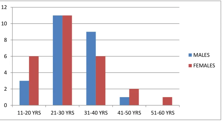
Fig 2 . Age and sex distribution .
In the study, the age group with maximum number of cases with chronic right lower quadrant pain was the 21 – 30 years , in both males and females , the total sum being 22 cases among the 50 cases , accounting to 44% of the total cases .
0 2 4 6 8 10 12
11-20 YRS 21-30 YRS 31-40 YRS 41-50 YRS 51-60 YRS
MALES
65
Fig 3. Pre – op symptoms
This fig shows that all the 50 patients ( 100% ) had right lower quadrant abdominal pain and along with that 5 patients (10% ) had fever and 6 patients ( 12% ) had vomiting.
0 10 20 30 40 50 60
FEVER VOMITING PAIN
66
0 5 10 15 20 25 30 35 40 45
1 2 3
NO OF PATIENTS
[image:78.595.139.469.92.199.2]Tab 2 . Pain improvement after 6 weeks .
Fig 4 . Pain improvement after 6 weeks .
This fig shows 40 patients ( 80% ) showed relief of pain and 8 patients ( 16% ) showed remarkable pain relief , while 2 patients (4%) still complained of right lower quadrant pain after 6 weeks.
67
0 5 10 15 20 25 30 35 40 45 50
1 2 3
NO OF PATIENTS
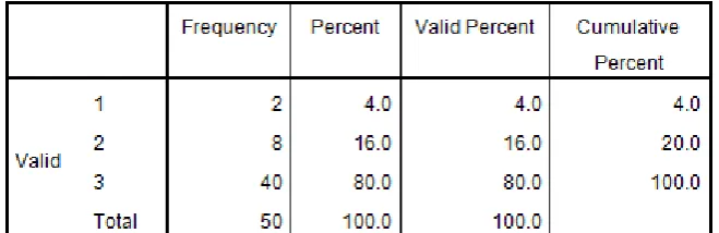
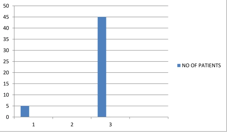
[image:79.595.115.511.276.513.2]Tab 3 . Pain improvement after 3 months
Fig 5 . Pain improvement after 3 months .
This fig shows 45 patients ( 90% ) showed relief of pain and 5 patients ( 10% ) complained of right lower quadrant pain
persistence after 3 months.
68
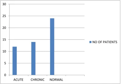
Fig 6. Histopathological features of removed Appendix.
This fig shows that 12 patients ( 24% ) had acute features of inflammed appendix, 14 patients (28%) had chronic histopathological features and 24 patients ( 48% ) had normal features.
0 5 10 15 20 25 30
ACUTE CHRONIC NORMAL
69 NULL
HYPOTHESIS
TEST SIGNIFICANCE DECISION
Histopathology
of the removed appendix is not
related to the pain relief at
6th week follow-up
Independent samples Mann – Whitney
U test
0.64552 Retains the null hypothesis
70 NULL
HYPOTHESIS
TEST SIGNIFICANCE DECISION
Histopathology
of the removed appendix is not
related to the pain relief at
3rd month follow-up
Independent samples Mann – Whitney
U test
0.50286 Retains the null hypothesis
p value < 0.05 is significant .
71
DISCUSSION
From our study, it has been shown that doing an appendectomy in patients presenting with chronic right quadrant pain is more likely to relieve the pain than in leaving the appendix in situ.
Right lower quadrant abdominal pain was the chief complaint in all of the 50 patients ( 100% ) and 5 patients ( 10% ) had fever and 6 patients ( 12% ) had vomiting . It has been identified that except chronic and recurrent right lower quadrant pain, no other clinical characteristics helps in establishing the diagnosis of chronic appendicitis, unlike in acute appendicitis. There were neither typical signs and symptoms nor routine diagnostic modalities to diagnose chronic appendicitis.
In our study, no post – op complications or post – op deaths were encountered.
72
In Netherlands, a study was performed in a teaching hospital between Sep 1994 to Nov 2004 by Dr. R.M.H. Roumen and his colleagues. This study was a single center, double – blinded randomized control trail and patients with chronic right lower quadrant abdominal pain ,were evaluated . Of the 40 patients, 18 patients had undergone laparoscopic appendectomy and 22 patients had undergone diagnostic laparoscopy only. Six months after surgery based on pain score, they observed that in the appendectomy group higher proportion of patients had significant relief of pain than patient who underwent only diagnostic laparoscopy only. In patients who still had RLQ pain and appendix in situ, second time diagnostic laparoscopic procedure intended for appendectomy was done.
73
In Fayez et al. study, 63 patients who had appendectomy for chronic RLQ pain were evaluated. In 92% of removed appendices, histopathological abnormalities were found and 95% patients were cured completely after appendectomy. This study concluded that the chronic appendicitis does exist as a separate entity and could be the cause of chronic RLQ pain.
Krone and Sperke analysed 1,718 prophylactic appendectomies during gynaecological operations. They found histopathological proof of acute appendicitis in 8 % and signs of chronic appendicitis in a whooping 65% and only in 21% were microscopically normal. The clinical relevance remains unclear in patients undergoing an ̏ en passant appendectomy with a high percentage of pathological appendices.
74
But in a study by Dr. Popovic et al. between 1999 and 2000 published in the Croatian medical journal, out of 53 patients 41 underwent laparoscopic appendectomy . The study concluded that laparascopic appendectomy had long – term results similar to the patients without appendectomy and also revealed that whether the removed appendix was normal or with pathological changes, the therapeutic results were similar. But based on the experience, they mentioned that the appendectomy must be performed , though there are no visible changes macroscopically due to changes intra – luminally.
In our study 45 patients ( 90% ) were rendered free from pain after laparoscopic appendectomy but 5 patients (10%) still complained of right lower quadrant abdominal pain.
75
CONCLUSION
BIBLIOGRAPHY
1. Hardin DM Jr. Acute Appendicitis: review and update. Am Fam Physician 1999; 60:2027-34.
2. Borushok KF, Jeffery RB Jr, Laing FC et al. Sonographic diagnosis of perforation in patients with acute appendicitis. AJR 1990;154:275-8.
3. Brinbaum BA, Wilson SR. Appendicitis at the millennium. Radiology. 2000;215:337-48.
4. Carr NJ. The pathology of acute appendicitis. Ann Diag Pathol 2000:4:46-58.
5. Schumpelick V, Dreua B, Ophoff K et al Appendix and caecum: Embryology, anatomy and surgical applications. Surg Clin North Am 2000;80:295-318.
6. Zinner MJ, Ellis H et al. Appendix and Appendicectomy. Maingot’s Abdominal Operations. Appleton and Lange 12th ed. 1997;39:1197-227.
8. Jansen FW, Kapiteyn K, Trimbos kemper TC et al. Complications of laparoscopy; a prospective multicentre observational study. Br J ObstetGynaec 1997; 104:595-600.
9. Giuliano V, Giuliano C, Pinto F, et al: Chronic appendicitis “syndrome” manifested by appendicolith and thickened appendix
presenting as chronic right lower abdominal pain in adults. Emerg Radiol 12:96-98, 2006.
10. Chiarugi M, Buccianti P, Decanini L, et al: “What you see is not what you get.” A plea to remove a “normal” appendix during
diagnostic laparoscopy. Acta Chir Belg 101:243-245. 2001.
11. Cobben LP, de Van Otterloo AM, Puylaert JP: Spontaneously resolving appendicitis: Frequency and natural history in 60 patients. Radiology 215:349-352, 2000.
12. Williams RG, Presidential address: a history of appendicitis Ann Surg. 1983; 197:495-506.
14. Frisch M, Pedersen BV, Andersson RE. Appendicitis, mesenteric lymphadenitis, and subsequent risk of ulcerative colitis: cohort studies in Sweden and Denmark. BMJ. 2009,338:b716.
15. Knight PJ, Vassy LE. Specific dieases mimicking appendicitis in childhood. Arch Surg. 1981;116:744-746.
16. Morrison JD. Yersinia and viruses in acute non-specific abdominall pain and appendicitis. Br J Surg. 1981;68:284-286.
17. Flum DR, Steinberg SD, Sarkis AY, Wallack MK. Appendicitis in patients with acquired immunodeficiency syndrome. J Am Coll Surg. 1997; 184:481-486.
18. de Kok HJ. Laparoscopic appendectomy: a new opportunity for curing appendicopathy. SurgLaparoscEndosc 1992; 2: 297–302.
20. Mattei P, Sola JE, Yeo CJ. Chronic and recurrent appendicitis are uncommon entities often misdiagnosed. JAm CollSurg 1994; 178: 385–389.
21. Falk S, Schutze U, Guth H, Stutte HJ. Chronic recurrent appendicitis. A clinicopathologic study of 47 cases. Eur JPediatrSurg 1991; 1: 277–281.
22. Krone HA, Sperke E. [Preventive appendectomy in gynecologic surgery. Report of 1718 cases ] GeburtshilfeFrauenheilkd 1989; 49: 1035–1038.
23. Lamps LW. Appendicitis & infections of the appendix. Semin DiagnPathol 2004; 2:86–97.
24. Andreou P, Blain S, DuBoulay CE. A histopathological study of appendix at autopsy and after surgical resection. Histopathology 1990;17: 427–431.
26. [Information Products, Hospital Statistics, Nationwide Medical Registration (LMR)] http://www.prismant.nl [ accessed 1 Dec 2006].
27. Leardi S, Delmonaco S, Ventura T, Chiominto A, DeRubeis G, Simi M. [Recurrent abdominal pain and chronic appendicitis] MinervaChir 2000:39-44.
28. Addiss DG, Shaffer N, Fowler BS, Tauxe RV. The epidemiology of appendicitis and appendectomy in US. Am J Epidemiol 1990; 132: 910–925.
29. Pieper R, Kager L. Incidence of acute appendicitis and appendectomy. An epidemiological study of 971 cases. ActaChirScand 1982; 148: 45–49.
PATIENT PROFORMA
Name : Age : Sex : IP No :
ON ADMISSION :
Main Complaints :
Duration of Complaints :
Co – Morbid Illness :
Significant Past History :
CLINICAL EXAMINATION :
Pulse : BP :
RR : Temp :
Pallor : Icterus :
CVS : RS :
INVESTIGATIONS :
CBC : Liver Function Test :
ESR : Renal Function Test :
CXR : USG Abdomen :
CECT ABDOMEN:
TREATMENT :
Intra - op Findings .
FOLLOW UP :
Pain Score : at 6 weeks :
at 3 months :
MASTER CHART
ABBREVIATIONS
P Pain
V Vomiting
F Fever
HPE Histopathology
A Acute appendicitis
N Normal
C Chronic appendiciits
P/V Pain/Vomiting
S.
no NAME AGE SEX I.P no D.O.A D.O.S D.O.D
Pre-OP sympto ms 6 weeks 3
S.
no NAME AGE SEX I.P no D.O.A D.O.S D.O.D
Pre-OP sympto ms 6 weeks 3