A STUDY ON “
CORRELATION OF
THYROID PROFILE WITH THE
COMPONENTS OF METABOLIC SYNDROME
”
Dissertation submitted in partial fulfillment of Requirement For the award of the Degree of
DOCTOR OF MEDICINE - BRANCH VII
GENERAL MEDICINE
APRIL 2016
TIRUNELVELI MEDICAL COLLEGE HOSPITAL.
THE TAMIL NADU DR.M.G.R. MEDICAL UNIVERSITY
CERTIFICATE
This is to certify that the Dissertation entitled a study on “CORRELATION OF THYROID PROFILE WITH
THE COMPONENTS OF METABOLIC SYNDROME” submitted
by Dr.P. GANESH KUMAR to The Tamilnadu Dr. M.G.R. Medical University, Chennai, in partial fulfillment for the award of M.D.Degree(GENERAL MEDICINE) is a bonafide work carried out by him under my guidance and supervision during the academic year 2013-2016. This dissertation partially or fully has not been submitted for any other degree or diploma of this university or other.
Prof.Dr.RAVICHANDRAN. MD., Prof. Dr.VAIRAMUTHURAJU. MD Chief, UNIT III, Professor and HOD,
Department of Medicine, Department of Medicine, Tirunelveli Medical College , Tirunelveli Medical College, Tirunelveli – 627011. Tirunelveli – 627011.
DECLARATION
I, Dr.P.GANESH KUMAR, solemnly declare that
the Dissertation titled “A STUDY ON CORRELATION OF
THYROID PROFILE WITH THE COMPONENTS OF
METABOLIC SYNDROME ” has been prepared by me.
This is submitted to the Tamilnadu Dr.M.G.R. Medical
University, Chennai, in partial fulfillment of the regulations
for the award of MD Degree Branch VII (MEDICINE).
It was not submitted to the award of any degree/diploma
to any University either in part or in full previously.
PLACE: TIRUNELVELI Dr.P.GANESH KUMAR, DATE : POST GRADUATE,
M.D.GENERAL MEDICINE,
ACKNOWLEDGEMENT
CONTENTS
S.NO. TITLE PAGE NO.
CERTIFICATE
DECLARATION
ACKNOWLEDGEMENT
1. INTRODUCTION 8
2. REVIEW OF LITERATURE 9
3. OBJECTIVES 57
4. MATERIALS AND METHODS 59
5. OBSERVATIONS AND RESULTS 61
6. DISCUSSION 84
7. CONCLUSION 86
8. REFERENCES 89
9. APPENDIX ABBREVIATION 93
PROFORMA 94
INTRODUCTION
Need for the study:
Metabolic syndrome has affected more than 25% of the population in the western civilisations. MetS is a major
determining factor for the early onset of insulin resistant diabetes & accelerated atherosclerosis. MetS is clinically a conglomeration of risk factors highlighted by the presence of systemic
hypertension, altered lipid profile, dysglycemia, proinflammatory & prothrombotic states.
Sub-clinical hypoT & MetS are well established risk factors for atheromatous-occlusive vascular diseases,
MetS and sub-clinical/overt thyroid dysfunction are independent risk factors, in the genesis of cardiovascular
diseases. Hence, it is plausible that persons affected with both these conditions, could have more than additive hazard.
This study is a step towards ascertaining the
possible positive link of thyroid dysfunction with the components of MetS.
In this study, TSH has been used as the prime indicator of thyroid dysfunction, as it increases before the elevation of FT4 and also before clinical manifestation.
Review of literature:
hand, modest increase in TSH could place people at excess risk for development of MetS.
Several studies have shown a significant association which links metabolic syndrome with subclinical and overt
hypothyroidism and the association seems to be more in females. Uzunlulu et al reported that the prevalence of subclinical
hypothyroidism was greater in women with MetS.
In a study by Bauer DC et al, it was concluded that among older hispanic women, elevated TSH levels were associated with hazardous changes in composition of lipids & that women with multitude lipid abnormalities were thrice as likely to have increased TSH levels.
The study conducted by Tromso and Basel, has
proven that thyroxine supplementation, in patients with borderline thyroid dysfunction, has a better effect on low density(LDL) cholesterol values. This also led to relative risk reduction in
A study conducted by HUNT, proposed that “Within the range of TSH that is considered clinically normal, increasing level of TSH was associated with less favorable lipid concentrations. The association with serum lipids was linear across the entire reference range of TSH”.
Research published in the February 2007 issue of the “Journal of Clinical Endocrinology and Metabolism” found a connection between thyroid function & metabolic syndrome. In euthyroid persons ie TSH within normal levels, the level of free T4 was important. Free T4 levels that were slightly low, but still within the normal range, significantly increased the risk of many risk factors for metabolic syndrome.
In hypothyroidism , energy metabolism is reduced leading to a decreased in appetite , cold intolerance, reduced protein
euthyroid , hypothyroid , hyperthyroid having metabolic syndrome and the group not having metabolic syndrome.
In a study conducted by Banaras University, atherogenic lipid abnormalities were observed in adult subjects with Subclinical hypothyroidism-2 (TSH > 10.0 Miu/L), and not in subjects with Subclinicalhypothyroidism-1 who had TSH ≤ 10.0 Miu/L in Indian population.
The Jaipur Heart Watch Study has shown that in urban adult populations, the prevalence of MetS was nearly 19% in men, 30% in women, and approximately 25% on the whole.
In a population based study conducted by,
“Prevention of Metabolic Disorders Research Center, Iran”, of over 900 reproductive aged women, subclinical thyroid
Metabolic syndrome is characterised by accumulation
of multiplex of risk factors that roots from insulin resistance,
henceforth accompanied by abnormal adipose tissue deposition
This is one of the most important problems faced
by the modern fraternity in the last two and half decades.
Problem statement:
About 25% of the world’s adult population has metabolic syndrome.
People with metabolic syndrome three times as likely to have a heart attack or stroke compared with people without the syndrome.
People with metabolic syndrome have a five-fold greater risk of developing type 2 diabetes.
People with metabolic syndrome two times as likely to have a cardio vascular mortality, as compared with
people without the syndrome.
This puts metabolic syndrome way ahead as a giant killer disease, yet the problem is not as well recognised.
HISTORY:
Long before the modern definition of MetS, the
Italian physician, Morgagni, described the association between
visceral obesity, arterial hypertension, atherosclerosis and high
levels of uric acid in blood, almost 250 years ago.
In 1920, Nicolae Pautescu, suggested diabetes and
obesity as the consequent phases of same pathological phases. In
1988, Reaven G., an endocrinologist, interpreted the association of
diabetes mellitus, hypertension and dyslipidemia, by their
pathogenic relationship with peripheral insulin resistance. He
named this “Syndrome X”.
Ferranini et al, confirmed this association and named
it ‘Insulin resistance syndrome’. Soon metabolic disturbances, was
found to be involving larger spectrum. Zimmet & co, put forward
hyperuricemia, obstructive sleep apnea etc. In 1998, WHO
formulated the first official definition of MetS.
IDF definition
:For a person to be diagnosed to have MetS, he/she
should have, c
Central obesity, defined as waist circumference with
ethnic specificity, >90cm for asian men and >80cm for asian
women.
Plus any two of the following criteria
Raised triglycerides
> 150 mg/Dl (1.7 mmol/L)
(Or) on specifi c treatment for this lipid abnormality
Reduced HDL cholesterol
< 40 mg/Dl (1.03 mmol/L) in males
Raised blood pressure
Systolic BP 130mm of Hg & above
(or) Diastolic BP 85 mm Hg & above
(or) 0n treatment of previously diagnosed hypertension
Raised fasting blood sugar
Table 1: The FPG >100 mg/Dl (5.6 mmol/L),
(or) previously diagnosed type 2 diabetes
If above 5.6 mmol/L or 100 mg/Dl, OGTT is strongly
recommended but is not necessary to define presence of the
Prevalence:
The exact prevalence varies according to extent
of westernisation, lifestyle patterns and economic & cultural
patterns prevailing in the area. Indian studies have shown that the
prevalence is more than 30% in urban population and is still
swelling. Gender difference is significant, with the women leading
the charts. Prevalence is 1.5- 2 times higher in women compared
to men.
There has been, also, a recent surge in prevalence
among the rural Indian population. Alarming increasing childhood
obesity rates, is a cause for concern. These factors indicate the
Risk factors for MetS:
Ethnicity
Asian Indian phenotype, is predisposed to the
development of MetS. This is characterised by higher body fat
with comparatively, lower BMI. The proportion of intra-abdominal
visceral fat is much higher compared to lean body mass, in asian
adults. Hence asian phenotype, has higher prevalence, earlier
onset and increased complications of T2DM at lower BMIs.
Obesity
Higher the body weight, higher the prevalence and
risk of development of MetS. Increased waist circumference is a
positive predictor of future development of MetS.
DIET
There has been a decline in the intake of
traditional foods, in the recent two to three decades. Traditional
Indian foods had higher fibre content and was lower in simple
sugars and saturated fat. The food composition has varied a lot
now-a-days. Higher consumption of animal fats, dairy products and
hydrogenated oils, has been shown to have a higher correlation
with the development of cardiovascular diseases. These changes in
dietary patterns may be implicated in the increased incidence of
metabolic syndrome.
Physical activity
Increased mechanisation of work, increased indoor entertainment activities and reduced outdoor activity, has
resulted in decreased physical activity on the whole. Finally
resulting in increased weight and waist circumference.
Migration
An adverse coronary risk profile has been
reported among rural-to-urban migrant population. This has been
suggested due to significant stress arising out of new environment,
job challenges, socio-economic disparities, lack of social support
etc.
Genetic and environmental factors
Adverse intrauterine environment, has been linked to insulin resistance and metabolic syndrome. Low birth
weight is associated with high SBP, insulin resistance, fasting
hyperinsulinemia. Catch up obesity seen in LBW offspring seems to
be important for adult onset insulin resistance and associated
cardiovascular risk factors.
Psychological factors
Stress activates sympathetic nervous system
leading to hormonal fluctuations. Stress has been associated with
hypertension, which is an integral part of metabolic syndrome.
Pathophysiology
Resistance to the action of insulin is the corner
stone for the altered metabolic state of the MetS. Altered free
fatty acid metabolism is the prime component involved in the
pathophysiology of dysglycemia and hyperlipidemia. Increased
plasma FFA concentration impairs the ability of insulin to
stimulate muscle glucose uptake and suppress hepatic glucose
production. In addition, high levels of free fatty acids delivered to
the liver increases hepatic very-low density lipoprotein triglyceride
production and plasma triglyceride concentration. An increase in
plasma triglycerides increases the transfer of triglyceride from
density lipoprotein clearance & decreased serum high density
lipoprotein(HDL) levels.
Adipose tissue secretes many pro-inflammatory
mediators (adipokines). These result in resistance to insulin action.
For example, TNF-alpha reduces insulin signaling, IL-6 increases
inflammatory reaction directly & stimulates hepatic-CRP
production, MCP-1 is a strong chemotactant for macrophages , and
IL-8 is activator for neutrophil granulocytes & chemoattractant for
most of the described migrating immune cells. This chronic
inflammatory process leads to heightened insulin resistance.
Excessive intrahepatic steatosis is associated with
impaired hepatic insulin action. This also cause reduced
insulin-dependent suppression of nocturnal glucose release from liver.
Excessive muscle fat is related to insulin resistance. This also
MetS and CVD risk:
Overall, the metabolic syndrome is associated
with a two-fold increase in risk of CVD, CVD mortality, and
stroke. There has been also a 1.5-fold increase in risk of
all-cause mortality. Patients with the metabolic syndrome, but without
type 2 diabetes mellitus, are still at high risk for CVD mortality,
MI, and stroke. The metabolic syndrome does not require type 2
diabetes mellitus in order to be closely associated with
cardiovascular risk.
Many meta-analyses have shown that the metabolic
syndrome is associated with higher cardiovascular risk in women relative to men. Several theories have been postulated to explain a potentially higher cardiovascular risk in women with the metabolic syndrome .
2) The cholesterol profile is different in women compared with men. HDL cholesterol decreases and LDL cholesterol
Increases post-menopausally. Also LDL particles becoming denser, and therefore, more atherogenic.
3) Elevated triglycerides are more highly associated with coronary artery disease in women than in men. In
a meta-analysis, it was shown that an increase in triglycerides of 18 mg/dl was associated with a 76% increased
cardiovascular risk in women compared with a 32% increased risk in men .
4) Presence of other unique risk factors like polycystic ovary syndrome, hormonal contraceptive use, and gestational diabetes may be responsible for a stronger association between
the metabolic syndrome and cardiovascular risk in women. The key mechanism behind elevated CVD risk is attributable to
Pro-inflammatory state:
Insulin resistance and obesity have been closely
linked to a pro-inflammatory condition. The main reason being excessive cytokine synthesis & release of acute phase reactants. C-Reactive protein is one of the important acute phase reactant, mainly produced in liver, after any noxious stimuli. Data from prior scientific researches have shown an association between serum CRP levels and features of MetS . CRP levels closely predicts the development of T2DM and Cardio-vascular diseases.
insulin action, the prime target being hepatic tissue and muscles.
Pro-coagulant state
Pro-coagulant state is being an important ally of MetS. Many factors have been suggested to be associated in hemostasis regulation in MetS. The main haematologic abnormality associated with MetS is the excessive levels of Plasminigen activator inhibitor-1. It is the important suppressor of fibrinolysis. Community level studies have concluded that excessive levels of PAI-1 is a fore-teller of myocardial ischaemia & CV mortality. Hepatic fat accumulation is the major reason for increased plasma PAI-1 levels in persons having MetS.
both the chronic persistent inflammatory process & resistance to insulin in MetS.
Even-then, the exact mechanism behind the excess production of fibrinogen from the hepatic tissue is yet to be
cleared. This has been proposed that Free fatty acids and
inflammatory mediators linked to insulin resistance, promote the hepatic production of clotting factors.
Pro-atherosclerotic state:
Fattyacid binding proteins by integrating
metabolic & immune responses, interlink the inflammatory and
lipid-mediated pathways. This mechanism plays a vital role in
MetS. Thus FABPs are causative in the process of atherosclerosis
, mainly by modifying cholesterol trafficking & inflammatory
processes. The FABPs thus, implicate a longlasting impact on
visceral adiposity, insulin resistance, diabetes mellitus &
Thus, MetS is a pro-inflammatory, pro-coagulant and pro-atherosclerotic state, resulting in elevated cardiovascular risk.
Management of MetS:
The efficient treatment, involves optimal control of
the risk factors promoting development of MetS. By
implementing lifestyle interventions remains the cornerstone of
management. Intensive life-style changes involving diet and
physical activity, is the recommended first-line treatment.
Pharmacological therapy is advised, when the lifestyle changes do
not produce optimal response. Pharmacological therapy, is needed
in persons with a cardiovascular risk score more than 20% .
Life style modifications:
Most of the persons with MetS are either
obese or overweight. They have to undergo a weight losing
Adult Treatment Panel III suggests a well
balanced diet, with reduced consumption of refined and simple
sugars. Trans fatty acids have to be avoided. Fresh fruits,
vegetables, and whole grains have to be consumed in large
proportions. Nutritionists have suggested, a daily reduction of
about 500–1000 kCal is the easiest way for a continued weight
reduction, along with a well-scheduled, well-adhered phsical
activity programme.
The most recent AHA recommendations, suggest a
moderate intensity exercise regimen, for atleast a period of not less
than thirty mts. Brisk walking and bicycling are the most favored
exercise programme. All the sedentary activities in our daily
routine life has to be avoided. Whenever possible, healthy
lifestyle habits have to be adopted, for example, going to shopping
by walk, utilizing steps instead of using lifts/travellators. All these
Specific Management of Metabolic Risk Factors:
. The exact objective of treating MetS is to decrease
the heightened risk of cardiovascular diseases & to slow down the
development of diabetes mellitus. A multitude of residual ongoing
these circumstances, pharmacological therapy, with a combination of
drugs, is necessary.
Pharmocologic interventions
:Metformin, TZDs – for dysglycemia management.
Fibrates &statins – for dyslipidemia and pleiotropic effects.
ACEI/ARBs - for BP control and micro angiopathy protection.
Aspirin - for negating the procoagulant state.
Insulin resistance and glucose intolerance:
Insulin resistance associated with impaired glucose
tolerance and elevated blood sugar levels is the hallmark feature
of MetS. Hence treatment of insulin insensitivity can modify both
reduction in simple sugars & saturated fats, in hands with easily
adoptable exercise program is the main modality of therapy .
Drugs that enhance insulin sensitivity, like the
biguanides (metformin) and the thiazolidenediones,
(PPARγagonists), have the efficiency to either slowdown/ prevent
the development of diabetes mellitus. These drugs also help in
modifying the metabolic profile. Thus they decrease risk of
atherosclerosis.
In a Diabetes Prevention Study conducted in
Finland, a total of 522 persons with MetS, rigourous lifestyle
modifications decreased the risk of diabetes to 58% , in
comparison to the controls. In the DPP study, metformin in the
dosage of 850 mg bid decreased the development of diabetes
mellitus by 31%. In the same study, vigorous lifestyle changes
reduced the risk to 58% in comparison to placebo. Inspite of
reduction in risk of CVD. Hence such drugs should not be
prescribed for the sole purpose of prevention.
Dyslipidemia:
Lifestyle intervention with dietary changes and physical
activity is the prime therapy of dyslipidemia in the MetS.
Eventhough raised Low Density cholesterol is a non-defining
aspect of the lipid constitution in these patients,
LDL-C and apo-B lipoproteins are pro-atherosclerotic. LDL
reduction therapy has to be as per the cardiovascular risk score.
-in persons with ≥20% risk as calculated by
the presence of CAD or CAD risk equivalents, the desired LDL
level is less than 100 mg/Dl.
-patients with medium risk ie 10 – 20% have to
achieve a desired LDL level <130mg/dl.
In persons with elevated TGL levels, more than
200 mg/dl, increased quantity of pro-atherosclerotic remnant
overlooked, by targeting mainly the Low Density cholesterol.
Hence , Adult Treatment Panel III generated the idea of non-HDL
cholesterol. This includes all the dangerous atheroma-prone
lipoprotein subdivisions. Non-High Density Cholesterol target is
30mgs% more than the LDL levels. This has to be used when
TGL level is more than 200 mgs%.
In such persons who fail to reach the desired
targets, with intensive lifestyle interventions, pharmacological
treatment with statins has to be started. Lipid lowering drugs
decreased unwanted cardiovascular events and CVAs in many
clinical trials. The lipid lowering therapy, had additional beneficial
effects in diabetes patients. This has been documented in
Collaborative Atorvastatin Diabetes Study &
Heart Protection Study. This has been proven, even when the
cholesterol is within normal limits. Statins decrease all the
apoB– lipoproteins . they successfully reach Adult Treatment
Fibric acid congeners modify majority of the
components of the atherogenic lipid consitution in MetS. This
improves mainly the decreased HDL , elevated TGLs and small,
dense LDL particles. Their use has to be thought of in all
persons with a risk of more than 20% for CAD. Nicotinic acid at
higher dosage, also modifies HDL & TGL levels, with the
comparable efficiency to fibrates. On the downside, nicotinic acid
causes impaired glucose tolerance & elevates serum uric acid
levels. Hence, this can not be used in persons with MetS.
Procoagulant & proinflammatory condition:
In MetS, inflammatory mediators such as ,
Interleukin-6 and Tumor Necrosis Factor–α and acute phase proteins
like, C-Reactive protein and fibrinogen are elevated. PAI-1 levels
decreased. This contributes to the characteristic
inflammatory-prone & procoagulant environment of the MetS.
The therapeutic and prognostic role measuring
inflammatory mediators is doubtful. This is an upcoming area of
scope in preventive cardiology. Eventhough assessment of
fibrinogen & procoagulation factors is not routinely advised.
AHA current recommendation is for anti-platelet prophylaxis in
SUB-CLINICAL HYPOTHYROIDISM
Definition:
SCH is defined as a serum
thyroid-stimulating hormone (TSH) level above the upper limit of
normal despite normal levels of serum free thyroxine. TSH
levels 5.5–10.0 Mu/l correspond to prevalence of SCH.
The incidence of subclinical hypothyroidism ranges
from 6 to 8% , based upon the sex , age and ethnicity of the
subjects studied. The effects of subclinical hypothyroidism
depends upon the duration and the degree of thyroid dysfunction
Effects of Thyroid Hormones on Lipid Metabolism:
potentiating the action of HMG co-A reductase, present in the hepatic parenchyma.
Thyroxine induces the action of LPL. Thus results in increased hydrolysis of TGLs into VLDL. Also the chylomicrons are hydrolysed as free fatty acids & glycerol. In thyroxine
deficiency, the LPL action level over the fatty tissue is
documented to be either normal or suppressed. In the mean time, hepatic lipase action is also suppressed, causing elevated measures of TGLs.
Thyroxine levels also directly affect the CETP
cholestrol clearance and hence, increased serum low density cholesterol levels in SCH.
Lipid metabolic disturbances in SCH:
Hyperlipidemia is a very common laboratory abnormality in persons with evidence of hypothyroidism,
including elevated levels of both total & Low density cholesterol. Researches pertaining to TGLs, Lp(a), HDL, apoB & apoA1
components in subclinical hypothyroidism are minimal. In addition to the quantitative changes, qualitatives changes like, smaller, denser and more oxidized Low density cholestrol is a routine feature of hypothyroidism.
controlled after therapy. This suggests a highly perplexed etiology for lipid abnormalities in SCH.
Not only overt hypoT, but also the SCH is also linked to profound lipid composition. The changes include the elevated total & low density cholesterol. Whereas HDL, TGLs, apoB, Lp-a & apoA1 levels did not show marked changes subclinical hypothyroidism when compared to the euthyroid controls , in most of the observations. Rondeau et al. noticed a negative correlation between HDL-C and TSH levels in obese and postmenopausal females.
Most of the researches have found that after thyroxine replacement treatment, both the total & Low density cholesterol levels are being normalized . Whereas hormone
& phospholipids in SCH was corrected after achieing euthyroid status.
Hemodynamic effects of thyroid dysfunction:
In overt hypoT, the prime modifications of cardiovascular functions, noticed are reduction in heart rate, increased peripheral vascular impedance, elevated diastolic blood pressure & hence, the afterload. Also there occurs reduced blood volume & hence, the preload. Put together, cardiac output is
Thyroid Dysfunction & Atherosclerosis:
Thyroid dysfunction is associated with the highly atherogenic dyslipidemia. In addition, insulin insensitivity,
hypertension, inflammation, oxidation stress & coagulation
Clinical hypoT is related to elevated diastolic blood pressure and hyperhomocysteinemia. Increased levels of hs-CRP and coagulation deficits have also been reported in patients with hypothyroidism. The proposed mechanisms being, impairment of intracellular glucose metabolism & translocation GLUT4. Enhancement of carotid artery intimal thickness, is also being noticed in persons with both clinical/subclinical hypoT.
In a survey conducted in Whickham , a positive correlation has been documented between the incidence of CAD and associated mortality in persons with SCH, followed over a period of two decades. This positive correlation was abolished after thyroxine replacement therapy .
Extending support to this observation, many
meta-analyses suggest that SCH is related to a marked rise in risk of CAD & associated mortality. In a meta-analysis done by
persons with SCH. This is more true in case of women. SCH is also causally, linked with the incidence of cerebral ischemia.
Rodondi et al. noticed a correlation between SCH & the risk of ischaemic stroke.
SCH and CV Disease Risk:
The CV modifications observed in SCH
patients qualitatively resemble those produced by overt hypothyroidism, quantitatively, to a lesser extent. Early alterations of cardiac performance, endothelial function, systemic blood pressure, and lipid profile have been documented in SCH patients. This supports a biologically plausible role for subclinical hypothyroidism in the development of early atherosclerosis.
cardiovascular disorders. Furthermore, it has been reported that SCH is associated with high serum total cholesterol and LDL
levels and low HDL which could increase the atherosclerosis risk. A significant correlation between SCH & CAD morbidity and death has been documented , although less evident in older people. The relationship between SCH and CHD persisted even after adjustment for traditional risk factors. This suggests an alternative mechanism by which SCH increases CV risk, at least in younger individuals.
Although the scientific literature is not uniform in the definition of the effect of SCH with mild TSH elevation (<10 Mu/L) in terms of CV events and mortality, available data suggest an impact of this clinical condition only in younger patients (<65 y), especially men.
Treatment guideline for SCH:
In most of the patients with subclinical hypo-T,
there is no need for treatment. Oral Levo thyroxine is the
drug of preference, in case of a decision to treat the deficiency.
Clinical trials of subclinical hypo-T patients, have proven that
thyroxine therapy is effecient in reducing TSH to the normal
reference range.
Ideally, thyroxine has to be started at a dose of
25-50 µg od. Followed up with a monthly dosage adjustment to
achieve the desired TSH value. In a study, 100 elderly persons
>65yrs of age, achieved the desired TSH level, with a
replacement dose of 50µg of thyroxine . In two more RCTs,
done in young subclinical hypo-T persons , it has been proven
that on an average, thyroxine at a dose of 50-75 µg is needed
In an another study, involving 35 subclinical
hypo-T persons , it has been that, on an average 100 µg of
thyroxine, is needed to maintain TSH levels between 0.5 - 2.5
Mu. In a large clinical study, all subclinical hypoT patients, when
started on 100 µg thyroxine od, discovered
that only 10% developed features of hyperthyroid as evidenced
by low serum TSH and increased T 4 levels . Henceforth, the
thyroxine dosage of 50 – 100 µg od is sufficient to bring
down TSH, to desired levels in most of the persons.
In many situations, the patient has to be started on
the approximate full dose of thyroxine . The exception being,
known patients with coronary arteriak heart disease. In such
patients, much lower doses have to be started, followed by
gradual stepwise dose escalation. For example, in a patient with
stable angina, thyroxine has to be started & increased slowly by
25 µg once in 2- 3 weeks. Eventhough a quiet identitical approach
has shown that such a cautious approach is not needed in elder
population , without any cardiac disease.
For maximal bioavailability, thyroxine has to be
consumed on an empty stomach. Studies have given varied results
with regard to the ideal time for thyroxine replacement therapy. A
study conducted in United States population, showed adequate
TSH control, if thyroxine is consumed on an empty stomach
state, one hour before food . But in a Denmark study, thyroxine
consumed just before bedtime is higher in efficacy, when
compared to that consumed one hour before breakfast.
There are many drug-food interactions. Milk,
coffee, soya & papaya especially hamper thyroxine absorption.
In reality, a lot of medications including
Salts of calcium & iron, antacids such as sucralfate,
H 2 blockers & PPIs, result in significant interference to
absorption of thyroxine .
atrophic gastritis, celiac sprue & pernicious anaemia. Both
impairment & improvement of thyroxine absorption are documented
following various types of bariatric surgery. Patients falling in
these situations tend to need increased dosage of thyroxine.
There is a possible theoretical advantage by
combining T 3 &T4 medication. But so far, studies using
Combination of T 3&T4 medications did not document any
definite advantage in patients with clinical hypoT.
Also, in a meta-analysis of studies involving
more than 1,500 hypoT persons, no convincing advantage of
T 3&T4 combination was found. In this setting, without any data
pertaining to subclinical hypo-T , this combination therapy should
not be prescribed in routine practice for them.
After starting replacement therapy with thyroxine,
patients have to undergo follow-up after 6–10 weeks. With the
repeat TSH value, thyroxine dose altered accordingly. This is to
the serum lipid profile has to be re-evaluated. This follow-up, is
to assess sufficient improvement or is there any need of
pharmacological therapy for dyslipidaemia. In the mean time,
re-evaluation of symptoms of hypoT, in patients with subclinical
hypo-t , who were commenced on thyroxine replacement for
possible symptoms of hypoT. This is very important, because if
at all any benefit from thyroxine replacement in subclinical
hypo-T, then it is worthwhile in considering prolonging treatment
for lifelong. If following a three-six months of therapeutic trial,
there has not been any significant symptomatic improvement, then
it is time to consider stopping replacement therapy.
A significant number of persons with subclinical
hypo-T will develop clinical hypoT. This progression has been
found to be around 6–10% per annum, degree of TSH elevation
being the important predictor . On the other side, sub-normal
thyroid production may normalize in 8–30% of subclinical
up. As a result, in most of the patients, thyroid function seems to
be stable.
If a patient is diagnosed with SCH, then the
thyroid function has to be re-evaluated after a period of 2-3
months, in addition to thyroid autoantibodies. If there is
normalization of thyroid secretion, no more testing is needed in
asymptomatic patients without either thyroid autoantibodies or
goitre.
In patients with persistent subclinical hypo-T,
thyroid function has to be re-evaluated , once in six months, at
least for the first two years & thereafter once a year only. This
Will assess any tendency to progress & to identify consequent
clinically evident hypoT.
a) To find out the type of thyroid dysfunction in metabolic syndrome.
b) To find out the association of thyroid dysfunction with the components of metabolic syndrome.
MATERIALS AND METHODS:
Source of DataPatients attending OPD of Dept of Internal Medicine, Tirunelveli Medical College Hospital, who are being diagnosed as metabolic syndrome and fulfill inclusion and exclusion criteria.
Method Of Collection of Data Sample size:
100 subjects with MetS & 50 controls.
Patients fulfilling the criteria for metabolic syndrome by International diabetic foundation[IDF] were taken into study. Patients with metabolic syndrome not on any
medications – newly detected metabolic syndrome patients.
Exclusion Criteria:
1) Known patients of hypothyroid or sub-clinical hypothyroid or hyperthyroidism
2) Patients on medications for diabetes mellitus , hypertension , thyroid disorders and dyslipidemia
3) Patients on steroids 4) Acutely ill patients
5) Individuals less than 18 years age, who can not give consent.
The purpose of the study was explained to the patient and informed consent was obtained. Data was collected using a pretested proforma meeting the objectives of the study. Detailed history and necessary investigations were undertaken. Patients were selected for study who satisfied all the inclusion and
exclusion criteria. Patients were diagnosed having metabolic syndrome by the,
“IDF criteria:
Central obesity – defined as waist circumference with ethnicity specific values (for south Asians : ≥90 cm for Men and
≥80cm for women were used)
AND any two of the following:
• Raised triglycerides: > 150 mg/Dl (1.7 mmol/L), or specific treatment for this lipid abnormality.
• Raised blood pressure: systolic BP > 130 (or) diastolic BP >85 mm Hg, or on treatment for previously diagnosed hypertension.
• Raised fasting plasma glucose FPG)>100 mg/dl , or
previously diagnosed type 2 diabetes mellitus”
All the patients enrolled for the study, were subjected to Thyroid Function Test. Test results were entered in a excel sheet. Meticulous analysis of the data was carried out.
INVESTIGATIONS
1. Fasting blood sugar
2. Lipid profile includes Triglycerides , HDL ,LDL, Total cholesterol 3. Thyroid assay includes T3 , T 4 , TSH
4. Blood pressure recording
RESULTS AND OBSERVATION
Statistical method:
All the compiled data were analysed using computer based software. By chi-square test p-value was calculated. P-value < 0.05 was considered as statistically significant.
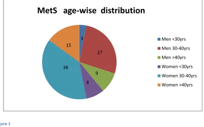
Age distribution among the subjects
:
Men Women
<30yrs 30-40yrs >40yrs <30yrs 30-40yrs >40yrs
MetS 3 27 9 8 38 15
Control 5 13 7 5 18 2
The mean age of the MetS subjects was around 36
years. The mean age of the controls was found to be 34.7 years.
This difference had no statistical significance. This implied that the
age. Thus the impact of age, on the incidence of SCH, was
negated, in the study population.
As clearly seen from the above chart, more than
two-thirds of the subjects fall in the 30-40yrs age category.
Clustering of MetS in the 30-40yrs age group, reveal the
deleterious effects of the wrong life-style patterns in the past two
decades.
The important thing to be considered is, this 30-40yrs population, is the backbone of a growing economy like
ours. Hence, any health related issue, affecting this population,
will produce a significant negative impact on the nation’s
Figure 1 3 27 9 8 38 15
MetS age-wise distribution
Men <30yrs Men 30-40yrs Men >40yrs Women <30yrs Women 30-40yrs Women >40yrs 5 13 7 5 18 2
Control age-wise distribution
[image:63.612.81.484.116.367.2]
Sex distribution of MetS subjects
:
Out of the 100 MetS subjects, 39 were male and 61 were
female. This is consistent with the results of many observational
studies, which found out that the incidence of metabolic syndrome is
1.5-2 times higher in females compared to males.
p-value <0.05, this means, the sex difference noted
in the prevalence of MetS is statistically significant.
The female sex, supposed to have protective
effect against, CV diseases, is the easy target for MetS. This strong
Met S in men 39
clustering of CV risk factors, negates the natural protection for
women, against cardiovascular diseases.
Figure 3
The popular belief that premenopausal women have
immunity against CV diseases, is becoming a thing of past. If not
taken seriously, this will haunt the future, of fertile female
population. This MetS, being
question mark over their fertility itself.
61
clustering of CV risk factors, negates the natural protection for
women, against cardiovascular diseases.
The popular belief that premenopausal women have
immunity against CV diseases, is becoming a thing of past. If not
taken seriously, this will haunt the future, of fertile female
This MetS, being associated with PCOS, will leave a
question mark over their fertility itself.
39
clustering of CV risk factors, negates the natural protection for
The popular belief that premenopausal women have
immunity against CV diseases, is becoming a thing of past. If not
taken seriously, this will haunt the future, of fertile female
associated with PCOS, will leave a
MetS men
Prevalence of SCH in MetS
:
Control with TSH<5.5 47 Control with TSH>5.5 3 Mets with TSH<5.5 79 Mets with TSH>5.5 21
Of the 100 MetS subjects, 21% had sub-clinical
hypothyroidism. Of the 50 controls, only 3 had SCH, which means
the prevalence among the control population is around 6% only.
p-value <0.05, this means, the difference in prevalence
of SCH in MetS & controls is statistically significant.
Both SCH and MetS, individually being CV risk factors,
their combination more than doubles the risk. This being the era
of non-communicable diseases, these two are the most easily
Figure 4
Figure 5
47 3
control with TSH<5.5
control with TSH>5.5
79 21
Mets with TSH<5.5
Sex-wise prevalence of SCH
:
Men Women
SCH in MetS 4 17
[image:68.612.73.536.268.558.2]SCH in control 1 2
Figure 6
More than 80% of the MetS with SCH were women.
p-value < 0.05, statistically significant.
4
17
SCH in MetS
BMI-wise distribution
MetS with SCH
MetS with euthyroid Control
Figure 7
BMI was calculated using the formula,
0 5 10 15 20 25 30
MetS with SCH MetS with euthyroid
distribution of Thyroid function:
avg BMI 26.7 25.9 21.4
BMI was calculated using the formula,
MetS with euthyroid Control
avg BMI
As expected, BMI was significantly higher in
MetS, compared to the controls. But the difference was
not significant, among MetS with SCH & MetS with euthyroid
status. P-value >0.05, implying, no statistical significance.
This reiterates the “
thin fat asian phenotype
”
concept. BMI is not an ideal marker, in asian population,
when compared to their european counterparts.
Visceral adiposity, is the determining factor, in
the definition of metabolic syndrome. The indian urban
population don’t indulge in regular physical activities. So,
their muscle mass & bone mass are on the lower side.
This the reason for their lower BMI, despite of their look
Comparing thyroid function with MetS components
:Waist circumference:
Men WC (cm) Women WC(cm)
MetS with SCH 100.3 93.6
MetS with euthyroid 95.5 87.5
Waist circumference is the essential
criteria for diagnosing metabolic syndrome. Both men and
women with MetS, have higher WC, compared to their
euthyroid counter-parts. But, this difference is subtle, with
a p-value>0.05, statistically insignificant.
Waist circumference is the indirect
measure of visceral adiposity. This has been also found to
Figure 8
Though there is a significant arithmetic
difference in the waist circumference between
groups, the p-value
significance.
80 82 84 86 88 90 92 94 96 98 100 102 Men WC(cm)Though there is a significant arithmetic
difference in the waist circumference between both
value is >0.05, conferring no statistical
Women WC(cm)Though there is a significant arithmetic
both the
, conferring no statistical
MetS with SCH
Thyroid function vs FBS:
Men FBS(mgs%) Women FBS(mgs%)
MetS with SCH 101 99.5
MetS with euthyroid 95.5 98.7
Fasting blood sugar, among MetS, was in the
range of 88-115mgs%, with the average value being 97.7mgs%.
Out of the 18 women with both MetS & SCH, 8 had a FBS value
more than 100mgs%. Out of the 3 men with both MetS & SCH, 2
had a FBS value more than 100mgs%.
The difference between the average FBS,
between the MetS with SCH and MetS with euthyroidism is only
figure 9
This difference in FBS, had no
the p-value was >0.05.
92 93 94 95 96 97 98 99 100 101
MetS with SCH MetS with
This difference in FBS, had no statistical significance, as
value was >0.05.
MetS with euthyroid
Men FBS(mgs%)
Women FBS(mgs%)
significance, as
Men FBS(mgs%)
Thyroid function vs TGL:
Men TGL(mgs%) Women TGL(mgs%)
MetS with SCH 182.3 182.2
MetS with euthyroid 139.5 141.2
Fasting Triglycerides, among
MetS, was in the range of 112-230mgs%, with the average value
being 148.7mgs%. Out of the 18 women with both MetS & SCH, all
18 had a TGL value more than 150mgs%. Out of the 3 men with
both MetS & SCH, all 3 had a TGL value more than 150mgs%.
This strongly implies that raised TGL, is an integral aspect of
Figure 10
This significant difference in TGL values, among
the groups is authenticated by the
TGL value, more than 150mgs%, in MetS should
0 20 40 60 80 100 120 140 160 180 200
MetS with SCH MetS with
euthyroid
This significant difference in TGL values, among
the groups is authenticated by the p-value <0.05. Hence r
TGL value, more than 150mgs%, in MetS should evoke
MetS with euthyroid
Men TGL(mgs%)
Women TGL(mgs%)
This significant difference in TGL values, among
Hence raised
suspicion
Men TGL(mgs%)
Thyroid function vs HDL:
Men HDL(mgs%) Women HDL(mgs%)
MetS with SCH 37 45.6
MetS with euthyroid 36.2 46.5
Out of the 18 women with both MetS &
SCH, 14 had a HDL value less than 50mgs%. Out of the 3 men
with both MetS & SCH, 2 had a HDL value less than 40mgs%.
When compared to their, euthyroid
counterparts, both men and women with MetS & SCH, had similar
HDL values. This rules out HDL, being a definite marker to screen
for thyroid dysfunction among MetS subjects.
This also stresses the fact that, in MetS,
Figure 11
Almost similar HDL values, among the groups,
leaves this criteria being insignificant, with the
0 5 10 15 20 25 30 35 40 45 50
MetS with SCH
MetS with euthyroid
Almost similar HDL values, among the groups,
leaves this criteria being insignificant, with the p-value >0.05
MetS with euthyroid
Men HDL(mgs%)
Women HDL(mgs%)
Almost similar HDL values, among the groups,
value >0.05.
Men HDL(mgs%)
Thyroid function vs SBP:
Men SBP(mm Hg) Women SBP(mm Hg)
MetS with SCH 132.7 129.9
MetS with euthyroid 136.6 131.4
Out of the 18 women with both MetS &
SCH, 7 had a SBP value more than 130mm of Hg. Out of the 3
men with both MetS & SCH, 1 had SBP value more than 130mm
of Hg.
When compared to their, euthyroid counterparts,
both men and women with MetS & SCH, had similar SBP values.
This rules out SBP, being a definite marker to screen for thyroid
Figure 12
In SCH, the heart rate and the stroke volume are
decreased. This results in a fall in SBP in cimparison to MetS with
euthyroidism. As a result, for the difference in SBP, among the
126 128 130 132 134 136 138
MetS with SCH
In SCH, the heart rate and the stroke volume are
is results in a fall in SBP in cimparison to MetS with
euthyroidism. As a result, for the difference in SBP, among the
MetS with euthyroid
In SCH, the heart rate and the stroke volume are
is results in a fall in SBP in cimparison to MetS with
euthyroidism. As a result, for the difference in SBP, among the
Men SBP(mm Hg)
Thyroid function vs DBP
:Men DBP(mm Hg) Women DBP(mm Hg)
MetS with SCH 88 84.5
MetS with euthyroid 88.5 85.7
Out of the 18 women with both MetS &
SCH, 9 had a DBP value more than 85mm of Hg. Out of the 3
men with both MetS & SCH, 1 had DBP value more than 85mm
of Hg.
The increase in DBP is due to increased arterial
stiffness in both MetS and SCH. But when compared to their,
euthyroid counterparts, both men and women with MetS & SCH,
Figure 13
Almost similar DBP values, among the MetS
with SCH & those with euthyroidism, leaves the
the association is statistically insignificant.
82 83 84 85 86 87 88 89
MetS with SCH MetS with euthyroid
Almost similar DBP values, among the MetS
with SCH & those with euthyroidism, leaves the p-value >0.05
the association is statistically insignificant.
MetS with euthyroid
Almost similar DBP values, among the MetS
value >0.05, ie,
Men DBP(mm Hg)
DISCUSSION:
In our study, of 100 MetS, majority were in the
30-40yrs age group, highlighting the at-risk population. Fast
changing food habits and sedentary life style pattern, in the last
two decades, could be the answer for this metabolic abnormality.
This means, economic backbone of our country, is amidst a crisis
regarding to health issues.
The prevalence of MetS in women is more than
two times, compared to men, in this study. The prevalence of SCH
in MetS, was found to be 21%, when compared to only 6% in
the control population. This association with SCH, is more
frequent among women. Due to increasing sedentary life style
changes, the natural immunity against cardiovascular diseases for
The thyroid dysfunction in MetS, is statistically
significantly associated with the serum triglycerides, followed
closely by the waist circumference. This association is not found
with the other components of MetS.
The almost nil difference among the subjects, in
regard to HDL, once again reiterates the fact that non-HDL
cholesterol has to be closely monitored.
Unless, strictly managed this double whammy of
SCH and MetS will result in a heavy toll, in our growing
economy. Intensive life-style, has to be initiated in a much
younger population, ie school going children. Only this primordial
intervention, can produce significant changes, helping to avert this
Conclusion:
Due to the alarming rise, in CV mortality and
morbidity, the people at risk have to be identified at the earliest
and their risk factors modified. Hence,
diagnosing MetS should
become a routine practice
among the medical fraternity.Even if routine screening for SCH among MetS
is not feasible among all patients,
Screening for thyroid
dysfunction, in MetS, especially those with elevated
triglycerides,
has to become a part of treatment.Patients diagnosed to have a double jeopardy, of
MetS with SCH, should be intensively treated
, with life-styleInterventions and if needed, with pharmacological therapy,
to
Limitations:
There are few limitations of the present study,
first is, this being a cross-sectional study, a cause and effect
relationship could not be determined. Further large scale cohort
study is needed to evaluate the deleterious effect of subclinical
hypothyroidism on cardiovascular disease and metabolic functions.
. Second, this study did not find the association
between TSH and many components of MetS , the reason might
be there were only few subjects with subclinical hypothyroidism.
Therefore, large epidemiological studies are needed to evaluate
the relationship between SCH in patients with MetS.
Though T3 and T4 values are within normal limits with isolated TSH elevation, without significant symptoms,
the diagnosis of SCH was made, in this study. But, FT4 was not
Scope for future study:
The reason for subclinical hypothyroidism, among MetS, could be cytokine mediated injury. MetS is a well known pro-inflammatory state, causing excess release of interleukins and interferons.
This augmented cytokine release may mediate injury
to thyroid follicles, exposing the enzymes on the apical border of
follicles to TPO antibodies which may then bind to autoantigens
and fix the complement leading to hypothyroidism. This proposed
mechanism, has to be scientifically studied, by comparing these
BIBILIOGRAPHY:
1. International Diabetes Federation: The IDF
consensus worldwide definition of the metabolic
syndrome. [updated 2014 Dec 17]. Available
from:.
2. Tehrani FR, Tohidi M, Dovom MR, Azizi F. A
population based study on the association of
thyroid status with components of the metabolic
syndrome. J Diabetes Metab. 2011;2:156–162.
3. Sharma SK, Ghimire A, Radhakrishnan J, Thapa
L, Shrestha NR, Paudel N, Gurung K, R M,
Budathoki A, Baral N, Brodie D. Prevalence of
hypertension, obesity, diabetes, and metabolic
syndrome in Nepal. Int J Hypertens.
2011;2011:821971.
4. Misra A, Khurana L. The metabolic syndrome in
South Asians: epidemiology, determinants, and
prevention. Metab Syndr Relat Disord.
5. Misra A, Misra R, Wijesuriya M, Banerjee D.
The metabolic syndrome in South Asians:
continuing escalation & possible solutions.
Indian J Med Res. 2007;125:345–354.
6. Diaz-Olmos R, Nogueira AC, Penalva DQ, Lotufo
PA, Bensenor IM. Frequency of subclinical
thyroid dysfunction and risk factors for
cardiovascular disease among women at a
workplace. Sao Paulo Med J. 2010;128:18–23.]
7. Liu YY, Brent GA. Thyroid hormone crosstalk
with nuclear receptor signaling in metabolic
regulation. Trends Endocrinol Metab.
2010;21:166–173.
8. Agarwal G, Sudhakar MK, Singh M, Senthil N,
Rajendran A. The prevalence of thyroid
dysfunction among south Indian women with
metabolic syndrome. J Clin Diagn Res.
2011;5:213–216.
9. Shantha GP, Kumar AA, Jeyachandran V,
Rajamanickam D, Rajkumar K, Salim S, Subramanian
KK, Natesan S. Association between primary
hypothyroidism and metabolic syndrome and the
10. Park SB, Choi HC, Joo NS. The relation of
thyroid function to components of the metabolic
syndrome in Korean men and women. J Korean Med
Sci. 2011;26:540–545.
11. Wong ND, Sciammarella MG, Polk D, Gallagher
A, Miranda-Peats L, Whitcomb B, Hachamovitch R,
Friedman JD, Hayes S, Berman DS. The metabolic
syndrome, diabetes, and subclinical
atherosclerosis assessed by coronary calcium. J
Am Coll Cardiol. 2003;41:1547–1553.
12. Ladenson PW, Singer PA, Ain KB, Bagchi N,
Bigos ST, Levy EG, Smith SA, Daniels GH, Cohen
HD. American Thyroid Association guidelines for
detection of thyroid dysfunction. Arch Intern
Med. 2000;160:1573–1575.
13. ClinLab Navigator: Thyroid function tests.
[updated 2014 Dec 17]. .
14. Meher LK, Raveendranathan SK, Kota SK,
Sarangi J, Jali SN. Prevalence of hypothyroidism
in patients with metabolic syndrome. Thyroid Res
Pract. 2013;10:60–64.
volume and nodule prevalence in a
mild-to-moderate iodine-deficient area. Eur J
Endocrinol. 2009;161:599–605.
16. Lai Y, Wang J, Jiang F, Wang B, Chen Y, Li
M, Liu H, Li C, Xue H, Li N, Yu J, Shi L, Bai X,
Hou X, Zhu L, Lu L, Wang S, Xing Q, Teng X, Teng
W, Shan Z. The relationship between serum
thyrotropin and components of metabolic
syndrome. Endocr J. 2011;58:23–30.
17. Tarcin O, Abanonu GB, Yazici D, Tarcin O.
Association of metabolic syndrome parameters
with TT3 and FT3/FT4 ratio in obese Turkish
population. Metab Syndr Relat Disord.
2012;10:137–142.
18. Garduno-Garcia Jde J, Alvirde-Garcia U,
Lopez-Carrasco G, Padilla Mendoza ME, Mehta R,
Arellano-Campos O, Choza R, Sauque L,
Garay-Sevilla ME, Malacara JM, Gomez-Perez FJ,
Aguilar-Salinas CA. TSH and free thyroxine
concentrations are associated with differing
metabolic markers in euthyroid subjects. Eur J
Endocrinol. 2010;163:273–278.