METAL COMPLEXES AS
POTENTIAL ANTICANCER
AGENTS
BY
MARK JAMES LYNCH
A THESIS
submitted for the degree of
DOCTOR OF PHILOSOPHY
of
THE AUSTRALIAN NATIONAL UNIVERSITY
The work described in this thesis is the candidates
own, except where otherwise noted.
Mark James Lynch
Department of Chemistry
Faculty of Science
The Australian National University
ACKNOWLEDGEMENTS
I would like to take this opportunity to give my most sincere thanks to my
supervisor, Dr. J. A. Broomhead, whose general guidance, ideas and encouragement
throughout this work has been greatly appreciated. I also thank him for providing me
with a very fulfilling project and for always being there when I needed assistance.
I also greatly indebted to Dr. M. Sterns, for not only her supervision and
assistance during the crystal structure determination, but for her support, encouragement
and valuable advice throughout my degree.
My thanks are extended to Prof. J. Elix, Dr. G. Salem and the rest of the
Department of Chemistry for their support, good humour and for providing an ideal
environment for study.
Many thanks to Dr. G. Robertson and Dr. A. Willis for allowing me access to their
diffractometer, structure solution programs and for the helpful advice that they were
always willing to give.
The help with the electrochemistry that Dr. G. Heath and Dr. S. Boyd have given •
me is greatly appreciated. For the help with the DNA work and for the generous gift of
DNA I am extremely grateful to Dr. N. Dixon.
Special thanks to Dr. L. Rendina whose learned advice I have relied upon not only
during this degree but right through my undergraduate degree. Good on you Lou.
For their friendship and help within the department, and for our fortnightly visits to
the Workers Club for lunch I thank Wayne Jealous and Geoff Deeble. Maybe our luck
will change one day.
My final and biggest thank you is extended to my Mother, Father, sister Carmel
and her family for all the love, encouragement and support they have shown me for all of
TABLE OF CONTENTS
CONTENTS...
iABSTRACT...
vABBREVIATIONS...
viiChapter 1:
APPROACHES TO THE TREATMENT OF CANCER BASED ON COORDINATION CHEMISTRY...11.1. IN T R O D U C T IO N ... 2
1.2. THE TREATMENT OF CANCER...2
1.3. THE CHEMOTHERAPY OF CANCER...3
1.4. PLATINUM ANTICANCER DRUGS... 5
1.4.1. Clinical aspects ofcisplatin...7
1.4.2. Disadvantages o f cisplatin... 8
1.4.3. Second generation platinum anticancer drugs... 9
1.4.4. Deoxyribonucleic acid as the target o f platinum d ru gs... 11
1.4.5. Dinuclear platinum complexes for use as anticancer d ru g s...15
1.4.6. Other transition metal complexes as potential anticancer d ru g s...18
1.5. THE TREATMENT OF CANCER BY BORON NEUTRON CAPTURE THERAPY... 21
1.5.1. I n t r o d u c t i o n ...21
1.5.2. Early clinical trials o f B N C T...22
1.5.3. The use o f the borocaptate anion in BN CT... 22
1.5.4. Other compounds used in BN CT... 24
1.5.5. Other nuclei fo r use in neutron capture therapy...27
1.5.6. Boronated transition metal complexes as potential agents fo r BNCT...29
Chapter 2:
THE PREPARATION AND CHARACTERIZATION OF DINUCLEAR
PLATINUM COMPLEXES BRIDGED BY THE
4,4'-DIPYRAZOLYLMETHANE LIGAND... 31
2.1. INTROD UCTIO N...32
2.2. RESULTS AND DISCUSSION...33
2.2.1. The synthesis and characterization o f c is - [ { P tC l2(N H 3)} 2( [ i- d p z m ) ]... 36
2.2.2. The synthesis and characterization o f trans-[{PtCl2(Me2SO)}2(\i-dpzm)]... 38
2.2.3. The synthesis and characterization o f c is-[{ P tC l2(M e 2S O )} 2( \i - d p z m ) ]... 40
2.2.4. Attempted synthesis o f [{Pt(mal)(Me2SO)}2(\^-dpzm)]... 41
2.2.4. The synthesis o f 4,4 '-dipyrazolylmethane, dpzm...42
2.3. FURTHER INVESTIGATIONS...43
Chapter 3:
THE PREPARATION AND CHARACTERIZATION OF COMPLEXES CONTAINING THE BOROCAPTATE LIGAND...443.1. INTROD UCTIO N... 45
3.2. RESULTS AND DISCUSSION...49
3.2.1. The synthesis and characterization o f [Ru( S B 12H 11)( N H 3)5] -2 H 20 ...49
3.2.2. The synthesis and characterization o f [Ru(SBi2H]j)(en)2(OH2) ]...54
3.2.3. The synthesis and characterization o f [Ru(SB]2H n)(terpy)(O H2)2]
... 57
3.3. CONCLUSIONS...59
iii
Chapter 4:
X-RAY STRUCTURE DETERMINATION OF PENT A AMMINE
(l-THIOLATO-c/aso-UNDECAHYDRODODECABORANE)
RUTHENIUM(III) DIHYDRATE, [Ru(SBi2Hn)(NH3)5]-2H20...61
4.1. BACKGROUND... 62
4.2. X-RAY STRUCTURE DETERMINATION OF [Ru(SBi2Hh)(NH3)5]-2H20... 64
4.2.1 Structure solution and refinement... 65
4.3. RESULTS AND DISCUSSION... 82
Chapter 5:
DNA BINDING STUDIES... 855.1. INTROD UCTIO N... 86
5.2. RESULTS AND DISCUSSION... 90
5.2.1. The binding to DNA o f cisplatin... 90
5.2.2. The binding to DNA o f cis-[{PtCl2(NH3) j 2(p - d p z m )]...91
5.2.3. The binding to DNA o f cis- and trans-[{PtCl2(Me2SO)}2(\i-dpzm )]... 94
5.2.4. The reaction o f DNA with borocaptate complexes... 96
5.3. DNA BINDING AND IN VITRO CYTOTOXICITY STUDIES...99
5.4. FURTHER STUDIES... 99
Chapter 6:
EXPERIMENTAL DETAILS... 1016.1. GENERAL... 102
6.2. SYNTHETIC PROCEDURES... 103
6.2.1. Materials and M ethods... 103
6.2.2. Preparation o f [PPh4][PtCls(NH3) ]... 104
6.2.3. Preparation o f 4,4 '-dipyrazolylmethane, d p z m...104
6.2.4. Preparation ofcis-[{PtC l2(NH3)}2([i-dpzm )]... 105
6.2.5. Preparation o f trans-[{PtCl2(Me2SO)}2(p-dpzm )]... 106
6.2.7. Attempted preparation o f[ (Pt(mal)(Me2SO))2(\L-dpzm)]... 108
6.2.8. Preparation o f [RuCl(NH3)5]Cl2... 108
6.2.9. Preparation o f [Ru(SBi2H n)(N H 3)5]-2H 20...109
6.2.10. Preparation o f [Ru(O3S C F t e r p y ) ]...109
6.2.11. Preparation o f [Ru(SB 12H u)(terpy)(O H2)2l ...HO 6.2.12. Preparation o f Xrans-[Ru(SBj2H ji)(en)2(OH2)]...I l l 6.2.13. Preparation o f [N(Bu)4]2[B]2H j j S H ]... I l l 6.2.14. The reaction o f [NiCl2(PEt3)2] with [NBu4][B12H n S H ]... 112
6.2.15. The reaction o f [PtCl(terpy)]Cl with CS2B11H12S H...112
6.2.16. The reaction o f cis-[RuCl2(Me2SO)4] with Cs2B12H n S H...113
6.3. PLASMID DNA-BINDING EXPERIMENTS... 113
6.3.1. Materials and M ethods... 113
6.3.2. Preparation o f saline-phosphate buffer... 114
6.3.3. P lasm id D N A -binding exp erim en ts...114
Appendix Al:
IN VITRO ANTICANCER STUDIES... 115A l.l. RESULTS OF IN VITRO ANTICANCER STUDIES... 116
A1.2. DISCUSSION...116
Appendix A2:
OBSERVED AND CALCULATED STRUCTURE FACTOR AMPLITUDES FOR [Ru(SBi2Hh)(NH3)5]-2 H 2 0... 118iv
ABSTRACT
The use of metal complexes as anticancer agents is at the present time a very active
area o f research. This thesis describes the synthesis, characterization and DNA binding
properties of two different classes o f m etal com plexes that have potential in cancer
treatment regimes. Dinuclear platinum complexes bridged by the dpzm ligand are one type
discussed, while the other class involves the use of the borocaptate anion as a ligand to
produce metal complexes with a high boron content for use in boron neutron capture
therapy.
Chapter One presents an overview of cancer chemotherapy with particular attention
paid to transition metal anticancer drugs. Topics discussed include the discovery, clinical
aspects and m ode o f action o f cisplatin as an anticancer agent. Second generation
platinum drugs, which appear likely to replace cisplatin in the clinic, are included. Also
discussed are novel approaches to new classes of metal chemotherapeutic agents, which
may display improved properties over cisplatin and its analogues, such as dinuclear
platinum com plexes, and com plexes with non-platinum m etals. The discussion is
extended to boron neutron capture therapy and the potential use transition metal
complexes may have in this form of cancer treatment.
C hapter Tw o discusses the synthesis and characterization o f the dinuclear
m onobridged platinum com plexes, cis-[{P tC l2(N H 3))2(/i-dpzm )], fr a n s -[{PtC l2- (M e2SO)}2(/f-dpzm )] and cis-[{PtCl2(M e2SO ))2(/f-dpzm )]. An attempt to produce [{Pt(mal)(M e2SO)}2(/f-dpzm)] is also discussed but due to its lack of solubility only a
very limited characterization of this complex could be made. An improved method for the
synthesis o f the dpzm ligand is also described.
A discussion of the known chemistry of the borocaptate anion, in relation to its
potential as a ligand in transition metal complexes, is presented in Chapter Three. This
chapter describes the synthesis and characterization o f the first complexes to feature
coordinated borocaptate; [R u (S B i2 H ii)(N H 3)5], [R u(S B i2H n )(terp y )(O H 2 )2 ] and
fra^5-[R u (S B i2H ii)(e n )2(O H2)]. The chemical properties of the borocaptate ligand,
vi
also discussed.
The details of the X -ray crystal structure determ ination of
[Ru(SBi2Hh)(NH3)5]-2H20 is presented in Chapter Four. The crystals of the complex are monoclinic, space group P2i/c, with unit cell parameters a = 8.056(1), b = 14.240(2), c = 15.172(2), ß = 98.48° and Z = 4. The structure was solved by the Patterson heavy atom method and was refined to R = 0.041, based upon 2196 reflections.The structure features discrete molecules of the complex separated from the
waters of crystallization by normal Van der Waals contacts. The geometry of the complex
can be described as a distorted octahedron. The distortions probably arise from the strong
electron donating properties and large steric bulk of the borocaptate ligand.
Chapter Five presents the results of time-dependent DNA binding experiments
conducted with the complexes described. The complex czs-[{PtCl2(NH3))2(/t-dpzm)]
shows a significantly faster rate of unwinding of supercoiled circular pUC9 DNA,
compared to cisplatin at equivalent concentrations, with the unwinding of Form I to Form
Io DNA occurring about 2.5 times more rapidly. The complex
trans-[ {PtCl2(Me2SO)} 2(M_dPzm)l shows a rate of unwinding of DNA that is comparable to
that of cisplatin, but after the convergence of the Form I and Form II bands little
separation due to rewinding is observed, indicating that this complex has a time limited
effect on DNA. Only very small effects on DNA are observed with
cis-[{PtCl2(Me2SO))2(/*-dpzm)] over the course of the experiment. This suggests that either
competitive reactions are involved, or that possible steric constraints prevent its binding to
DNA. The interaction of the borocaptate anion and of the complexes
[Ru(SBi2Hi i)(NH3)5] and [Ru(SBi2H ii)(en)2(OH2)] with DNA is also discussed. Although no binding occurred, these compounds did act as DNA cutting agents. The rate
of this cutting, as monitored by the appearance of linear Form
in
DNA, was significantlyslower in the complexes than for the free ligand. The relevance of these results when
compared to the data obtained from in vitro anticancer screens presented in Appendix One is also discussed.
V ll
ABBREVIATIONS
Anal. Calc. analysis calculated
ATPase adenosine 5'-triphosphatase
B N C T boron neutron capture therapy
Bu butyl group
ca. circa (about)
D deuterium
dach 1,2-diam inocyclohexane
dien A -(2-am inoethyl)-1,2-ethanediam ine
dm f N ,N -dim ethylform am ide
DNA deoxyribonucleic acid: A, adenine; C, cytosine; G,
guanine; T, thym ine
dpzm 4,4'-dipyrazolylm ethane
dtpa [[(carboxym ethyl)im ino]-
bis(ethanediylnitrilo)]tetracetate
E. coli Escherichia coli
e .s .r. electron spin resonance
en 1,2-ethanediamine
Et ethyl group
et al. et alii (and others)
f.a .b .-m .s . fast atom bom bardm ent-m ass spectrometry
g .c .-m .s. gas chrom atography-m ass spectrom etry
i.e. id est (that is to say)
int. intensity
IR infrared: s, strong; m, m edium ; w, weak; br,
broad; sh, shoulder
LDL low density lipoprotein
mal malonate
vm
min.
n.a.
n.m .r.
NCI
NHE
ppm
R
r.p .m .
rel.
RNA
solv.
terpy
tmdpz
tmdpzm
tms
triflic
vs.
w/v
X
x.r.d .
minutes
not applicable
nuclear magnetic resonance: s, singlet; d, doublet;
br, broad.
National Cancer Institute
normal hydrogen electrode
parts per million
alkyl group
revolutions per minute
relative
ribonucleic acid
solvent
2 ,2 ',5 ',2 ,'-trip y rid in e
3,3',5,5'-tetram ethyl-4,4'-dipyrazole
3,3\5,5'-tetram ethyl-4,4'-dipyrazolylm ethane
tetramethylsilane
trifluoromethanesulfonic
versus
weight per volume
anionic group
Chapter One
I , — B M B m ■ ~
APPROACHES TO THE TREATMENT OF
CANCER BASED ON COORDINATION
Chapter One 2
1. 1.
INTRODUCTION
This chapter presents an overview of the treatment of cancer, with particular
attention paid to platinum based chemotherapeutic agents and to the use of boron
compounds in boron neutron capture therapy. It includes the discovery, clinical aspects
and mode of action of platinum anticancer drugs, as well as the potential dinuclear
platinum complexes have as anticancer treatments. As an extension, other metal
complexes that have shown promise as anticancer drugs are included. The use of boron
neutron capture therapy in the treatment of cancer is also discussed. This includes the
rationale behind the technique, the success it has had in the treatment of brain tumours
and an overview of the boron compounds that have been investigated for use in the
therapy.
This thesis describes the synthesis and characterization of transition metal
complexes that have a potential use in the treatment of cancer. Two distinct approaches
have been investigated. The first involved mono-bridged dinuclear platinum complexes,
where the bridging ligand is 4,4'-dipyrazolylmethane. The other approach was to
investigate the possible use of mercaptoundecahydro—c/oso-dodecaborate as a ligand to
produce complexes with a high boron content that could be of use in boron neutron
capture therapy. The DNA binding properties of these complexes, along with results from
some preliminary in vitro anticancer screens are also presented.
1.2.
THE TREATMENT OF CANCER
Cancer is defined as “a cellular tumour the natural course of which is fatal”1.
Cancer cells exhibit the properties of invasion and metastasis, and are characterized by
reversed development. More than 270 types of human cancer have been recognised and
defined histologically, but the degrees of variation within a single tumour type can be
infinite. The clinical spectrum of these various diseases can be likewise infinite2.
Cancer can be described as cured only when all cancerous cells have been
-Chapter One 3
surgery, radiotherapy, chemotherapy and immunotherapy3. Surgery and radiotherapy are
particularly useful when the tumour is solid and localised. Often, at the time of diagnosis
the cancer is dissem inated throughout the body as m icroscopic foci, which are very
difficult to detect and treat by these methods. Chemotherapy has the advantage, in theory
at least, that it can selectively attack these foci. The problem is in obtaining the selectivity,
as both healthy cells and disease cells are fundamentally the same. Fortunately there are
some differences which can be exploited to gain the necessary selectivity. Cancer cells are
characterized by their more rapid rates of replication, and thus they utilize biosynthetic
precursors, such as amino acids, purines and pyrim idines, at an enhanced rate. This
intensified uptake means that certain anticancer drugs can be accumulated preferentially in
tumour cells.
Another difference, which can be used to gain selectivity, is that a tumour grows as
a cellular mass without any real form o f vasculature, thus it is generally undersupplied
with oxygen, making it hypoxic. This lack of oxidizing potential makes the tumour
resistant to the effects of radiation, but this may be exploited by certain chemotherapeutic
agents which are activated under reducing conditions to give toxic effects.
Im m unotherapy is still an experim ental form of treatm ent, relying upon the
stimulation o f natural mechanisms to fight the disease. It is not uncommon for cancer
patients to experience what is known as a spontaneous cure, whereby their own immune
system fights and destroys the cancer without any external intervention. What triggers the
immune system to recognise and attack cancer cells is still unknown, and much work is
still necessary in order to produce an effective treatment4.
All these forms of treatment are commonly used in combination to combat and cure
cancer3. One particular combination treatment is boron neutron capture therapy (BNCT),
which will be discussed in detail later.
1.3.
THE CHEMOTHERAPY OF CANCER
Chemotherapy involves the use of chemicals that will destroy disease without
Chapter One
4treating a disease dates to the Incas of Peru, who treated malaria with a tea made from
cinchona bark which is a rich source of the drug quinine3. Many centuries later Paul
Ehrlich5 laid the foundations of modem chemotherapy when he discovered that the dye
trypan red could inhibit the growth of the trypanosoma parasite, which causes sleeping
sickness. He later introduced Salvarsan, which was successful in the treatment of
syphilis6»7. In 1935 Gerhard Domagk8 reported that Prontosil, the first of the
sulfonamide drugs, could cure streptococcal infections. Shortly after, the development of
penicillin was achieved by Chain, Florey and Gardner9, thereby establishing
chemotherapy as an effective means of disease management.
The use of chemicals to treat cancer was originally based on mustard gas, 1,1 —
thiobis[2-chloroethane]. It was observed that individuals heavily gassed with this during
World War I suffered damage to bone marrow and lymphoid tissue. Animal studies
performed with the nitrogen mustards (N-methyl-2,2'-<üchlorodiethylamine and similar
molecules) showed that these compounds selectively destroyed lymphoid cells and trials
were undertaken for the treatment of cancers of this tissue, such as lymphosarcoma and
Hodgkin’s disease10. Initially these drugs were highly effective in treating tumours of
this type, but because of their severe toxicity to bone marrow it was impossible to
continue the treatment and completely cure the patient. Many derivatives have been
subsequently synthesized, the most successful being cyclophosphamide11’12, which is
still used frequently in the treatment of lymphosarcoma and Hodgkin’s disease, as well as
breast, ovarian and lung cancers3.
Drugs of this type are known as alkylating agents as they covalently bind organic groups to DNA, RNA and certain important enzymes that are necessary for cellular
function. Another class of anticancer drugs are the antimetabolites. An antimetabolite is a drug that is structurally quite similar to a compound essential to the organism. These
agents act by inhibiting key enzymes in metabolic pathways vital to the cell. As cancer
cells have generally a higher uptake of metabolites, these drugs are selectively
accumulated, to the detriment of the cell. The antimetabolites of folic acid, such as
Chapter One
5treatm ent13. Other antimetabolites are employed in many drug regimes and are selective
for a variety of cancer cells.
Certain antibiotics have been found to also have antineoplastic activity. Some of the
m ore successful antibiotic-anticancer drugs are actinom ycin D 14, daunorubicin15,
doxorubicin (adriam ycin)1^ and mitomycin C 17. Other natural sources have provided
many active agents. For example, vinblastine and vincristine, alkaloids derived from the
periw inkle plant (Vinca rosea), are particularly useful against leukem ias and lymphomas18, while semisynthetic derivatives of podophyllotoxin derived from the May
apple (Podophyllum peltatin) have been used against solid tum ors19.
The discovery o f the antineoplastic properties of certain platinum compounds by
Rosenberg et al-20 in 1969 introduced yet another class o f anticancer drugs - those based on coordination chemistry.
1.4.
PLATINUM ANTICANCER DRUGS
Rosenberg, while conducting experiments on the effects electric fields have on cell
division, noted that when such a field was applied to a culture o f the bacterium
Escherichia coli (E. coli), unusual growth characteristics were observed. The cells grew up to 300 times their normal length, but failed to divide21. Further experiments showed
that the growth was not in fact due to the electric field, but could be attributed to the
presence of platinum-ammine complexes, formed in an electrochemical reaction between
the platinum electrodes and the growth medium used in the experiment21-23.
The m ajor species identified was the hexachloroplatinate(IV) anion, [PtClö]2-.
Sequential replacem ent o f the chloro ligands by am m onia, over the period of the
experim ent, gave com plexes o f the form c/.s-[PtCl6-x(N H3)x](2-x)-, where x < 2.
Subsequent studies into the effects these com plexes had on the growth o f E. coli
demonstrated that [PtCl^]2- was bacteriostatic, the amminepentachloroplatinum(TV) anion
[P tC l5(N H3)]_ had little effect on cell growth or division, but the uncharged
cis-diamminetetrachloroplatinum(IV) cis-[PtC4(NH3)2] was found to be a potent inhibitor of
Chapter One
6rrfl/i5-[PtCl4(NH3)2], was shown to be ineffective in inhibiting cell division, and thus it
was concluded that ds-[PtCl4(NH3)2] was the species responsible for the unusual growth
observed in the original experiments2^.
The cytotoxic activity of the platinum(II) analogues ds-[PtCl2(NH3)2] and
trans-[PtCl2(NH3)2] was examined in vitro. Again, the cis isomer was found to be active, whereas the trans isomer was essentially inactive. Rosenberg next took the intuitive step of examining the anticancer properties of platinum complexes on the premise that the
inhibition of cell division would be an advantage in halting the progress of cancer cells20.
It was found that the complexes c/5-[PtCl4(NH3)2], c/.s-[PtCl2(NH3)2], [PtCL^en)] and
[PtCl2(en)] (en = 1,2-ethanediamine, Figure 1.1) were highly active against Sarcoma 180
and L1210 leukemia in mice with ds-[PtCl2(NH3)2] displaying the best activity. The
corresponding fra/is-diammine isomers were once again found to be inactive.
Figure 1.1. The molecular structures o f neutral chloroammineplatinum complexes, which were the first transition metal complexes to display anticancer activity, (a) cis-[PtCl2(NH3)2] or cisplatin. (b) The platinum(TV) analogue cis-[PtCl4(NH3)2]. (c) The 1,2-ethanediamine-Pt(II) analogue [PtCl2(en)J. (d) The 12 -ethanediamine-Pt(TV) complex [PtCl4(en)J.
Further animal studies conducted with cw-[PtCl2(NH3)2], or cisplatin as it is now
commonly known, demonstrated that it has a broad spectrum of activity against slowly or
rapidly growing solid, disseminated or ascitic tumours24. When compared with
established and clinically available organic drugs in the National Cancer Institute (NCI)
murine tumour panel, cisplatin was found to have comparable or better activity (Table
Chapter One
7Table 1.1. Comparison of cisplatin25 with other drugs in the NCI murine tumour panelf
Drug Substantial
Activity
Minimal Activity
No Activity Erratic Activity
Cisplatin L1210
B16 CD8F1
LL Colon 38
5-Fluorouracil L1210
CD8F1 Colon 38
B16 LL
Doxorubicin L1210
B16 CD8F1
Colon 38 LL
Bis(chloroethylnitrosourea) L1210 CD8F1
B16 LL Colon 38
Methotrexate L1210 B16
CD8F1 LL Colon 38
t L1210 = L1210 lymphoid leukemia, B16 = B16 melanotic melanoma, CD8F1 = CD8F1 mammary carcinoma, LL = Lewis lung carcinoma and Colon 38 = Colon 38 carcinoma.
1
.
4
.
1
.
Clinical aspects of cisplatin
With the completion of successful animal trials, the next step was to investigate the
applicability of cisplatin to human patients. Clinical trials were started in 1971 under
sponsorship from the National Cancer Institute26. During these early stages the complex
showed little or no activity against common tumours and its use was associated with
severe toxicity to the patient. Cisplatin was virtually abandoned as a drug but incidental
testing in patients afflicted with testicular cancer produced favourable results.
Subsequently, the drug was advanced as a treatment for this cancer in combination with
other drugs, with the results obtained demonstrating its excellent activity27. Clinical trials
of cisplatin were com pleted in 1978. It was soon approved for use in a num ber of
Chapter One
8advanced testicular cancer when used in com bination w ith other drugs, such as
vinblastine and doxorubicin. Its effectiveness against this form o f cancer can be seen in
the 70% o f patients who are apparently cured and the 90% who are in long term
remission after this form o f treatment. The drug is also used to treat other cancers of the
genitourinary region, notably bladder and ovarian, as w ell as those o f the head and
neck28.
1.4.2.
Disadvantages of cisplatin
Despite its success, cisplatin has a num ber o f draw backs. Testicular cancer
accounts for less than 1% of all malignancies encountered, thus its actual spectrum of
activity is limited. Dose limiting toxic side effects are also a problem associated with the
use of cisplatin. It is nephrotoxic27»29 and neurotoxic20, although its nephrotoxicity can
be largely overcome by the administration o f large amounts of intravenous fluids and by
the introduction of diuretics such as D-mannitol31 and furosemide29»32»33. The neurotoxic
effects are manifested as demyelenation and axonal degeneration o f peripheral nerves
when high or prolonged doses of the complex are administered. High frequency hearing
loss (ototoxicity) can also occur, due to damage to the hair-cells o f the organ o f Corti.
This occurs especially in young children and in some cases has resulted in total hearing
loss. O ther side-effects that have been reported include Raynauds phenom enon (a
disorder causing chilblain like symptoms), im pairm ent o f sex horm one production,
psycho-sexual difficulties, elevated blood pressure and elevated levels o f blood
ch o lestero l34. Cisplatin also displays the toxic effects, which are common to other
anticancer drugs, such as severe nausea and vomiting. M ild haematological toxicity is
observed as well, but this is not as severe as is observed with other anticancer drugs or
with radiation therapy35.
Yet another major obstacle to the clinical utility of cisplatin is that cancer cells may
develop resistance to its cytotoxic effects3^. Because of these limitations many thousands
of platinum derivatives have been synthesized in the hope that better pharmacological
Chapter One
91.4.
3.
Second generation platinum anticancer drugs
O f all the platinum complexes screened over the past two decades or so, only very
few have actually progressed to the stage of clinical trials. In order to justify the time and
expense required for such a trial, the compound must show at least comparable activity to
cisplatin and should display some improvement in either the spectrum of activity or the
minimization of toxic side effects. One particular drug, carboplatin37 (d ia m m in e (l,l-
cyclobutanedicarboxylato-OjOOplatinumCII)), appears to be the successor to cisplatin for
clinical use38»39 (Figure 1.2(a)). Although its activity is much the same as that of
cisplatin, it can be given safely at a much higher dose40. Its nephrotoxicity is
considerably lower and as a result the hydration regim e used with cisplatin can be
eliminated. Furthermore, it is now possible to give platinum chemotherapy in the course
of a short visit to the outpatient's department. U nfortunately, cancer cells which are
resistant to cisplatin appear to be similarly resistant to carboplatin, so this problem still
remains unsolved. It would appear that both cisplatin and carboplatin act upon the same
intracellular target30.
O
II
(a) (b)
O
o ch3
\\ I
c c6h13
'c- c- c6h13
o ch3
Chapter One 10
Another group of analogues which has received a great deal of attention involves the replacement of the ammine groups of cisplatin with 1,2-diaminocyclohexane (dach), on the basis that platinum complexes containing this ligand are known to be effective agents against the L1210 leukemia cell line resistant to cisplatin41. However, considerable doubt now exists as to the utility o f the murine leukemias as a model o f cisplatin-refractory disease, since dach compounds are frequently cross-resistant with cisplatin in other platinum resistance models; for example human ovarian carcinoma xenografts42. A total o f eleven different dach complexes have entered clinical trials in the past two decades, but most have failed early in development because o f either formulation difficulties or unacceptable clinical toxicity. Clinical evidence for the activity of platinum-dach complexes against resistant cells has yet to be found. However, three platinum-dach compounds remain in clinical trials30 (Figure 1.2).
Both cisplatin and carboplatin are administered intravenously. The compliance and quality of life o f patients receiving platinum chemotherapy could be enhanced if the treatment could be administered orally. The Institute o f Cancer Research, along with Johnson Matthey and Bristol-Myers Squibb Oncology are currently developing a cisplatin analogue which can be absorbed by the gastrointestinal tract. One of the more promising of these agents is the platinum(IV) complex d5,rra«5,cw-[PtCl2(0 2CPr)2(NH2C6H n )-(NH3)], or as it is generically known JM 216 (Figure 1.3), which has recently entered clinical trials30. Animal studies, involving a panel o f four human ovarian carcinoma xenografts in rodents, have shown that after oral administration JM 216 has an activity comparable to cisplatin and carboplatin. The toxic side effects that have been observed are similar to those observed for carboplatin30.
Chapter One
111.4.4 .
Deoxyribonucleic acid as the target of platinum
drugs
Deoxyribonucleic acid (DNA) is the primary genetic material of all forms of life,
with the possible exception of a few viruses42. Clearly DNA is vitally important for the
survival of the cell. Aberrations in the function of DNA are thought to be responsible for
the changes that occur when a healthy cell becomes cancerous44»45.
It was realized by Rosenberg that the filamentous growth of bacterial cells under
the influence of cisplatin was due to the inhibition of DNA synthesis21. Other
fundamental functions of the cell, such as RNA and protein syntheses were unaffected.
Further experiments with cultured human cells46 and Ehrlich ascites cells47 showed that
at therapeutically relevant doses cisplatin caused the inhibition of DNA synthesis. It was
found that this inhibition was due to a reaction with the DNA template and not to the
inhibition of the important replication enzyme DNA polymerase48. Measurement of the
amount of platinum bound to various macromolecular fractions of cultured HeLa cells
demonstrated that a far greater amount of platinum would bind to DNA than to either
RNA or protein, when the molecular weights of macromolecules were considered49. The
amount of cisplatin that was bound to either the RNA or protein fractions could not be
lethal to the cell. Additional experiments showed that mutant cell lines50-54, deficient in
DNA-excision repair mechanisms, are far more susceptible to the action of cisplatin. The
complex is mutagenic toward both eukaryotic55»56 and prokaryotic cells50»52 and
produces chromosomal abnormalities in eukaryotic cells57-59. All this evidence strongly
implicates DNA as the intracellular target of cisplatin.
A two hour lag time exists between intravenous injection of cisplatin into animals
and the onset of its cytotoxic action46»47. A similar lag time is observed on incubation of
cisplatin with bacterial cells52. This suggests that cisplatin requires metabolic activation
Chapter One 12
decreased chloride concentration (c a. 4 mM). Under these conditions a number of hydrolysis products are formed by the replacement of the labile chloro ligands61*62. The rate limiting step for the binding of cisplatin to DNA is believed to be this hydrolysis65.
DNA offers a number of potential sites where a metal could coordinate66. Covalent binding can occur to the phosphodiester backbone, to the sugar residues or to the purine or pyrimidine bases. A non-covalent mode that can occur is the intercalation of planar molecules between the base pairs of DNA, which is promoted by Van der Waals forces and in many cases by electrostatic interactions. It is expected that the sp2-hybridised nitrogen atoms of the purine and pyrimidine bases would be the most favourable sites for the binding o f the metal. It has been found that the N-7 atom of guanine is the most preferred site for platinum(II) coordination, but the N-7 atom o f adenine is also favoured67. These results have been confirmed by numerous studies60*68-73.
The binding o f cisplatin to DNA (Figure 1.3) can occur in either a mono- or bifunctional manner66. The monofunctional manner involves the coordination of one donor atom from DNA to the platinum centre. Evidence suggests that this mode is not responsible for the anticancer activity observed. The complexes rra«s-[PtCl2(NH3)2] and
[PtCl(dien)]+ (dien = V-(2-aminoethyl)-l,2-ethanediamine), for example, bind to DNA in this manner, yet are both inactive74. Bifunctional binding involves the coordination of two donor atoms to adjacent sites in the square-plane of platinum(II) and it is this mode which is most likely responsible for the anticancer properties of cisplatin. Bifunctional binding can occur in a number of possible ways66. These include chelation to a single base, DNA-protein crosslinking, interstrand crosslinking and intrastrand crosslinking.
Chapter One 13
Figure 1.3. Schematic representation of the binding modes of cisplatin to DNA. 1. Monofuctional binding. 2. DNA-protein crosslinking. 3. Interstrand crosslinking. 4. Intrastrand crosslinking d(GpG) 5. Intrastrand crosslinking d(ApG) 6. Intrastrand crosslinking d(GpNpG) where N is adenine, cytosine or thymine66.
Both cisplatin and frarts-[PtCl2(NH3)2] are capable of forming DNA-protein
crosslinks in vivo. It has been estimated, however, that this mode accounts for only 0.15% of the total cisplatin-DNA adducts formed in mammalian cell lines55. As
trans-[PtCl2(NH3)2] is more capable of forming these crosslinks, it is highly unlikely that the
activity of cisplatin is due to this mode of binding77’78.
Interstrand crosslinks are formed when there is covalent binding of cisplatin to
guanine N-7 donor atoms which are on opposite strands of the DNA duplex. Estimates
suggest that this accounts for less than 1% of the total adducts formed in mammalian
cells55. The role, if any, interstrand crosslinking plays in the anticancer activity of
cisplatin has yet to be shown60.
The majority of adducts formed by the binding of cisplatin to DNA involve
intrastrand crosslinks. These occur when cisplatin binds to adjacent adenine and guanine
bases d(ApG), to two adjacent guanine bases d(GpG), or to two guanine bases separated
Chapter One
14Interestingly, the attachment o f cisplatin to d(GpA) sequences has not been observed.
M olecular m echanics m odelling suggests that this is due to a highly unfavourable
interaction between the ammine ligand and the 0 -6 atom of the 3' guanine79.
The predominant adduct of bifunctional cisplatin binding has been identified as
cis-[Pt(N H3)2(d(GpG))]. In vitro studies80»81 suggest that this adduct accounts for about 65% of all platinum-DNA adducts found, whereas in vivo studies82 indicate that this figure is about 50%. It is believed that adducts involving intrastrand crosslinks are
primarily responsible for the biological effects o f cisplatin. Experiments have shown that
only a very low level of cisplatin is required to inhibit the replication of a platinated S V40
viral genome; 50% inhibition occurs when only about four platinum atoms are bound per
genome. It is highly unlikely that the low frequency adducts would be present in such
system s77. Another experim ent has been conducted where one cw -Pt(N H3)2d(G pG )
intrastrand crosslink is incorporated into a E. coli bacteriophage M l 3 genome. The viability of the genome was only 10% of normal, demonstrating that just one platinum
intrastrand crosslink can be lethal to the bacteriophage83.
Although cisplatin inhibits DNA replication, thereby blocking cell division, the
exact m echanism by which it does this and so kills the cell rem ains obscure84. The
biochemical basis by which cisplatin is selectively toxic toward only certain cancer cells is
also uncertain. It is thought85 that the binding o f cisplatin induces changes in the tertiary
structure of the DNA duplex, such as unwinding or kinking o f the m olecule8^ 87. This
can be caused by the hydrogen-bonding o f the ammine ligands with either the phosphate
backbone or with adjacent nucleotide bases. These structural changes may prevent the
machinery o f DNA synthesis from functioning. The actual stage o f the cell cycle when
inhibition occurs may also be important in placing the cancer cell in a non-viable state88.
Cells which are deficient in DNA repair mechanisms are far more susceptible to the
actions of cisplatin than are normal cells54»89. Conversely, in cells which are resistant to
cisplatin, the platinum-DNA adducts that are formed have been found to be repaired at an
enhanced rate. These observations may partly explain the selectivity cisplatin has for
Chapter One
15mechanisms present within normal cells, then they would indeed be more susceptible to
the effects of cisplatin.
Recently, proteins that bind to DNA structurally modified by cisplatin have been
isolated from yeast and human cells90. It has been found that a strain of yeast, which is
deficient in the gene that encodes for this protein, is two to three times more resistant to
cisplatin than a normal yeast cell91.
While these results have provided clues, the actual biochemical mechanisms for the
effects cisplatin has on DNA replication are still largely unknown. It should, however, be
noted that the mechanisms of replication in eukaryotic cells are not fully understood
either. Until such time as the full process of replication has been elucidated, it is likely
that the mechanism of cisplatins action on cancer cells will remain obscure.
1.4.5.
Dinuclear platinum complexes for use as anticancer
drugs
A class of platinum complexes has been recently described which appear to be
quite effective against cisplatin resistant cell lines. These complexes involve the linking of
two cisplatin-like centres with a bridging ligand that replaces one or both of the ammine
groups. The types of bridging ligands that have given complexes, which have shown
promising anticancer results, involve diaminoalkanes, bis(dimethylaminomethyl)-
ferrocene and 4,4'-dipyrazolylmethane.
A series of diaminoalkane bridged diplatinum complexes (Figure 1.3) have been
described92«93 and their solution chemistry94, nucleotide binding95, DNA binding96,
antitumour activity and cytotoxicity97*98 has been explored (Figure 1.4). The cis/cis
isomers have shown in vitro activity against L1210 leukemia cell lines that is comparable to cisplatin, with the 1,4-diaminobutane (n = 4) bridged complex showing the greatest
activity98. In vivo results with L1210 and P388 murine leukemia screens suggest that this complex is also the most toxic. The 1,5-diaminopentane (n = 5) bridged complex gives
Chapter One
16than that of the 1,4-diaminobutane bridged analogue98. As expected for this type of
complex, the trans/trans analogue does not give any improved activity over
trans-[PtCl2(NH3)2], which has been noted previously as being inactive. However, the cationic
trans isomer that has just one chloro group per platinum centre does show an in vitro
toxicity that is much improved in the cisplatin resistant cell line97. This is the first
example of a platinum species which is at least equally as active in cell lines sensitive and
resistant to cisplatin. The mixed cisjtrans analogue does show reasonable activity in vitro, which suggests that the presence of one cis moiety is sufficient to impart anticancer properties to dinuclear platinum complexes.
H3N H2N-(CH2)n-NH2 NH3
\ ✓ \ /
H3N HoN-CCH^n-Nu NH31 2 +
(C)
Cl n h3 h3n Cl
Figure 1.4. D iam inoalkane bridged dinuclear platinum complexes studied as potential anticancer drugs. The alkane linkers used have been fo r n - 4, 5 or 6 and the analogues indicated are (a) cis/cis, (b) trans/trans, (c) bis(chloroplatinum) trans/trans and (d) mixed cis!trans.
Mt^SO
I
^ N M e jCl
[image:27.553.52.535.236.803.2]Chapter One
17A novel diplatinum complex with a ferrocene derivative as the bridging ligand has
been reported recently. This com pound com bines features o f anticancer platinum
com plexes with those o f m etallocene derivatives that have also shown anticancer
properties. The water-soluble complex that has been prepared (Figure 1.5.) exhibits low
toxicity and has significant activity against P388 tumours in m ic e " .
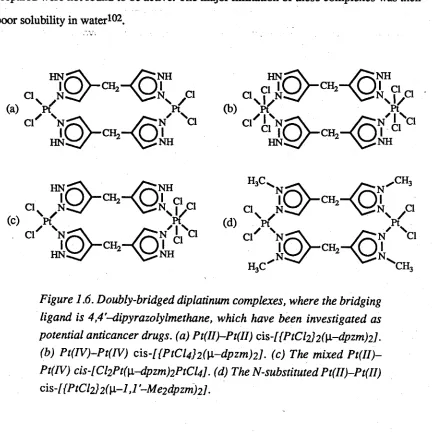
Recent work has produced a series o f doubly-bridged diplatinum complexes,
where the bridging ligand was 4,4'-dipyrazolylm ethane (dpzm )100 (Figure 1.6). It has
been found that the Pt(II>—Pt(II) analogue possesses significant in vivo activity against the P388 leukemia cell line, when the complex was administered in M e2S 0101. Mass-spectral
evidence suggests that the m ajor species was a M e2SO adduct. The other complexes
prepared were not found to be active. The major limitation o f these complexes was their
[image:28.553.78.510.329.761.2]poor solubility in water102.
Figure 1.6. Doubly-bridged diplatinum complexes, where the bridging ligand is 4,4,-dipyrazolylmethane, which have been investigated as potential anticancer drugs, (a) Pt(II)-Pt(II) cis-[{PtCI2]2([i-dpzm)2l.
Chapter One
18The potential o f these dinuclear-platinum anticancer drugs is quite encouraging.
Dinuclear platinum complexes which are bridged by just one dpzm ligand have not been
examined to date. One of the aims o f this research has been to investigate such complexes
as possible anticancer drugs. It was hoped that complexes o f this type would have better
solubilities, greater degrees o f freedom with respect to their possible binding modes with
DNA, and thus improved anticancer activity.
1.4.6.
Other transition metal complexes as potential
anticancer drugs
Numerous complexes with metals other than platinum, have been investigated as
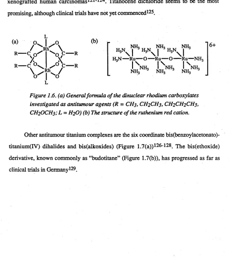
potential anticancer agents. O ctahedral dinuclear rhodium (II) carboxylates103-105
(Figure 1.6(a)) have been found to be potent inhibitors o f tum our growth, with the
butyrate (R = CH2CH2CH3, L = H2O) and the propionate (R = CH2CH3, L = H2O)
being the most effective106. In the series o f rhodium(III) compounds which have been
examined, antitumour activity has been found in species o f the octahedral type m
er-[RI1X3L3] (L = NH3 or another monodentate amine, X = anionic ligand)107.
Numerous ruthenium (II) and (III) com plexes are characterized by antitumour
a c tiv ity 103. The m ain representatives are the ruthenium (III)-am m ine com plexes,
[RuCl3(NH3)3] (which is a mixture o f fa c and mer isomers), c/5-[RuCl2(NH3)4]Cl, [RuC1(N H 3)5]C12, [Ru(0 0 C C H 2CH3)(NH3)5](C104)2 and the salt [Ru(04C2)(en)2] [R u (0 4 C 2 )2 ( e n )]108-1J1. The neutral ruthenium (II) com plexes cis- and
trans-[RuCl2(Me2SO)4] are also active, with the trans isomer displaying the higher activity112- 115. Experimental evidence indicates that the mode o f action o f these complexes is the
inhibition of DNA synthesis and that DNA itself is the target site, as has been found for
the platinum anticancer drugs116. Another ruthenium complex, the so-called “ruthenium
Chapter One 19
against various experimental tumour systems117»118. Its mode o f action is fundamentally
different to that o f other cytostatic ruthenium com plexes. R uthenium red inhibits
specifically the cell m em brane Ca2+-A T P a se and im pairs Ca2+ transport at the
mitochondrial and cellular m embrane119»120. It is also selectively accumulated in tumour
cells103»117.
As m entioned previously m etallo cen e com plexes (F igure 1.7(a)) show
antiproliferative activity against various experimental tumours, including Ehrlich ascites
tumour, sarcoma 180, B16 melanoma, colon 38 carcinoma, Lewis Lung carcinoma and
xenografted hum an carcinom as121-124. Titanocene dichloride seems to be the most
promising, although clinical trials have not yet commenced126.
(a)
R-/ ° '
cy?
■ C ° '
V
;V
'OV
■R
■R
H,N NH, NIL NH,
\ l H s N \ l H 3 \ l
H,N — Ru— O — Ru— O ---Ru— NIL
\
NH,NH,
\
NH,NH,
\
[image:30.553.59.522.267.795.2]NH,NH,
Figure 1.6. (a) General formula o f the dinuclear rhodium carboxylates investigated as antitumour agents (R = CH3, CH2CH3, CH2CH2CH3, CH2OCH3; L = H2O) (b) The structure of the ruthenium red cation.
Other antitumour titanium complexes are the six coordinate bis(benzoylacetonato)-
titanium (IV ) dihalides and bis(alkoxides) (Figure 1.7(a))126-128. The bis(ethoxide)
derivative, known commonly as “budotitane” (Figure 1.7(b)), has progressed as far as
Chapter One 20
Figure 1.7. (a) General formula for metallocene complexes which have displayed anticancer properties. (M = Ti, V, Nb, Mo; e.g. X = Cl) (b)
General formula o f bis(benzoylacetonato)titanium complexes which have been advanced as cancer treatments.
Novel anticancer agents are described on almost a daily basis but few progress
further than initial screening. There is how ever a great deal o f scope left for the
Chapter One
211.5.
THE TREATMENT OF CANCER BY BORON
NEUTRON CAPTURE THERAPY
1.5.1.
Introduction
Boron neutron capture therapy (BNCT) is a type of cancer treatment that combines
elements of both radiotherapy and chemotherapy for the destruction of tum ours130-133.
The basic strategy of this therapy is to introduce selectively a chemical species rich in 10B
into tumour cells that are then irradiated with a stream o f thermal energy neutrons. The
nuclear reaction that takes place when the 10B nucleus captures a thermal energy neutron
yields prim ary fission fragm ents that have a short range, high linear energy transfer
(Scheme 1.1), and will destroy biological function o f any m acromolecule within then-
flight distance of about 10 p.m. Because o f the relatively inert nature o f neutron radiation
to biological material, healthy cells that have not been dosed with 10B are unaffected,
unlike standard methods o f radiation treatment that invariably cause damage to healthy
neighbouring tissue.
7Li + y + 0.48 MeV
Scheme 1 .1 .10B neutron capture processes1^ .
The advantages o f using 10B, rather than some other nuclide, are that it is non
radioactive, it comprises about 20% o f naturally occurring boron, the pathlength of the
fission products is such that they are confined to a radius approximately the same as that
of a single cell, and the chemistry o f boron suggests that it may be incorporated into a
multitude of different chemical structures133.
For BNCT to be effective it has been calculated that if just 17 boron-neutron 4He + 7Li + 2.79 MeV (6%)
10B + *n
Chapter One
22capture reactions occur within a single tumour cell, the cell will be destroyed. In order to
attain this goal, a minimum of 2.5 x 10¹² neutrons per gram of tumour have to be
delivered when the average concentration of 10B is 25 jig per gram of tumour132. This
figure would be of the order of two to five times lower if the 10B were to be localized in
the nucleus of the cell, where the most harm could be inflicted134.
1.5.2 .
Early clinical trials of BNCT
The basic strategy for BNCT was probably conceived in 1936, just four years after
the discovery of the neutron131. The first clinical trials of this treatment were conducted
from 1951 to 1961 at the Brookhaven National Laboratory and at the Massachusetts
Institute of Technology. However, these early studies were less than encouraging, as
they failed to achieve any regression in the tumours treated132. This was due mainly to
the poor uptake by tumour cells of the 10B compounds used in the study. This led to
severe radiation induced side-effects on the central nervous and vascular systems that
were due to high blood concentrations of boron compounds1311.
Subsequent work by Hatanaka in the late 1960's established BNCT as a viable
means of cancer control. His work involved the treatment of brain tumours and in
particular glioblastoma multiforme (gliomas of Grades III-IV). This disease accounts for
45% of all brain cancers and prior to the work of Hatanaka it was invariably fatal with
one- and five-year survival rates of 4.6 and 0 % respectively. Using BNCT, Hatanaka
has dramatically increased these survival rates to 58 and 29 %, as has been determined
from a group of 107 patients treated between 1968 and 1990. Recent results, where the
techniques involved have become more refined, suggest that the survival rate after five
years could be as high as 60%135.
1.5.3 .
The use of the borocaptate anion in BNCT
The boron-10 carrier used by Hatanaka was the mercaptoundecahydro-c/oj'o-
Chapter One
23This com pound was first described in the early 1960’s, in w o rk 136 detailing the
derivative chemistry of the anions dodecahydro-c/oso-dodecaborate [B i2H i2]2~ and
decahydro-c/oso-decaborate [B ioH io]2 -. Together with the anion [BioCl8(SH)2]2_,
borocaptate was shown to be capable of achieving a significant concentration differential
between gliomas and normal brain-blood in rodents. These results were the stimulus for
the work conducted subsequently by Hatanaka. The actual biochemical and physiological
processes by which these compounds localize in neoplasm s rem ains unclear. It is
apparent that the thiol group has a significant role in the observed activity, as there are
m ajor differences between the physiological effects o f B ^ H n 2', which is essentially
inert having a LD50 of 7 g/kg131, and its mercapto derivative.
Studies show that borocaptate will form disulfide linkages with various plasma
p ro te in s 137, but it is unclear w hether this is the basis for its accum ulation within
m alignant cells. It has been suggested that such boron containing proteins become
endocytosed, possibly in an manner analogous to the incorporation of antibodies into
m alignant cells. Recent work has dem onstrated a significantly greater accretion and
dim inished degradation of these proteins by cancer cells, when com pared with the
corresponding normal cells138, although further experiments confirming this hypothesis
have yet to be conducted.
It was discovered139 that the preparations of cesium borocaptate used in the initial
stu d ie s w ere s ig n ific a n tly c o n ta m in a te d w ith the o x id a tio n p ro d u c ts
[B12H11SSB12H11]4- and [Bi2HhS S ( 0 ) Bi2Hi i]4 -. These compounds have been shown to achieve higher concentrations in tum our cells than does borocaptate.
Unfortunately, these agents display an increased toxicity and so do not have any clinical
v a lu e 140. H ow ever, know ledge o f the m echanism s by which sulfur-containing
polyhedral boranes achieve selective accumulation in tumour cells would be of interest for
Chapter One 24
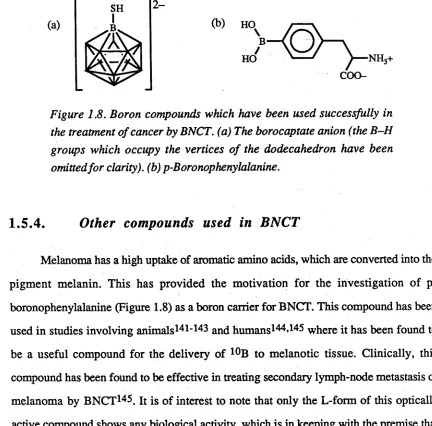
Figure 1.8. Boron compounds which have been used successfully in the treatment of cancer by BNCT. (a) The borocaptate anion (the B-H groups which occupy the vertices o f the dodecahedron have been omitted for clarity), (b) p-Boronophenylalanine.
1.5.4.
Other compounds used in BNCT
Melanoma has a high uptake of aromatic amino acids, which are converted into the
pigment melanin. This has provided the motivation for the investigation of p-
boronophenylalanine (Figure 1.8) as a boron carrier for BNCT. This compound has been
used in studies involving animals14 1-143 and humans144»14^ where it has been found to
be a useful compound for the delivery of 10B to melanotic tissue. Clinically, this
compound has been found to be effective in treating secondary lymph-node metastasis of
melanoma by BNCT146. It is of interest to note that only the L-form of this optically
active compound shows any biological activity, which is in keeping with the premise that
it is used as a biosynthetic precursor. The investigation of other boron analogues of
amino acids146 has been conducted based on the observation that cancer cells, which
grow more vigorously, require greater amounts of these biochemical building blocks that
will be thus selectively incorporated into such cells.
Another approach for the delivery of boron compounds to melanomas is based on
the fact that thiouracil is uniquely and selectively incorporated into such cancers during
melanin formation. This has led to the investigation of a boronated thiouracil147»148
(Figure 1.9). Similarly, chlorpromazine has exhibited a high degree of localization in
tissue that contains preformed melanin and efforts have been directed toward the
production of boronated promazine derivatives149; such as that shown in Figure 1.9.
[image:35.553.64.496.64.490.2]Chapter One
25variety o f tum our types. In vivo studies involving a boronated porphyrin derivative, M n(III)BOPP (Figure 1.10), have shown that it can produce boron concentrations o f 65
[ig / g of tumour in murine gliomas, with no toxic effects. In addition, the compound may
be used as a contrast agent for m agnetic resonance im aging, enabling the direct
monitoring of the tumour. Little, if any, of the agent is seen in normal brain tissue, so it
would appear to be an ideal candidate for further study150.
Figure 1.9. Agents advanced fo r use in BNCT. (a) 5-Dihydroxyboryl-2-thiouracil. (b) A boronated promazine derivative.
In an attempt to produce a boron compound that can be incorporated directly into
the DNA o f rapidly dividing cells, the boronated nucleoside 5-dihydroxyboryl-2'-
deoxyuridine has been prepared151 (Figure 1.10). In vitro studies done on cells grown in the presence of this compound suggest that it replaces thymidine to the extent of 5-13%,
with a boron concentration reaching about 6 |ig B / g cell, which is approaching the level
required for a useful therapeutic action133. A number of other nucleosides and nucleotides
have been also investigated134»15^ '155.
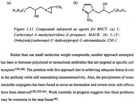
In vitro studies done on a carboranylaziridine derivative, M ACB(Figure 1.11), have shown relatively high growth inhibition toward B16 melanoma and Hep G 2 liver
cancer cells, when compared to results obtained from TIG -1-20 normal human foetal lung
cells. This indicates that MACB possesses a selective toxicity toward certain cancer cells
in its own right. Such selective toxicity has not been observed in any of the boron carriers
Chapter One 26
that this agent may serve as a anticancer drug, independent of any other form of
treatment156.
Figure 1.10. Agents advanced fo r use in BNCT. (a) Boronated porphyrin-manganese(III) complex MnBOPP. (b) 5-Dihydroxyboryl-2'-deoxy uridine.
Boronated derivatives of 2-nitroimidazole have been prepared for use as hypoxia
selective agents157»158. One of these derivatives
l-(3'-ortho[nido]c2irboTa.ny\-2'-hydroxy)propyl-2-nitroimidazole, CNI-1 (Figure 1.11), has been found to produce in vitro toxicity toward KHTn rodent tumour cells only under anaerobic conditions157. The magnitude of this toxicity is similar to that of misonidazole, which is used as a hypoxia-
selective radiosensitizing drug. As a number of tumours are characterized by poor
vasculature, and hence an oxygen deficiency, it would appear that compounds of this sort
Chapter One 27
Figure 1.11. Compounds advanced as agents fo r BNCT. (a) 1 -Carboranyl-3-methylaziridino-2-propanol, MACB. (b) l-(3 '~ Ortho[nidolcarboranyl-2 '-hydroxy)propyl-2-nitroimidazole, CNI-1.
Rather than use small molecular weight compounds, another approach attempted
has been to boronate polyclonal or monoclonal antibodies that are targeted at specific cell
receptors159»160. The problem with this approach lies in achieving adequate boron levels
in the antibody while still maintaining immunoreactivity. Also, the precipitation of water
insoluble conjugates has been found to occur on boronation and certain toxic side-effects
have been observed130’159*161. Work currently in progress suggests that these problems
may be overcome in the near future161.
The use of liposom es and lipoproteins has been investigated as a means of
delivering boron to cancer cells. Low density lipoprotein (LDL) is the major cholesterol
transport protein within the human body and it is recognised by high affinity receptors on
the surface membranes of cells162. By modifying LDL to carry small molecular weight
lipophilic boron molecules, it seems possible to achieve an uptake of boron that is about
100 times that o f the borocaptate an io n 163»164. The levels o f 10B, which can be
incorporated into cancer cells, necessary for BNCT can be easily achieved with this
approach.
1.
5.
5.
Other nuclei for use in neutron capture therapy
Nuclei other than 10B have been also considered for neutron capture therapy. In
order to be considered, the nuclei must have a high cross section for capture of thermal
neutrons (Table 1.2). Originally all potential nuclei, other than 10B, were discounted, as
they were either radioactive or did not yield particles that have a high linear energy
[image:38.553.73.513.62.394.2]Chapter One 28
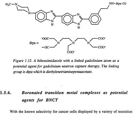
50 times greater than 10B, has produced some very promising results. The process involved with neutron capture by 157Gd is associated with electron internal conversion that produces Auger electron cascade that as a consequence can cause lethal damage to tumour cells. Early experiments involved the use o f GdCl3 in a low ionic strength buffer that allowed ionic association o f Gd3+ with the phosphate groups of DNA. Subsequent neutron bombardment initiated a capture reaction that led to double strand cleavage of the DNA. A more effective agent for the delivery o f 157Gd to DNA has been recently developed. This compound was produced by linking a bibenzimidazole, which is known to intercalate into DNA, to the chelating agent diethylenetriaminepentaacetic acid (dtpa), which acts as a carrier for gadolinium (Figure 1.12). Preliminary results show that this compound does indeed increase the rate o f DNA strand breakage on irradiation with neutrons. The investigation of compounds of this type is still at a very early stage165.
Table 1 2 . Thermal neutron capture cross section values o f potential nuclides fo r neutron capture therapy.
Nuclide Cross Section (a )
(bams) Nuclide
Cross Section (a) (bams)
3He 5500 155Gd 58000
6I i 953 157Gd 240000
10ß 3837 174Hf 400
113Cd 20000 199Hg 2000
135Xe* 2720000 2 3 5 |j* 678
149S m 41500 241pu * 1375
151Eu 5900 242Am* 8000
[image:39.553.22.516.400.793.2]Chapter One 29 NH-dtpa-Gd
dtpa =
coo-Figure 1.12. A bibenzimidazole with a linked gadolinium atom as a potential agent fo r gadolinium neutron capture therapy. The linking group is dtpa which is diethylenetriaminepentaacetate.
1.5.6.
Boronated transition metal complexes as potential
agents for BNCT
W ith the known selectivity for cancer cells displayed by a variety of transition
metal complexes, it is somewhat surprising that no investigation into the use of such a
com pound as a tum our selective boron delivery system for use in BNCT, has been
forthcoming. The complexes that could be envisaged would feature a non-labile ligand
that had a high boron content and possibly a num ber of labile ligands that could be
substituted for any of the possible sites on DNA. The borocaptate anion appears to be an
ideal candidate for use as boron rich ligand. The thiol group is well known in inorganic
chemistry as a good donor function, and coordination should be analogous to that of an
aromatic thiol. One of the aims of the present work was to investigate the possible use of
the borocaptate anion in a transition metal complex that may have an affinity for the DNA
of cancer cells.
1. 6.
SUMMARY
The major aspects of the chemotherapy of cancer using coordination complexes
[image:40.553.77.506.60.431.2]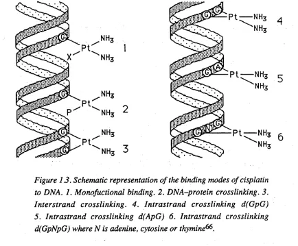


![Table 3.1. Infrared bands of the complex [Ru(SBj2H n)(^H ^)^]-2H2 0 , with](https://thumb-us.123doks.com/thumbv2/123dok_us/7922061.191885/62.553.56.504.113.377/table-infrared-bands-complex-ru-sbj-h-h.webp)
