Binding Domain with Site I Reveals a Correlation between GAGGC
Spacing and Spiral Assembly
Gretchen Meinke, Paul J. Phelan, Celia J. Harrison, Peter A. Bullock
Department of Biochemistry, Tufts University School of Medicine, Boston, Massachusetts, USA
Polyomavirus origins of replication contain multiple occurrences of G(A/G)GGC, the high-affinity binding element for the viral
initiator T-antigen (T-ag). The site I regulatory region of simian virus 40, involved in the repression of transcription and the
en-hancement of DNA replication initiation, contains two GAGGC sequences arranged head to tail and separated by a 7-bp AT-rich
sequence. We have solved a 3.2-Å costructure of the SV40 origin-binding domain (OBD) bound to site I. We have also
estab-lished that T-ag assembly on site I is limited to the formation of a single hexamer. These observations have enabled an analysis of
the role(s) of the OBDs bound to the site I pentanucleotides in hexamer formation. Of interest, they reveal a correlation between
the OBDs bound to site I and a pair of OBD subunits in the previously described hexameric spiral structure. Based on these
find-ings, we propose that spiral assembly is promoted by pentanucleotide pairs arranged in a head-to-tail manner. Finally, the
possi-bility that spiral assembly by OBD subunits accounts for the heterogeneous distribution of pentanucleotides found in the origins
of replication of polyomaviruses is discussed.
P
olyomaviruses are small DNA tumor viruses implicated in
hu-man diseases, particularly among the immunocompromised
and the elderly (
1
). For example, the polyomavirus JC causes
pro-gressive multifocal leukoencephalopathy (PML) (
2
), BK virus is
correlated with renal dysfunction in kidney transplant patients
(
2
), and Merkel cell polyomavirus (MCV) is implicated in a rare
but aggressive form of skin cancer (
3
,
4
). The recent identification
of several new human polyomaviruses (e.g., KI and WU
polyoma-viruses [
5
]) have provided additional incentives to establish
fun-damental aspects of the polyomavirus life cycle.
The best-studied member of the polyomavirus family is simian
virus 40 (SV40). Advances made with SV40 include the
identifi-cation of several of the tumor suppressor proteins targeted during
viral transformation (reviewed in reference
6
). It has also been
used to identify many of the proteins required for eukaryotic DNA
replication (reviewed in references
7
to
9
). In related studies, it was
used to establish basic mechanisms involved in the replication
process (reviewed in references
10
to
12
). A current focus of
re-search in this field is providing a molecular understanding of
ini-tiation events (reviewed in reference
13
). In the long term, it is
anticipated that progress made in terms of understanding SV40
replication will lead to therapies for treating polyomavirus
infec-tions.
Among the highly conserved features of the circular,
double-stranded DNA genomes of polyomavirus family members are the
origins of DNA replication. The minimal or “core” origin of SV40
is a 64-bp region that is necessary and sufficient for DNA
replica-tion (reference
14
and references therein) (
Fig. 1A
). The central
region of the core origin (termed site II) contains a palindromic
arrangement of four GAGGC pentanucleotides (termed P1 to P4).
The pentanucleotides are the high-affinity binding sites for T
an-tigen (T-ag), the virally encoded initiator protein (
15
–
17
).
Flank-ing the central site II region of the core origin are the AT-rich and
early palindrome (EP) regions.
The T-antigens encoded by polyomaviruses are
multifunc-tional, modular proteins that contain an N-terminal DNA J
do-main, a central origin binding domain (OBD), and a C-terminal
helicase domain (reviewed in references
10
to
12
). The OBDs are
necessary and sufficient for site-specific binding to the
pen-tanucleotides. Once site-specific binding is completed, the
heli-case domains are positioned over the flanking regions (
18
,
19
).
The EP region of the origin is melted by a motif in the helicase
domain termed the beta-hairpin (
20
–
23
). We have proposed that
the beta-hairpin-dependent melting of the flanking sequences is
coupled to the assembly of hexameric rings of T-ag around one of
the newly generated single strands of DNA (
21
,
24
). Once the
oligomerization process is completed, two hexamers of T-ag are
formed on the core origin (reviewed in references
13
and
25
).
Recent crystallographic studies of the individual domains of
T-ag have greatly advanced our understanding of its dynamic
in-teractions with the origin of replication. For example, the
costruc-tures of the OBD bound to site II established the basis for
site-specific binding to the pentanucleotides (
24
,
26
,
74
). In brief,
residues in the OBD A1 and B2 motifs (
27
,
28
) make numerous
contacts with both the sugar-phosphate backbone and the major
groove of individual pentanucleotides (reviewed in reference
13
).
Related structural studies revealed how the OBD interacts with
single-stranded DNA (ssDNA) (
29
,
30
) and with duplex DNA in a
non-sequence-specific manner (
26
). Moreover, crystal structures
of the SV40 helicase domain were solved in the presence and
absence of nucleotide (
20
,
31
). The structures of the helicase
domain, and related structures of the bovine E1 helicase
do-main (
32
), established fundamental principles regarding the
Received17 September 2012Accepted19 December 2012
Published ahead of print26 December 2012
Address correspondence to Peter A. Bullock, [email protected]. Copyright © 2013, American Society for Microbiology. All Rights Reserved.
doi:10.1128/JVI.02549-12
on November 7, 2019 by guest
http://jvi.asm.org/
mechanisms employed by eukaryotic helicases (reviewed in
reference
33
).
The crystal structure of the SV40 T-ag OBD has also been
solved in the absence of DNA. In one frequently detected crystal
form, the OBDs adopt a left-handed helical filament having six
molecules per turn (
34
,
35
; reviewed in reference
13
). This
struc-ture was termed the “spiral” or “lock washer” form of the T-ag
OBD. It was postulated that one turn of the spiral (containing six
OBDs) occurs in the context of full-length T-ag hexamers. The
OBDs in the spiral structure cannot interact with double-stranded
DNA (dsDNA) in a sequence-specific fashion due to
conforma-tional changes in the A1 loops, as well as steric collisions predicted
to occur between the DNA and neighboring T-ag OBDs (reviewed
in reference
13
). These and related observations led to the
pro-posal that the lock washer conformation is adopted by the OBDs
subsequent to site-specific binding of the GAGGCs (
24
). An
ad-ditional interesting feature of the OBD spiral is a “gap” through
which ssDNA may transit (
34
,
36
).
While the recent structures have provided many snap shots of
the initiation process, it is not understood how T-ag’s interactions
with the origin translate into the assembly of hexamers and
dou-ble-hexamers. For example, origin subfragments containing
sin-gle pentanucleotides support hexamer formation (
37
–
39
) and
certain pairs of pentanucleotides support double-hexamer
assem-bly (
38
–
40
). Therefore, it is not clear why the central region of the
core origin has four pentanucleotides. Moreover, it is not obvious
why the pentanucleotides in polyomavirus origins are separated
by spacers of different lengths (
41
).
To continue our investigation of T-ag’s interactions with the
SV40 regulatory region, we have determined the costructure of the
OBD bound to a regulatory region termed site I (
Fig. 1A
). Site I is
adjacent to the EP region and contains two GAGGC
pentanucle-otides (herein termed P5 and P6) arranged in a head-to-tail
fash-ion and separated by a 7-bp spacer [containing 6 deoxythymidines
and 1 deoxyguanosine and termed the poly(dT)·poly(dA) spacer].
The primary physiological function of site I is the repression of
early gene transcription, although it has also been reported to
facilitate viral replication (
42
–
47
). The function(s) of site I are
completely dependent upon its exact sequence (see, e.g.,
refer-ences
48
and
49
) as well as its orientation and spacing relative to
site II (
48
).
Analyses of the costructure formed between the OBDs and site
I indicated that a single hexamer of T-ag forms on this site, a
possibility confirmed by additional EMSAs (see below).
There-fore, the OBDs initially bound to P5 and P6 must contribute to the
formation of the site I hexamer. Regarding their roles in hexamer
assembly, we have observed a surprising spatial relationship
be-tween the OBDs bound to P5 and P6 and a pair of OBDs in the
left-handed spiral structure. Moreover, structure-based modeling
studies demonstrate that a similar correlation can be made
be-tween an additional pair of OBDs within the spiral and the OBDs
bound to a pentanucleotide pair in site II that is separated by a
much shorter spacer length (
34
). In view of these findings, we
propose a unifying hypothesis regarding pentanucleotide pairs
that are arranged in a head-to-tail manner in polyomavirus
ori-gins of DNA replication; namely, they promote the formation of
OBD spirals.
MATERIALS AND METHODS
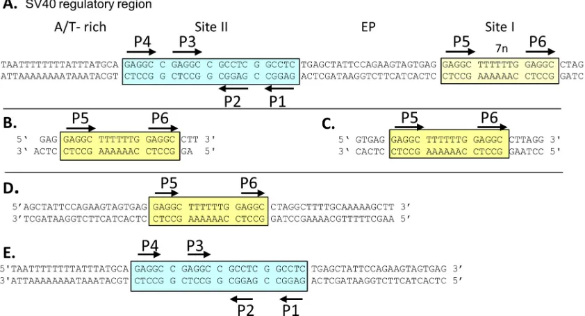
Molecular biology techniques. (i) Purification of SV40 T-ag and the T-ag OBD.Protocols for the purification of the T-ag OBD, amino acids (aa) 131 to 260, were previously reported (24,50); they were used as described previously with the exception that the Sephacryl S-100 column was replaced with a smaller Superdex 75 column. Selenomethionine (Se-FIG 1(A) DNA sequence of the core origin of replication and the site I regulatory region. Site II is indicated by a cyan box, and site I is indicated by a yellow box. Black arrows indicate both the positions and the orientations of the GAGGC pentameric sequences. The locations of the AT-rich and EP regions are also indicated. (B) Site I-containing oligonucleotide used to determine the OBD-site I costructure. Note that the oligonucleotide has a 1-bp overhang; this is commonly used to promote formation of a pseudocontinuous helix within the crystal. (C) Twenty-eight-base-pair site I-containing oligonucleotide used in the ITC studies. DNA substrates used in the EMSAs included the 59-bp site I-containing oligonucleotide (D) and the 64-bp core origin oligonucleotide (E).
on November 7, 2019 by guest
http://jvi.asm.org/
[image:2.585.82.503.70.298.2]Met)-containing T-ag OBD was also prepared as described previously (34). To summarize the purification protocol, the T-ag OBD is expressed as a glutathioneS-transferase (GST) fusion inEscherichia coliBL21(DE3), purified by affinity chromatography with glutathione Sepharose 4B resin (GE, Inc.), digested with thrombin to remove the GST, and further puri-fied by anion and cation exchange chromatography followed by gel filtra-tion. Upon purification, the T-ag OBD was stored in storage buffer (20 mM Tris, pH 8.0, 50 mM NaCl, 10% glycerol, 1 mM EDTA, 0.1 mM phenylmethylsulfonyl fluoride [PMSF], and 0.1% beta-mercaptoethanol) at⫺80°C until ready for use.
Full-length SV40 T-ag (aa 1 to 708) was generated using a baculovirus expression vector containing the T-ag-encoding A gene (51). It was sub-sequently isolated using immunoaffinity techniques with monoclonal an-tibody PAb 419 as previously described (52,53). Once purified, T-ag was dialyzed into T-ag storage buffer (see above) and stored at⫺80°C until ready for use.
(ii) EMSA of T-ag and DNA.The electromobility shift assay (EMSA) reactions with T-ag and32P-labeled doubled-stranded DNA (reference54
and references therein) were conducted under replication conditions (55) as previously reported (38). The double-stranded DNA substrates used in these assays were prepared as described previously (38). After 20 min of incubation at 37°C, the reaction mixtures were cross-linked via the addi-tion of glutaraldehyde (0.1% final concentraaddi-tion) for 5 min. Once the cross-linking was completed, the samples were applied to a 4-to-12% gradient polyacrylamide gel (19:1 acrylamide/bisacrylamide ratio) and electrophoresed in 0.5⫻Tris-borate-EDTA (TBE) for⬃2 h (⬃600 V and 20 mA). Gels were then dried and subjected to autoradiography.
(iii) 1,10-Phenanthroline– copper footprinting.A single-stranded oligonucleotide (⬃25 pmol), derived from the previously described 47-bp “site I⫹wt 30” oligonucleotide (50), was32P labeled at its 5=terminus using standard methods (56). It was then hybridized to its unlabeled com-plementary strand, and the asymmetrically labeled substrates were puri-fied by electrophoresis (38).
The gel retardation 1,10-phenanthroline– copper reactions were per-formed as described by Kuwabara and Sigman (57); T-ag-specific proto-cols are presented in references38and58. In brief, the products of stan-dard band shift reactions with the 47-bp site I⫹wt 30 oligonucleotide and T-ag (54,59) or the T-ag OBD (50) were separated on 4-to-12% gradient polyacrylamide gels in 0.5⫻TBE for⬃2 h (⬃500 V and 20 mA). Follow-ing electrophoresis, the gel was rinsed in 50 mM Tris-HCl (pH 8.0); cleav-age of the DNA was catalyzed by soaking the gel at room temperature in 1,10-phenanthroline–CuSO4–3-mercaptopropionic acid (final
concen-trations, 0.17 mM, 38M, and 5 mM, respectively) for 20 min. The DNA cleavage reaction was quenched by the addition of 2,9-dimethyl-1,10-phenanthroline (final concentration, 2.2 mM) for⬃2 min. The gel was then rinsed in H2O, wrapped in plastic wrap, and subjected to
autoradiog-raphy for⬃1.5 to 2 h. Polyacrylamide slices containing complexes formed between the 47-bp site I⫹wt 30 oligonucleotide and either T-ag or the T-ag OBD or containing free DNA were excised and eluted overnight at 37°C in⬃0.5 ml of elution buffer (0.3 M sodium acetate [pH 5.2], 0.2% sodium dodecyl sulfate [SDS], 10 mM magnesium acetate, 10g of pro-teinase K per ml [56]). Polyacrylamide fragments were removed by cen-trifugation, and the DNA-containing supernatants were subjected to phe-nol-chloroform-isoamyl alcohol (25:24:1) and chloroform-isoamyl alcohol (24:1) extractions; the DNA fragments were then precipitated with 100% (vol/vol) ethanol. The DNA pellets were washed with 70% (vol/vol) ethanol, dried, and resuspended to⬃2,000 cpm/l in loading dye (80% [vol/vol] formamide, 10 mM NaOH, 1 mM EDTA, 0.1% bro-mophenol blue, and 0.1% xylene cyanol). Aliquots were boiled for⬃3 min and applied to a 14% polyacrylamide-8 M urea gel in 1⫻TBE at
⬃2,000 V and 28 mA. Sequencing markers were obtained after perfor-mance of Maxam-Gilbert (60) G and G⫹A reactions on the asymmetri-cally labeled 47-bp site I⫹wt 30 oligonucleotide.
Biophysical techniques. (i) Crystallography.(a) DNA purification.
Synthetic oligonucleotides were ordered from Integrated DNA
Technol-ogies (IDT). The sequence of the double-stranded site I-containing oligo-nucleotide used in crystallization is shown inFig. 1B. The two strands used to form this oligonucleotide were purified by anion exchange chromatog-raphy (DNAPak; Vydac Inc.). The eluted DNA was lyophilized, desalted on a PD-10 column (GE Inc.), and lyophilized to dryness. The oligonu-cleotides were resuspended in annealing buffer (10 mM Tris, pH 7.5, 50 mM NaCl), and the complementary strands were mixed in a 1:1 molar ratio based on calculated molar extinction coefficients. The DNA was annealed by heating it to 95°C for 3 min, followed by slow cooling to 4°C. The duplex DNA was stored at⫺20°C.
(b) Crystallization.The complexes of site I DNA with either the sel-enomethione-containing or the nonlabeled T-ag OBD were prepared in a 1.2:2 DNA/protein molar ratio. The complex was concentrated via ultra-filtration using a Vivaspin concentrator (10,000 molecular weight cutoff) to a protein concentration of⬃10 mg/ml, aliquoted, quick-frozen in liq-uid nitrogen, and then stored at⫺80°C.
Crystals of the T-ag OBD in complex with the site I-containing oligo-nucleotide were grown by vapor diffusion over a 1-ml reservoir using a Linbro plate at 18°C by mixing 1l of the complex with 1l of the reservoir solution (16.875% [wt/vol] polyethylene glycol 3350 [PEG 3350], 0.18 M CaCl2, and 3% [wt/vol] sucrose). Crystals of the
SeMet-T-ag OBD–site I DNA complex were grown by vapor diffusion over a 1-ml reservoir using a Linbro plate at 18°C by mixing 2l of the complex with 2l of the reservoir solution (0.1 M sodium acetate, pH 4.6, 0.2 M am-monium sulfate, 30% [wt/vol] PEG 4000, and 10 mM CaCl2).
(c) X-ray data collection and structure solution.The crystals were trans-ferred to a cryogenic solution (25.5% [wt/vol] PEG 4000, 15% ethylene glycol, 0.085 M sodium acetate, pH 4.6, 0.17 M ammonium sulfate, and 8.5 mM CaCl2) using a cryoloop (Hampton Research Inc.) and then
fro-zen in liquid nitrogen. X-ray data were collected at 100 K at NSLS Beam-line X29 (Brookhaven National Laboratory, NY). The X-ray data were processed with HKL2000 (61). The costructure was solved by using single anomalous diffraction and molecular replacement (SAD and MR) and T-ag OBD coordinates bound to GAGGC as a search model (RCSB Pro-tein Data Bank [PDB] identification number [ID] 2NTC) and the program PHASER-EP (62). The structure was refined using the program REFMAC5 (63), within the CCP4 suite of programs (64), and the PHENIX suite of pro-grams (65). The resulting maps and models were visualized and improved with the molecular graphics program COOT (66).
(ii) ITC.Isothermal titration calorimetry (ITC) data were collected with a VP-ITC system (MicroCal, Northampton, MA); the data were an-alyzed with Origin software provided by the manufacturer. The site I-con-taining dsDNA used in the ITC experiments is presented inFig. 1C. Prior to our conducting the experiments, the dsDNA oligonucleotides and pro-teins were buffer exchanged into 10 mM sodium phosphate buffer, pH 7.0, and 50 mM NaCl, using PD-10 columns (GE Healthcare). Protein and DNA concentrations were determined spectrophotometrically, using cal-culated extinction coefficients from the ProtParam Web server and the IDT website, respectively.
(iii) Molecular modeling.Unless otherwise indicated, all molecular graphic images were generated using the program PyMOL (67). Superpo-sitions were generated using the program LSQMAN (68). DNA parame-ters were analyzed using either the program CURVES⫹(69) or 3DNA (70). Protein-DNA interactions were analyzed using the program PDBSUM (71). The previously solved structures used in the molecular modeling discussed here are the apo-T-ag OBD and T-ag helicase do-main (RCSB PDB IDs 2FUF and 1SVM, respectively).
RESULTS
Our previous structures of the SV40 OBD revealed that it is highly
versatile in terms of the complexes it forms (reviewed in references
10
and
13
). Given this property and the unique features of site I,
we elected to investigate the structural basis for the interaction of
the OBD with this region.
(i) The T-ag OBD–site I costructure.
(a) Overview of T-ag
on November 7, 2019 by guest
http://jvi.asm.org/
OBD bound to site I.
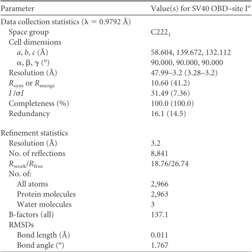
The T-ag OBD–site I crystal structure was
refined to 3.2 Å, with an
R
workof 18.76 and an
R
freeof 26.74. The
refinement statistics are presented in
Table 1
. The complex
crys-tallized in space group C222
1, with two T-ag OBD molecules and
one 23-mer site I-containing oligonucleotide in the asymmetric
unit. The DNA substrate used in this study, shown in
Fig. 1B
,
contains two GAGGC sequences (i.e., P5 and P6) arranged as
head-to-tail direct repeats and separated by the previously
de-scribed 7-bp spacer (molecule A is bound to P5, and molecule B is
bound to P6). The N-terminal residues (i.e., 131 to 133 within
molecule A and 131 to 134 within molecule B) and the C-terminal
residues (i.e., 258 to 260 within molecule A and 257 to 260 within
molecule B) were not visible in the electron density, and therefore
they are not included in the final model. Side chain electron
den-sity was also missing from several lysines (i.e., K136 in molecule A
and K136, K167, K214, and K228 in molecule B). Therefore, in the
final model (
Fig. 2A
), the side chains of these residues were
trun-cated. The bound T-ag OBDs are on the same face of the site I
DNA and are oriented in a head-to-tail fashion. This orientation is
in contrast to that of our previous structure of a T-ag OBD pair on
a DNA derivative of site II (i.e., on P1 and P3), wherein the
GAGGC sequences were oriented in a head-to-head fashion
(RCSB PDB ID 2NTC). As a result, when OBDs are bound to site
I, the N terminus of each OBD is distal to the core origin, while the
C terminus is proximal.
In the site I costructure, superposition of T-ag OBD molecule
A upon molecule B requires a rotation of
⬃
60° and a translation of
approximately 30 Å (data not shown). The superposition of the
two T-ag OBDs revealed that they have nearly identical structures,
as indicated by a low root mean square deviation (RMSD) (0.72 Å
over 115 C
␣
s). Additional notable features of the costructure
in-clude the following.
(b) Protein-protein interactions.
The crystal structure indicates
that the OBDs bound to site I do not interact (
Fig. 2A
). However,
a region of the OBD bound to P5 (i.e., residues 214 to 218) is
juxtaposed next to the C-terminal portion of the OBD bound to
pentamer P6 (i.e., residues 253 to 256). (The closest point of
in-teraction [
⬃
4.2 Å] is between Asp 256 and Lys 214). Of interest,
Thr 217 and Phe 218 are among the residues comprising the B3
motif (
72
,
73
) (aa 213 to 218). The B3 motif is a flexible region of
the T-ag OBD, and we previously proposed that this flexibility
contributes to the plasticity of T-ag as it transits to larger
com-plexes during oligomerization (
34
). Consistent with this proposal,
T-ags containing mutations in the B3 regions are known to be
defective in oligomerization (reviewed in reference
30
). Thus, in
[image:4.585.40.287.86.333.2]FIG 2Costructure of the T-ag OBD bound to site I. (A, top) Ribbon diagrams of the two T-ag OBDs (yellow and cyan) bound to the site I oligonucleotide (shown as a surface representation). Pink arrows indicate the orientations of the GAGGC sequences; P5 and P6 are labeled. The GAGGC sequences are pink, and their complements are light green. The DNA-binding A1 (aa 147 to 159) and B2 (aa 203 to 207) motifs are shown in red. The N and C termini of the OBDs are labeled when visible. The B3 residues (aa 213 to 220) of the OBD bound to P5 are orange and shown as sticks. The C-terminal residues of the OBD bound to P6 are light blue and shown as sticks. (Bottom) Same as in panel A, but the view is rotated 90 degrees. Mol A and Mol B, molecules A and B, respectively. (B) Closeup of the protein-DNA interactions in which residues in the A1 and B2 motifs are selectively depicted. Residues in A1 making site-specific contacts with the GAGGCs include N153, R154, and T155. Residues in B2 making site-specific contacts include H203 and R204.
TABLE 1X-ray data collection and refinement statistics for the T-ag OBD–site I costructure
Parameter Value(s) for SV40 OBD–site Ia
Data collection statistics ( ⫽0.9792 Å)
Space group C2221
Cell dimensions
a,b,c(Å) 58.604, 139.672, 132.112
␣,,␥(°) 90.000, 90.000, 90.000 Resolution (Å) 47.99–3.2 (3.28–3.2)
RsymorRmerge 10.60 (41.2)
I/I 31.49 (7.36)
Completeness (%) 100.0 (100.0) Redundancy 16.1 (14.5)
Refinement statistics
Resolution (Å) 3.2 No. of reflections 8,841
Rwork/Rfree 18.76/26.74 No. of:
All atoms 2,966
Protein molecules 2,963 Water molecules 3 B-factors (all) 137.1 RMSDs
Bond length (Å) 0.011 Bond angle (°) 1.767
aValues in parentheses are for the highest-resolution shell.
on November 7, 2019 by guest
http://jvi.asm.org/
[image:4.585.316.523.238.592.2]the context of full-length T-ag, or under solution conditions, the
OBDs bound to P5 and P6 may interact.
(c) Protein-DNA interactions.
As in the previously described
T-ag OBD–site II costructures (
24
,
26
), the DNA-binding motifs
(A1 and B2) interact deep in the major groove. A closeup view of
the residues in the A1 and B2 motifs interacting with the major
groove is presented in
Fig. 2B
. A schematic of the set of
protein-DNA interactions involved in protein-DNA binding, calculated using the
program PDBSUM (
71
), is shown in
Fig. 3A
. A superposition of
either OBD bound to a GAGGC on site I upon either OBD bound
to a GAGGC in site II (PDB ID 2NTC [
24
]) established that they
are virtually identical (RMSDs
⫽
0.49 to 0.73 over 115 C
␣
s for all
possible combinations of OBD pairs). Thus, as one might predict,
the network of protein-DNA interactions between the OBD and
the GAGGCs are nearly identical to those observed in the previous
T-ag OBD–site II DNA structure (e.g., RCSB PDB ID 2NTC).
However, differences are observed in the contacts, most of which
can be attributed to the lower resolution of this structure.
Never-theless, it is apparent that the A1 motif makes preferential
inter-action with one strand and that the B2 motif interacts with the
opposite strand.
(d) DNA distortions.
The duplex DNA forms a continuous
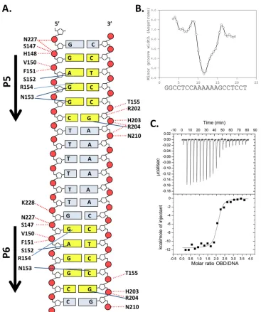
FIG 3Set of interactions between the T-ag OBD and site I and related biophysical data. (A) Schematic of the protein-DNA interactions. Dashed red lines indicate interactions with the phosphate backbone; solid blue lines indicate base-specific interactions. Residues from the A1 motif extend between residues 147 and 159, while those from the B2 motif extend between residues 203 and 207. (B) Analysis of DNA structure within the complex. The minor groove width is shown on theyaxis; the DNA sequence is presented on thexaxis. (C) ITC-based measurement of the thermodynamic parameters for the interaction of the T-ag OBD with site I. The 28-bp duplex DNA used in these experiments, used at a concentration of⬃1.5M, is presented inFig. 1C. Titration of the T-ag OBD into the site I-containing oligonucleotide took place at 25°C in reaction buffer (Materials and Methods). The protein concentration in the syringe was⬃37.5M. The actual calorimetric trace is shown in the top panel. Theyaxis of the isotherm is power incal/s; thexaxis is time in minutes. The stoichiometry and association constants were determined from curve fitting the integrated calorimetric trace presented in the bottom panel.
on November 7, 2019 by guest
http://jvi.asm.org/
[image:5.585.109.476.62.501.2]pseudohelix in the crystal lattice. The conformation of the DNA is
basically B-form. Analysis of each of the GAGGC duplexes,
mea-sured using the program CURVES
⫹
(
69
), revealed consistent
structural distortions (data not shown), such as a low helical twist
and a low helical rise in the center of the sequence (20°
and 2.6 Å,
respectively). These structural distortions were also observed in
previous OBD-GAGGC-containing structures (see, e.g., reference
24
). There is also a small kink on either side of the A-tract.
Fur-thermore, there is an overall curvature of the helical axis of 10.2
degrees; this bend decreases to 4.3 degrees in the A-tract region. A
compression of the minor groove between the T-ag OBDs is also
observed (
Fig. 3B
). Owing to these distortions, the T-ag OBDs are
positioned closer together than they would be on perfectly straight
DNA. A compression of the minor groove was also observed in the
costructure of the OBD bound to pentanucleotides 1 and 3 in site
II (
24
).
(e) Comparison with apo T-ag OBD.
Structures of the T-ag
OBD in the absence of DNA are also available (
34
,
74
).
Compar-ison of the apo form (RCSB PDB ID 2FUF) to the OBD structure
when it is bound to the GAGGCs in site I revealed that the main
conformational shift occurs in the DNA-binding region termed
the A1 loop. In the present structure, the DNA-binding A1 motif is
in the bound state (i.e., extended away from the protein core), a
conformation seen previously in other T-ag OBD–GAGGC
co-structures (
24
; reviewed in reference
13
).
(ii) ITC studies of the interaction of the T-ag OBD with site I.
It was previously reported that full-length T-ag binds site I
ap-proximately 10 times more tightly than site II [e.g., site I
dissoci-ation constant (
K
d)
⫽
0.7 nM; site II
K
d⫽
5 nM (
75
)]. Therefore,
we investigated whether this relationship extends to the isolated
T-ag OBD. To conduct these studies, we used the technique of
isothermal titration calorimetry (ITC), which allows for
determi-nation of both the stoichiometry of binding and the binding
af-finity of the components. The data (
Fig. 3C
) show that two T-ag
OBDs bind to the 2 pentamers in site I with a
K
dof
⬃
12.3 nM. This
K
dis
⬃
8-fold tighter than that determined from the recently
re-ported ITC data of the T-ag OBD bound to a
4-GAGGC-contain-ing site II DNA target (
K
d⫽
93.5 nM) (
76
). Thus, the tighter
affinity of full-length T-ag for site I than for site II is largely a
function of the OBD-DNA interactions.
(iii) A single hexamer of T-ag assembles on site I.
(a) EMSAs
establish that T-ag forms single hexamers on site I.
To continue the
characterization of T-ag assembly on site I, EMSAs were
con-ducted using full-length T-ag and adenylyl imidodiphosphate
(AMP-PNP) (
Fig. 4A
). The site I-containing 59-bp
oligonucleo-tide used in these experiments is shown in
Fig. 1D
. As a positive
control, experiments were conducted with an oligonucleotide
containing the 64-bp core origin (
Fig. 1E
). T-ag forms both single
and double hexamers on this substrate (
Fig. 4A
, lanes 5 to 6). In
contrast, T-ag oligomerization on the 59-bp site I
DNA-contain-ing substrate is limited to hexamer formation (lanes 2 to 3). (There
is a trace amount of a lower-molecular-weight species [lanes 2 and
3] that is presumed to be an assembly intermediate). These studies
establish that T-ag assembly on this site I-containing
oligonucle-otide is limited to formation of a single hexamer.
EMSAs were also previously conducted with the shorter 47-bp
site I
⫹
wt 30 oligonucleotide and both T-ag and the OBD (
50
).
The products formed in these EMSA reactions were footprinted
using the 1,10-phenanthroline– copper footprinting technique
(
57
). These data were not previously published; therefore, a
rep-resentative footprint is shown in
Fig. 4B
. The product formed in
the reaction conducted with full-length T-ag and the site I
⫹
wt 30
oligonucleotide is presented in lane 3. The protected region starts
at the 5
=
end of P5, extends through the poly(dT)·poly(dA) spacer,
and terminates at the 3
=
end of P6. The footprint that resulted
when the experiment was repeated with the purified T-ag OBD is
presented in lane 2. It is apparent that the same
⬃
18-nucleotide
FIG 4Full-length T-ag forms single hexamers on site I-containing DNA. (A) EMSA of full-length SV40 T-ag bound to a 59-bp oligonucleotide containing site I (lanes 2 and 3) or the 64-bp core origin oligonucleotide (lanes 5 and 6). The presence (⫹) or absence (⫺) of T-ag is indicated at the bottom. Lanes 2 and 5, 1.5 pmol T-ag; lanes 3 and 6, 3 pmol T-ag. The reaction products include single hexamers (SH) and double hexamers (DH). The DNA that did not enter the gel is labeled “well,” and the unbound DNA substrates are labeled “input.” (B)In situfootprinting of full-length T-ag and the OBD, when complexed to site I. The footprints were obtained using the gel retardation 1,10-phenanthro-line– copper ion footprinting technique (57). The initial EMSAs were con-ducted with the previously described 47-bp site I⫹wt 30 oligonucleotide (50). Free DNA (i.e., DNA obtained from reactions conducted in the absence of protein and used as a control) is presented in lane 1. The locations of sequence features, including P5 and P6, are indicated. (C) Structure-based modeling of hexamers of the T-ag helicase and OBD domains on site I. The OBDs initially bound to P5 and P6 are shown as yellow and cyan, respectively. The OBDs are represented by single spheres, which are centered at the geometric center of mass, and the radius is approximately the radius of gyration of the domain (i.e.,⬃17.5 Å). The helicase domains are represented by 2 spheres; those helicase domains connected to the initially bound OBDs are also shown as yellow and cyan. Dotted black lines represent the flexible linkers connecting the N termi-nus of the helicase domains to the C termitermi-nus of the OBDs. Finally, an ideal-ized three-dimensional model of site I DNA, positioned along the 6-fold screw axis of the OBD spiral, is shown as a ribbon representation. (D) Molecular modeling studies indicating that two independent T-ag hexamers cannot form on P5 and P6. Two models of T-ag hexamers were constructed; one nucleated at P5 and one at P6. In the resulting model, significant collisions occur between the helicase domains (the collisions are shown in green).
on November 7, 2019 by guest
http://jvi.asm.org/
[image:6.585.319.525.63.372.2](nt) footprint is formed by both T-ag and the OBD. Of interest,
the same-sized footprint was generated when T-ag or the OBD was
footprinted on a site II-containing oligonucleotide (
38
,
50
). The
significance of these observations is considered in Discussion.
Fi-nally, it is noted that the T-ag footprint does not extend into the
flanking regions. We previously attributed this pattern to the
rout-ing of ssDNA over the helicase domain, where it would be subject
to cleavage by oxygen radicals (
21
,
24
).
(b) Molecular modeling of hexamer formation.
It is possible to
generate structural models for full-length T-antigen assembled on
site I using the available crystal structures (i.e., the structures of the
T-ag OBD [aa 131 to 260] bound to site I and the crystal structure
of the T-ag helicase domain [aa 266 to 627] [
31
]). A schematic of
a hexamer bound to site I, based on these studies, is shown in
Fig.
4C
. (The N-terminal J domain is omitted from this model since it
is not necessary for replication
in vitro
[
77
,
78
].) The OBD and
helicase domains are connected via a flexible hinge
(approxi-mately aa 250 to 266, shown as a dotted line). Additional
molec-ular modeling studies indicate that extensive molecmolec-ular clashes
would prevent two independent hexamers from forming on P5
and P6 (
Fig. 4D
). Thus, the modeling studies support the
conclu-sion that T-ag binding to site I results in the formation of a single
hexamer. Finally, the C termini of the OBDs bound to site I are
proximal to the EP region (
Fig. 2A
); this indicates that the helicase
domains are positioned over the EP region.
(iv) Crystal-structure-based modeling suggests that during
hexamer formation on site I, the T-ag OBDs form a spiral
struc-ture.
Given that T-ag assembly on site I is limited to the formation
of single hexamers, the OBDs bound to P5 and P6 must participate
in the assembly of this oligomer. Therefore, we considered how
the OBDs bound to these pentanucleotides might participate in
hexamer formation. In particular, we compared the site I
costruc-ture with an additional struccostruc-ture that is adopted by the SV40 T-ag
OBD, the apo OBD lock washer structure (RCSB PDB ID 2FUF).
A depiction of the OBD-site I costructure is presented in
Fig. 5
(top). Initially, we examined the spatial relationship between the
OBDs bound to site I. Superimposition of monomer A at P5 upon
monomer B at P6 requires a linear translation of approximately 31
Å and a rotation of 60
o. The previously reported left-handed OBD
spiral (
34
) is presented at the bottom of
Fig. 5
. Of interest, OBD
subunits
a
and
f
, the OBD pair proximal to the spiral gap, are
separated by 29.6 Å and offset by an angle of 60
o. Thus, the OBDs
bound to P5 and P6 have a spatial relationship that is very similar
to that of subunits
a
and
f
of the left-handed spiral structure.
A simplified rendering of the OBD-site I costructure, which
further highlights its relationship to the spiral, is presented in
Fig.
6A
. In this figure, the DNA-binding A1 and B2 loops are depicted
as red spheres. Inspection of the two structures presented in
Fig.
6A
(top and bottom) reveals that the positions of the A1 and B2
loops are significantly different. Indeed, in order for the OBDs
bound to P5 and P6 to become subunits
a
and
f
of the spiral, the
bound OBDs must rotate as well as translate away from the DNA
(movements modeled in
Fig. 6B
). Nevertheless, it is apparent that
a spatial relationship exists between the OBDs bound to P5 and P6
and spiral subunits
a
and
f
.
(v) Modeling spiral formation on a pentanucleotide pair
hav-ing a 1-bp spacer.
We previously proposed (
24
) that the OBDs
bound to pentanucleotides 1 and 2 in site II, a GAGGC pair
sep-arated by 1 base pair, could also assemble via a spiral. In this
instance, superimposition of the monomer bound to P1 upon the
monomer bound to P2 requires a linear translation of
approxi-mately 19 Å and a rotation of 156
o(reproduced in
Fig. 6C
, top). As
shown in
Fig. 6C
(bottom), spiral subunits
a
and
d
have a very
similar spatial relationship. Therefore, we suggested that the
OBDs bound to P1 and P2 become subunits
a
and
d
of the spiral in
a T-ag hexamer (
24
). Moreover, as with spiral formation on site I
(
Fig. 6A
), the relative positions of the A1 and B2 loops
(symbol-ized by the red spheres) indicate that the initially bound OBDs
must rotate as well as translate away from the pentanucleotides
during spiral formation.
Based on these structure-based modeling studies, it is
con-cluded that the left-handed spiral can accommodate OBD pairs
bound to GAGGC pentanucleotides separated by either 1 or 7 base
pairs. This indicates that by simply utilizing different pairs of OBD
subunits, the left-handed OBD spiral can accommodate all of the
GAGGC pairs, arranged in a head-to-tail manner, present in the
SV40 regulatory region. Thus, the helical properties of the DNA
appear to be correlated to the helical properties of the OBD spiral.
DISCUSSION
The cocrystal structure presented herein has significantly
in-creased our understanding of SV40 T-ag’s interactions with site I.
As predicted by methylation interference studies (see, e.g.,
refer-ences
79
and
80
), the two T-ag OBDs contact the major groove of
the GAGGC sequences. Moreover, the specific interactions
be-tween the OBDs and the pentanucleotides in site I are very similar
to those seen in previous T-ag OBD–GAGGC crystal structures
(
24
,
26
). Limited contacts with the phosphate backbone of the
poly(dT)·poly(dA) spacer region were also observed (
Fig. 3A
, via
K228, N210, and R204). These backbone interactions were,
however, also observed in previous studies of the T-ag OBD–
GAGGC interaction (
24
,
81
). Therefore, these findings are
con-sistent with the proposal that the site I spacer does not function
with the pentanucleotides to form an extended recognition
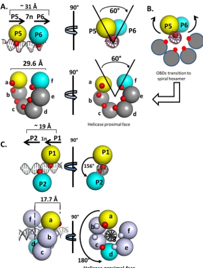
FIG 5Spatial correlations between the T-ag OBDs site-specifically bound to pairs of GAGGC pentanucleotides, with OBD pairs within the left-handed spiral. (Top) Two views (rotated by⬃90°) of the OBDs bound to site I are presented as translucent surface representations; the DNA is shown as a ribbon diagram. Values describing the relationships between the OBDs (i.e., the trans-lation and angular rotation) are indicated. (Bottom) Two views of the left-handed spiral structure of the T-ag OBD rotated by⬃90° (the subunits are labeledatof). Spiral subunitais yellow, while subunitfis cyan; they are offset by the same angle that separates the OBDs bound to P5 and P6 (i.e., spiral subunitsaandfare also 60 degrees apart).on November 7, 2019 by guest
http://jvi.asm.org/
[image:7.585.338.504.64.225.2]motif (
49
) and that T-ag has limited interactions with the bases
in the poly(dT)·poly(dA) spacer (
17
,
79
,
82
).
A curve with a single binding constant fits the ITC data for the
T-ag OBD–site I interaction; therefore, this result is consistent
with those of previous studies indicating that P5 and P6 contribute
to T-ag binding to roughly the same extent (
82
). The affinity of the
T-ag OBD for site I is
⬃
8-fold greater than that for site II.
Regard-ing the molecular basis for this tighter interaction, DNA bendRegard-ing
is known to occur at adenine-thymine tracts (
83
). Indeed, our
costructure demonstrates that there is a slight bend in the site
I-containing DNA (
⬃
10 degrees) and that the minor groove in the
adenine-thymine tract is compressed. It is noted, however, that
A-tract-containing DNA crystal structures (PDB IDs 1BDN,
1D98, and 1D89), solved in the absence of binding proteins,
ex-hibit related features (e.g., little bending of the A-tract [2.4 to 3.0
degrees] and a narrowing of the minor groove in the A-tract
re-gion [3.4 to 3.5 Å]) (reference
84
and references therein). In the
current costructure, the DNA bends 4.3 degrees in the A-tract
region, and the minor groove compresses to a minimum value of
2.4 Å in the center of the A-tract. Although the free A-tract DNA
structures exhibit similar distortions, they are more pronounced
in the OBD-site I costructure. Thus, the intrinsic structural
fea-tures of the A-tract may contribute to the high-affinity binding of
the OBD to site I, and conversely, OBD binding may increase the
structural perturbations.
A model for hexamer-dependent spiral formation.
Based on
our EMSAs, it was concluded that only a single hexamer forms on
site I, a hypothesis that is supported by our structure-based
mod-eling studies. Therefore, since T-ag assembly is limited to hexamer
formation, it was of interest to consider how the OBDs bound to
P5 and P6 contribute to hexamer formation. As shown in
Fig. 5
,
the OBDs docked to P5 and P6 have a spatial relationship similar
to that of subunits
a
and
f
in the left-handed spiral structure. These
and related observations have led to a model that accounts for the
roles of pentanucleotide pairs, arranged in a head-to-tail manner,
in T-ag assembly (
Fig. 7
, with site I as the example).
Hexamer formation is initiated (
Fig. 7A
) when the OBD on a
T-ag monomer interacts with a single pentanucleotide (P5 used in
this example), using the repertoire of bonds reported herein (
Fig.
3A
). One consequence of site-specific binding is that the helicase
domain is positioned over the adjacent EP region (e.g.,
Fig. 4C
).
Regarding the next step, recall that single pentanucleotides are
known to support the formation of full-length T-ag hexamers
(see, e.g., references
37
,
38
, and
40
). Therefore, it is proposed that,
following monomer binding, five additional T-ag molecules are
recruited, a reflection of the high affinity of helicase domains for
each other (
31
). The product formed in this step would contain
the helicase domains assembled into a hexamer and five unbound
OBDs (
Fig. 7B
, left). Based on previous studies, it is predicted that
one strand of DNA goes through the central channel, while the
second traverses the outer surface of the helicase domain (
21
,
24
,
85
). An alternative possibility is, however, that a second OBD
binds to the second pentanucleotide in site I, resulting in the
for-mation of a T-ag dimer (
Fig. 7B
, right). In either case, it is
pro-posed that the next intermediate has the helicase domains
oli-gomerized into a hexamer, with the DNA routed as described
above, and two OBDs bound to pentanucleotides P5 and P6 (
Fig.
7C
). This intermediate would have four unbound OBDs; their
local concentration would, however, be increased. According to
the spiral hypothesis, left-handed spiral formation is nucleated by
subsequent interactions between the four unbound OBDs with
the OBDs associated with P5 and P6 (
Fig. 7D
). The
protein-pro-tein interactions needed for spiral formation may include those in
the previously described OBD-OBD interface (
34
).
Spiral formation is incompatible with OBDs that are
site-spe-cifically bound to DNA (
13
) (
Fig. 6B
). Therefore, it is proposed
that spiral assembly is coupled to the disruption of site-specific
binding to P5 and P6, perhaps as a result of the structural
distor-tions of the flanking sequences stemming from helicase assembly.
Consistent with this possibility, Gutierrez et al. reported that T-ag
binding to site I stimulated DNA unwinding (
86
). Once hexamer
FIG 6Simplified renderings of the OBD interactions with pairs ofpen-tanucleotides. (A, top) Two views of the OBD interactions with site I. The OBDs are represented by spheres centered at the geometric center of mass. The T-ag OBDs are yellow on P5 and cyan on P6. The smaller red spheres represent the DNA-binding A1 and B2 motifs. (Bottom) Rendering of the left-handed spiral in which the OBDs proximal to the gap are represented by spheres colored as described above. This depiction serves to further illustrate that spiral subunitsaandfhave the same spatial relationship as the OBDs bound to P5 and P6. As in the top images, the red spheres indicate the relative locations of the A1 and B2 motifs. (B) Modeling studies indicate that the OBDs initially bound to P5 (yellow) and P6 (cyan) undergo a significant transition during spiral formation. This involves rearrangement of the A1 loop from the bound to the free form, subsequent rotation and translation away from the major groove of the dsDNA, and interaction(s) with other OBDs (reviewed in refer-ence13). (C, top) Two views of the OBD interactions with site II, rotated by
⬃90°, used to illustrate the generality of the relationship (24,26) (RCSB PDB ID 2ITL). P1 and P2 are separated by a 1-bp spacer; therefore, the OBDs bound to P1 (yellow) and P2 (cyan) are⬃156 degrees apart and separated by a trans-lation of⬃19 Å. (As in previous examples, the DNA-binding A1 and B2 motifs are red and magenta, respectively). (Bottom left) Side view of the left-handed OBD spiral hexamer with DNA modeled within the central channel (the 6 subunits are labeled a to f). It is apparent that in this instance, spiral subunitsa
andd(yellow and cyan, respectively) have a spatial relationship that is analo-gous to that of the OBD subunits bound to P1 and P2. (Bottom right) Helicase-proximal view of the left-handed spiral structure with duplex DNA modeled in the central channel.
on November 7, 2019 by guest
http://jvi.asm.org/
[image:8.585.61.264.65.333.2]assembly is completed, the OBDs assemble into the previously
discussed left-handed spiral. The left-handed direction of this
quaternary structure is opposite to that of right-handed B-form
DNA. Therefore, one consequence of left-handed spiral
forma-tion by the OBD may be to promote DNA melting, a process in
which the OBDs are known to play a role (
87
). Characterization of
the left-handed conformation of the MCM2-MCM7 complex also
led to the suggestion that this conformation is adopted to promote
DNA melting (
88
).
In support of the spiral hypothesis, Cuesta et al. (
36
) analyzed
the T-ag fragment from aa 108 to 627 assembled on the SV40
origin via electron microscopy (EM) and concluded that the OBD
forms a spiral. In addition, the footprint formed by either T-ag or
the OBD on the 47-bp site I
⫹
wt 30 oligonucleotide was
deter-mined; these studies revealed that both T-ag and the OBD form a
footprint that extends over
⬃
18 nt (
⬃
61.2 Å) (
Fig. 4B
). The
diameter of an OBD subunit is
⬃
35 Å (see, e.g., reference
24
);
thus, if the OBDs in T-ag form a single closed or planar ring on site
I, the complex would form a footprint of approximately 11 bp
(
⬃
35.2 Å). Therefore, the footprinting studies are consistent with
the hypothesis that upon oligomerization on site I, the OBDs
within T-ag adopt a structure that extends between P5 and P6.
Implications of the spiral assembly hypothesis to OBD
inter-actions with site II.
A single OBD spiral can accommodate the
OBDs bound to pentanucleotides P1 and P2 (or P3 and P4) in site
II. Of further interest, the pentanucleotide containing regulatory
regions of polyomaviruses are highly conserved (see, e.g.,
refer-ence
89
). Therefore, the spiral assembly model may be generally
applicable to the assembly of hexamers on polyomavirus origins of
replication. However, since the spiral formed by the SV40 OBD is
the only one reported to date (
34
,
35
), this hypothesis remains to
be verified.
Furthermore, given that site II contains four pentanucleotides,
an interesting question is, in the context of a double hexamer
assembled on the core origin, are two OBD spirals formed?
Mo-lecular modeling demonstrates that a double spiral of the OBD fits
into the central region of electron density derived from EM images
of T-ag double hexamers (
Fig. 8A
). Indeed, we previously
pro-posed (
34
) the relative orientations of the double spiral formed on
site II based on earlier mutagenesis studies (
73
). Moreover, the
model presented in
Fig. 8B
demonstrates that a double OBD spiral
may explain the
⬃
18-nt footprint generated by double-hexamer
formation on site II (
38
). However, phenanthroline-copper
foot-printing studies of T-ag double hexamers revealed that
pen-tanucleotide 4 is not protected from cleavage by oxygen radicals
(
38
). Thus, in the context of a T-ag double hexamer, all four
pen-tanucleotides may not be simultaneously bound by OBDs.
There-fore, it is uncertain if site II supports the concurrent formation of
two OBD spirals. Consistent with double-spiral plasticity on site
II, EM studies indicate that wild-type T-ag bound to the core
origin is present in two conformations; in one, the central module
is an open ring with a gap, while in the second, the central module
is a compact closed ring (
36
). Furthermore, the formation of both
open and closed OBD rings may be related to previous biochemical
experiments indicating that a feature of the two hexamers formed on
site II is asymmetry. (For instance, one hexamer forms in a manner
that is independent of the phosphorylation of Thr 124, while
forma-tion of the second hexamer is enhanced by Thr 124 phosphorylaforma-tion
(reviewed in reference
37
); additional examples of asymmetric
as-sembly of T-ag hexamers are presented in references
22
and
25
).
Fi-nally, asymmetric assembly events involving the OBD (i.e., formation
of single spirals) may help to explain related observations, such as the
recent finding that only three pentanucleotides are needed for the
assembly of the Merkel cell T-antigen (
76
).
Considerations regarding T-antigen’s dynamic interactions
with site I and site II.
The shifting of T-ag hexamers from site I to
site II is coupled to the repression of transcription of the early
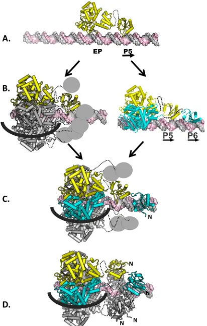
FIG 7Model depicting hexamer-dependent OBD spiral formation on site I.(A) Initially, a monomer of T-ag, shown in yellow, binds to site I (the J-domain is not shown). The flexible linker that connects the two domains is shown as a dashed line. (B) Intermediates in hexamer formation. (Left) Following the binding of the initial monomer, additional T-ag molecules are recruited to site I owing to the high affinity of the helicase domains for each other. Upon recruitment, the helicase domains assemble into a hexamer (18). The five newly recruited monomers are shown in gray; the OBDs are symbolized by the small gray spheres. The black line represents one strand of DNA going over the outer surface of the helicase domain; the second strand is routed through the central channel. (Right) An alternative possibility is that a second monomer of T-ag binds to the second pentanucleotide in site I. (C) Formation of an immature hexamer. In this intermediate, the helicase domains have formed a hexamer over the flanking region, and P5 and P6 are bound by two of the OBDs. The four unbound OBDs are shown as gray spheres. The routing of the DNA is as described above. (D) Maturation of the hexamer to form the spiral structure. It is proposed that the four free OBDs shown in panel C interact with the OBDs bound to P5 and P6. As a result of this interaction, the OBDs adopt the left-handed spiral structure. (A left-handed helix is defined as turning in a counterclockwise fashion as it progresses forward. The N termini of the OBDs in the spiral are indicated.) Once formed, the spiral has a gap though which ssDNA may pass. Finally, it is noted that the isolated T-ag OBDs are monomeric in solution even at high concentrations (74). This is one indi-cation that the protein-protein interactions that occur in the OBD spiral are not strong; therefore, the spiral structure is presumably dynamic in nature.
on November 7, 2019 by guest
http://jvi.asm.org/
[image:9.585.61.267.67.391.2]region and the initiation of DNA replication (see, e.g., references
45
,
46
,
90
, and
91
). Given the importance of these events, an
in-teresting question is what causes the transition from binding to
site I to site II? Previous studies using full-length T-ag established
that one of the key determinants is phosphorylation of T-ag,
par-ticularly at two clusters located at the N and C termini of the
molecule (
92
,
93
). How these phosphorylation events regulate the
interaction(s) between the OBD and the pentanucleotides in site I
and site II has yet to be determined. However, a prediction of the
assembly events described herein is that a hexamer formed on site
I would block hexamer formation on the proximal side of site II.
As shown in
Fig. 9
, collisions would occur between the opposing
helicase domains. This prediction is consistent with the results of
previous studies that demonstrated that T-ag assembly on site I
precedes its assembly on site II (see, e.g., reference
45
).
Initiator proteins and spiral formation.
Helical protein
fila-ments are known to be formed by other replication initiators
(re-viewed in reference
94
). For instance, EM studies provided
evi-dence for a helical filament in the
Drosophila melanogaster
origin
recognition complex (ORC) (
95
). As previously noted, the
left-handed lock washer structure is also a feature of certain MCM
complexes (
88
) and MCM complexes contain gaps through which
DNA may pass (
96
–
98
). Moreover, the prokaryotic initiator DnaA
forms a right-handed superhelix that orients DNA on the outside
of the helical assembly (
99
,
100
). Importantly, the protein filament
formed by DnaA provides a simple explanation for the
heteroge-neous arrangement of DnaA boxes found in bacterial origins
(
100
). Reasons for suggesting that the spiral formed by the T-ag
OBD may also explain the heterogeneous distribution of
pen-tanucleotides in polyomavirus origins of replication were
previ-ously discussed. An important difference between DnaA and T-ag
is, however, that the AAA
⫹
domain is the primary determinant of
filament formation in DnaA, whereas the available evidence
indi-cates that the OBD is the primary unit of spiral formation in T-ag.
Thus, a universal feature of all cellular initiator proteins may be
spiral formation (
100
), but spiral formation may be a property of
different initiator domains. It is also important to note that the
hexameric helicase DnaB adopts a closed spiral staircase
quater-nary structure during translocation (
101
). This observation has
led to the proposal of the hand-over-hand mechanism for DNA
helicase activity. If the spiral structure of the T-ag OBD is shown to
be a feature of the replication fork, it will be of interest to
deter-mine if the OBDs are engaged in a similar hand-over-hand
mech-anism.
Finally, it is noted that other proteins known to form
open-ring spirals have been suggested to recruit protein substrates in a
spiral-dependent manner (e.g., DnaA [
100
] and the ParR protein
involved in DNA segregation [reviewed in reference
102
]).
There-fore, it will be of interest to determine if spiral assembly by the
T-ag OBDs is related to the recruitment of the cellular factors
needed to initiate viral DNA replication.
ACKNOWLEDGMENTS
We thank Howard Robinson at the National Synchrotron Light Source, Brookhaven National Laboratory, for X-ray data collection at Beamline X29. We thank Jacques Archambault and Andrew Bohm for helpful discussions.
This work was supported by a grant from the National Institutes of Health to P.A.B. (9R01GM55397).
REFERENCES
1.Jiang M, Abend JR, Johson SF, Imperiale MJ.2009. The role of polyo-maviruses in human disease. Virology384:266 –273.
2.Eash S, Manley K, Gasparovic M, Querbes W, Atwood WJ.2006. The human polyomaviruses. Cell. Mol. Life Sci.63:865– 876.
3.Feng H, Shuda M, Chang Y, Moore PS.2008. Clonal integration of a polyomavirus in human Merkel cell carcinoma. Science319:1096 –1100. 4.Shuda M, Feng H, Kwun HJ, Rosen ST, Gjoerup O, Moore PS, Chang Y.2008. T antigen mutations are a human tumor-specific signature for Merkel cell polyomavirus. Proc. Natl. Acad. Sci. U. S. A.105:16272– 16277.
5.Babakir-Mina M, Ciccozzi M, Bonifacio D, Bergeallo M, Costa C, Cavallo R, DiBonito L, Perno CF, Ciotti M.2009. Identification of the novel KI and WU polyomaviruses in human tonsils. J. Clin. Virol.46:75–79.
6.Ahuja D, Saenz-Robles MT, Pipas JM. 2005. SV40 large T antigen FIG 9T-ag hexamers cannot exist simultaneously on site I and the proximal side of site II. As reported herein, a T-ag hexamer nucleated through the OBDs bound at P5 and P6 positions the helicase domains over the EP region. Fur-thermore, when T-ag assembles into a double hexamer on site II, the helicase domains span both the AT-rich and EP regions (19). Therefore, if both site I and the EP-proximal side of site II were occupied by T-ag hexamers, the heli-case domains would completely overlap and suffer steric clashes.
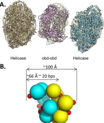
FIG 8OBD oligomerization in the context of T-ag double hexamers formed on the core origin. (A) The PDB coordinates for the helicase domain (1SVM) and the OBD (2FUF) were fit into the EM density map (EMD-1681 [36]). The figure was generated using the program CHIMERA (103); the helicase do-mains were fit using the CHIMERA fit-in-map option. A double-hexamer spiral of the OBDs, shown as a ribbon representation, was generated and man-ually positioned into the central density. (B) Model of a T-ag OBD double spiral. The individual spirals are shown in yellow and cyan. The smaller arrow illustrates that its diameter (⬃66 Å) is similar to the area protected in previous footprinting studies (see, e.g., reference38).
on November 7, 2019 by guest
http://jvi.asm.org/
[image:10.585.348.493.67.175.2] [image:10.585.80.247.68.266.2]targets multiple cellular pathways to elicit cellular transformation. On-cogene24:7729 –7745.
7.Hurwitz J, Dean FB, Kwong AD, Lee S-H.1990. The in vitro replication of DNA containing the SV40 origin. J. Biol. Chem.265:18043–18046. 8.Kelly TJ.1988. SV40 DNA replication. J. Biol. Chem.263:17889 –17892. 9.Waga S, Stillman B.1998. The DNA replication fork in eukaryotic cells.
Annu. Rev. Biochem.67:721–751.
10. Bullock PA.1997. The initiation of simian virus 40 DNA replication in vitro. Crit. Rev. Biochem. Mol. Biol.32:503–568.
11. Fanning E, Knippers R.1992. Structure and function of simian virus 40 large tumor antigen. Annu. Rev. Biochem.61:55– 85.
12. Simmons DT.2000. SV40 large T antigen functions in DNA replication and transformation. Adv. Virus Res.55:75–134.
13. Meinke G, Bullock PA.2012. Structural “snap-shots” of the initiation of SV40 replication, p 195–215.InGaston K (ed), Small DNA tumor vi-ruses. Horizon Scientific Press, Norwich, United Kingdom.
14. Deb S, DeLucia AL, Baur C-P, Koff A, Tegtmeyer P.1986. Domain structure of the simian virus 40 core origin of replication. Mol. Cell. Biol. 6:1663–1670.
15. DeLucia AL, Lewton BA, Tjian R, Tegtmeyer P.1983. Topography of simian virus 40 A protein-DNA complexes: arrangement of pentanucle-otide interaction sites at the origin of replication. J. Virol.46:143–150. 16. Titolo S, Welchner E, White PW, Archambault J.2003.
Characteriza-tion of the DNA-binding properties of the origin-binding domain of SV40 large T antigen by fluorescence anisotropy. J. Virol.77:5512–5518. 17. Tjian R.1978. Protein-DNA interactions at the origin of simian virus 40 DNA replication. Cold Spring Harbor Symp. Quant. Biol.43:655– 662. 18. Mastrangelo IA, Hough PVC, Wall JS, Dodson M, Dean FB, Hurwitz
J.1989. ATP-dependent assembly of double hexamers of SV40 T antigen at the viral origin of DNA replication. Nature338:658 – 662.
19. Valle M, Gruss C, Halmer L, Carazo JM, Donate LE. 2000. Large T-antigen double hexamers imaged at the simian virus 40 origin of rep-lication. Mol. Cell. Biol.20:34 – 41.
20. Gai D, Zhao R, Li D, Finkielstein CV, Chen XS.2004. Mechanisms of conformational change for a replicative hexameric helicase of SV40 large tumor antigen. Cell119:47– 60.
21. Kumar A, Meinke G, Reese DK, Moine S, Phelan PJ, Fradet-Turcotte A, Archambault J, Bohm A, Bullock PA.2007. Model for T-antigen-dependent melting of the simian virus 40 core origin based on studies of the interaction of the beta-hairpin with DNA. J. Virol.81:4808 – 4818. 22. Reese DK, Sreekumar KR, Bullock PA.2004. Interactions required for
binding of simian virus 40 T antigen to the viral origin and molecular modeling of initial assembly events. J. Virol.78:2921–2934.
23. Shen J, Gai D, Patrick A, Greenleaf WB, Chen XS.2005. The roles of the residues on the channel-hairpin and loop structures of simian virus 40 hexameric helicase. Proc. Natl. Acad. Sci. U. S. A.102:11248 –11253. 24. Meinke G, Phelan PJ, Moine S, Bochkareva E, Bochkarev A, Bullock PA, Bohm A.2007. The crystal structure of the SV40 T-antigen origin binding domain in complex with DNA. PLoS Biol.5:e23. doi:10.1371 /journal.pbio.0050023.
25. Borowiec JA, Dean FB, Bullock PA, Hurwitz J. 1990. Binding and unwinding-how T antigen engages the SV40 origin of DNA replication. Cell60:181–184.
26. Bochkareva E, Martynowski D, Seitova A, Bochkarev A.2006. Struc-ture of the origin-binding domain of simian virus 40 large T antigen bound to DNA. EMBO J.25:5961–5969.
27. Simmons DT, Wun-Kim K, Young W.1990. Identification of simian virus 40 T-antigen residues important for specific and nonspecific bind-ing to DNA and for helicase activity. J. Virol.64:4858 – 4865.
28. Wun-Kim K, Upson R, Young W, Melendy T, Stillman B, Simmons DT.1993. The DNA-binding domain of simian virus 40 tumor antigen has multiple functions. J. Virol.67:7608 –7611.
29. Meinke G, Phelan PJ, Fradet-Turcotte A, Bohm A, Archambault J, Bullock PA.2011. Structure-based analysis of the interaction between the simian virus 40 T-antigen origin binding domain and single-stranded DNA. J. Virol.85:818 – 827.
30. Reese DK, Meinke G, Kumar A, Moine S, Chen K, Sudmeier JL, Bachovchin W, Bohm A, Bullock PA.2006. Analyses of the interaction between the origin binding domain from simian virus 40 T-antigen and single stranded DNA provides insights into DNA unwinding and initia-tion of DNA replicainitia-tion. J. Virol.80:12248 –12259.
31. Li D, Zhao R, Lilyestrom W, Gai D, Zhang R, DeCaprio JA, Fanning E, Jochimiak A, Szakonyi G, Chen XS.2003. Structure of the replicative
helicase of the oncoprotein SV40 large tumour antigen. Nature423:512– 518.
32. Enemark EJ, Joshua-Tor L.2006. Mechanism of DNA translocation in a replicative hexameric helicase. Nature442:270 –275.
33. Brewster AS, Chen XS.2010. Insights into the MCM functional mech-anism: lessons learned from the archaeal MCM complex. Crit. Rev. Biochem. Mol. Biol.45:243–256.
34. Meinke G, Bullock PA, Bohm A.2006. The crystal structure of the T-ag origin binding domain. J. Virol.80:4304 – 4312.
35. Meinke G, Phelan P, Fradet-Turcotte A, Archambault J, Bullock PA. 2011. Structure-based design of a disulfide-linked oligomeric form of the simian virus 40 (SV40) large T antigen DNA-binding domain. Acta Crys-tallogr. D Biol. CrysCrys-tallogr.67:560 –567.
36. Cuesta I, Nunez-Ramirez R, Scheres SHW, Gai D, Chen XS, Fanning E, Carazo JM.2010. Conformational rearrangements of SV40 large T antigen during early replication events. J. Mol. Biol.397:1276 –1286. 37. Barbaro BA, Sreekumar KR, Winters DR, Prack AE, Bullock PA.2000.
Phosphorylation of simian virus 40 T-antigen on Thr 124 selectively promotes double-hexamer formation on subfragments of the viral core origin. J. Virol.74:8601– 8613.
38. Joo WS, Kim HY, Purviance JD, Sreekumar KR, Bullock PA.1998. Assembly of T-antigen double hexamers on the simian virus 40 core origin requires only a subset of the available binding sites. Mol. Cell. Biol. 18:2677–2687.
39. Kim HY, Barbaro BA, Joo WS, Prack A, Sreekumar KR, Bullock PA. 1999. Sequence requirements for the assembly of simian virus 40 T-an-tigen and T-anT-an-tigen origin binding domain on the viral core origin of replication. J. Virol.73:7543–7555.
40. Sreekumar KR, Prack AE, Winters DR, Barbaro BA, Bullock PA.2000. The simian virus 40 core origin contains two separate sequence modules that support T-antigen double-hexamer assembly. J. Virol.74:8589 – 8600.
41. Allander T, Andreasson K, Gupta S, Bjerkner A, Bogdanovic G, Persson MAA, Dalianis T, Ramqvist T, Andersson B.2007. Identifi-cation of a third human polyomavirus. J. Virol.81:4130 – 4136. 42. Alwine JC, Reed SI, Stark GR.1977. Characterization of the
autoregu-lation of simian virus 40 gene A. J. Virol.24:22–27.
43. Hansen U, Tenen DG, Livingston DM, Sharp PA.1981. T antigen repression of SV40 early transcription from two promoters. Cell27:603– 612.
44. Khoury G, May E.1977. Regulation of early and late simian virus 40 transcription: overproduction of early viral RNA in the absence of a functional T-antigen. J. Virol.23:167–176.
45. Myers RM, Rio DC, Robbins AK, Tjian R.1981. SV40 gene expression is modulated by the cooperative binding of T antigen to DNA. Cell25: 373–384.
46. Rio D, Robbins A, Myers R, Tjian R.1980. Regulation of simian virus 40 early transcriptionin vitroby a purified tumor antigen. Proc. Natl. Acad. Sci. U. S. A.77:5706 –5710.
47. Tegtmeyer P, Schwartz M, Collins JK, Rundell K.1975. Regulation of tumor antigen synthesis by simian virus 40 gene A. J. Virol.16:168 –178. 48. Guo ZS, Heine U, DePamphilis ML.1991. T-antigen binding to site I facilitates initiation of SV40 DNA replication but does not affect bidirec-tionality. Nucleic Acids Res.19:7081–7088.
49. Ryder K, Silver S, DeLucia AL, Fanning E, Tegtmeyer P. 1986. An altered DNA conformation in origin region 1 is a determinant for the binding of SV40 large T antigen. Cell44:719 –725.
50. Joo WS, Luo X, Denis D, Kim HY, Rainey GJ, Jones C, Sreekumar KR, Bullock PA.1997. Purification of the simian virus 40 (SV40) T-antigen DNA binding domain and characterization of its interactions with the SV40 origin. J. Virol.71:3972–3985.
51. O’Reilly DR, Miller LK.1988. Expression and complex formation of simian virus 40 large T antigen and mouse p53 in insect cells. J. Virol. 62:3109 –3119.
52. Dixon RAF, Nathans D.1985. Purification of simian virus 40 large T antigen by immunoaffinity chromatography. J. Virol.53:1001–1004. 53. Simanis V, Lane DP.1985. An immunoaffinity purification procedure
for SV40 large T antigen. Virology144:88 –100.
54. Dean FB, Dodson M, Echols H, Hurwitz J. 1987. ATP-dependent formation of a specialized nucleoprotein structure by simian virus 40 (SV40) large tumor antigen at the SV40 replication origin. Proc. Natl. Acad. Sci. U. S. A.84:8981– 8985.
55. Wobbe CR, Dean F, Weissbach L, Hurwitz J.1985.In vitroreplication
on November 7, 2019 by guest
http://jvi.asm.org/
of duplex circular DNA containing the simian virus 40 DNA origin site. Proc. Natl. Acad. Sci. U. S. A.82:5710 –5714.
56. Sambrook J, Fritsch EF, Maniatis T.1989. Molecular cloning: a labo-ratory manual, 2nd ed. Cold Spring Harbor Labolabo-ratory, Cold Spring Harbor, NY.
57. Kuwabara MD, Sigman DS.1987. Footprinting DNA-protein com-plexes in situ following gel retardation assays using 1,10-phenanthroline-copper ion: Escherichia coli RNA polymerase-lac promoter complexes. Biochemistry26:7234 –7238.
58. Sreekumar KR, Barbaro BA, Prack A, Bullock PA.2000. Methods for studying interactions between simian virus 40 T-antigen and the viral origin of replication. Methods Mol. Biol.165:49 – 67.
59. Murakami Y, Hurwitz J.1993. DNA polymerase␣stimulates the ATP-dependent binding of simian virus tumor T antigen to the SV40 origin of replication. J. Biol. Chem.268:11018 –11027.
60. Maxam AM, Gilbert W.1980. Sequencing end-labeled DNA with base-specific chemical cleavages. Methods Enzymol.65:499 –560.
61. Otwinowski Z, MW.1997. Processing of X-ray diffraction data collected in oscillation mode. Methods Enzymol.276:307–326.
62. McCoy AJ, Grosse-Kunstleve RW, Adams PD, Winnn MD, Storoni LC, Read RJ.2007. Phaser crystalographic software. J. Appl. Crystallogr. 40:658 – 674.