Abstract: Plant disease interrupts the normal or Ordinary condition of a plant and it alters the essential functionality of a plant. Which intern impacts the productivity of the crop. Speedy observation, recognition and categorization of the plant pathogens will increase the crop yield more than 60% of the total productivity. Disease analysis is more evident on the leaves when compare to the other parts of the plants. Automated methods are most commonly available in different image processing techniques to detect the pathogen attack which can be made more efficient by combining multiple domain, that utilizes computer vision technologies. Most modern techniques or technologies are analyzed to identify the various disease on several crops or crop types. The paper summarizes about types of plants, types of plant diseases and the standard methodologies or technique that would help gaining knowledge about Computer Vision and its applications on plant disease identification and classification. Performance of the Classifiers are analyzed to recognize and classify the better method that typically works among different plant groups and different types of pathogen attack.
Keywords: Classification, Neural Network, Optimization, Accuracy, Efficient
I. INTRODUCTION
Agriculture or Crop Production depends on the quality and quantity of plant growth Worldwide. The pathogen detection on the effected plants or plant leaves engages different image processing techniques to alleviate the challenging task [1]. Multiple factors like Climatic condition, pest attack or diseases will cause the plant to have specific type of infection. Manual process of disease detection method is very long and tedious process, it is impossible for the farmers to identify the disease and factor of impact. So, it is highly recommended to adapt automatic disease analysis system Multiple techniques are applicable in identifying and classifying the effected portion of the plant [2,10]. Multi Crop, Multi disease are proposed in our System [3,22].
Revised Manuscript Received on December 05, 2019.
Kapilya Gangadharan, Research scholar in Saveetha School of Engineering, Saveetha Institute of Medical and Technical Sciences, Chennai. She is currently Utility Analyst at Fidelity National Information Services, Durham, NC, USA
G. Rosilne Nesa Kumari, Professor in the Department of Computer Science and Engineering, in Shadan women’s college of Engineering and Technology, Chennai, India
D. Dhanasekaran, Professor in the Department of Computer Science and Engineering, Saveetha School of Engineering, Saveetha Institute of Medical and Technical Sciences, Chennai, India
Another method is proposed in [4] which is an automatic Citrus Canker detection from leaf images and proved that the accuracy is in the higher-level using Image Processing techniques when compared to that of Human experts on screen. The accuracy is calculated as 87.99% for Imaging Techniques. The Techniques detects the pathogens on the plants or plant leaves automatically at their early stage. In this study we analyzed both simple and complex pathogen attacks on the plant leaves using automatic systems in its primary stage which improves the amount and nature of yield generation and management.
From the analysis we observed that the disease detection under closed environment will face many difficulties due to unevenness of the circumstances, which is undergone by the agricultural field. There is a high necessity to adapt an automatic system or an algorithm to address the factor of change [5]. This paper has multiple methodologies to make the system more feasible, reliable and accurate which helps the research to move on a focused dimension [6]. Our work is structed or arranged as follows. Section 2 briefs about the various types of Crop on research for disease detection, Section 3 illustrates various types of diseases that effects plant leaves, Section 4 describes Architecture of leaves disease detection system using Image processing techniques, Section 5 presents the types of Classification methods on Multiple plant groups, Section 6 Consists of Performance Evaluation of Classifiers, section 7 has the conclusion and future work.
II. TYPESOFCROPONRESEARCH
The major crops which is under research are broadly categorized into four major types based on the current state of research explored on the crops during last 10 years [6] which is mainly dependent on the usage. The four main categories of crops are 1. Food Grain Crops- 43%, 2. Cash Crops-10%, 3. Horticulture Crop-25% and Floriculture- 6%, 4. Others-16%. Fig [1] shows the research of Crops on the recent times. Which clearly shows that the research is mostly on the Food Grain Crop when compared to the other crops. Over all 43% of the total study is based on food crops [6]. The food Grain crop represented on research are Rice-14%, Wheat-6%, Legumes-14%, Corn- 7% and Clover leaves – 2%. Cash Crops are Cotton- 6% and Peanut 4%. Horticulture Crops are broadly classified into fruits-12% and vegetables- 13%, Fruits are Grape-10%, Orange- 1%, Lemon- 1% and
Classification and Functional Analysis of Major
Plant Disease using Various Classifiers in Leaf
Images
Fig 1 Percentage of Plant groups on research [6] vegetables are Chili- 1%, Cucumber-6%, Tomato-6%. Floriculture Crops are Maple and Hydra-1%, Oil Palm Plant 4%, Phalaenopsis 1%.
III. TYPESOFPLANTDISEASES
Plant disease is broadly classified into two types, Biotic and Abiotic. Disease in the plants that is caused by any living organism is known as Biotic plant disease. Which can be further classified as Fungal disease, Bacterial disease, Viral
[image:2.595.316.565.124.325.2]Disease and Insect Attack. When the disease that are formed due to the environmental change i.e., Non-living factors like weather, inadequate moisture, Chemical Substances, Freezing/Very hot temperature etc. Fig [2] and Fig [3] shows the types of plant diseases in different form.
Fig 2: Classification of Plant Diseases
Fig 3: Biotic Plant Diseases Biotic plant disease affect several species or culture of the
same age. Abiotic diseases are non-infectious, not progressive and it will not spread from one host to other. We are focusing more on the Fungal, Bacterial disease in our paper which effects the food grain like Rice, Wheat, Soybean and Cash Crops like Cotton and Peanut.
Fungus is one of the main Biotic plant diseases, the pores created by fungus when it contacts with other plants or leaves it creates infection. It can be spread by means of wind, insects, water, direct contact etc. The other plant diseases like
Virus, Bacteria are also vital. The diseases vary based on the plant group or types. The common categories of plant leaf diseases are as follows [7].
A. Leaf Spots
They are regular spots or blemishes which vary in its structure like shape, size and color. They are found in the multiple varieties of plant or leaves. The spots may be generally
[image:2.595.61.539.347.586.2]blotch. The leaf spots are caused by fungus or bacteria. It can also be called as frog eye leaf spot. It appears on all the varieties of plants.
B. Leaf Blight or Blotch
They are irregular in shape, size and color. They can be due to complete chlorosis, browning and
death of leaf tissues. They are the advanced stage of spot disease. It can be caused by fungal attack. It includes
corn leaf blight or early blight. It effects flowers, fruits and varieties of plants.
C. Rust
It is caused by fungus. The infection effects wheat, barley, rice etc., They look more or less similar to leaf spots with different color, shape and size. It can be bright amber, orange red, brownish red, black, grey or ash in color. It is very aggressive during winter.
D. Mildew
There are two types of Mildew they are powdery Mildew and Downy Mildew. Powdery Mildew is a
fungal disease that effects wide variety of plants. Which would be powdery yellow substance on the leaves. Downy Mildew is also a fungal disease. It appears like a yellow or white patch on the surface of the leaves. It effects vegetables.
E. Leaf Cankers
It is caused by numerous species of fungus and bacteria. It is of various color, shape and size. The symptoms look like irregular sunken, uneven colored areas, cracked portion. It mainly targets woody species.
F. Leaf Rots
They are caused by fungus/ Bacteria effects cauliflower coconut plants, cotton etc., The leaves turn into yellow, reddish, brown or grey. Once it effects the leaves it will Wilt and die.
G. Leaf Curl
It is a most dangerous and destructive disease in tomatoes. It is caused by virus. It is a deformation and reciprocity of leaves. It also makes the leaves yellow or red. In peach it is caused by fungus.
H. Mosaic Virus
These are type of plant virus that will have sparkled appearance on the leaves. It is caused by several virus and spread by insects, fungus, nematodes, pollen etc.
I. European Corn borer
It is also known as European Corn Worm or European high-flyer. It forms a moth family. The insect infects grains like corn, rice wheat etc. It also includes grass moths. The infectious insect increases its production due to
climate changes. It will damage the leaves by chewing tunnels on various parts of the leaves.
J. Boll Weevil
It is an insect that targets cotton plants. It destroys the cotton-based agriculture. It causes severe damage to the cotton production. It eats all the buds and lays egg in the plant.
K. Fall Army Worm
It refers to several species in the order of Lepidoptera. It is a pest that attacks large number of leaves, stems and other parts of plants. It damages peanut, corn, small grain crops etc.
L. Aphid
Russian wheat Aphid is one of the major insect pests. The affects the small grain family. It injects the toxin into the plant and damages the growth. It results in white Stripe all over the leaf.
IV. PLANTDISEASEANALYSISANDDETECTION
THROUGHIMAGEPROCESSINGTECHNIQUES
Fig 4: Basic Image Processing Methodologies
A. Image Acquisition
The algorithm/methodology is evaluated on the basis of input images used as the trained dataset. The Image quality is depending on the cameras used to capture the pictures. Most of the images were captured using Digital cameras, Multispectral cameras, Android mobile Cameras, iPhone Mobile cameras. Initial set of images were self-collected image captured using Drone. Few sets of Images were taken from a dataset which was available in the website [8]. Some images were scanned Images [9]. The accuracy of the output varies with the background of the input image and the capturing condition [2].
B. Image Pre-processing
Preprocessing techniques involve multiple methods like Color space conversion, Standardization, Smoothening, Noise filtering, Image Enhancement etc. It is a method to reduce the distortion/noise in the image, to enrich the sample or to wrench out any necessary information from the image. Image can be noised or distorted during the capture and transmission. HSI, HSV, YCbCr, CIE, RGB are the common color spaces available. Converting RGB to new Color space is more accurate [12]. Denoising an Image is very crucial factor in Imaging technique [11]. Filters like Mean, Median, Average or Sigma filters are applied to denoise the image. Contrast Limited Adaptive Histogram Equalization (CLAHE) method is used to adjust background of the image to produce better contract lighting condition [13].
C. Image Segmentation
It is an operation that divides the image into numerous segments or regions. Which is mostly used to identify the object or any feature that is present in the particular portion of the Image [48]. The border of the infected region could be identified using Edge detection method. Sobel, Prewitt, canny edge detection are popular methods [14]. K-means Clustering algorithm is the best when compared to Sobel algorithm, Prewitt algorithm and Canny methods [15,43]. K-means Algorithm could be combined with Watershed algorithm to Show accuracy. Determining appropriate Threshold value is very important in Segmentation process [16]. OTSU’s method are popular among the threshold-based segmentation methods [17]. OTSU’s method is used to derive the segmentation, Where the image is generated by the object and to differentiate it from the background, A gray value is introduced to the background and threshold is adopted to segment the image into two groups [18].
D. Feature Extraction
Feature indicated the characteristics of the image. It depends purely on the color of the image, texture or outline, shape of the image. Graphical representation of the image is plotted with the intensity of the color as histogram. Texture is used to identify the feature of the Image and the Shape is based on the template.
[image:4.595.63.549.613.836.2]
Efficient disease detection methods adapt hybrid feature which combines Discrete Cosine Transform, Wavelet packet decomposition, Fourier transform and multiple different operators [19]. Color histograms and areas or other features are combined to detect the infected portion of the plant or leaf [20,21]. Average key points are obtained using (SIFT) Scale-Invariant feature Transform and SURF Speedup powerful feature method [23]. SIFT, Dense SIFT, Histogram of Oriented Gradient (HOG), SURF, Pyramidal Histograms of Visual words (PHOW) are compared to detect and classify the leaf disease [24]. Fungal disease can be detected in the early stage using Eigen Feature regularization and extraction techniques [25].
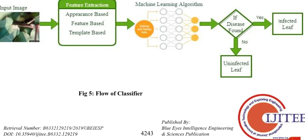
E. Image Classification
Image Classification is categorizing the image into number of predefined groups. Classification is a process that relay on the
characteristic of the Image dataset, Accuracy purely depends on the trained dataset. It is an Important process in the disease detection. The trained datasets are classified first and tested with the test set of Images. The main point in Classification is it should Identify healthy and unhealthy portion of the leaf Images. Classification of Image can be done using Machine learning methodologies. Fig [5] shows the
V. CLASSIFICATIONMETHODSONMULTIPLE
PLANTGROUPS
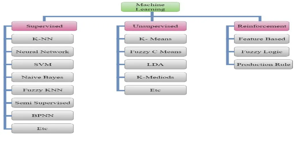
[image:5.595.41.550.276.522.2]Fig [6] Shows the types of Machine Learning algorithms used as classification methods to identify the infection in the plant leaves. We are focusing on two types of Infection or disease they are Fungal and Bacterial diseases on Food grain crops and Cash Crops.
Fig 6: Types of Machine Learning Algorithms
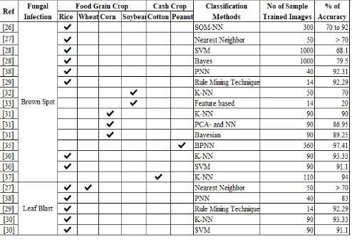
A. Fungal Disease Classification
The Infections in the food grain crop are easily detected through Visualization. The visual aspect of these kind of infection is spots around the infected portion. From the analysis we found that most of the rice field is infected by Brown spot diseases. Research by Phadikar S, Sil -2008 [26] stated that the diseased images of paddy leaves are categorized using SOM-NN (Self Organizing Map Neural Network). In which samples are trained by discovering the characteristic of effected leaves. 4 different types of samples were used to train and 300 samples were tested. Accuracy range was between 70% to 90% with different cases. The research [27] has been carried out using Nearest Neighbor Classification to which recognizes the Sheath disease, Rice Blast diseases, Brown Spot and Bacterial-Bligh diseases. The average classification accuracy is more than 70% to recognize the infection and it utilized less than 2 seconds for
computation. Phadikar S, Sil -2012 [28], proposed an
automatic system to detect two types of rice disease. Bayes classifier algorithm and SVM (Support Vector Machine) assorts the Brown Spot and Blast Diseases. Where they used 1000 number of samples as training and test images. The accuracy of 79.5% and 68.1 % has been achieved for Bayes’ Classifier and SVM.
Another Method which is faster version of Neural Network called as PNN (Probabilistic Neural Network) is proposed by using Fractal Texture descriptor [38]. Over all accuracy of 91.80 % is achieved by the system. Where Brown Spot accuracy is 92.31%, Leaf Blast Accuracy is 83.0%. Phadikar S, Sil -2013 [29] designed rice disease identification system has been proposed with different Visual Properties. They have used Threshold based segmentation, to Calculate the threshold they have adapted Fermi energy method to extract regions. Classification is derived using Rule Mining Technique. They have used Dataset of 14 test images and obtained Accuracy of 92.29%.
SIFT to extract the characteristics from infected region of Paddy leaves. This work compares the performance of two methods, they are KNN (K- Nearest Neighbors) and SVM. It identifies Brown Spot, Leaf Blast fungal diseases in the Paddy leaves [30]. KNN – accuracy 93.33% performs well when compared to SVM – accuracy 91.10%.
Another system is designed which compares KNN, PCA and NN, Bayesian classifier to classify and recognize Brown spots on corn leaves. which resulted in 90% accuracy using KNN algorithm and 86.85% and 89.25% using PCA and Bayesian classifier [31]. Research by Srivastava S, Hooda DS (2014) [32] was on the crop Soybean, they have used image captured by Mobile phone and employed 50 testing samples of KNN classifier to detect and classify Brown Spots and Frog eye disease on the plant leaves. KNN classifier resulted in 70 % accuracy on Brown Spots and 80% accuracy on frog eye. Another research has been carried out in Soybean leaves to detect Brown Spot and Rust diseases using feature-based classifier [33]. The results are extracted using confusion Matrix. The accuracy for Brown Spot disease is 20% and Rust is 71% with the training dataset of 14 and 7 each.
The K-Means algorithm and NN is proposed to detect the Scorch disease in plants like peanut [34]. The algorithms developed proves that the efficiency of 94% has been achieved. The method has been tested using 32 Samples and the threshold is computed with the global thresholding method called OTSU’s method. A new method is formulated to identify 4 phases of Cercospora disease. BPNN (Back Propagation Neural Network) is proposed in the system [35]. The accuracy of the method was 97.41% with the training dataset of 360 images on Cercospora spots in the peanut plants. Geometric moment algorithm was introduced to detect the inadequacy in the peanut leaves to identify Mineral deficiencies [36]. The system generated 93% accuracy. Another method is proposed by using PCA/ KNN algorithm to analyze leaf spot disease in Cotton Leaves. 110 images where trained to classify the disease and achieved 95% accuracy [37].
B. Bacterial Disease Classification
A method called PNN (Probabilistic Neural Network) is proposed which uses Fractal Texture descriptor to identify the bacterial diseases in the paddy leaves. [38]. Over all accuracy of the system is 91.80 %. Where Bacterial leaf blight accuracy is 96.25%. Another work is proposed by [30] SIFT (Scale Invariant Feature Transform) to extract the features of Infected leaves. It identifies Bacterial Blight disease in the Paddy leaves. SVM predicts the result with accuracy of 91.10% and KNN with 93.33%. [33] Researchers have employed feature-based classifier to identify Bacterial Blast disease in Soybean leaves. They trained around 38 samples and tested around 18 samples and obtained the accuracy of 37.5%. [39]A system is developed that differentiates KNN,
PCA and NN, Bayesian classifier which identifies Bacterial Blight on corn leaves. 90% accuracy is obtained using KNN algorithm and 86.85% and 89.25% using PCA and Bayesian classifier.
Decision Tree Classifier is employed to classify the health and diseases tomato leaves with bacterial leaf spots and Bacterial canker. 26 images where tested to obtain the accuracy of 69.2% and 84.6% of both the diseases. Phalaenopsis is a plant which is infected mostly by Bacterial soft rot and Bacterial brown spot [21], A method based on BPNN classifier is designed to identify the diseased leaves and they have BPNN classifier trained 144 datasets on color texture features which resulted in accuracy of 90.9% and 88.8%.
Bacterial blight on Cotton plant leaves has been identified and classified using Feed Forward BPNN Classifier. Which resulted in 85.52% of accuracy with the training set of 50 Images [40]. Another method is proposed to detect Bacterial Blight disease in Cotton leaves using ANN classifier [41] which provides around 85 to 91% of accuracy by training 120 good quality images. [37] proposed KNN algorithm to analyze leaf Blight disease in Cotton Leaves which proves 95% accuracy in classifying. 110 images where used to train and test the infection
VI. PERFORMANCEEVALUATIONOF
CLASSIFIERS
A. Fungal Disease
a. Brown Spot/Leaf Spot
[image:6.595.306.553.539.709.2]We have taken two major types of Fungal diseases for observation Brown Spot / leaf Spot and Leaf blast fungal disease. Fig [7] denotes the graphical representation the performance evaluation of Brown Spot/Leaf Spot classification using different types of Classifiers that is applied in Multiple plant groups like Food grain crop and Cash Crops. From the analysis it is observed that Backpropagation Neural Network (BPNN) proves to be the best
Fig 7: Performance Analysis of Multiple Classifiers in Identifying Fungal Leaf Spot disease
classifier which provides 97.41% accuracy which uses trained dataset of 360 images and tested on 40 Images.
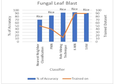
b. Leaf Blast
Fig [8] represents the graphical representation of observed Fungal Leaf Blast disease on Paddy leaves. Where multiple classifiers are utilized to
the leaves and its accuracy is calculated based on the of total number of training sets. From the analysis it is shown that KNN algorithm is more accurate than the other algorithms.
[image:7.595.325.567.47.215.2]
Fig 8: Performance Analysis of Multiple Classifiers in Identifying Fungal Leaf Blast disease
B. Bacterial Blight
The analysis has been made on plant varieties like Rice, Soybean, Corn and cotton. The infected leaves had been trained and tested for identifying the Bacterial blast disease. And it is observed that the KNN algorithm is better among the other classifier algorithms with the training dataset of 110 Images. Which is graphically represented in the Fig [9].
Table 1: Comparison of Fungal Diseases (Brown Spot and Leaf Blast) Detection and dentification using
Multiple Classifiers
[image:7.595.57.280.93.257.2]Table 2: Comparison of Bacterial Disease Detection Identification using Multiple Classifiers
Fig 9: Performance Analysis of Multiple Classifiers in Identifying Bacterial Blight disease
VII. CONCLUSIONANDFUTUREWORK
The paper compares various image processing methodologies like Image Acquisition, Preprocessing, Segmentation and Classification to analyze, detect and classify disease in the leaf image of multiple plants or culture. Where we mainly focused on the Classifiers to evaluate the performance. The performance of a system is purely based upon number of data-set used for training. It depends on the number of images used for training. From the analysis it is observed that well trained framework or system is profoundly effective. Over trained systems might produce inappropriate result. Proposing an inclusive method for each module and also comparing particular disease in a particular set of plant is very hard and difficult. Hence, we have compared different set of algorithms used to test different set of plants and diseases. From the analysis it is observed that using Back Propagation Neural Network Classifier performed better when compare to other algorithms like KNN, SVM, Bayesian, Feature based etc., But when the samples of multiple plant groups were tested using multiple Algorithms, KNN proved to be the best Algorithm. In the future to provide better performance we are focusing on developing a system which includes combination of best algorithms in both Image processing and Machine learning techniques. As a future enhancement more training dataset can be included
and tested using new Hybrid Algorithm to detect diseases which is combined with KNN and RBPNN classifier to produce a better result, reduced false positive results and to classify multiple diseases on variety of plant groups.
REFERENCES
1. Bagde S, Patil S, Patil S, Patil P (2015) Artificial neural network-based plant leaf disease detection. Int J Comput Sci Mob Comput 4(4):900–905
2. Barbedo JGA (2016) A review on the main challenges in automatic plant disease identification based on visible range images. Biosyst Eng 144:52–60
3. Martinelli F, Scalenghe R, Davino S et al (2015) Advanced methods of plant disease detection. A review. Agron Sustain Dev 35(1):1–25 4. Zhang M, Meng Q (2011) Automatic citrus canker detection from leaf
images captured in field. Pattern Recognit Lett 32(15):2036–2046 5. X.E. Pantazi, D. Moshou, A.A. Tamouridou, Automated leaf disease
detection in different crop species through image features analysis and One Class Classifiers,Computers
November 2018; Accepted 2 November 2018 , 0168-1699/ © 2018 Elsevier
6. Sukhvir Kaur, Shreelekha Pandey, Shivani Goel, Plants Disease Identification and Classification Through Leaf Images: A Survey, Received: 26 June 2017 / Accepted: 11 January 2018 _ CIMNE, Barcelona, Spain 2018, Archives of Computational Methods in Engineering
https://doi.org/10.1007/s11831-018-9255-6(0123456789().,-volV)(01 23456789().,-volV)
7. Babu Kumar S, J Nagaraja, S Venkatesan , An Insight into Plant Disease Detection Using Image Processing- A Review, International Journal of Engineering Research in Computer Science and Engineering (IJERCSE), Vol 5, Issue 4, April 2018, ISSN (Online) 2394-2320
8. University of Minnesota Extension. https://www.extension.umn. edu/. Accessed 16 May 2017
9. Barbedo JGA (2014) An automatic method to detect and measure leaf disease symptoms using digital image processing. Plant Dis 98(12):1709–1716
10. Prashar K, Talwar R, Kant C (2015) A review on efficient identification of american cotton leaf diseases through training set. Int J Comput Appl 132(7):32–39
11. Malathi K, Nedunchelian R., Comparison of Various Noises And Filters For Fundus Images Using Pre-Processing Techniques, International Journal of Pharma and Bio Sciences Int J Pharm Bio Sci 2014 July; 5 (3): (B) 499 – 508
12. Camargo A, Smith JS (2009) An image-processing based algorithm to automatically identify plant disease visual symptoms. Biosyst Eng 102(1):9–21
13. Malathi K, Nedunchelian R. An Automated Detection of Optic Disc From Digital Retinal Fundus Images Using Region Based Segmentation Technique, International Journal of Applied Engineering Research , ISSN 0973-4562 Volume 10, Number 1 (2015) pp. 285-296 © Research India Publications
14. Xia C, Lee JM, Li Y, Song YH, Chung BK, Chon TS (2013) Plant leaf detection using modified active shape models. Biosyst Eng 116(1):23–35
15. Rastogi A, Arora R, Sharma S (2015) Leaf disease detection and grading using computer vision technology and fuzzy logic. In: IEEE 2nd international conference on signal processing and integrated networks SPIN, pp 500–505
16. Kurniawati NN, Abdullah SNHS, Abdullah S, Abdullah S (2009) Investigation on image processing techniques for diagnosing paddy diseases. In: IEEE international conference on soft computing and pattern recognition SOCPAR., Malacca, pp 272–277
17. Wang L, Dong F, Guo Q, Nie C, Sun S (2014) Improved rotational kernel transformation directional feature for recognition of wheat stripe rust and powdery mildew. In: IEEE 7th international conference on image and signal processing CISP, Dalian, pp 286–291
18. Weizheng S, Yachun W, Zhanliang C, Hongda W (2008) Grading method of leaf spot disease based on image processing. In: IEEE international conference on computer science and software engineering, Wuhan, Hubei, December 8, pp 491–494
19. Shrivastava S, Singh SK, Hooda DS (2014) Statistical texture and normalized discrete cosine transform-based automatic soya plant foliar infection cataloguing. Br J Math Comput Sci 4(20):2901–2916 20. Yao Q, Guan Z, Zhou Y, Tang J, Hu Y, Yang B (2009) Application of
support vector machine for detecting rice diseases using shape and color texture features. In: IEEE international conference on engineering computation ICEC, Hong Kong, pp 79–83
21. Huang KY (2007) Application of artificial neural network for detecting Phalaenopsis seedling diseases using color and texture features. Comput Electron Agric 57(1):3–11
22. Sankaran S, Mishra A, Ehsani R, Davis C (2010) A review of advanced techniques for detecting plant diseases. Comput Electron Agric 72(1):1–13
23. Malathi K, Nedunchelian R. Efficient Method To Detect And Classify Diabetic Retinopathy Using Retinal Fundus Images, International Journal Of Pure And Applied Mathematics, Volume 116 No. 21 2017, 89-97, ISSN: 1311-8080 (Printed Version); ISSN: 1314-3395 (On-Line Version)
24. Pires RDL, Gonc¸alves DN, Orue ˆ JPM, Kanashiro WES, Rodrigues JF, Machado BB, Gonc¸alves WN (2016) Local descriptors for soybean disease recognition. Comput Electron Agric 125:48–55 25. Gurjar AA, Gulhane VA (2012) Disease detection on cotton leaves by
eigenfeature regularization and extraction technique. Int J Electr Commun Soft Comput Sci Eng 1(1):1–4
26. Phadikar S, Sil J (2008) Rice disease identification using pattern recognition techniques. In: IEEE 11th international conference on computer and information technology ICCIT, Khulna, pp 420–423 27. [33] Anthonys G, Wickramarachchi N (2009) An image recognition
system for crop disease identification of paddy fields in Sri Lanka. In: IEEE international conference on industrial and information systems ICIIS, Sri Lanka, pp 403–407
28. Phadikar S, Sil J, Das AK (2012) Classification of rice leaf diseases based on morphological changes. Int J Inf Electron Eng 2(3):460–463 29. Phadikar S, Sil J, Das AK (2013) Rice diseases classification using feature selection and rule generation techniques. Comput Electron Agric 90:76–85
30. Mohan KJ, Balasubramanian M, Palanivel S (2016) Detection and recognition of diseases from paddy plant leaf images. Int J Comput Appl 144(12):34–41
31. Zhang SW, Shang YJ, Wang L (2015) Plant disease recognition based on plant leaf image. J Anim Plant Sci 25(Suppl. 1):42–45
32. Shrivastava S, Hooda DS (2014) Automatic brown spot and frog eye detection from the image aptured in the field. Am J Intell Syst 4(4):131–134
33. Barbedo JGA, Godoy CV (2015) Automatic classification of soybean diseases based on digital images of leaf symptoms. SBI AGRO 34. H. Al-Hiary, S. Bani-Ahmad, M. Reyalat, M. Braik and Z.
ALRahamneh, Fast and Accurate Detection and Classification of Plant Diseases, International Journal of Computer Applications (0975 – 8887), Volume 17– No.1, March 2011
35. Ramakrishnan M (2015) Groundnut leaf disease detection and classification by using back probagation algorithm. In: IEEE international conference on communications and signal processing (ICCSP), April, pp 0964–0968
36. Nisale SS, Bharambe CJ, More VN (2011) Detection and analysis of deficiencies in groundnut plant using geometric moments. World Acad Sci Eng Technol 5:608–612
37. Viraj A. Gulhane, Maheshkumar H. Kolekar, Diagnosis of Diseases on Cotton Leaves Using Principal Component Analysis Classifier, 2014 Annual IEEE India Conference (INDICON), 978-1-4799-5364-6/14 38. Asfarian A, Herdiyeni Y, Rauf A, Mutaqin KM (2013) Paddy diseases
identification with texture analysis using fractal descriptors based on Fourier spectrum. In: IEEE international conference on computer, control, informatics and its applications IC3INA, Jakarta, pp 77–81 39. Lu J, Cui D, Jiang H (2013) Discrimination of tomato yellow leaf curl
disease using hyperspectral imaging. American Society of Agricultural and Biological Engineers, Kansas City, Missouri, July 21-July 24, p 1 40. Rothe PR, Kshirsagar RV (2015) Cotton leaf disease identification using pattern recognition techniques. In: IEEE international conference on pervasive computing (ICPC), January, pp 1–6 41. Mr. V. A. Gulhane & Dr. A. A. Gurjar, Detection of Diseases on Cotton
Leaves and Its Possible Diagnosis, International Journal of Image Processing (IJIP), Volume (5): Issue (5): 2011
42. Shrivastava S, Singh SK, Hooda DS (2014) Statistical texture and normalized discrete cosine transform-based automatic soya plant foliar infection cataloguing. Br J Math Comput Sci 4(20):2901–2916 43. Prasad S, Peddoju SK, Ghosh D (2016) Multi-resolution mobile vision
system for plant leaf disease diagnosis. Signal Image Video Process 10(2):379–388
44. https://www.growerexperts.com/aphids-in-potatoes/
45. https://www.kaggle.com/vipoooool/new-plant-diseases-dataset
46. https://data.mendeley.com/datasets/3f83gxmv57/2
47. https://plantvillage.psu.edu/diseases
AUTHORSPROFILE
Kapilya Gangadharan is a Research scholar in Saveetha School of Engineering, Saveetha Institute of Medical and Technical Sciences, Chennai. She is currently working as Utility Analyst at Fidelity National Information services, Durham, NC, USA with total of 6 years’ teaching experience as Assistant Professor and 2 years’ experience in IT. She received B.Tech degree in Information Technology from CSI College of Emgineering, Ketti, Nilgiris and M.E in Computer Science and Engineering from Periyar Maniammai University, Thanjavur. Her current research includes Digital Image Processing, Machine learning, Precision farming, Plant Technology and Production Engineering.
Dr. G Rosline Nesa Kumari received her Ph.D degree in Computer Science and Engineering at Dr MGR University Chennai, and received her M.E. Degree from Sathyabama University Chennai in 2005. She is having Sixteen Years of teaching experience. At present she is working as a Professor, Department of Computer Science and Engineering in Shadan women’s college of Engineering and Technology, Chennai. She published sixty-five research publications in various International, National Conferences and Journal. She is a life member of Indian Science Congress Association (ISCA), IAENG, IET, CSI (Life Member), Red Cross. Her research interest includes Image processing, Steganography, Network security and Information Security. includes Image processing, Digital Watermarking and Security
![Fig 1 Percentage of Plant groups on research [6]](https://thumb-us.123doks.com/thumbv2/123dok_us/8155446.248296/2.595.61.539.347.586/fig-percentage-plant-groups-research.webp)