0022-538X/06/$08.00⫹0 doi:10.1128/JVI.01465-06
Copyright © 2006, American Society for Microbiology. All Rights Reserved.
The
␣
Isoform of Protein Kinase CKI Is Responsible for Hepatitis C
Virus NS5A Hyperphosphorylation
䌤
Manuela Quintavalle, Sonia Sambucini, Chiara Di Pietro,
Raffaele De Francesco, and Petra Neddermann*
Istituto di Ricerche di Biologia Molecolare “P. Angeletti,” 00040 Pomezia (Roma), Italy
Received 11 July 2006/Accepted 24 August 2006
Hepatitis C virus (HCV) has been the subject of intensive studies for nearly two decades. Nevertheless, some aspects of the virus life cycle are still a mystery. The HCV nonstructural protein 5A (NS5A) has been shown to be a modulator of cellular processes possibly required for the establishment of viral persistence. NS5A is heavily phosphorylated, and a switch between a basally phosphorylated form of NS5A (p56) and a hyperphos-phorylated form of NS5A (p58) seems to play a pivotal role in regulating HCV replication. Using kinase inhibitors that specifically inhibit the formation of NS5A-p58 in cells, we identified the CKI kinase family as a target. NS5A-p58 increased upon overexpression of CKI-␣, CKI-␦, and CKI-, whereas the RNA interference of only CKI-␣reduced NS5A hyperphosphorylation. Rescue of inhibition of NS5A-p58 was achieved by CKI-␣ overexpression, and we demonstrated that the CKI-␣ isoform is targeted by NS5A hyperphosphorylation inhibitors in living cells. Finally, we showed that down-regulation of CKI-␣attenuates HCV RNA replication.
The discipline of “chemical genomics” has progressively be-come more and more important for the elucidation of the molecular basis of complex phenotypes. Chemical genomics implies a combination of medicinal chemistry and genetics in which small molecules are used to perturb biological pathways by modulating the activity of individual gene products. Such compounds may then assist scientists in identifying target pro-teins and genes. We used this combination of methodologies to better understand the mechanisms by which the hepatitis C virus (HCV) is able to replicate its genome in the host cells.
The positive-sense single-stranded RNA genome of HCV encodes a single polyprotein that is co- and posttranslationally processed into at least four structural and six nonstructural (NS) proteins (22). Virus-encoded enzymes are attractive tar-gets for the development of antiviral agents, and the HCV NS2-3 and NS3-4A proteinases, the NS3 helicase, and the RNA-dependent RNA polymerase NS5B have been investi-gated for many years. NS5A is a nonstructural protein without any yet-identified biochemical activity.
NS5A has been described to influence many different cellu-lar pathways involved in cell cycle control and cellucellu-lar growth, apoptosis, and inflammatory and immune responses (24). In most cases, the activity of cellular kinases is directly or indi-rectly influenced by the presence of NS5A (24). While all this information indicates that NS5A can modulate the activity of cellular kinases, cellular kinases might also affect the function of NS5A itself. NS5A is phosphorylated in three main clusters (1), and two phosphorylated forms of NS5A, termed p56 (ba-sally phosphorylated) and p58 (hyperphosphorylated), can be distinguished (32). The three residues S2197, S2201, and S2204 and the presence of other HCV nonstructural proteins (2, 21,
28) have been reported to be implicated in the formation of NS5A-p58.
To date, the function of the differentially phosphorylated forms of NS5A in HCV genome replication can only be spec-ulated upon. However, some observations point to a direct role in RNA replication: NS5A interacts with the HCV RNA poly-merase NS5B (24) as well as with RNA (17) and colocalizes with all other HCV nonstructural proteins in a cytoplasmic membrane structure termed the “membranous web” (26). Be-sides the physical participation of NS5A within the HCV rep-lication complex, there is additional evidence suggesting that the hyperphosphorylated form of NS5A plays an important role in RNA replication. It has been observed that cell culture adaptive mutations, which dramatically increase the replication efficiency of the Con1 HCV subgenomic RNA within the he-patic cell line Huh7 (23), map predominantly to the NS5A-coding sequence (4). Notably, the most effective mutations are those which reduce the formation of NS5A-p58. This observa-tion led to the hypothesis that the levels of hyperphosphory-lated NS5A affect the efficiency of replication. In fact, an in-verse correlation between NS5A phosphorylation and HCV replication has been demonstrated (10).
Direct evidence that efficient replication depends on the proper expression levels of p58 came from our recent work, where we demonstrated that pharmacological inhibition of cel-lular kinases responsible for the formation of NS5A-p58 acti-vates cell culture replication of nonadapted Con1 subgenomic replicons that otherwise do not replicate efficiently in this sys-tem (29). One possible explanation is that the production of NS5A-p58 is deregulated such that too much p58 is produced. On the other hand, complete abrogation of p58 production obtained by the concomitant mutation of two serine residues important for NS5A hyperphosphorylation, S2197 and S2204, is also detrimental for HCV RNA replication (5). Thus, the hypothesis was formulated that a well established ratio be-tween NS5A-p56 and NS5A-p58 is required for productive replication/infection. In agreement with this idea are data * Corresponding author. Mailing address: Istituto di Ricerche di
Bio-logia Molecolare “P. Angeletti,” 00040 Pomezia (Roma), Italy. Phone: 39-06-91093221. Fax: 39-06-91093654. E-mail: Petra_Neddermann@merck .com.
䌤Published ahead of print on 30 August 2006.
11305
on November 8, 2019 by guest
http://jvi.asm.org/
showing that adaptive mutations known to alter the phosphor-ylation pattern of NS5A prevent productive infection of HCV full-length RNA in the chimpanzee model (7).
In order to sustain this hypothesis, it is important to identify the cellular kinase(s) required for the production of NS5A-p58: changing the expression or activity of these kinases within the cell could in turn influence the outcome of HCV infections. Several kinases which use NS5A as a substrate in vitro (8, 18, 19) or which associate with NS5A in living cells have been identified (30). However, which cellular kinase(s) are physio-logically relevant for NS5A hyperphosphorylation still remains to be proven.
In this study, we used the previously reported inhibitors of NS5A hyperphosphorylation in order to identify a cellular ki-nase important for the formation of NS5A-p58. This kiki-nase turned out to be the␣isoform of casein kinase I, and here we show the effects of this kinase on NS5A hyperphosphorylation and HCV replication.
MATERIALS AND METHODS
Plasmids, antibodies, and compounds.The plasmid pHCV-AT is similar to plasmid pHCVNeo17.wt described previously (35) but contains the mutation A2199T in NS5A. The plasmid pcD-BLA-wt contains the entire HCV sequence as described for plasmid wt-BLA (29) cloned into the pcDNA3 expression vector (Invitrogen). The coding regions for casein kinase I␣,␦,ε, and␥1 were taken from Image clones presented to the NCBI data bank with the accession numbers BQ641640, BC015775, BC006490, and BC017236, respectively, and cloned into the EcoRV restriction site of the vector pcDNA3. A FLAG tag (DYKDDDD KGG) was added in frame to the N terminus of the CKI-␣protein by conven-tional techniques, resulting in plasmid pCD-Flag-CKI-␣. The expression vectors for CKI-␥2 and GSK3were purchased from Invitrogen. The antibodies against CKI-␣, CKI-␦, and CKI-εwere purchased from Santa Cruz Biotechnology, against p38 MAP kinase␣from Cell Signaling Technology, and against GSK3 or the FLAG tag from Sigma. The specific antibodies and the NS5A-specific kinase inhibitors have been described previously (29). CKI-7 was bought from US-Biological, SP 600125 and SB203580 from Sigma, and IC261 from Calbiochem. The screening of the panel of kinases was performed by Upstate.
In vitro kinase assay.Purified rat CKI-␦and its substrate peptide were pur-chased from BioLabs. CKII and its substrate peptide were purpur-chased from Upstate. The kinase assay was performed as follows: 20 ng of CKI-␦(14 nM) or 50 ng of CKII (6 nM) was incubated with 100M substrate peptide in the presence of 125M cold ATP, 0.1l of [␥-33P]ATP (1Ci/point), and 18 mM MgCl2in reaction buffer (10 mM MOPS [morpholinepropanesulfonic acid; pH 7.2], 12.5 mM-glycerolphosphate, 2.5 mM EDTA, 0.5 mM Na-orthovanadate, and 0.5 mM dithiothreitol) in a final volume of 40l for 1 h at room temperature with or without the indicated compounds. The reaction was stopped with 8l of 3% phosphoric acid, and 20l of the reaction mixtures was spotted onto a P30 Filtermat (Wallac). The filter was washed three times for 5 min with 75 mM phosphoric acid and once with methanol. Radioactivity was counted in 5 ml of Ready Protein scintillation cocktail (Beckman Coulter).
RNA transfection, protein expression, and metabolic labeling of proteins.
RNA transcripts were prepared from plasmid pHCV-AT and transfected into 10A-IFN cells as described previously (29). Protein expression using the vaccinia virus-T7 infection/transfection system was performed as previously described (28). The day before the experiment, 3.5⫻105
10A-IFN cells/35-mm diameter dish were plated. If not stated otherwise, 2 g of total plasmid DNA was transfected, using Fugene6 (Roche) as the transfection reagent. Metabolic la-beling and immunoprecipitation were performed as previously described (28). Inhibitors were added during the starvation reaction and were present during metabolic labeling of the cells.
Immunoblotting was performed using the corresponding horseradish peroxi-dase-conjugated secondary antibodies, and enzymatic reactions were developed using the ECL system (Amersham).
RNA interference.Small interfering RNAs (siRNAs) were transfected by elec-troporation. The protocol used for electroporation was previously described (29). Briefly, 10M of siRNA was electroporated into 1⫻10610A-IFN cells in a volume of 0.1 ml. After electroporation, 4.5⫻105
cells were plated in a 35-mm-diameter dish and incubated for 2 days. Protein expression with the T7-vaccinia
described below: CKI-␣ (sense), 5⬘-GAAACAUGGUGUCCGGUUUTT-3⬘; CKI-␦(sense), 5⬘-CCUGCUGCUUGCUGACCAATT-3⬘; CKI-ε(sense), 5⬘-G UAUGAACGGAUCAGCGAGTT-3⬘; and p38-␣(sense),5⬘-CUCCUGAGAU CAUGCUGAATT-3⬘.
Inhibition of HCV replication.In order to measure inhibition of HCV repli-cation, Huh7 cells stably expressing a HCV subgenomic replicon containing the adaptive mutation S2204R (SR3) were used. Selection of clones was performed as described previously (23), except that 10A-IFN cells instead of naı¨ve Huh7 cells were used. siRNAs were electroporated into SR3. After electroporation, the cells were plated in 6-well plates (5⫻105cells for day 1, 3⫻105cells for day 3, and 2⫻105
cells for day 5). Quantitative PCR was performed as previously described (29). Briefly, 10 ng of RNA (HCV) or 100 ng (each kinase) was used for the reaction. The siRNAs, primers, and probes are described below: CKI-␣ sense, 5⬘-CATCTATTTGGCGATCAACATCA-3⬘; CKI-␣antisense, 5⬘-GCCT GGCCTTCTGAGATTCTA-3⬘; CKI-␣probe, 5⬘-CAACGGCGAGGAAGTGG CAGTGA-3⬘; HCV sense, 5⬘-CGGGAGAGCCATAGTGG-3⬘; HCV antisense, 5⬘-AGTACCACAAGGCCTTTCG-3⬘; and HCV probe, 5⬘-CTGCGGAACCG GTGAGTACAC-3⬘.
For the detection of CKI-␦mRNA (see Fig. 3), the following primers and probes were used: CKI-␦sense, 5⬘-CCCCCATCGAAGTGTTGTGT-3⬘; CKI-␦
antisense, 5⬘-CTGAATTTCTGCCGTTCCTTG-3⬘; and CKI-␦probe, 5⬘-AGG CTACCCTTCCGAATTTGCCACA-3⬘.
All probes contained 6-carboxyfluorescein dye at the 5⬘end and 6-carboxytetra-methylrhodamine quencher at the 3⬘end. As the endogenous standard, we used a-actin probe containing VIC dye (Applied Biosystems) at its 5⬘end. Reactions were conducted in three stages under the following conditions: stage 1, 30 min at 48°C; stage 2, 10 min at 95°C; stage 3, 15 s at 95°C and 1 min at 60°C for 40 cycles. The total volume of the reaction was 50l.
RESULTS
NS5A hyperphosphorylation inhibitors H479, A852, and F495 are inhibitors of casein kinase I. We have previously identified three 2,4,5-trisubstituted imidazole kinase inhibitors that specifically inhibit the formation of hyperphosphorylated NS5A in cell culture (29). In order to identify cellular kinases targeted by these compounds, we tested their inhibitory activity in vitro on a panel of protein kinases. We chose a fixed con-centration of 4M, sufficient to inhibit NS5A hyperphosphor-ylation in cell culture. Kinases inhibited byⱖ70% are high-lighted in Table 1. The spectrum of inhibitory activity is different for each compound, and only three kinases were potently inhibited by all three compounds. Mitogen-activated protein kinases p38-␣and p38-were excluded from the list of candidate targets, since SB 203580, a known p38 inhibitor, has no effect on NS5A hyperphosphorylation in cell culture (29). The third kinase is yeast (Schizosaccharomyces pombe) casein kinase I (CKI) (20).
To confirm inhibition of the mammalian CKI, we titrated the three compounds on the rat␦ isoform of CKI (CKI-␦), obtained through a commercial source (Fig. 1). As a negative control, we used casein kinase II (CKII), which has also been reported to phosphorylate NS5A in vitro. While CKII was not inhibited at up to 10M by any of the three compounds, all of them efficiently inhibited CKI-␦, with 50% inhibitory concen-tration values of 1.4M, 0.4M, and 0.1M for H479, A852, and F495, respectively. Due to high cell toxicity of compound F495, possibly associated with its broad spectrum of action
(Table 1), we decided to continue our study with only H479 and A852.
Inhibitors of CKI reduce NS5A hyperphosphorylation in cell culture.From the results of the in vitro kinase screening, CKI was identified as a possible candidate responsible for NS5A phos-phorylation. Compounds CKI-7, IC261, and SP600125 have been
on November 8, 2019 by guest
http://jvi.asm.org/
reported to specifically inhibit CKI (20–22). These inhibitors were used to assess the effect of CKI inhibition on NS5A hyperphosphorylation in cell culture. A subgenomic Con1 HCV RNA containing the A2199T adaptive mutation (4) was electro-porated into 10A-IFN cells (35), and replication was allowed to proceed for 3 days. This adaptive mutation was chosen because replicons bearing this mutation are a good tool to examine effects on NS5A hyperphosphorylation, since these replicons express both NS5A phosphoisoforms. The compounds were then added to the cells at the indicated concentrations, the cells were incubated for an additional 24 h, and NS5A hyper-phosphorylation was detected by Western blot analysis (Fig. 2). SB203580 and c1 were the control compounds. Compound c1 is a nonnucleoside inhibitor of the HCV RNA-dependent RNA polymerase NS5B, with an inhibitory potency compara-FIG. 1. H479, A852, and F495 are inhibitors of mammalian CKI-␦ in vitro. Kinase reactions were performed with recombinant CKI-␦or CKII and synthetic peptides as substrates, as described in Materials and Methods. Enzymatic activity was monitored by33P incorporation, using [␥-33P]ATP as the phosphate donor. M, molar concentration.
FIG. 2. NS5A hyperphosphorylation in cells is inhibited by known CKI inhibitors. In vitro-transcribed HCV subgenomic RNA from plas-mid pHCV-AT was transfected into 10A-IFN cells. After 3 days, dif-ferent compounds were added at the indicated concentrations, and the cells were incubated for an additional 24 h. Fifty micrograms of cell extract was subjected to 7.5% sodium dodecyl sulfate-polyacrylamide gel electrophoresis, and NS5A was detected by Western blotting, using NS5A-specific polyclonal antibodies. The positions of NS5A-p56 and NS5A-p58 are indicated by arrows on the right side of the figure. The ratios between p58 and p56 concentrations are indicated below.⫺, no compound added.
TABLE 1. Inhibition of kinases by NS5A hyperphosphorylation inhibitors in vitroa
Protein kinase % Inhibition by indicated compound
A852 H479 F495
c-RAF (h) 55 36 89
MEK1 (h) 6 5 32
MAPK2 (m) 23 5 77
p38-␣(h) 98 100 100
p38-(h) 90 97 91
p38-␥(h) 4 0 0
p38-␦(h) 5 0 8
MAPKAP-K2 (h) 9 0 77
MSK1 (h) 83 20 60
MKK4 (m) 36 27 37
MKK7b (h) 0 14 46
JNK1a1 (h) 12 16 9
JNK2a2 (h) 34 10 65
SGK (h) 0 17 10
PKCa (h) 15 20 65
ROCK-II (r) 70 21 27
Fyn (h) 52 49 91
ZAP-70 (h) 1 0 0
CHK2 (h) 25 72 100
PRK2 (h) 13 9 25
PKCbII (h) 54 14 78
PKCg (h) 5 0 20
Blk (m) 41 58 91
CaMKIV (h) 9 0 0
CDK3/cyclinE (h) 0 8 15
CDK5/p35 (h) 0 0 1
CK1 (y) 91 74 97
CSK (h) 31 1 3
IKKa (h) 0 15 0
IKKb (h) 0 28 16
PKCq (h) 0 12 27
Syk (h) 0 60 95
p70S6K (h) 25 17 40
CHK1 (h) 7 42 78
AMPK (r) 4 2 81
CDK2/cyclin A (h) 0 0 0
JNK3 (r) 78 57 99
cSRC (h) 60 57 98
CK2 (h) 0 7 29
Lck (h) 50 41 92
PRAK (h) 2 0 26
PDK1 (h) 2 63 73
Lyn (m) 92 59 98
GSK3b (h) 7 13 0
PKA (b) 75 14 76
PKBa (h) 18 6 27
CaMKII (r) 0 0 3
CDK1/cyclinB (h) 6 8 12
MAPK1 (h) 20 14 80
CDK2/cyclinE (h) 0 0 23
CDK6/cyclinD3 (h) 0 0 5
RSK3 (h) 11 23 42
IGF-1R (h) 0 0 9
IR (h) 5 8 2
PKBb (h) 50 25 11
FGFR3 (h) 22 10 9
PDGFRa (h) 9 6 37
PDGFRb (h) 5 0 56
CDK7/cyclinH/MAT1 (h) 3 18 14
a
The compounds were tested at a fixed concentration of 4M, and the numbers in the columns indicate the percent inhibition of the kinases. The numbers in bold indicate inhibitory activity ofⱖ70 %.
on November 8, 2019 by guest
http://jvi.asm.org/
[image:3.585.45.286.104.676.2] [image:3.585.305.541.508.624.2]ble to that of our NS5A hyperphosphorylation inhibitors (50% effective concentration [EC50],⬃5M) (data not shown). As expected, the NS5A hyperphosphorylation inhibitors H479 and A852 showed a marked reduction in p58 (p58/p56, 0.3 to 0.4) (Fig. 2, lanes 2 and 3), while the control lanes showed a p58/ p56 ratio of around 0.8. All CKI inhibitors tested here showed inhibition of NS5A hyperphosphorylation, even though with different efficiencies (Fig. 2, lanes 5 to 9). The least active compound was CK1-7, possibly because of its poor cellular uptake. This experiment suggests that pharmacological inhibi-tion of CKI causes a reducinhibi-tion of NS5A hyperphosphorylainhibi-tion.
The CKI-␣isoform is important for NS5A hyperphosphor-ylation.The results shown above demonstrate that kinases of the CKI family are promising candidates for NS5A hyperphos-phorylation. In mammals, the CKI protein kinase family con-sists of seven distinct isoforms:␣,,␥1,␥2,␥3,␦, andε(14). In order to test which of the CKI isoforms affects the phos-phorylation pattern of NS5A in cells, we performed “gain-of-function” and “loss-of-“gain-of-function” experiments. With these ex-periments, the expression pattern of a single gene was specifically modulated, and the effect on NS5A hyperphosphor-ylation could be directly attributed to this gene product. This method has an advantage over that using kinase inhibitors, which can have more- or less-pronounced off-target activity.
First, we overexpressed the different CKI isoforms in the presence of the HCV nonstructural polyprotein, using the T7-vaccinia virus-T7 infection/transfection system (Fig. 3A). As a negative control, we used the nonrelated kinase GSK3. Pro-teins were metabolically labeled with [35S]-methionine, and NS5A was immunoprecipitated with an NS5A-specific anti-serum. The only kinases which did not alter the ratio between basally (p56) and hyperphosphorylated (p58) NS5A in this experiment were the CKI␥1 and ␥2 isoforms and GSK3. In
contrast, overexpression of the␣ isoform and, to a lower ex-tent, the␦- andεisoforms increased levels of NS5A-p58. Ex-pression of active␦andεisoforms of CKI in Huh7 cells was confirmed on the natural substrate dvl (16; also data not shown). Overexpression of CKI-␣/ε/␦ and GSK3 was con-firmed by Western blotting (Fig. 3A, lower panel), whereas overexpression of the␥isoforms was confirmed by quantitative PCR, due to the lack of appropriate antibodies (data not shown).
Next, we tested whether the opposite effect could be ob-served upon silencing of individual kinase genes. As we could not detect any change in NS5A phosphorylation upon overex-pression of the CKI-␥isoforms, we focused our attention on the␣,␦, andεisoforms. 10A-IFN cells were transfected with siRNAs directed to CKI-␣, CKI-␦, CKI-ε, or p38 as the neg-ative control. After 48 h, the HCV subgenomic replicon was expressed and NS5A was metabolically labeled and immuno-precipitated (Fig. 3B). A clear reduction of NS5A hyperphos-phorylation was observed only upon silencing of CKI-␣ expres-sion, suggesting that CKI-␣is the isoform responsible for the modulation of NS5A hyperphosphorylation in cells. The silenc-ing efficiencies of p38 and CKI-␣/εwere monitored by Western blot analysis, while the silencing efficiency of CKI-␦was con-firmed by quantitative RT-PCR, due to the lack of appropriate antibodies, which are not able to detect endogenous levels of CKI-␦(Fig. 3B, bottom panels).
[image:4.585.59.528.66.208.2]In order to further strengthen this result, we investigated whether NS5A hyperphosphorylation could be rescued by overexpression of the different CKI isoforms in a CKI-␣ -silenced cellular background (Fig. 3C). The CKI-␣isoform was silenced as shown in Fig. 3B, and the HCV replicon was expressed together with the different CKI isoforms as de-scribed above. As expected, silencing of CKI-␣ reduced FIG. 3. CKI-␣plays an important role in NS5A hyperphosphorylation. (A) Overexpression of CKI-␣, CKI-␦, and CKI-εincreases NS5A-p58 levels. Plasmid pcD-Bla-wt (2g) was transfected together with plasmids expressing the indicated kinases (each 1g) in 10A-IFN cells, and proteins were expressed using the vaccinia virus-T7 infection/transfection system. The proteins were labeled, and NS5A was immunoprecipitated from 20g of total protein extract. The proteins were subjected to 7.5% sodium dodecyl sulfate-polyacrylamide gel electrophoresis, and an autoradiogram (AR) is shown in the upper panel. The lower panel shows a Western blot (WB) of the specific kinases. CKI-␣was detected with ␣-FLAG antibody. NS5A and the kinases are indicated. (B) RNAi of CKI-␣decreases NS5A hyperphosphorylation. The indicated kinases were silenced in 10A-IFN cells as described in Materials and Methods. Forty-eight hours after siRNA transfection, 2g of pcD-Bla-wt was transfected, and the proteins were expressed, using the vaccinia virus-T7 infection/transfection system. The proteins were labeled, and NS5A was immuno-precipitated as described above. The upper panel shows the autoradiogram. Silencing of the different kinases is shown in the Western blot for CKI-␣/εand p38 and by quantitative RT-PCR for CKI-␦in the lower panels (QP). The numbers indicate mRNA expression levels of CKI-␦with respect to that of untransfected cells (100%). (C) Overexpression of CKI-␣rescues inhibition of NS5A hyperphosphorylation. CKI-␣expression was silenced as described for panel B. After 48 h, 2g of pcD-Bla-wt and 0.5g (lanes 3, 5, and 7) or 1g (lanes 4, 6, and 8) of plasmids expressing the indicated kinases were transfected. Proteins were expressed and labeled as described above, and NS5A was immunoprecipitated. The upper panel shows an autoradiogram. The lower panels show a Western blot of the overexpressed kinases. CKI-␣was detected with␣-FLAG antibody.
on November 8, 2019 by guest
http://jvi.asm.org/
NS5A-p58 (Fig. 3C, compare lanes 1 and 2). Upon concom-itant overexpression of CKI-␣, a clear increase in NS5A hyperphosphorylation was observed (Fig. 3C, lanes 3 and 4), whereas overexpression of CKI-␦or CKI-ε did not signifi-cantly affect the NS5A p58-to-NS5A p56 ratio (Fig. 3C, lanes 5 to 8). Overexpression of the respective kinases is shown in the bottom panels.
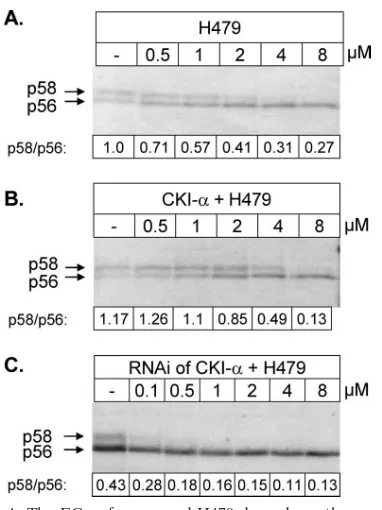
Overexpression or silencing of CKI-␣affects the potency of the NS5A-specific kinase inhibitors. Another set of experi-ments was performed in order to further demonstrate that the CKI-␣isoform is the target of the NS5A hyperphosphorylation inhibitors. We measured the effective compound concentration required to inhibit 50% of NS5A hyperphosphorylation in cell culture. We anticipated that overexpression of the target ki-nase should increase the EC50, whereas silencing of this kinase should decrease it. HCV proteins were expressed, using the vaccinia virus-T7 infection/transfection system, and compound H479 was present in increasing concentrations during HCV protein expression.
The EC50for compound H479 was between 1 and 2 M (Fig. 4A). Upon overexpression of CKI-␣, the EC50increased and could be estimated at around 4M (Fig. 4B). An opposite effect was observed upon silencing of CKI-␣, where the EC50 clearly dropped below 1 M (Fig. 4C). Overexpression and silencing of CKI-␦or CKI-εisoforms did not show the same correlation between kinase expression level and the EC50of the compound (data not shown).
Inhibition of HCV replication upon silencing of CKI. We have previously shown that the NS5A-specific compounds in-hibit HCV replication (29). In this work, we have demon-strated that cellular CKI-␣is targeted by compound H479 (Fig. 4). We next investigated whether HCV replication is inhibited as a consequence of reduced expression of the CKI-␣isoform by RNA interference (RNAi). To perform this experiment, we used Huh7 cells which stably express an HCV subgenomic replicon containing the adaptive mutation S2204R. This mu-tation shows a reduced formation of hyperphosphorylated NS5A (29). We chose this adaptive mutation for the following experiments because this replicon is more potently inhibited by the compound H479 (data not shown). Mock-transfected cells or siRNA-transfected cells were collected 1, 3, and 5 days after electroporation and controlled for HCV RNA and silencing efficiency of the kinase by quantitative PCR (Fig. 5). mRNA levels of HCV or CKI-␣ in the mock-transfected cells were arbitrarily set to 100%. Throughout the duration of the exper-iment, the mRNA levels of CKI-␣ in those cells transfected with the specific CKI-␣siRNA remained below 30% of that of the mock-transfected cells (Fig. 5, right panel). At the same time points, HCV RNA slowly decreased and reached a min-imum of 40% with respect to that of the mock-transfected cells at day 5, which means a 60% inhibition. This experiment shows that reduction of CKI-␣ expression results in inhibition of HCV replication.
DISCUSSION
The aim of this work was the identification of the cellular kinase(s) which is required for the hyperphosphorylation of NS5A. Identification of this kinase(s) would contribute signif-icantly to further understanding of the HCV life cycle and could possibly lead to novel therapeutic strategies for the treat-ment of hepatitis C patients.
We used three previously identified NS5A hyperphosphory-lation inhibitors as tools to screen a limited panel of cellular kinases in vitro. The most promising hit within the panel was represented by the yeast protein kinase CKI, which was po-tently inhibited by all three compounds. The CKI protein ki-nase family is evolutionary conserved and ubiquitously ex-FIG. 4. The EC50of compound H479 depends on the expression
[image:5.585.67.258.64.319.2]level of CKI-␣. pcD-Bla-wt (2g) was expressed using the vaccinia virus-T7 infection/transfection system either alone (A) or together with 1g of plasmid expressing CKI-␣(B). For panel C, CKI-␣was silenced as described in Materials and Methods and pcD-Bla-wt (2g) was expressed 48 h after RNAi. H479 was added at the indicated concentrations, the proteins were labeled, and NS5A was immunopre-cipitated as described above.
FIG. 5. Silencing of CKI-␣ inhibits HCV replication. CKI-␣was silenced by transfection of siRNA in SR3 cells. After 1, 3, and 5 days, RNA was isolated and mRNA for CKI-␣(right panel) and HCV RNA (left panel) were detected using quantitative PCR. Shown are the relative RNA quantities expressed as percentages of those of the mock-transfected control cultures (c) at each time point. The data shown represent the averages of the results of three independent experiments, and the error bars indicate the experimental standard deviations.
on November 8, 2019 by guest
http://jvi.asm.org/
[image:5.585.312.532.69.164.2]pressed in eukaryotic organisms (14). In mammals seven distinct isoforms (␣,,␥1,␥2,␥3,␦, andε) are expressed, and members of this family are involved in many different physio-logical and cellular processes (20). Some characteristic fea-tures of CKI make it an especially interesting candidate for NS5A phosphorylation. CKI prefers acidic target sites and has a high preference for substrates containing phosphoserine or phosphothreonine within the consensus sequence (12). NS5A is an acidic protein with an isoelectric point of around 5 and is heavily phosphorylated. In fact, NS5A contains at least 20 potential CKI phosphorylation sites. Interestingly, the region around the NS5A hyperphosphorylation sites is a hotspot for CKI recognition, suggesting that one or more of the serine residues situated in this region might be a substrate for CKI. To further confirm that CKI is an important kinase for the formation of NS5A-p58 in cells, we inhibited NS5A hyperphos-phorylation using known CKI inhibitors. All CKI inhibitors clearly reduced the formation of p58. These inhibitors, how-ever, may also affect other kinases at similar concentrations and therefore this type of experiment cannot be taken as con-clusive proof for the involvement of CKI.
The catalytic domain of CKI is highly conserved throughout different species and among different isoforms. However, the different CKI␣,␦,ε, and␥isoforms have been shown to play important roles in distinct cellular pathways, and therefore we aimed to identify the isoform(s) important for NS5A hyper-phosphorylation, using typical “gain-of-function” and “loss-of-function” experiments. While overexpression of the isoforms
␣,␦, andεincreased NS5A hyperphosphorylation, RNA inter-ference of only the␣isoform was able to diminish the expres-sion levels of NS5A-p58 (Fig. 2). Our results thus confirm the importance of the␣isoform for NS5A hyperphosphorylation. This experiment, however, does not rule out the possibility that, although diminished, the residual kinase activity of CKI-ε or CKI-␦after RNAi may be sufficient for NS5A hyperphos-phorylation. The exceptions were the␥isoforms, which did not seem to influence NS5A phosphorylation. These results indi-cate that all three isoforms are capable of recognizing NS5A as a substrate when ectopically overexpressed. Two additional experiments support CKI-␣as being the physiologically rele-vant isoform. (i) Rescue of the inhibited formation of p58 was achieved only upon overexpression of the␣isoform, but not upon expression of the␦orεisoforms. The fact that overex-pression of CKI-εor CKI-␦increased p58 in a normal cellular background, while this effect cannot be observed in a CKI-␣ -silenced cellular background, might indicate that different phosphorylation sites in NS5A are involved and that hyper-phosphorylation by CKI-εor CKI-␦is facilitated through pre-phosphorylation by CKI-␣. (ii) A clear indication of whether a kinase is the target of a specific inhibitor is a change of EC50 dependent on the expression level of the kinase. We have shown that this correlation was confirmed for the␣isoform. Even though our data support a link between CKI-␣and NS5A hyperphosphorylation, one cannot exclude the possibility that NS5A is not a direct substrate of CKI but that an enzyme downstream of CKI or under its control may be responsible for NS5A modification.
With these results, one of the most interesting questions was whether the reduction of active CKI-␣affects HCV replication. Inhibition of HCV replication upon incubation with the known
CKI inhibitors could not be tested due to the high cytotoxicity of these compounds. We addressed this question by RNAi. Attenuation of CKI-␣expression inhibited production of HCV RNA in cells containing actively replicating HCV subgenomes up to 60% after 5 days of CKI-␣silencing. This result strongly supports a direct correlation between CKI-␣ expression and HCV replication. We also tried to activate replication of Con1 wild-type subgenomes in cells upon silencing of CKI- ␣, as described for the NS5A hyperphosphorylation inhibitors (29). However, transfection of siRNAs and subgenomic RNA at different time points drastically increased cell mortality, and silencing efficiency might not be high enough for the establish-ment of replication. This type of experiestablish-ment has to await the production of efficient small hairpin RNAs, which can be in-troduced into the cells by viral vectors.
All data obtained so far suggest that the CKI-␣ isoform is important for NS5A hyperphosphorylation. Four different splice variants have been characterized biochemically, and the most important difference resided in their subcellular localiza-tions (20). In addition, the␣isoform has also been found to be associated with cellular membranes (3) and vesicular struc-tures (15) and is important for vesicle biogenesis (11). This is of particular interest because it has been demonstrated that all HCV nonstructural proteins are associated with intracellular membranes, including the endoplasmic reticulum and Golgi apparatus (27), and that active replication most likely takes place in lipid rafts of the plasma membrane or internal mem-brane compartments such as the Golgi apparatus (31).
Unlike CKI-␦and CKI-ε, which are regulated by autophos-phorylation of the C-terminal tail of the protein (13), CKI-␣is constitutively active because it misses this regulatory domain and regulation of enzymatic activity has therefore to be achieved by other means. Subcellular localization and a hier-archical order of substrate phosphorylation are two possible explanations. Another interesting mechanism of activity regu-lation has been observed for the membrane-associated CKI-␣ isoform. This form is potently inhibited by phosphatidylinositol 4,5-bisphosphate (PIP2) (6), present only in some membrane compartments. It is tempting to speculate that NS5A hyper-phosphorylation varies according to the cellular compartment where it resides. The activity of membrane-bound CKI-␣might be different in lipid rafts, where replication takes place, and at the cellular membrane, where virus assembly and/or virus exit is organized.
Until recently, this hypothesis was difficult to prove, due to lack of a suitable infection system. Fortunately, an HCV strain has recently been isolated which is able to infect and replicate in cells in culture (36), and this system now offers the oppor-tunity to study protein functions for replication as well as virus assembly and virus exit.
Hyperphosphorylation might have a number of structural and functional consequences for NS5A. Interestingly, the hy-perphosphorylation region lies between two protease-resistant domains (33) and is therefore easily accessible for regulatory proteins such as kinases. Recently, the crystal structure of the N-terminal domain of NS5A has been published (34), and two interesting features were observed. First of all, NS5A crystal-lized as a dimer, and even though the stoichiometry of NS5A within the replication complex is not known, one could imagine that hyperphosphorylation changes the conformation of NS5A,
on November 8, 2019 by guest
http://jvi.asm.org/
which might provoke a switch between a monomeric and a dimeric state. Such regulation has already been demonstrated for NSP5, a rotavirus nonstructural protein phosphorylated by CKI (9). Second, the NS5A dimer forms a groove which could easily accommodate single- as well as double-stranded RNA. In fact, NS5A has been shown to bind RNA in vitro (17). Also, in this case phosphorylation of the flexible linker region be-tween the N-terminal and the C-terminal domains could change their relative positions, resulting in different capabili-ties to bind RNA. NS5A hyperphosphorylation could also be involved in a switch between translation of the plus-strand RNA and production of the minus-strand RNA by the NS5B polymerase (25). Finally, the phosphorylation state of NS5A might regulate the interaction with cellular partners important for some aspects of HCV replication. It has been shown that NS5A hyperphosphorylation disrupts the interaction of NS5A with hVAP-A, which is thought to be involved in RNA repli-cation complex assembly (10).
The roles of NS5A for viral replication and/or infection still remain a mystery. What becomes increasingly evident is that regulation of NS5A hyperphosphorylation plays an important role. We started to investigate which of the cellular kinases are important for NS5A hyperphosphorylation, using small mole-cule inhibitors as well as genetic tools. Here we have identified the casein kinase I family of kinases as possible targets of our NS5A hyperphosphorylation inhibitors and demonstrated that the CKI-␣isoform is the kinase involved in NS5A hyperphos-phorylation. Even though our experiments suggest that the CKI-␣isoform is targeted by our compounds, we cannot ex-clude the possibility that other cellular kinases are also inhib-ited and possibly implicated in the hyperphosphorylation of NS5A. It seems obvious that NS5A is phosphorylated on many sites, and many different cellular kinases might be involved in general NS5A phosphorylation. One additional candidate could be a member of the CMGC family of kinases, as sug-gested previously (30). Identification of CKI-␣ as one of the cellular kinases important for NS5A hyperphosphorylation is just the first piece within the complicated puzzle of NS5A phosphorylation. We are currently trying to identify additional cellular kinases which might also play important roles in NS5A hyperphosphorylation, using inhibitor affinity chromatography with the NS5A-hyperphosphorylation inhibitors. Detailed dis-section of NS5A phosphorylation and hyperphosphorylation might well facilitate understanding of the role of NS5A-p58 within the viral life cycle of HCV and reveal novel therapeutic points of intervention.
ACKNOWLEDGMENTS
We acknowledge Mauro Cerretani and Sergio Altamura for their precious contribution to the in vitro screening of the kinase panel. In addition, we thank Paola Gallinari, Licia Tomei, and Janet Clench for critical readings of the manuscript and Giovanni Migliaccio for helpful discussions.
REFERENCES
1.Appel, N., T. Pietschmann, and R. Bartenschlager.2005. Mutational analysis of hepatitis C virus nonstructural protein 5A: potential role of differential phosphorylation in RNA replication and identification of a genetically flexible domain. J. Virol.79:3187–3194.
2.Asabe, S. I., Y. Tanji, S. Satoh, T. Kaneko, K. Kimura, and K. Shimotohno.
1997. The N-terminal region of hepatitis C virus-encoded NS5A is important for NS4A-dependent phosphorylation. J. Virol.71:790–796.
3.Bazenet, C. E., J. L. Brockman, D. Lewis, C. Chan, and R. A. Anderson.1990. Erythroid membrane-bound protein kinase binds to a membrane component and is regulated by phosphatidylinositol 4,5-bisphosphate. J. Biol. Chem.
265:7369–7376.
4.Blight, K. J., A. A. Kolykhalov, and C. M. Rice.2000. Efficient initiation of HCV RNA replication in cell culture. Science290:1972–1974.
5.Blight, K. J., J. A. McKeating, and C. M. Rice.2002. Highly permissive cell lines for subgenomic and genomic hepatitis C virus RNA replication. J. Vi-rol.76:13001–13014.
6.Brockman, J. L., and R. A. Anderson.1991. Casein kinase I is regulated by phosphatidylinositol 4,5-bisphosphate in native membranes. J. Biol. Chem.
266:2508–2512.
7.Bukh, J., T. Pietschmann, V. Lohmann, N. Krieger, K. Faulk, R. E. Engle, S. Govindarajan, M. Shapiro, M. St. Claire, and R. Bartenschlager.2002. Mutations that permit efficient replication of hepatitis C virus RNA in Huh-7 cells prevent productive replication in chimpanzees. Proc. Natl. Acad. Sci. USA99:14416–14421.
8.Coito, C., D. L. Diamond, P. Neddermann, M. J. Korth, and M. G. Katze.
2004. High-throughput screening of the yeast kinome: identification of hu-man serine/threonine protein kinases that phosphorylate the hepatitis C virus NS5A protein. J. Virol.78:3502–3513.
9.Eichwald, C., G. Jacob, B. Muszynski, J. E. Allende, and O. R. Burrone.
2004. Uncoupling substrate and activation functions of rotavirus NSP5: phos-phorylation of Ser-67 by casein kinase 1 is essential for hyperphosphoryla-tion. Proc. Natl. Acad. Sci. USA101:16304–16309.
10.Evans, M. J., C. M. Rice, and S. P. Goff.2004. Phosphorylation of hepatitis C virus nonstructural protein 5A modulates its protein interactions and viral RNA replication. Proc. Natl. Acad. Sci. USA101:13038–13043.
11.Faundez, V. V., and R. B. Kelly.2000. The AP-3 complex required for endosomal synaptic vesicle biogenesis is associated with a casein kinase I␣-like isoform. Mol. Biol. Cell11:2591–2604.
12.Flotow, H., P. R. Graves, A. Q. Wang, C. J. Fiol, R. W. Roeske, and P. J. Roach.1990. Phosphate groups as substrate determinants for casein kinase I action. J. Biol. Chem.265:14264–14269.
13.Graves, P. R., and P. J. Roach.1995. Role of COOH-terminal phosphory-lation in the reguphosphory-lation of casein kinase I delta. J. Biol. Chem.270:21689– 21694.
14.Gross, S. D., and R. A. Anderson.1998. Casein kinase I: spatial organization and positioning of a multifunctional protein kinase family. Cell. Signal.
10:699–711.
15.Gross, S. D., D. P. Hoffman, P. L. Fisette, P. Baas, and R. A. Anderson.1995. A phosphatidylinositol 4,5-bisphosphate-sensitive casein kinase I alpha as-sociates with synaptic vesicles and phosphorylates a subset of vesicle pro-teins. J. Cell Biol.130:711–724.
16.Hino, S., T. Michiue, M. Asashima, and A. Kikuchi.2003. Casein kinase Iε enhances the binding of Dvl-1 to Frat-1 and is essential for Wnt-3a-induced accumulation of-catenin. J. Biol. Chem.278:14066–14073.
17.Huang, L., J. Hwang, S. D. Sharma, M. R. Hargittai, Y. Chen, J. J. Arnold, K. D. Raney, and C. E. Cameron.2005. Hepatitis C virus nonstructural protein 5A (NS5A) is an RNA-binding protein. J. Biol. Chem.280:36417– 36428.
18.Ide, Y., A. Tanimoto, Y. Sasaguri, and R. Padmanabhan.1997. Hepatitis C virus NS5A protein is phosphorylated in vitro by a stably bound protein kinase from HeLa cells and by cAMP-dependent protein kinase A-alpha catalytic subunit. Gene201:151–158.
19.Kim, J., D. Lee, and J. Choe. 1999. Hepatitis C virus NS5A protein is phosphorylated by casein kinase II. Biochem. Biophys. Res. Commun.257:
777–781.
20.Knippschild, U., A. Gocht, S. Wolff, N. Huber, J. Lohler, and M. Stoter.2005. The casein kinase 1 family: participation in multiple cellular processes in eukaryotes. Cell. Signal.17:675–689.
21.Koch, J. O., and R. Bartenschlager.1999. Modulation of hepatitis C virus NS5A hyperphosphorylation by nonstructural proteins NS3, NS4A, and NS4B. J. Virol.73:7138–7146.
22.Lindenbach, B. D., and C. M. Rice.2005. Unravelling hepatitis C virus replication from genome to function. Nature436:933–938.
23.Lohmann, V., F. Korner, J. Koch, U. Herian, L. Theilmann, and R. Bartenschlager.1999. Replication of subgenomic hepatitis C virus RNAs in a hepatoma cell line. Science285:110–113.
24.Macdonald, A., and M. Harris.2004. Hepatitis C virus NS5A: tales of a promiscuous protein. J. Gen. Virol.85:2485–2502.
25.McCormick, C. J., D. Brown, S. Griffin, L. Challinor, D. J. Rowlands, and M. Harris.2006. A link between translation of the hepatitis C virus polyprotein and polymerase function; possible consequences for hyperphosphorylation of NS5A. J. Gen. Virol.87:93–102.
26.Moradpour, D., M. J. Evans, R. Gosert, Z. Yuan, H. E. Blum, S. P. Goff, B. D. Lindenbach, and C. M. Rice.2004. Insertion of green fluorescent protein into nonstructural protein 5A allows direct visualization of functional hepatitis C virus replication complexes. J. Virol.78:7400–7409.
27.Mottola, G., G. Cardinali, A. Ceccacci, C. Trozzi, L. Bartholomew, M. R. Torrisi, E. Pedrazzini, S. Bonatti, and G. Migliaccio.2002. Hepatitis C virus
on November 8, 2019 by guest
http://jvi.asm.org/
cells expressing viral subgenomic replicons. Virology293:31–43.
28.Neddermann, P., A. Clementi, and R. De Francesco.1999. Hyperphosphor-ylation of the hepatitis C virus NS5A protein requires an active NS3 pro-tease, NS4A, NS4B, and NS5A encoded on the same polyprotein. J. Virol.
73:9984–9991.
29.Neddermann, P., M. Quintavalle, C. Di Pietro, A. Clementi, M. Cerretani, S. Altamura, L. Bartholomew, and R. De Francesco.2004. Reduction of hep-atitis C virus NS5A hyperphosphorylation by selective inhibition of cellular kinases activates viral RNA replication in cell culture. J. Virol.78:13306– 13314.
30.Reed, K. E., J. Xu, and C. M. Rice.1997. Phosphorylation of the hepatitis C virus NS5A protein in vitro and in vivo: properties of the NS5A-associated kinase. J. Virol.71:7187–7197.
31.Shi, S. T., K. J. Lee, H. Aizaki, S. B. Hwang, and M. M. Lai.2003. Hepatitis C virus RNA replication occurs on a detergent-resistant membrane that cofractionates with caveolin-2. J. Virol.77:4160–4168.
32.Tanji, Y., T. Kaneko, S. Satoh, and K. Shimotohno.1995. Phosphorylation of
3986.
33.Tellinghuisen, T. L., J. Marcotrigiano, A. E. Gorbalenya, and C. M. Rice.
2004. The NS5A protein of hepatitis C virus is a zinc metalloprotein. J. Biol. Chem.279:48576–48587.
34.Tellinghuisen, T. L., J. Marcotrigiano, and C. M. Rice.2005. Structure of the zinc-binding domain of an essential component of the hepatitis C virus replicase. Nature435:374–379.
35.Trozzi, C., L. Bartholomew, A. Ceccacci, G. Biasiol, L. Pacini, S. Altamura, F. Narjes, E. Muraglia, G. Paonessa, U. Koch, R. De Francesco, C. Steinkuhler, and G. Migliaccio.2003. In vitro selection and characterization of hepatitis C virus serine protease variants resistant to an active-site peptide inhibitor. J. Virol.77:3669–3679.
36.Wakita, T., T. Pietschmann, T. Kato, T. Date, M. Miyamoto, Z. Zhao, K. Murthy, A. Habermann, H. G. Krausslich, M. Mizokami, R. Bartenschlager, and T. J. Liang.2005. Production of infectious hepatitis C virus in tissue culture from a cloned viral genome. Nat. Med.11:791–796.