S H O R T R E P O R T
Open Access
Distinct expression patterns of mitochondrially
localized YFP in neuronal subsets in the retina of
three transgenic mouse lines
Robert W Burgess
1, Peter G Fuerst
1,2*Abstract
Background:Transgenic labels that allow the visualization of specific populations of neurons have proven to be powerful tools for research. Further developing such resources to label additional cell types and specific organelles within these cell will provide additional experimental opportunities.
Findings:The retinal expression profile of a mitochondria-localized yellow fluorescent protein (YFP) in each of three transgenic mouse lines was determined. Each line, Mito-R, Mito-Y and Mito-Z, expresses YFP in distinct and reproducible populations of retinal neurons. In the Mito-R line, YFP is expressed in most or all retinal ganglion cells (RGCs) and photoreceptors making this line useful for studying axonal transport in diseases such as glaucoma and photoreceptor degeneration related to transport of mitochondria into the inner segments. In the Mito-Y line, YFP is expressed in many cell types in the dorsal retina and in a rough mosaic population of RGCs in the rest of the retina, making this line useful for study of how retinal mosaics are organized. In the Mito-Z line, YFP is expressed in a subset of RGCs, amacrine cells, bipolar cells and photoreceptors. The Mito-Z line is inserted on the
X-Chromosome, resulting in X-inactivation mosaicism in female mice carrying a single copy of the transgene. In the female hemizygous retina, expression is present in distinct clonal columns, making this transgenic line useful for analysis of clonal proliferation and lateral migration of retinal neurons.
Conclusion:The retinal expression profiles of three transgenic mouse lines that express a mitochondrially localized YFP were characterized in this study. These lines will allow researchers to isolate and identify cell types within the retina and to study retinal mitochondrial trafficking and disease.
Introduction
The retina contains at least fifty-five distinct types of neurons that interact to form the functional circuitry of vision [1,2]. Retinal cell types are defined by a mixture of physiology, morphology, stratification of processes and antigenic markers [2-4]. Transgenic mouse strains that express fluorescent proteins have greatly enhanced understanding of the development and function of var-ious populations of retinal neurons [3,5-7]. To further the development of such resources, three transgenic mouse lines that express YFP fused to the cytochrome oxidase 8 (COX8) mitochondrial localization sequence, under control of the Neuron Specific Enolase (Eno2) promoter, were previously generated [8]. The Eno2
neural promoter used to drive expression of YFP in these transgenic lines is sensitive to position effect related to the genomic insertion site of the transgene, similar to other transgenic strains [9]. In this study the retinal expression profile of YFP was assayed for each line. Each transgenic line expresses YFP in a distinct and reproducible set of retinal neurons, making each useful for different applications related to retinal biology.
Results
Three strains of transgenic mice expressing YFP fused to the COX8 mitochondrial localization sequence and under control of the Eno2promoter were generated by pronuclear injection as previously described [8]. Each strain was found to express YFP in reproducible, dis-crete subpopulations of retinal neurons. Marker analysis * Correspondence: [email protected]
1The Jackson Laboratory, Bar Harbor, ME 04609, USA
Full list of author information is available at the end of the article
and immunohistochemistry was performed to determine what cell populations express YFP in each transgenic line (Summarized in Table 1).
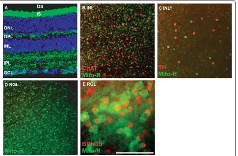
In the Mito-R line, YFP is expressed in retinal gang-lion cells, as evidenced by the presence of YFP in retinal ganglion cell axons, some amacrine cells, horizontal
cells and photoreceptors (Figure 1A and 1C and Table 1 N > 10). The retinal ganglion cell layer (RGL) also con-tains displaced amacrine cells, which may represent as many as half the cells present in this layer. To deter-mine the extent to which YFP-labeled cells in the RGL were amacrine cells versus RGCs, the colocalization of YFP with the transcription factor BRN3b, a marker of approximately 80% of retinal ganglion cells, was assayed. The majority of BRN3b-positive cells were also YFP-positive, indicating that YFP is expressed in the majority of RGCs in the Mito-R retina (Figure 1E). YFP is not expressed in cholinergic or dopaminergic amacrine cells in the Mito-R retina, although other cells within the inner nuclear layer were YFP-positive (Figure 1B).
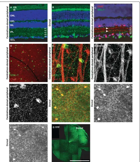
[image:2.595.57.291.100.243.2]In the Mito-Y line, YFP is differentially expressed in retinal domains (N > 10). In the dorsal retina, YFP is expressed in a large number of cell types compared to the rest of the retina (Table 1 and Figure 2G) [10]. In the dorsal retina YFP is expressed in a subpopulation of retinal ganglion, amacrine, bipolar and horizontal cells, Table 1 Mito-YFP expression in retinal cell types
Mito-Z Mito-Y (central/non-dorsal peripheral)
Mito-Y (dorsal)
Mito-R
Ganglion Cells ++ + ++ +++
Smi-32 (+) Cells ++ - + +++
Amacrine Cells ++ - + +
Dopaminergic Amacrine Cells
+++ - +
-Cholinergic Amacrine Cells
- - -
-Bipolar cells +++ - ++
-Horizontal Cells + - ++ +
Photoreceptors + - +++ +++
[image:2.595.58.544.356.674.2]as well as all or nearly all photoreceptors (Figure 2B and 2G). In the central and non-dorsal peripheral retina, YFP is expressed in a very small number of retinal gang-lion cells, which are organized in a loose mosaic pattern (Figure 2A, G and Table 1) [10]. Colabeling sections of Mito-Y retina with ChAT was performed to label the ON-OFF cholinergic bands. YFP positive RGCs lami-nated dendrites adjacent to S2 and S4, and YFP expres-sion was also observed in S1 (Figure 2C). These cells are not cholinergic amacrine cells, as evidenced by the lack of ChAT immunoreactivity, and do not overlap with non-phosphorylated neurofilament (Smi-32) positive alpha cells (Figure 2D and 2E). Dopaminergic cells are YFP-positive in the dorsal Mito-Y retina, but not other portions of the retina (Figure 2F and data not shown).
In the Mito-Z line, YFP is expressed in a subset of ret-inal ganglion, amacrine, horizontal and bipolar cells as well as a small number of photoreceptors (Figure 3A N > 10). Based on the absence of male to male inheritance of the transgene, and exclusive male to female inheri-tance of the transgene, the Mito-Z line insertion site is on the X-Chromosome and is inactivated in roughly half of neurons in the retinas of hemizygous female mice (no transgenic male pups and all transgenic female pups out of over 100 pups from greater than twenty pairs consisting of Mito-Z hemizygous male mice crossed to wild type females) (Figure 3B and 3C) [11]. YFP expression overlapped with non-phosphorylated neurofilament, a marker of alpha retinal ganglion cells, and tyrosine hydroxylase (TH), a marker of dopaminer-gic amacrine cells, but not ChAT, a marker of starburst amacrine cells (data not shown; Figure 3C and Table 1).
Discussion
Three transgenic mouse lines expressing mitochondrially localized YFP in different populations of retinal neurons are described in this study. YFP is expressed in distinct and reproducible cell populations in the retinas of each transgenic line, but the expression pattern is different for each transgenic line, Therefore, these strains are use-ful to researchers seeking to assay different aspects of retinal biology and disease.
Study of Mitochondrial Diseases
The YFP transgene is fused to the mitochondrial locali-zation signal of mouse COX8 (the N-terminal 34 amino acids). Defective mitochondrial transport within retinal neurons is speculated to underlie diseases of both retinal ganglion cells and retinal photoreceptors [12]. The dif-ferential expression of YFP in subsets or all RGCs and photoreceptors offers a tractable system by which researchers studying these processes can visualize mito-chondria either in vivo or in vitro. Such approaches have been applied to motor neurons, and facilitate
studies of mitochondrial morphology, trafficking, and fusion. In the eye, autosomal dominant optic atrophy 1 is caused by mutation of theOPA1gene. OPA1 encodes a mitochondrial inner membrane protein involved in mitochondrial fusion. For studies of diseases such as optic atrophy, the mice described in this study would provide an excellent research tool.
Study of Retinal Neuron Subtype Development
The expression of various combinations of transcrip-tion factors are required for specificatranscrip-tion of different populations of retinal neurons. Transcription factors required for generation of the various basic types of retinal neurons, such as rods or amacrine cells, have been identified; however, it is largely unknown how subtypes of retinal neurons are specified. The availabil-ity of transgenic labels that are expressed in specific subtypes of retinal neurons will simplify identification of such factors by allowing the isolation of specific subtypes of retinal neurons. Purified cell populations can also be used for gene expression profiling to obtain a better molecular definition of the differentia-tion state of the cells, to provide addidifferentia-tional markers for studying the cells, and to better understand the molecular code of surface proteins that confer cell identity. Furthermore, being able to visualize specific cell types in vivo is a great boon to studies of retinal patterning and mosaic formation.
Study of Clonal Proliferation and Lateral Migration
reporter, for example by using both reporters in tandem to compare horizontal migration into and out of differ-entially labeled clonal columns [17]. The X-Chromo-some insertion site of the transgene also simplifies maintaining Mito-Z mice, as male carries crossed to wild type females will sire only transgenic female and wild type male offspring.
In this report we characterize the retinal expression pattern of three transgenic mouse lines. These lines will be useful for a number of various studies, including ana-lysis of mitochondrial trafficking, and will complement a similar line of mice that express of mitochondrial loca-lized CFP in different populations of retinal neurons [18]. An increasing number of transgenic mouse lines offers the ability to label more retinal cell types, permit-ting an increasing amount of experimental flexibility and control [3,7,9,19]. The mice described here add value by labeling new subsets and by labeling mitochon-dria allowing new fields of study.
Materials and methods
Animal care and handling
Mice were used in accordance with protocols approved by the Animal Care and Use Committee at The Jackson Laboratory. The Jackson Laboratory is accredited by AAALAC. Animals were housed in PIV caging and given food and water ad libitum, and maintained on a 14 hour:10 hour light:dark cycle.
Mouse strains
The following mouse strains were used in this study: Mito-R: Tg(Eno2-YFP/Cox8a)RRwb/J [8], Mito-Y: Tg (Eno2-YFP/Cox8a)YRwb/J (Jax stock number 007857) [8], Mito-Z: Tg(Eno2-YFP/Cox8a)/ZRwb (Jax stock number (007860) [8].
Tissue Preparation and staining
[image:5.595.59.536.91.408.2]Mice were transcardially perfused with PBS followed by perfusion with 4% paraformaldahyde buffered with PBS.
Eyes were then fixed for an additional one hour in 4% paraformaldahyde buffered with PBS. Eyes were enu-cleated and sunk in 30% sucrose overnight and then fro-zen in cyropreservation media. Sections of retina were cut at 8 μm thickness with a cyrostat. Sections were rinsed in PBS and mounted with antifade or stained with antibodies.
Immunofluorescence was performed by blocking sec-tions in PBS with 3% normal horse serum and 0.1% tri-ton x-100, and then incubating sections overnight at 4°C with primary antibodies diluted in blocking buffer. Sec-tions were then washed three times for ten minutes in PBS and incubated with secondary antibodies diluted in blocking buffer. Sections were washed three times for ten minutes in PBS, with DAPI included in the last wash, and mounted with antifade media. The following commercially available antibodies were used: mouse anti-nonphosphorylated neurofilament (1:200; Covance Research Products), rabbit anti-tyrosine hydroxylase (1:500 Invitrogen), goat anti-ChAT (1:500, Chemicon) and goat anti-BRN3b (1:200; Santa Cruz Biotechnology).
Whole retinas were prepared by fixing as described above. Retinas were then blocked in PBS, 3% normal horse serum and 0.4% triton X-100. Retinas were incu-bated in primary antibodies diluted in the blocking solu-tion for four days at 4°C and then washed overnight in blocking buffer. Retinas were incubated overnight with secondary antibodies diluted in blocking buffer and washed in blocking buffer for an additional day and then mounted with antifade media. All images were col-lected on a Leica SP5 confocal microscope.
Acknowledgements
This research was funded by research grants to Robert W. Burgess from the ALS Association and NIH EY018605.
Author details
1The Jackson Laboratory, Bar Harbor, ME 04609, USA.2Department of
Biological Sciences and WWAMI Medical Education Program, The University of Idaho, Moscow ID 83844, USA.
Authors’contributions
PGF: Microscopy and writing of manuscript. RWB: Design of Mito-Y transgenic mice and editing of manuscript. All authors have read and approved the final manuscript.
Competing interests
The authors declare that they have no competing interests.
Received: 10 June 2010 Accepted: 6 October 2010 Published: 6 October 2010
References
1. Masland RH:Neuronal diversity in the retina.Curr Opin Neurobiol2001, 11(4):431-6.
2. Coombs J,et al:Morphological properties of mouse retinal ganglion cells.Neuroscience2006,140(1):123-36.
3. Siegert S,et al:Genetic address book for retinal cell types.Nat Neurosci 2009,12(9):1197-204.
4. Badea TC, Nathans J:Quantitative analysis of neuronal morphologies in the mouse retina visualized by using a genetically directed reporter.J Comp Neurol2004,480(4):331-51.
5. Yonehara K,et al:Expression of SPIG1 reveals development of a retinal ganglion cell subtype projecting to the medial terminal nucleus in the mouse.PLoS ONE2008,3(2):e1533.
6. Kim IJ,et al:Molecular identification of a retinal cell type that responds to upward motion.Nature2008,452(7186):478-82.
7. Huberman AD,et al:Genetic identification of an On-Off direction-selective retinal ganglion cell subtype reveals a layer-specific subcortical map of posterior motion.Neuron2009,62(3):327-34.
8. Misgeld T,et al:Imaging axonal transport of mitochondria in vivo.Nat Methods2007,4(7):559-61.
9. Feng G,et al:Imaging neuronal subsets in transgenic mice expressing multiple spectral variants of GFP.Neuron2000,28(1):41-51.
10. Fuerst PG,et al:DSCAM and DSCAML1 function in self-avoidance in multiple cell types in the developing mouse retina.Neuron2009, 64(4):484-97.
11. Fuerst PG,et al:Neurite arborization and mosaic spacing in the mouse retina require DSCAM.Nature2008,451(7177):470-4.
12. Carelli V, Ross-Cisneros FN, Sadun AA:Mitochondrial dysfunction as a cause of optic neuropathies.Prog Retin Eye Res2004,23(1):53-89. 13. Young RW:Cell differentiation in the retina of the mouse.Anat Rec1985,
212(2):199-205.
14. Young RW:Cell proliferation during postnatal development of the retina in the mouse.Brain Res1985,353(2):229-39.
15. Reese BE, Tan SS:Clonal boundary analysis in the developing retina using X-inactivation transgenic mosaic mice.Semin Cell Dev Biol1998, 9(3):285-92.
16. Reese BE, Galli-Resta L:The role of tangential dispersion in retinal mosaic formation.Prog Retin Eye Res2002,21(2):153-68.
17. Reese BE, Harvey AR, Tan SS:Radial and tangential dispersion patterns in the mouse retina are cell-class specific.Proc Natl Acad Sci USA1995, 92(7):2494-8.
18. Emanuela Voinescu P, Kay JN, Sanes JR:Birthdays of retinal amacrine cell subtypes are systematically related to their molecular identity and soma position.J Comp Neurol2009,517(5):737-50.
19. Kim IJ,et al:Laminar restriction of retinal ganglion cell dendrites and axons: subtype-specific developmental patterns revealed with transgenic markers.J Neurosci2010,30(4):1452-62.
doi:10.1186/1756-0500-3-253
Cite this article as:Burgess and Fuerst:Distinct expression patterns of mitochondrially localized YFP in neuronal subsets in the retina of three transgenic mouse lines.BMC Research Notes20103:253.
Submit your next manuscript to BioMed Central and take full advantage of:
• Convenient online submission
• Thorough peer review
• No space constraints or color figure charges
• Immediate publication on acceptance
• Inclusion in PubMed, CAS, Scopus and Google Scholar • Research which is freely available for redistribution