JOURNALOF VIROLOGY,Apr. 1990, p. 1657-1666 0022-538X/90/041657-10$02.00/0
Copyright C 1990,American Society for Microbiology
Cell-Type-Specific Control
Elements of the Lymphotropic
Papovavirus Enhancer
JORN R. ERSELIUS, BIRGIT JOSTES, ANTONIS K. HATZOPOULOS, LUITGART MOSTHAF,t ANDPETERGRUSS*
Department of Molecular Cell Biology, Max-Planck-Institute of Biophysical Chemistry,
Am
Fassberg,
3400Gottingen,
FederalRepublic of Germany
Received 2 October1989/Accepted 26 December 1989
Lymphotropic papovavirus (LPV) exhibits a highly restricted host range, inwhich only cells of primate B-lymphocyte origin are permissive for infection. Its enhancer element contributes to this tropism, since
transcriptional potentiationis confinedtocells ofthe hematopoietic lineage. Nuclearextracts from B and T
cells, but notfrom HeLa cells, contain protein factors that interact specifically with the LPV 63-base-pair
enhancerrepeat,asdemonstratedby DNase Ifootprintingand gelretardation experiments.Within therepeat
three sequence motifs were identified: the core motif, the Pu box, and a novel element named T motif. Functionalanalysis demonstrated that these motifsaswellas some sequencesupstreamoftherepeatcontribute
totheoptimal activityof theenhancer. Therearecleardifferences between thepatternsof binding of theBand
T lymphocyte nuclear proteins tothe enhancerwhichare alsoreflected inthetranscriptional activity ofthe
enhancer in both cell types. Furthermore, the activity of theLPVenhancer and its interaction with nuclear proteinsseemtobe regulated during B-celldifferentiation.
Enhancers are cis-acting regulatory elements that
poten-tiate transcription in a relatively position- and orientation-independentmanner(forareview, seereference 17). One of
the best studied examples is the simian virus 40 (SV40) enhancer (3, 19, 23, 47). It is located in the nontranscribed regulatory region of the SV40 viral genome between the earlyandlategenes. Its major functional region consists ofa 72-base-pair (bp) direct repeat, but approximately 120 bp
upstream of the repeat are also required for optimal
en-hanceractivity (47). The whole enhancerregion consists ofa variety of sequence motifs. Although most single motifs
cannot activate transcription to a considerable degree, oli-gomerizationofamotifcancreate powerfulenhancers (11, 26, 33). Interestingly, these artificial enhancers have distinct
cell specificities. While an oligomer of the core sequence
shows almost the same pattern of activity as the entire enhancer, anoligomerof the"pseudocore" isactive onlyin
CV-1 cells and an oligomerof the Sph-I motifis lymphoid
cell specific (26, 33).
Enhancers have been characterized in alarge number of
cellular genes as well (17). Besides being necessary for appropriate levels of expression,inmany casestheyarealso responsiblefortissue-specific expression of the correspond-ing gene. The best-studied example of a tissue-specific
cellular enhancer is the immunoglobulin heavy-chain gene enhancer, which is activeonlyin Blymphocytes (2, 15, 25).
Like viralenhancers, cellular enhancers consist of different sequence motifs (13). Techniques that can probe DNA-proteininteractions (e.g.,DNase Ifootprinting, gel
retarda-tion, etc.) have shown that these motifs are the targets of
specificnuclear factors (17).
Lymphotropic papovavirus (LPV) belongs to the same familyofpapovavirusesasdoSV40,BKvirus,andJC virus
(27, 37, 48). LPV infectsonly mature B lymphocytes from
primates (41). Itsgenomestructureis almost identicaltothat
* Correspondingauthor.
tPresent address: Max-Planck-Institute for Biochemistry, 8033 Martinsried,FederalRepublicofGermany.
of SV40; the genomes share about 80% homology in their
coding regions (28). Themajor regionsofnonhomologyare
confined to the sequences between the early and late
tran-scriptionalstartsiteswhich, inLPV,asinSV40,encompass
an enhancer. The main feature ofthe LPV enhancer is a 63-bp direct repeat. In contrast to the SV40 enhancer, the LPV enhancer is only active in cells of the hematopoietic lineage and notin cells of fibroblastoid orepithelioid origin (24). In this respect, the LPV enhanceractivity is broader thantheinfectivity ofthevirus itself. Inaccordancewith the in vivo results, itwas shown that the LPV enhancer
acti-vates transcription in vitro when incubated with nuclear
extracts of B and T cells but not HeLacells, ahuman cell line ofepithelial origin (35).
Toexplainthe host range restriction ofLPVand the cell type specificityof the LPVenhancer, wefound itnecessary first to characterize the cis-acting regulatory elements
con-tained within the enhancer and the trans-acting nuclear
proteins that recognize these sequences. Using DNase I footprint analysis, we identified several sequence motifs within the LPV enhancer which interact with nuclear factors present in B and T lymphocytes. Mutational analysis of these motifs demonstrated thattheyarecrucial for enhancer activity. Two of these regulatory elements interact with nuclear factors from both B and Tcells (30, 43). A newly
identifiedsequence motif isspecifically protectedin Tcells.
Oligomerization of the area of the LPV enhancerwhich contains the Pu box and the core sequences creates a powerful cell-type-specificenhancer. Furthermore,both the
activity and the interaction of the LPV enhancer with nuclearproteins areregulated duringB-cell differentiation.
MATERIALS AND METHODS
Cell culture and extract preparation. HeLa cells were
grownin MEM Spinner medium(Flow Laboratories, Inc.)
withlx nonessential amino acids(GIBCO Laboratories);
B-and T-cell linesweregrownin RPMI 1640(GIBCO)with 3 g ofNaHCO3perliter. The mediawere supplementedwith 2 mM glutamine, 50 ,ug of streptomycin per ml, 50 IU of 1657
Vol. 64, No. 4
on November 10, 2019 by guest
http://jvi.asm.org/
1658 ERSELIUS ET AL.
4A
0Cp
3X)
T CP T 200 10MF f- - I .__
on-core ori-core
-- 234bp-LPV-Enhancer
Hac III HacI11
B HA1
'I
\ O-, . NIO,rL 4 -prot.1) C . a I) c
Apflup
ifre4dMA
-
BE~~~~~~
-. - [4
uSl
rCj
14
A
-5|. __
-.
o--
OW-J
C4
L ATAGTGATTTTGCAGACTTG
E TATCACTAAAACGTCTGAAC
BJA-B
p c
CAGACTTCAATAACAGGTTGTTTTTGCAGAATCAACTGAAAGAGGAAGCTGTGGTTAGACTCG
GTCTGAAGTTATTGTCCAACAAAAACGTCTTAGTTGACTTTCTCCTTCGACACCAATCTGAGC
_ AA AA '_ 2
CTCACCGCCTCCAAAG
GAGTGGCGGAGGTTTC
MOLT4
T P C
--- . , __
L ATAGTGATTTTGCAGACTTG CAGACTTCAATAACAGGTTGTTTTTGCAGAATCAACTGAAAGAGGAAGCTGTGGTTAGACTCG
_- _
E TATCACTAAAACGTCTGAAC GTCTGAAGTTATTGTCCAACAAAAACGTCTTAGTTGACTTTCTCCTTCGACACCAATCTGAGC
L.As . A A
lAI'
2CTCACCGCCTCCAAAG
GAGTGGCGGAGGTTTC
J. VIROL.
0/5270
AUG L* early
C
T-TF
on November 10, 2019 by guest
http://jvi.asm.org/
CELL-TYPE-SPECIFIC CONTROL ELEMENTS OF LPV ENHANCER
penicillin per ml, and 10% fetal calf serum. Nuclear extracts were prepared as described before (6, 46), except that for some extracts the nuclei were prepared by the addition of
0.05% Nonidet P-40 instead of mechanical disruption. No
difference in the binding pattern was observed in these cases.
DNase I footprinting assays.The 234-bpHaeIII fragment of LPV (seeFig. 1A) was excised, ligated to BglII linkers, and clonedinto the BamHI site of the SP65 polylinker such that the LPV HaeIII site at position 158 was located near the EcoRI site and the HaeIII site at position 392 was located neartheXbaI site of the polylinker. After restriction within the polylinker, the plasmid was dephosphorylated and end labeled, and the LPV enhancer fragment was subsequently excised by a second restriction enzyme digestion. The uniquely end-labeled fragment was used for DNase I foot-printing assays and sequencing.
DNase Ifootprinting reactions were carried out essentially
as described previously (34, 45). Briefly, 8 ,ul of nuclear extract(80
Rig)
waspreincubated for 20 min on ice with 28 ngofpBR322 (restrictedwith HpaII) in a10-pdvolume
contain-ing20 mM N-2-hydroxyethylpiperazine-N'-2-ethanesulfonic acid (HEPES) (pH 7.9), 20 mM KCl, 1 mM MgCl2, 2 mM
dithiothreitol,and17%glycerol. After preincubation, 2
[lI
of thelabeled template (104 cpm per reaction; approximately 3fmol)wasadded and the samples were incubated for another 10min onice. The samples were heated to 20°C, and DNase
I (1mg/ml; freshly diluted in20%glycerol-1 mM MgCl2-20
mMKCl-1 mM dithiothreitol) was added for 90 s in different amounts (1, 0.5, and 0.25 ,ug per reaction). Reactions were
terminatedby the addition of 200p,l of 0.2% sodium dodecyl sulfate-30 ,g of tRNA per ml-0.6 M sodium acetate and
subsequentextraction with phenol-chloroform. After
precip-itation withisopropanol, the samples were analyzed on 5% urea-polyacrylamide gels with a G-C sequence ladder run in
parallel. Thecontrolreactions, which contained noextract, were treated as described for the samples except that the DNase Iconcentrations used were 20 and 40 ng per reaction. In every case, theprocedure wascarried out with two to four
independentlyprepared extracts.
Gel retardation assays. Oligonucleotideswere end labeled with polynucleotide kinase and purified by electrophoresis
on 10% polyacrylamide gels. Oligonucleotides (5,000 to
10,000 cpm;approximately 0.15 fmol) were incubated with 5 to 8
p.g
of nuclear extract in a total volume of 20pul
of 10 mM HEPES (pH 8.0)-5 mM MgCl2-50 mM KCl-1 mMspermi-dine-0.5 mM
dithiothreitol-17%
glycerol-1 Lg of poly(dI-dC) double-stranded heteropolymer (BoehringerMannheimBiochemicals) for15 minatroom temperature.DNA-protein
complexes were resolved on 5 to 6% polyacrylamide gels
(31). The gels were run for 3 to 4 h at 200 V, dried, and
exposed for 4to 12 hto XARfilm (Eastman KodakCo.).
Cell transfections. Cells were transiently transfected with the Gene Pulser electroporation apparatus (Bio-Rad Labo-ratories) (5, 32). On the day before transfection, the cells were split to adensity of 3 x 10⁵cells per ml. Onthe next day, 2 x 10⁷cells werecollected by centrifugation, washed
once with phosphate-buffered saline, and suspended in 0.8 ml ofphosphate-buffered saline. SupercoiledDNA (20 pug) was added, and the cellsuspension was pulsed at a voltage setting of 250 to 400 V and a capacitance setting of 960 ,uF. The average time constantwas12 to 14 ms. The cells were
kept for10min at room temperatureandtransferred to 50 ml of medium. Approximately48 hfollowing transfection, cells wereharvested bycentrifugation and washed first in 20 ml of 25 mM Tris hydrochloride (pH 7.5)-137 mM NaCl-5 mM KCl-0.7 mM CaCI2-0.5 mM MgCl2-0.6mM Na2HPO4 and subsequently in 1 ml of 0.25 M Tris hydrochloride (pH 7.8). The cells were suspended in 0.15 ml of 0.25 M Tris hydro-chloride (pH 7.8) and sonicated. Cell debris was removed by
centrifugation, and the supernatant(extract)wasassayed for
chloramphenicol acetyltransferase (CAT) activity (16). As-saymixtures for CAT activity determinations contained 70 pIofextract,0.2,Ci of['4C]chloramphenicol(NewEngland Nuclear Corp.), 20
RI
of 4 mM acetyl coenzyme A (Boehr-inger), and 80 pl of 0.25 M Tris hydrochloride (pH 7.8).Sampleswere incubated for 90 minat 37°C, extracted with 0.4 mlofethylacetate,dried under vacuum, andsuspended
in 10
Ku
of ethyl acetate. The acetylated forms of[14C]chloramphenicol
were separated bythin-layer chroma-tography.Oligonucleotides. All oligonucleotides were synthesized
(BioSearch 8700 DNA synthesizer) with threeguanidine or
cytidineresiduesattheendstoallow unidirectionalligation.
Theywere used either forgelretardation experiments or to
clone distinct parts of the LPV enhancer. Forcloning, the
two corresponding single strands were heated together to
95°C and cooled down slowly to room temperature for
annealing. The double-stranded forms were separated from single-stranded forms on 10% polyacrylamidegels, cutout, andelectroelutedin TE(10mMTrishydrochloride [pH 8],1 mM EDTA [pH 8]) buffer. Subsequently, the
oligonucleo-tides were phosphorylated (polynucleotide kinase;
Boehr-inger), ligatedtoBglIIlinkers,and clonedinto theBglII site of p209A21 (16). For gel retardation experiments,
[y-32P]ATP (Amersham Corp.) was usedfor phosphorylation.
The double-stranded forms were isolated after separation
from free nucleotides andsingle-strandedforms on 10 to15%
polyacrylamide gels.
RESULTS
Interaction of cell-type-specific factors with the LPV en-hancer invitro. Transient transfection experiments showed
FIG. 1. (A)Structure andposition ofthe LPV enhancer. The LPV enhancer is situated in thenoncoding region (700 bp)which separates theearlyandlate codingregions oftheviralgenome. Itspositionandorientationwith respecttotheearly regionareindicated. The HaeIII fragment (positions158 to392)containinga63-bprepeat wasshownto act asanenhancerinvivo(24)andin vitro(35).Theprotectedregions
in the DNase Ifootprint analysisaremarkedC,P, and T. ori,Origin ofreplication. (B)Comparisonofprotected regions in the DNase I footprintassayof the LPV enhancerearlystrand with nuclearextractsfromBJA-B,MOLT4,and HeLa cells. The LPV enhancerfragment
wasexcised from the SP65vectorby restriction withEcoRIandXbaI.Thestrandcodingfor the viralearlyproteinswasradioactivelylabeled byafill-in reaction with the Klenowpolymerase. After incubationwith the extracts, the labeled strandwastreatedwithincreasingamounts of DNase 1(0.25, 0.5, and 1.0 ,ug in lanes a,b,and c,respectively). Inthe control lanes(-prot.),DNAwasincubatedonlywith 20to40ng of DNase I. The digestion products were separated on 5% sequencing gels. Protected areas are marked C, P, and T. A, DNase
I-hypersensitive sites. Thepositionsof the63-bprepeats areindicated. (C) Summaryof the results of the DNase Ifootprint analysisof the LPVenhancerfragmentin B- andT-cell nuclearextracts.Thereactions for the late strandwerecarriedout asdescribed for theearlystrand inpanelB.Land E mark the late andearlystrands,respectively.A,DNaseI-hypersensitivesites. The Pu box(P)and thecore(C)sequences areunderscored, theprotected regions (P, C,andT)arebracketed,and broken lines indicateweakerprotection.The resultsareshown for onerepeat. Inthe second repeat thecorresponding regionswereprotected (subscript 2).Thenumberingsystem isthat ofPawlitaetal.(28).
VOL.64, 1990 1659
on November 10, 2019 by guest
http://jvi.asm.org/
that a 234-bp fragment including the LPV enhancer with the 63-bp repeat (Fig. 1A) wasactivein cells of the hematopoi-etic lineagebut not in epithelial cell lines (24). To identify
factors that interact with the LPV enhancer, we prepared nuclear extracts from the human cell lines BJA-B, a
Bur-kitt'slymphoma cell linerepresenting mature Blymphocytes
(20), MOLT 4, an acute T-cell lymphoblastic leukemia cell
line (22), and HeLa cells of epithelial origin (14) by
previ-ouslydescribedmethods (6, 45) and used them in the DNase Ifootprint analysis (12). The 234-bpfragment was radioac-tively end labeled, incubated with the different nuclear extracts, and partially digested with increasing
concentra-tions of DNase I.
Figure 1B shows the protected areas within the 63-bp repeat onthe strandcoding for theearlyviral proteins.With
the BJA-B extracts, an 18-bp regioncontaining the Pu box (marked P) and the coremotif (marked C) wasprotected in
bothrepeats.Thisregionincluded a DNaseI-hypersensitive
site consistingof twocytidineresidues (motifsPand C;see
below). When the strand coding for the late viral proteins
was labeled, a regionspanning 22nucleotides was protected. At the same position as in the strand coding for the early proteins two DNase I-hypersensitive sites were observed
(data not shown). Incubation with the MOLT 4 extracts
resulted in a footprint in the same area (Fig. 1B), but the protected sequence was more extended, indicating that a
modifiedfactor or a different factor was binding in T cells. In
addition, asecond protected region (marked T) of 16
nucle-otides was found (Fig. 1B). In contrast to the results
ob-tainedwith the B- and T-cell extracts,noprotected domains
could be seen in footprint experiments with HeLa nuclear
extracts (Fig. 1B). The same results were observed with different extract preparations. The data from the DNase I
footprint analysis aresummarized in Fig. 1C.
The protected region common to B and T cells contains
the core motifand the Pu box. The core motifis found in many viral and cellular enhancers (43), and the Pu box, a
purine-rich region, is found in many lymphoid cell-specific transcriptionalelements, suchastheimmunoglobulin
heavy-chain gene enhancer (1), the 5' areaofthe major histocom-patibility complex class II gene (7), and the SV40 enhancer (30). In contrasttothecoremotifand the Pubox,theTmotif
has notbeen described before.
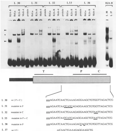
Mutational analysis of the LPV enhancer in vitro. To analyze the sequence requirements for the binding of the nuclear factors in more detail, we synthesized and tested several 39-bp oligonucleotides spanning the area of the Pu box and the core motif in gel retardationassays with nuclear extractsfrom BJA-B, MOLT 4, and HeLa cells (Fig. 2). The
oligonucleotide termed L 30 represents the wild-type
se-quence(Pubox and core), while L 31, L 32,L33,and L38
contain different introduced mutations. L 37 contains only
the Pu box wild-type sequence.
The wild-type oligonucleotide (L 30) formed a specific
complexwithanuclearprotein present in BJA-B cells (lower arrowhead in Fig. 2). The same complex was found with nuclear extracts from the B-cell line Namalwa, a human Burkitt's lymphoma cell line (data not shown). This factor was present only in very small amounts in MOLT 4 and HeLa extracts. On the other hand, the MOLT 4 extract containedadifferentfactor that formed a weak protein-DNA complex and that was not seen with the other extracts (upper arrowhead inFig. 2). No specific complex could be detected with HeLa nuclear extracts. The specificity of binding was tested in competition experiments in which only the wild-typeoligonucleotide (L 30)competed effectively forbinding
(data not shown). One band was detected with all oligonu-cleotides (asterisk in Fig. 2) and was most likely due to
binding of a protein to threeprotruding guanosineorcytidine residues which were introduced into all oligonucleotides to
facilitate unidirectional cloning (36). The amount of this proteinvaried according to the conditions of cellgrowthand nuclear extractpreparation (see, forexample, thetwo BJA-B nuclear extracts in Fig. 2).
Twoindependent mutations in the Pu box region(L 31 and L 38;Fig. 2) led to adramatic decrease in thebinding of the B- and T-cell-specific factors, confirming the specificity of thefactorsbinding to the Pu box. Surprisingly,amutation in the core motif (L 32) had no effect on the binding of the cell-specific factors. The altered G residue was previously shown to be critical for the function of the core motif in the SV40 enhancer (43). Simultaneous mutations in both the Pu box and the core motif (L33) resulted inapatternsimilarto
the one observed with oligonucleotides L 31 and L 38, in which only the Pu box was changed. These results demon-strate that the Pu box sequences were sufficient for the
binding of the cell-type-specific factors seen in the gel
retardation assays. Toconfirmtheseresults,wesynthesized
a shorteroligonucleotide (L 37)containing thewild-type Pu box but not the core motif. This oligonucleotide was suffi-cient to form theB-cell-specific complex with BJA-B nuclear extracts (Fig. 2).
Inadditiontothedescribedcomplexes,faintprotein-DNA
interactionswerefound with the L 30wild-type oligonucle-otide butwerenotfurther investigated. The introduction of mutations into thisoligonucleotide (L31, L32, L33,and L 38) gave rise in some cases to new, faint complexes which had mobilities different from those of the complexes elimi-nated by the mutations. Thesecomplexes might have been due to weak interactions withproteinsthat tookplace when the specific factors did not bind or, alternatively, to the creation ofnew bindingsites.
Anoligonucleotide spanningtheTmotifwasalsotested in gel retardation assays. Nospecific complexes were formed with any of the extracts, indicating that this sequence by itselfwas not sufficient forbinding (datanot shown).
Characterization of the LPV enhancer in vivo. To test
whether the binding sites identified in vitro contributed to
the activity of the LPV enhancer in vivo, we introduced
deletions and specific mutations and cloned the
correspond-ing fragments in front of the SV40 promoter of plasmid
p209A21 driving the cat gene (21). The constructs were
introduced by electroporation into BJA-B, MOLT 4, and HeLa cells. In all transfection experiments, the parental
vector p209A21 was used as a negative control and pSV2CAT (16), which contains the SV40 enhancer, was
used as apositive control. Plasmid pLPV CATS(24), which contains the entire LPVenhancer,wasalso used. In B and T cells, all constructs were compared with pLPV CATS, the activity of which was set arbitrarily at 100%. The different
constructs are shown in Fig. 3A.
As previously described, pLPV CATS is active in BJA-B andMOLT4cells butnotin HeLacells (24). InHeLacells, activity was not detectable with any of the LPV enhancer constructs,whereas theSV40 enhancerstimulated
transcrip-tion considerably (data not shown). The two63-bp repeats alone without further 5' or 3' sequences (pLPV 1) were
almostas active as thewild-type enhancer in MOLT4cells
(Fig. 3A). In BJA-B cells, the activity was reduced to80%.
A single 63-bp repeat without any surrounding sequences (pLPV 4) was not able to stimulate transcription in BJA-B
cells, andonly 10%of theactivitywas maintained in MOLT
on November 10, 2019 by guest
http://jvi.asm.org/
CELL-TYPE-SPECIFIC CONTROL ELEMENTS OF LPV ENHANCER
1 30
1.
311.
32 1,33 1 38r ----
I.~~l
_- 1r- - IIr-
I1
I 1I
-V
---,Cl;-,
ALaf
=V -r
_ _ t
c- - :: :;
=
-_
C_, , ;z
_ V -r
_ o _~~~~~~~~~~
U _ _i
C
_~
_ oa "p
-; -
-a
A_
W a * .s
a a SP
_ t
* __NMW
_ uO
.~e
_--A
iso asup aNoN
A
T %
Pn'I
C~r.55^^55 +tv 55 Z55^ ^ mnm', oo^oRsRR^5^^ ^)5^x R R R
IJ 30 wL[P+C]
L 31 mutationin P
L 32 mutation inC
L 33 mutation inP+C
L 38 mutation inP
L, 37 wt{Pl
gggAGAATCAACTGAAAGAGGAAGCTFGTGG1T7AGACTCG
gggAGAATCAATCATCAGAGGAAGCTGTGGfTAGACTFCG
gggAGAATCAACTGAAAGAGGAAGCJTGTAATTAGACTCG
gggAGAATCAATCATCAGAGGAAGCTGTAATTAGACrCG
gggAGAATCAACT7GAAAGAGTA5§GCT7GTGGTTAGACTCG
[image:5.612.101.509.70.530.2]ccCAACTGAAAGAGGAAGCFG
FIG. 2. Comparison of thepatternsof binding of nuclearextractsfromBJA-B, MOLT4,andHeLacellstothe Pu box-core regionofthe LPVenhancer ingel retardationassays.The gel retardationassays werecarriedoutasdescribed in Materialsand Methods. Thesequences
ofthesynthetic double-stranded oligonucleotides spanning the Pu box andcore sequences areshown. Mutated nucleotides in oligonucleotides
L31,L32, L33, and L 38 areunderlined. L 37contains only Pu box sequencesandwasusedtodetermine whetherthePu box itself is sufficienttobindtheB-cell-specific factor (rightmosttwolanes). For thewild-type(wt)oligonucleotide L 30, the control (-protein) is shown in Fig. 4, and the result for two independently prepared BJA-B extracts is shown here instead. The upper arrowhead indicates the
T-cell-specific protein-DNA complex, and the lower arrowhead indicates the B-cell-specific complex. Thecommonband(asterisk)inall lanes
is duetononspecific binding ofafactortothe protrudingggg or ccc(corresponding strand) ends oftheoligonucleotides.L 37contains only twoc's.
4 cells. In contrast, one repeat plus 32 bp of upstream sequences (pLPV 2) was active in BJA-B cells (30%) and
MOLT 4 cells(85%). Thus, thepresenceoftwocopies of the
repeat is necessary for full activity of the enhancer, but
upstream sequences contribute tothetranscriptional
stimu-lation aswell. The activity of theupstream sequences was
mostpronouncedin Bcells,whereas in Tcells the effectwas
observed only when one of the repeats was present. The sameresultswereobtainedindependent of the orientation of the inserted fragment.
Mutations in the Pu box (pLPV 3 and pLPV 6; same
mutationsasin the L38oligonucleotide;seeabove)resulted
in a complete loss ofactivity inboth BJA-B and MOLT 4 cells, confirmingtheimportanceof the Pu box for enhancer
activityinvivo. Mutationswereintroduced into thecoreand T motifs in pLPV 4 (constructs pLPV 7 and pLPV 5, respectively). Both constructs were inactive in MOLT 4
cells, demonstrating that both the T and the core motifs contribute toenhancer activity in Tcells.
It was previously shown that enhancers with a distinct BJA-B
Is
0 f fl"I e
It.
"I..
4
_
VOL.64, 1990 1661
on November 10, 2019 by guest
http://jvi.asm.org/
pSV2CAT
EcoRI Bg611
p209A21 polylinker
pLPV
CATS
_ 6 3 3,pLPV 1
EoIj6
3'I
:3""1j
T PC
pLPV 2s pLPV 2as
C T
pLPV3
-pLPV 4s T PC
pLPV 4as 6
PC
pLPV 5
T C
pLPV 6
T P
pLPV 7 Liii
PCPCPCPC pLPV 8
c c c c
pl,PV 9
in
BJA-B MOLT 4
400 85
2
80
30
4
2
2
2
2
2
2 5
2
1u
100
85
2
10
2
2
2
100
30
B
TP
C
pLPV 2 GCTAGGGTTGCCATAGTGATTTTGCAGACTTG
I CAGACTTCAATAACAGGTTGTTTTTGCAGAATCAACTGAAAGAGGAAGCTGTGGTTAGACTCGI
T p C
T P C
pLPV 4 GCTAGGGTTGCCATAGTGATTTTGCAGACTTG CAGACTTCAATAACAGGTTGTTTTTGCAGAATCAACTGAAAGAGtAcGCTGTGGTTAGACTCG
T P C
pLPV 4 LCAGACTTCAATAACAGGTTGTTTTTGCAGAATCAACTGAAAGAGGAAGCTGTGGTTAGACTCG]
T P C
pLPV 5 [CAGACTTCAcTcACAGGTTGTTTTTGCAGAATCAACTGAAAGAGGAAGCTGTGGTTAGACTCG]
T P C
pLPV 6 [
CAGACTTCAATAACAGGTTGTTTTTGCAGAATCAACTGAAAGAGtAcGCTGTGGTTAGACTCGI
T P C
pLPV 7 [CAGACTTCAATAACAGGTTGTTTTTGCAGAATCAACTGAAAGAGGAAGCaG2GGTTAGACTCG]
1662
on November 10, 2019 by guest
http://jvi.asm.org/
CELL-TYPE-SPECIFIC CONTROL ELEMENTS OF LPV ENHANCER
tissue specificity could be obtained by oligomerization of certain sequence motifs (13, 26, 33). We were interested in determining whetheroligomerization of the area containing the Pu box and core motif could result in an enhancer
element. The wild-type oligonucleotide L 30 (Pu box and core)and the mutant oligonucleotide L 31 (mutated in the Pu
box) were oligomerized four times, inserted in p209A21 (constructs pLPV 8 and pLPV 9, respectively), and
trans-fected into BJA-B, MOLT 4, and HeLa cells. Both con-structs were inactive in HeLa cells (data not shown). The wild-type tetramer was a very strong enhancer in T cells but wasinactive in BJA-B cells. The mutated tetramer was only 30% asactive asthe wild-type tetramer in T cells. Surpris-ingly, the mutated tetramer showed comparable activity in B and Tcells (Fig. 3A).
LPVenhancer activity duringB-lymphocyte differentiation. B lymphocytes follow a precise program of differentiation. In pre-Bcells, onlyimmunoglobulin,.heavy-chain genes are expressed. In later stages, transcription of the K or X light
chains followsandimmunoglobulin Mmolecules appear on the surface of virgin B,] cells. These cells give rise first to mature
B,
+,
cells and, following antigen stimulation, to blast and plasmacells (4).The transcriptionalfactor NF-KB, involved in the
expres-sionof theimmunoglobulingenes, is only present in mature B cells, whereas the factor OTF-2 is also present in earlier stages (34, 38-41, 44). Since it has been demonstrated that sequences homologous to the Pu box are also present in the immunoglobulin heavy-chain enhancer and are required for
activity(30), we wereinterestedin determining whether the LPVenhancer is regulated during B-cell differentiation and whether the activity correlates with the presence of the
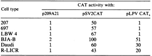
corresponding nuclear factors.To answerthefirst question,
wetransfected several cell lines representing different stages
ofB-lymphocyte differentiation with plasmids pLPV
CAT,
andpSV2CAT(asacontrol). Theactivityinall celllineswascompared with the activity of pSV2CAT in BJA-B cells, which was set at 100. The SV40 enhancer strongly stimu-lated transcription in all cell lines, whereas the LPV
en-hancerwasonly activeinmatureBcells (BJA-B andDaudi)
aswellasin theplasma cell line R-LICR (Table 1). In these three cell lines, the activity of the LPV enhancer was
significantlylower compared with that of the SV40 enhancer, but theratioof SV40 to LPV enhanceractivity was constant. In pre-B cells (cell lines 207 and 697) or in cells in an early stage of differentiation (LBW 4) (8, 10, 18), the LPV
en-hancerwas notactive.
Subsequently,
nuclearextracts from BJA-B cells(repre-senting mature B cells) and 207 cells (representing pre-B
cells) were compared in gel retardation assays. The
B-cell-specific factorbinding tothe Pubox(arrowhead in Fig. 4) was much more abundant in the mature B-cell line BJA-B. The pre-B cell line 207 contained only low levels of the
factor. Asa control, an oligonucleotide containing the
XP-TABLE 1. LPV enhanceractivityduringB-lymphocyte differentiationa
CATactivitywith: Celltype
p209A21 pSV2CAT pLPVCAT,
207 1 50 1
697 1 57 1
LBW 4 1 67 1
BJA-B 2 100 51
Daudi 1 60 30
R-LICR 1 43 20
aCell linesindifferent stages ofB-lymphocyte differentiationwere
trans-fectedwithplasmid pLPVCAT,.Cell lines207 and697 arepre-B cells.LBW 4 is a cell lineinan early stageofdifferentiation, while Daudi and BJA-B
represent mature B cells. R-LICR is a plasma cell line. p209A21 and
pSV2CATwretransfectedinparallel as controls. The activity of pSV2CATin
BJA-B cells was set at 100, corresponding to 85% CAT conversion (for details, seeMaterialsand Methods).
1-binding site of the immunoglobulin ,. heavy-chain gene enhancer was used in these experiments (9). This
oligonu-cleotidedetected equalamountsof thecorresponding factors inbothcelllines,indicatingthat thedifference observedwas
specific forthe Pubox-binding protein.
DISCUSSION
LPV represents an ideal system for studying cell-type-specific gene expression. Its enhancer, a 234-bp fragment containing a direct repeat of 63 bp, is active only in cells of the hematopoietic lineage ofprimates (24). The aim of this study was to identify and characterize the cis-acting ele-ments and theircorresponding trans-acting factors that are responsible for this tissue specificity and to compare the requirements for enhanceractivity in B and T lymphocytes. The results presented here demonstrate that three sequence
elements, namely, the core, the Pu box, and the T motif, bind nuclearproteins present only inlymphoid cells. Muta-tional analysis showed that these motifs all contribute to
enhancer activity. Besides the two 63-bp repeats which contain these threetranscriptional elements, sequences out-side the repeats also contributetooptimal enhancer activity. Thus, the LPVenhancer,likemosttranscriptionalelements, consists of different sequence motifs. Since it iscomposedof
only a small number of motifs which are active only in lymphoid cells, it is not surprising that the activity of the whole enhancer isalso restrictedtothesecells. Incontrast to
this very specific enhancer, the SV40 enhancer is highly active in a variety of cell types. In this case, many motifs, such as the core, the pseudocore, the Sph-I motif, the octamer, the Pu box,andthe Pmotif, contribute in various
extents toenhancer activity (19, 26).
The three motifsprotected in thefootprint analysisof the LPVenhancerarethe Pubox,the core, and theTmotif. The
FIG. 3. (A)Functional analysisof the LPV enhancerby theCATassay. For alltransfectionexperiments,20p.gofsupercoiledDNAwas used. The DNA wasintroducedinto the cellsbyelectroporation, and 48 h aftertransfectiontheCATextracts wereprepared(fordetails,see Materials andMethods).TheCATactivityof the LPV enhancer (pLPV
CATJ)
was setin all celllinesat100%. pSV2CAT andp209A21were used aspositiveandnegative controls,respectively. Onthe left side of thepanel, the LPV constructs usedfor transfectionareshown.The threemotifs,Tmotif(T), Pubox (P), and core motif(C),areindicated. Asterisks indicate mutated sequences. On therightside of thepanel,the CAT activities ofthedifferentconstructs(aspercentages)arecomparedwith thatofpLPV
CAT,.
pLPVCAT5represents thewild-typeenhancer, pLPV1contains thetwo63-bpwild-typerepeatswithout anysurroundingsequences,pLPV8isa tetramerofoligonucleotide L 30,andpLPV9isatetramerofoligonucleotide L 31(Fig. 2).Thesubscripts "s"and "as" refertosenseandantisense,respectively. (B) Sequences ofthe LPVCATconstructsusedfortransfection inBJA-B,MOLT4,and HeLa cells.pLPV2andpLPV4containonlywild-type
sequences. Inthe other constructs, theintroducedmutationsareunderlined. Allconstructs weremade withsyntheticoligonucleotides. The brackets indicatetheprotectedareasin the DNase Ifootprintanalysis (Fig. 1B and C).
VOL.64, 1990 1663
on November 10, 2019 by guest
http://jvi.asm.org/
[image:7.612.310.554.98.189.2]_ -
-mu1
.-FIG. 4. Binding of nuclear factors tothe Pubox-core regionof the LPVenhancer in thepre-B cell line 207and the matureB-cell lineBJA-B. ThesequenceofoligonucleotideL30isgiveninFig.2. Intheleftmost lanes of each panel theoligonucleotidewasincubated withoutextract.207 isapre-B cellline, and BJA-B isamatureB-cell line. The arrowhead indicates the B-cell-specific complex formed with Puboxsequences. TheXP-1-binding site of the immunoglob-ulin ,. heavy-chain gene enhancer was incubated with the same
extractsasacontrol.
Pubox isfoundin alarge numberoflymphoid cell-specific transcriptionalelements, suchasthemajor histocompatibil-ity complexclass IIgenesand theimmunoglobulinenhancer (1, 7). Itwasfirstidentified in theSV40enhancer, inwhich it is found approximately 120 bp upstream of the 72-bp
repeats. Duplication ofa DNA segment containing the Pu box creates a lymphoid cell-specific enhancer (30). In the case ofLPV, oligomerization of the Pu box-core sequence alsocreates acell-type-specific enhancerbut,incontrastto that in SV40, it is active only in T cells. As the DNase I
footprinting and gel retardation experiments showed, dif-ferentfactorsarebound in B and Tlymphocytes. It hasbeen shown for theoctamermotif that the samesequencebindsa plethora of factors which can, in turn, mediate different
tissue specificities (36). Recently, a 100-kilodalton protein
from mouse lymphocytes that interacted with sequences similartothe Puboxandpresentintheimmunoglobulin
CR
enhancerwas purifiedand showntomediate
immunoglobu-linenhanceractivity (1); however,it is unclear whether this factor binds the LPV Pu box. As the in vivo experiments demonstrated, the Pu box is essential for B- and T-cell activity,althoughtheoligomer works onlyin Tcells. Muta-tions inthePuboxleadtoadramatic decrease in activity in
both cell types. So, although the Pu box is required for activity in B cells, it probably requires othersequences for
itsactivity.Ifthesesequences arenotpresent,thebinding of
thefactormightbe inhibitory.
The Pu box factor seems to be regulated during B-cell differentiation. Strongbinding activityis detected inmature B cells but not in pre-B cells. In this respect, the Pu
box-binding
protein
resembles nuclearfactorNF-KB,whichisinduced inmature B cells(38, 42),andnot
OTF-2,
which isalsopresentinpre-B cells. Itisnotyetclearwhetherthe Pubox-binding
factorcanbeinduced inother cell types, like NF-KBcan(38).
The LPVenhancerisonly
activein Bcells,
such asBJA-B,
that show strongbinding
of thefactor,
providing
additional evidence that the Pu box is a crucial elementfortranscriptional
activity.
It willbeinteresting
toseeif the Pu box interacts withproteins ofother cells of the
hematopoietic lineage.
Moreover,it willbeinteresting
tosee ifoligomerizedL30 is active in these cellsorif itsactivityisrestricted toT
lymphocytes.
Thecore motif isfoundinmanytranscriptional
elements,
and oligomers of the coremotifare active inawidevariety
of cells (26). However, the core motif found in the LPV enhancer binds
lymphoid cell-specific proteins since,
incontrast to the results with nuclear extracts of
lymphoid
cells, it is not
protected
in the DNase I footprintanalysis
with HeLaextracts. Inaddition,anoligomerof the mutated
oligonucleotide
L 31, which contains a tetramer of thewild-typecoresequence, isactive inB and Tcells butnotin HeLa cells. Interestingly, the LPV core is identical to the core in the immunoglobulin enhancer, and mutational
anal-ysis
showed that the core contributes toimmunoglobulin
enhancer
activity
(29). It is noteworthythat in theimmuno-globulinC,u enhancerthe Pu box and thecoremotifarealso
found in close
proximity
andthecore representsthestrong-est homology between these two lymphoid
cell-specific
elements (34).
Although the core motif is clearly protected in the
foot-printing experiments,
anoligonucleotide
consisting only of the core sequence does not bind proteinsin thegelretarda-tionexperiments. Itisprobablethat thecore-bindingprotein
needs additional sequences not present in the
oligonucleo-tide and/or that the binding of proteins outside the core sequenceisrequired. This might alsobetruefor the T motif: the same sequence which is protected in the DNase I
footprinting experiments with T-cell nuclear extracts does
notformacomplex with nuclearproteins in the gel
retarda-tion assays. Mutational analysis proved that this sequence,
like the Pu box and thecore motif, contributes toenhancer
activity in T lymphocytes. However, computer analysis
shows thatno sequence homologous tothe T motif hasbeen
described todate in any regulatory element.
Thus, allthree motifsidentified in the 63-bp repeatsof the LPV enhancer by in vitro experiments are involved in mediating transcriptional activityinacell-type-specific
man-ner. Inaddition, sequencesupstream of the repeats
contrib-ute to the overall activity. The results presented here de-scribe the activity of the LPV enhancer in B and T cells. Since the LPV enhancer is also active in other celltypes of the hematopoietic lineage, such as erythrocytes, macro-phages, and granulocytes (24), furtherinsights into its activ-ity should comefrom characterizingcis-acting elements and the corresponding trans-acting factors present in these cells and comparing them with the ones analyzed in B and T
lymphocytes. Moreover, purification and cloning of the proteins interacting with the LPVenhancer andregulating its activity will helptobetter understandits celltypespecificity and regulation oftranscription in cells of the hematopoietic lineage.
ACKNOWLEDGMENTS
The firsttwoauthors contributed equally tothis work.
We thank Steven R. Bauer for providing several mouse and
humanB-cell lines. We alsothank R. Altschaffel forphotographic
I.3
.=I_
-- ..
-AM"I
...G:.-, :-'ad
f.
.1
on November 10, 2019 by guest
http://jvi.asm.org/
[image:8.612.67.307.64.334.2]CELL-TYPE-SPECIFIC CONTROL ELEMENTS OF LPV ENHANCER work, H.-P. Geithe for synthesis of oligonucleotides, and G.
Dressler, M. Goulding, A. Puischel, and H. Scholer for helpful commentsonthemanuscript.
J.R.E. and B.J. were supported by the Max-Planck Society. A.K.H. was a recipient of a postdoctoral fellowship from the
European Molecular Biology Organization. This work was
sup-ported byBundesministeriumfurForschung undTechnologie. LITERATURECITED
1. Araki, K., H.Maeda, J. Wang, D. Kitamura, andT.Watanabe. 1988. Purificationofnucleartrans-actingfactorinvolvedin the regulatedtranscriptionofahumanimmunoglobulinheavychain
gene.Cell53:723-730.
2. Banerji, J., L. Olson, and W.Schaffner. 1983. A lymphocyte-specific cellular enhancer is located downstream of the joining region inimmunoglobulinheavychaingenes.Cell33:729-749. 3. Banerji, J., S. Rusconi, and W. Schaffner. 1981.Expressionofa
P-globin gene is enhanced by remote SV 40 DNA sequences.
Cell27:299-308.
4. Blackwell, T. K., and F. W. Alt. 1988.Immunoglobulingenes, p.
1-60. In B. D. Hames and D. M. Glover (ed.), Frontiers in molecular biology. Molecular immunology. IRL Press Ltd., Washington, D.C.
5. Chu, G., H.Hayakawa, and P. Berg. 1987. Electroporationfor theefficienttransfectionofmammaliancells with DNA. Nucleic Acids Res. 15:1311-1326.
6. Dignam, J. D., R. M. Lebovitz, and G. Roeder. 1983. Accurate transcription initiation by RNA polymerase II in a soluble
extract from isolated mammalian nuclei. Nucleic Acids Res. 11:1475-1489.
7. Dorn,A., H.J. Fehling, W. Koch, M. Le Meur, P. Gerlinger, C. Benoist,and D. Mathis. 1988. B-cellcontrol regionatthe 5'end ofamajorhistocompatibility complex class IIgene:sequences
and factors. Mol.Cell. Biol. 8:3975-3987.
8. Edwards, P. A. W. 1981. Someproperties and applications of monoclonal antibodies. Biochem. J. 200:1-10.
9. Ephrussi,A.,G. M. Church, S. Tonegawa, andW.Gilbert. 1985. B lineage-specific interaction ofan immunoglobulin enhancer withcellular factors in vivo. Science227:134-140.
10. Findley, H. W., Jr., M. D. Cooper, T. H. Kim, C. Alvarado, and A.H.Ragab. 1982. Twonewacutelymphoblasticleukemia cell lines withearlyB-cell phenotypes. Blood60:1305-1309. 11. Fromental, C., M. Kanno, H. Nomiyama, and P. Chambon.
1988. Cooperativity andhierarchical levels of functional
orga-nizationinthe SV 40enhancer. Cell 54:943-953.
12. Galas,D.,and A.Schmitz. 1978. DNase Ifootprinting:asimple
method for the detection ofprotein-DNA binding specificity. Nucleic Acids Res. 5:3157-3170.
13. Gerster,T., P.Matthias, M.Thali, J. Jiricny, and W. Schaffner. 1987. Cell type-specificity elements of the immunoglobulin heavy chaingeneenhancer. EMBO J. 6:1323-1329.
14. Gey, G. O., W. D.Coffman, andM. K. Kubicek. 1952. Tissue culture studies of the proliferative capacity of cervical
carci-nomaandnormalepithelium. Cancer Res. 12:264-265.
15. Gillies, S. D.,S. L. Morrison, V. T. Oi, and S.Tonegawa. 1983. Atissue-specifictranscription enhancer element is located in the majorintronofarearrangedimmunoglobulin heavy chaingene.
Cell33:717-728.
16. Gorman, C. M., L. F. Moffat, and B. H. Howard. 1982. Recombinantgenomes which express chloramphenicol
acetyl-transferase inmammalian cells. Mol. Cell. Biol. 2:1044-1051. 17. Hatzopoulos, A. K., U. Schlokat, and P. Gruss. 1988. Enhancers
and other cis-acting regulatory elements, p. 43-96. In B. D. HamesandD.M.Glover(ed.),Frontiersinmolecularbiology. Transcription andsplicing. IRLPressLtd., Washington, D.C. 18. Hendershot, I., and D. Levitt. 1982. Differentialregulation of
membrane and secretory 1L chain synthesis in human B cell lines.J.Exp. Med. 156:1622-1634.
19. Herr,W., and J. Clarke. 1986.TheSV 40enhancer iscomposed ofmultiple functional elements that can compensate for one
another. Cell45:461-470.
20. Klein, G., B. Giovanella, A. Westman, J. S. Stehlin, and D. Munford. 1975. An EBV-genome-negativecell lineestablished
fromanAmerican Burkitt
lymphoma;
receptorcharacteristics,EBV infectibilityand permanentconversion intoEBV-positive
sublinesbyin vitroinfection. Intervirology5:319-334.
21. Laimins, L. A., G. Khoury, C. Gorman, B. Howard, and P. Gruss. 1982.Host-specificactivation oftranscription bytandem repeats from simian virus 40 and Moloney murine sarcoma virus. Proc. Natl.Acad. Sci. USA 79:6453-6457.
22. Minowada, J., T. Ohnura, and G. E. Moore. 1972. Rosette-forminghuman
lymphoid-cell
lines. I. Establishment and evi-dencefororiginofthymus-derivedlymphocytes.J. Natl. Can-cerInst. 49:891-895.23. Moreau,P.,R.Hen,B.Wasylyk,R.Everett,M. P.Gaub,and P. Chambon. 1981.The SV40 72bprepeathasastrikingeffecton geneexpressionbothinSV40and otherchimericrecombinants. Nucleic AcidsRes. 9:6047-6068.
24. Mosthaf, L., M. Pawlita,and P. Gruss. 1985. A viral enhancer element specificallyactivein human haematopoieticcells. Na-ture(London)315:597-600.
25. Neuberger, M. S. 1983.Expression andregulation of
immuno-globulin heavy chain gene transfected into lymphoid cells. EMBOJ. 2:1373-1378.
26. Ondek, B.,A.Shepard,and W. Herr. 1987. Discreteelements withintheSV40enhancerregiondisplay differentcell-specific
activities. EMBOJ.6:1017-1025.
27. Padgett,B.L.,C. M.Rogers,and D. L. Walker. 1977.JCvirus,
a human polyomavirus associated with progressivemultifocal
leukoencephalopathy: additional biological characteristics and
antigenic relationships. Infect. Immun. 15:656-662.
28. Pawlita, M.,A.Clad, andH.zurHausen. 1985.CompleteDNA sequence of
lymphotropic
papovavirus: prototype of a newspeciesofthe polyomavirusgenus. Virology
143:196-211.
29. Perez-Mutul,J.,M. Macchi,and B.Wasylyk. 1988.Mutational
analysis ofthe contribution of sequence motifs within the Ig
enhancer to tissue specific transcriptional activation. Nucleic AcidsRes.
16:6085-6096.
30. Petterson, M., and W. Schaffner. 1987. A purine-rich DNA sequencemotifpresentinSV 40and
lymphotropic
papovavirusbinds a
lymphoid-specific
factor and contributes toenhanceractivity inlymphoidcells. GenesDev. 1:962-972.
31. Piette,J.,M. H. Krysze, and M. Yaniv. 1985. Specific interac-tion of cellularfactors with the B enhancerofpolyomavirus.
EMBOJ.4:2675-2685.
32. Potter, H., L. Weir, and P. Leder. 1984. Enhancer-dependent expression ofhuman K immunoglobulin genes introduced into mousepre-Blymphocytes byelectroporation.Proc.Natl.Acad. Sci. USA 81:7161-7165.
33. Schirm, S., J. Jiricny, and W. Schaffner. 1987. The SV 40 enhancercan bedissected into multiple segments,each witha different cellspecificity. GenesDev. 1:65-74.
34. Schlokat, U., D. Bohmann, H. Scholer, and P. Gruss. 1986. Nuclear factorsbindingspecificsequences within the
immuno-globulin enhancer interact differentially with other enhancer elements. EMBOJ. 5:3251-3256.
35.
Scholer,
H.R.,andP. Gruss. 1985.Celltype-specifictranscrip-tionalenhancement in vitrorequiresthepresenceoftrans-acting
factors. EMBOJ.4:3005-3013.
36.
Scholer,
H.R.,A. K.Hatzopoulos,R.Balling,N.Suzuki,andP. Gruss. 1989. A family of octamer-specific proteins presentduring mouse embryogenesis: evidence for
germline-specific
expressionofanOct factor. EMBOJ. 8:2543-2550.
37. Seif, I., G.Khoury,and R. Dhar. 1979. Thegenomeofhuman
papovavirus BKV.Cell 18:963-977.
38. Sen, R.,andD.Baltimore. 1986. InducibilityofK immunoglob-ulin enhancer binding protein NF-KB by a
posttranslational
mechanism.Cell47:921-928.39. Singh, H., R. Sen, D. Baltimore, and P. A. Sharp. 1986. A nuclear factor that binds aconserved sequence motifin tran-scriptional control elements ofimmunoglobulin genes. Nature
(London)319:154-157.
40. Staudt,L.M.,H.Singh,R.Sen,T.Wirth,P. A.Sharp, andD. Baltimore. 1986. A lymphoid specific protein binding to the octamermotifofimmunoglobulingenes. Nature
(London)
323: 640-643.1665 VOL. 64,1990
on November 10, 2019 by guest
http://jvi.asm.org/
41. Takemoto, K. K., A. Furuno, K. Kato, and K. Yoshiike. 1982. Biological and biochemical studies of African green monkey lymphotropic papovavirus. J. Virol. 42:502-509.
42. Wall, R., M. Briskin, C. Carter, H. Goven, A. Taylor, and P. Kincade.1986. A labile inhibitor blocksimmunoglobulinKlight
chaingenetranscription in pre-B leukemic cell line. Proc. Natl.
Acad. Sci. USA83:295-298.
43. Weiher, H., M. Konig, and P. Gruss. 1983. Multiple point mutations affecting the simian virus40enhancer. Science219: 626-631.
44. Weinberger, J., D. Baltimore, and P. A. Sharp. 1986. Distinct factorsbindtoapparently homologous sequences in the
immu-noglobulin heavy chain enhancer. Nature (London) 322:846-848.
45. Wildeman, A.,P.Sassone-Corsi,T. Grundstrom, M.Zenke, and
P.Chambon. 1984. Stimulation of in vitrotranscriptionfrom the SV 40 early promoter by the enhancer involves a specific
trans-acting factor. EMBO J. 13:3129-3133.
46. Wildeman, A. G., M. Zenke, C. Schatz, M. Wintzerith, T. Grundstrom, H. Matthes, K.Takahashi, and P. Chambon. 1986. Specific protein bindingtothe simian virus 40 enhancer invitro. Mol. Cell. Biol. 6:2098-2105.
47. Zenke, M., T. Grundstrom, H. Matthes, M. Wintzerith, C. Schatz,A.Wildeman, and P. Chambon.1986. Multiplesequence
motifs are involved in SV 40 enhancer function. EMBO J.
5:387-397.
48. zurHausen,H., andL.Gissmann.1979. Lymphotropic
papova-viruses isolated from African green monkey and human cells.
Med. Microbiol. Immunol. 167:137-153.