JOURNAL OF VIROLOGY, May1989, p.2169-2179 0022-538X/89/052169-11$02.00/0
Copyright © 1989, AmericanSocietyfor Microbiology
Physical
Mapping and Nucleotide Sequence of
a
Herpes
Simplex
Virus
Type 1 Gene Required for
Capsid Assembly
B. PERTUISET,' M. BOCCARA,'tJ. CEBRIAN,' N. BERTHELOT,' S. CHOUSTERMAN,'t
F. PUVION-DUTILLEUL,' J. SISMAN,2 AND P. SHELDRICK1*
Institut de Recherches Scientifiques surle Cancer, 94802 VillejuifCUdex,' and Instiitut Pasteur, 75015 Paris,2 France Received 28 June 1988/Accepted13 January 1989
In thisreport,wedescribesomephenotypic properties ofatemperature-sensitive mutantof herpes simplex type1(HSV-1) andpresentdata concerning the physical location and nucleotidesequenceof the genomic region harboringthemutation.Theeffect of shiftsfromthe permissivetothenonpermissivetemperatureoninfectious
virus production by themutantA44ts2 indicatedthat the mutatedfunctionisnecessarythroughout,orlate in, the growth cycle. At the nonpermissive temperature, no major differences were detected in viral DNA or protein synthesiswith respect totheparentA44ts+. Ontheother hand, electronmicroscopy of mutant-infected
cells revealed that neither viral capsids nor capsid-related structures were assembled at the nonpermissive temperature. Additional analyses employingthe Hirtextraction procedureshowed that A44ts2is also unable to mature replicated viral DNA into unit-length molecules under nonpermissive conditions. The results of
marker rescue experiments with intact A44ts2 DNA and cloned restriction fragments of A44ts+ placed the lesioninthe coordinate interval 0.553to0.565(1,837basepairs in regionUL)oftheHSV-1physicalmap.No function has previously been assigned to this region, although it is known to be transcribed into two 5'
coterminal mRNAs which code in vitro for a 54,000-molecular-weight polypeptide (K. P. Anderson, R.J. Frink, G. B. Devi, B. H. Gaylord, R. H. Costa, and E. K. Wagner, J. Virol. 37:1011-1027, 1981), We sequenced theinterval0.551to0.565andfoundanopenreadingframe(ORF)fora50,175-molecular-weight polypeptide. Thepredicted productof this ORF exhibits stronghomologywith theproduct ofvaricella-zoster
virus ORF20 and lower, butsignificant, homology withthe product ofEpstein-Barr virus BORFI. For the three viruses, the corresponding ORFs liejust upstream of the gene coding for the large subunit of viral
ribonucleotide reductase. The ORFdescribed here corresponds tothe ORFdesignated UL38 in therecently
published nucleotidesequenceof the HSV-1 UL region (D. J. McGeoch, M. A.Dalrymple, A.J. Davison, A.
Dolan,M. C. Frame,D.McNab,L. J. Perry, J. E.Scott,and P.Taylor, J. Gen. Virol.69:1531-1574, 1988).
Inthe nucleusoftheherpesvirus-infected cell,viral DNA and several virus-specified proteins are assembled into nu-cleocapsids that subsequently acquire composite protein-lipid envelopesastheyleave the nucleus and migrate tothe exterior of the cell. The chain of events leading to the formation ofnucleocapsids is incompletely understood,
al-though there is suggestive evidence that one of the steps involves thepackagingof viral DNA intoperformed capsids (reviewed in references 7 and 67). A frequent observation taken insupport of this is thatcapsid assembly stilloccurs,
albeit at reduced efficiency, when DNA synthesis is
pre-vented either by chemical means (22, 51, 56) or by growth
under nonpermissive conditions for various temperature-sensitive (ts) mutants (4, 7, 9, 13, 59). In fact, the great majorityof allherpesvirustsmutantsfor which thisaspectof the phenotype has been studied are apparently able to
assemblesomeform ofcapsidlikestructure,whetherempty, partial cored, oraberrant, during nonpermissive growth (1, 4, 6, 7, 9, 13, 35, 52, 59, 63, 70).
The capsid-negative phenotype, in which no
capsid-re-lated structures are produced at all, has been observed
among ts mutants ofherpes simplex virus type 1 (HSV-1) (13, 59,70), HSV type 2 (HSV-2) (4, 9), and pseudorabies virus (PrV) (7, 35, 36). Certain ts capsid-negative mutants
*Correspondingauthor.
t Present address: Laboratoire de Pathologie Vegetale I.N.A.. 75005Paris, France.
tPresent address: Unite245,Centre INSERM Saint Antoine184, 75751Paris, France.
affectcapsid assembly by grossly interferingwith viralgene
expression. These mutants produce athermolabile immedi-ate-early regulatory protein (175K/ICP4for HSV-1 [13, 18]; 180K IE for PrV [7]) and are blocked for the subsequent synthesis ofallearlyand late viralgeneproducts,aswellas
for viral DNA replication. Others, however, appear to interfere with thecapsid assembly pathway inamoredirect
way.Thesemutantsexpressmost,ifnotall, viral proteinsat
normal levels throughout the infectious cycleand replicate viral DNA. Thus far, such DNA-positive capsid-negative ts
mutants have been assigned to a single complementation group in both HSV-1 and HSV-2 (60) and to two or three
complementation groups in PrV (36). For HSV-1, the
rele-vant complementationgroup, 1-7, has been mapped to the
genefor the 155,000-molecular-weight major capsid protein
in thecoordinate interval 0.235 to0.271of the genome(70).
Here we describe a ts mutant, A44ts2, of HSV-1 which also displays the DNA-positive capsid-negative phenotype
but whose mutation maps by marker rescue to the coordi-nateinterval 0.553to0.565. The nucleotidesequenceof this
region as determined in this work, and independently by
McGeochetal. (42),contains anopen readingframe (ORf)
for a 50,000-molecular-weight protein previously identified as a 54,000-molecular-weight product of in vitro-translated
HSV-1 RNA(2).
(A preliminary account of the marker rescue and nucle-otide-sequencing portionsof this workwas presentedatthe 11th International Herpesvirus Workshop, Leeds, England,
inJuly 1986.)
2169
Vol.63, No. 5
on November 10, 2019 by guest
http://jvi.asm.org/
2170 PERTUISET ET AL.
MATERIALS AND METHODS
Cellsandviruses. The RS537 line of rabbit skin fibroblasts
(61) used throughout this work was propagated in Eagle minimal essential medium (MEM) supplemented with 5%
fetal calf serum in the absence of antibiotics. High-titered stocks of the parental strain A44ts+ were prepared after
infectionat 37°C in MEM-2% fetal calfserum as described previously (61). Stocks ofthemutantA44ts2wereprepared following growth at 30°C.
Plaque titration of viruswasperformedoncellmonolayers in 60-mm plastic petri dishes overlaid with MEM-2% fetal
calfserum containing 0.6% carboxymethyl cellulose.
Incu-bation inan atmosphere of 95%air-5%CO2 wasat37°Cfor
A44ts+ and 30°C for A44ts2. Plaques were developed by treatmentof the monolayerwith 0.05% neutral red in phos-phate-buffered saline (PBS).
Mutagenesis and screeningfortsmutants.Confluent
mono-layers were infected with A44ts+ at 10 PFU per cell and
incubated at 34°C for 4 h. The medium was replaced with
PBS containing N-methyl-N'-nitro-N-nitrosoguanidine (Ald-rich Chemical Co., Inc., Milwaukee, Wis.) at a concentra-tion of 1 mg/ml, and incubationwascontinuedat34°Cfor 30
min. After removal of the mutagen and several rinses with
PBS, fresh medium was added and the monolayers were
incubated for 48 h at 30°C. Final yields of infectious virus
werereduced by afactor of
104
in mutagen-treated cultures as compared with PBS-treated controls. Dilutions of muta-genized stockswere usedtoinfect cellmonolayers inmicro-dilution wells(Falcon Microtest)at30°Csuch that 50to75%
of the wells showed advanced cytopathic effects after
sev-eraldays. Thecontentsofthe wellswerethen transferredto duplicate microdilution plateswithareplica platingdeviceas described by Robb and Martin (57)for incubation at38°C.
Single-cycle virus growth and temperature shift experi-ments. Confluent cell monolayers in 60-mm plastic petri dishes(2 x 10⁶ cells perdish)wereinfectedat amultiplicity of 10 PFU per cell in 1 tnl of MEM at 37°C for 30 min. Followingtwo washes with MEM, the dishes were flooded
with 2 mlof MEM-2% fetal calfserum (takenas t = 0)and incubatedat30or38°C for the desiredtimes. Cultureswere plaque titrated after three freeze-thaw cycles followed by clarificationby low-speed centrifugation.
Viral DNA extraction. Stocks of CsCl-purified virus (61) were lysed with 1% (final concentration) sodium dodecyl sulfateinthepresenceof10 mM EDTA and extractedthree times withequal volumes of phenol preequilibrated with 100 mM Tris hydrochloride (pH 8).DNAtobe used in
transfec-tion experiments was exhaustively dialyzed against 10 mM Trishydrochloride (pH 7.5)-i mMEDTA, and itsinfectivity was assayed as described below. For cloning, DNA was furtherpurified by sedimentation through 5 to20% sucrose
gradients as previously described (61).
Radiolabeling and buoyant density centrifugation of in-fected cell DNA. Monolayers infected at 38°C with 10 PFU
percellwerelabeledwith [3H]thymidineat 5,uCi/mlfrom 6 to 9 h postinfection. After freezing (-70°C) and thawing,
pronase(50 ,g/ml) in 100 mM NaCI-10mM Tris
hydrochlo-ride (pH 7.2)-10 mM EDTA-0.5% sodium dodecyl sulfate
wasadded (2 ml/60-mmdish) for 30 minat37°C. Thelysates were adjusted to a density of 1.71 g/cm2 with CsCl and centrifugedinaBeckmanSW50rotor at40,000rpmfor 60 h at 20°C. Fractions were collected for determination of re-fractive index and, following trichloroacetic acid precipita-tion, determination of radioactivity.
Radiolabeling and polyacrylamide gel electrophoresis of
infected cell
polypeptides.
Infectedmonolayers
at37°C
in MEMlacking
methionine(30-mm
dishes,
5 x 10⁵ cells perdish)
were labeled with 100,uCi
of[35S]methionine
per ml from 22 to 24 hpostinfection.
Cells were harvestedby
scraping
and washedby
centrifugation
atlowspeed
inPBS,
andthe final
pellet
wassuspended directly
insample
appli-cation buffer
(60
mM Trishydrochloride
[pH
6.8],
2%sodiumn dodecyl sulfate,
5%2-mercaptoethanol,
10%glyc-erol) (37).
Electrophoresis
was done on 10%(wt/wt)
poly-acrylamide gels
cross-linked with0.27%(wt/wt)
N,N'-meth-ylene-bisacrylamide by
the method of Laemmli(37).
Fixed anddriedgels
wereexposed
toHyperfilm
Betamax(Amer-sham
Corp.,
Arlington
Heights,
Ill.).
Markerrescue. Allmarkerrescue
experiments
weredone with cloned(see
below)
DNAfragments.
The method used for transfectionis basedonthecalciumphosphate
technique
of Graham and Van Der Eb
(29)
with modifications(48).
Subconfluent
monolayers
of RS537 cells in 50-mmpetri
disheswere
pretreated
for3minatambienttemperature
with 0.5 mlofaDEAE-dextransolution(200
,ug/ml
inPBS).
After dextranwasremoved,
cellswerewashed with5 mlofMEM and covered with5 mlofMEMsupplemented
with 5% fetal calfserumand antibiotics(100
unitsofpenicillin
per mland 100 ,ug ofstreptomycin
perml).
Calcium chloride(200
mM finalconcentration)
wasaddedtotheviralDNA and linear-izedplasmid
DNAs in HEPES-buffered saline(140
mMNaCl,
5 mMKCl,
5 mMglucose,
0.7mMNa2HPO4,
20mM HEPES[N-2-hydroxyethylpiperazine-N'-2-ethanesulfonic
acid] [pH
7.08]),
andcoprecipitates
weredirectly
pipetted
intothe
growth
medium.The cellswereallowedtoincubate at37°C
for20h,
at whichtime themediumwasreplaced by
a
carboxymethyl
celluloseoverlay
(1% carboxymethyl
cel-lulose in MEMsupplemented
with 5% fetal calfserum andantibiotics). Plaques
developed
after 3 to4days
of incuba-tionat37°C
or4to5days
at30°C
for
thecontrols. For each transfectionexperiment,
theequivalent
of100to200 PFUof viral DNA(as
assayed
at30°C)
and 0.5 ,ug of linearizedplasmid
were used in a final volume of 300,ul.
In theseconditions,
rescue wasconsidered tobepositive
when 10to 100plaques
were scored after incubation at37°C.
Control transfections withA44ts2 DNAaloneproduced
noplaques.
All markerrescue assays were
performed
at least indupli-cate.
During
theseexperiments,
we found thatsingle-stranded viral DNA from M13
hybrids (see below)
was asefficientin rescue as double-stranded
replicative
forms and thereforeuseditforsome oftheassEiys.
Cloning
andsequenceanalysis
ofDNA. The HSV-1 DNAlibrary
was agift
fromR. Sandri-Goldinand M. Levine;it iscomposed
ofEcoRIfragments
from strain KOSDNAcloned intothe EcoRI site ofpBR325
(28).
PlasmidpSG124,
which carriesthefragment EcoRI-A,
wasdigested
with theappro-priate
restrictionenzymes, andsubfragments
were isolatedby
electrophoresis
inlow-melting-point
agarosegels
fol-lowedby
purification through
NACS-52 minicolumns(Be-thesda Research
Laboratories, Inc.,
Gaithersburg,
Md.).
In the same way, aSaII-KpnI
fragment (0.545
to 0.565 mapunits)
wasisolatedfrompurified A44ts+
virion DNA. EcoRI-Asubfragments
were inserted intopBR327 (66)
togenerate
the six
plasmids pBPA2
topBPA7 (Table 1).
M13mpl8
andM13mpl9 (72)
were used to clone theA44ts+
SalI-KpnI
fragment
andfragments
derived from it. The constructspBPA1
andM13mpA44-2
(Table 1)
weregenerated
by
par-tially
digesting
theparental
hybrid
andrecircularizing
the truncated forms.Ligations
wereperformed
with T4 DNAligase,
and when necessary, DNA was made blunt-ended with T4 DNApolymerase.
Recipient
bacterial strains wereon November 10, 2019 by guest
http://jvi.asm.org/
HSV-1 GENE REQUIRED FOR CAPSID ASSEMBLY 2171
TABLE 1. Marker rescueof A44ts2by cloned HSV-1
DNA fragments
Plasmid or M13 Mapcoordinates of Efficiency of
construct" fragment or restrictionsite" rescue(%)'
pSG1 (JK) 0.000-0.086 <0.15
0.966-1.000
pSG10 (D) 0.086-0.194 <0.15
pSG18 (F) 0.315-0.422 <0.15
pSG3(L) 0.457-0.493 <0.15
pSG124 (A) 0.493-0.636 8
pSG22 (1) 0.636-0.724 <0.15
pSG28 (EK) 0.724-0.865 <0.15
pSG25 (H) 0.865-0.966 <0.15
pBPAl EcoRl (0.493)-BainHI (0.521) <0.2 pBPA2 BamnHI (0.521)-BainHI (0.570) 18 pBPA3 BamHI (0.570)-BainHI (0.575) <0.2 pBPA4 BamHI (0.575)-BanmHI (0.600) <0.2 pBPA5 BamnHl (0.600)-EcoRI (0.636) <0.2 pBPA6 BamtiHI (0.521)-Sall (0.545) <0.1 pBPA7 Sall (0.545)-BatnHI (0.570) 35 M13mpA44-I" Sall(0.545)-Kpnl (0.565) 52 M13mpA44-2" Accl (0.555)-Kpnl (0.565) 44
M13mpA44-3d Ncol(0.558)-Kpnl (0.565) <0.1
"ThepSG plasmids containingthedesignatedstrainKOSEcoRIfragments (inparentheses) aredescribedbyGoldin et al (28). The pBPAplasmidscarry
subfragments of KOS EcoRI-A. and the M13mpA44 constructs carry the indicated A44ts+fragments.
"Coordinatesarebased on those usedby Weller et al.(70).
Valuesobtained from the number of plaques observed on monolayers cotransfected at37°C dividedbythenumber ofplaquesinparallel
transfec-tions at 30'C with an equivalent amount of A44ts2 DNA alone. Positive cotransfections gave 10 to 100plaques.whereas noplaqueswereobserved in
negative cotransfections. All cotransfections wereperformed at leasttwice.
andthe values are averages.
dSingle-strandedvirion DNAwasused forcotransfections.
Escheri(chia (coliK-12HB101 forpBRhybridsand JM105 for M13 derivatives. Bacteria were transformed bythe calcium chloride procedure. Detailed protocols for these techniques
aredescribed by Maniatis etal. (39). Biohazards associated with the experiments described in this report have been examined previously by the French National Control Com-mittee.
M13 clones were sequenced by the dideoxynucleotide-chain termination method(58).Sequencedataanalyseswere performed bythe CITI2 computer facilities in Paris. Homol-ogy searches were performed on Genbank (release 48). EMBL (release 10), and NBRF(release 11) data bases with the programofGoad and Kanehisa (27). Codon preference was analyzed with the program of Staden and McLachlan (68) with codon frequencies calculated from 12 HSV-1 codingsequences present in the Genbank data base. Amino
acid sequences were aligned with the program BESTFIT (65). The nucleotide sequence ofFig. 8 has been submitted to EMBL-Genbank under the accession numberM18454.
RESULTS
Phenotypic characterization of A44ts2. The mutant A44ts2
was found among the progeny of nitrosoguanidine-treated cells (Materials and Methods) infected with the A44 strain (61) of HSV-1. At 38°C, the nonpermissive temperature. plaque formation byA44ts2 was decreasedbyafactor ofat least
105
relative to that at thepermissive temperature,30°C. althoughtheparental strain A44ts+ plated equally wellat30and 38°C. Subsequently, it was noted that 37°C served
equally well as the nonpermissive temperature and, for purposes ofconvenience, was used in some of the experi-ments which follow. In mixed infections of parent and
mutant,nosignificant depression of virus yieldwasobserved atthe nonpermissive temperature, indicating that the
muta-tionaffectingA44ts2 is recessive. Infection of cell
monolay-ers with A44ts2 at greater than 0.05 PFU per cell at the
nonpermissive temperature resulted in a generalized cyto-pathic effect, and we were unable to directly determine a
reversion frequency to ts+. However, the absence of ts+ plaques at lower multiplicities of infection in several trials indirectlyindicatedareversion frequencyof less than
10-5.
Single-cycle growth kinetics (Fig. la) showed comparable virusyields (10 to 50 PFU percell) at 30°C for both parent andmutant, butnoinfectiousvirus wasproducedbyA44ts2 at 38°C. The growth kinetics of A44ts2 in experiments in
whichpermissive-to-nonpermissive temperature shifts were
carried out at various times during the infectious cycleare
shown in Fig. lb. Efficient inhibition of infectious virus
productionwasobtainedintemperature shifts from 8to18h postinfection.Thisinhibitionwas notduetothe inactivation
of completed virus, because under similar experimental conditionsinfectious virions of A44ts2 were notsignificantly
morethermolabile than those of theparentalstrain(datanot shown). The results of these experiments thus showed that the function affected in A44ts2 is not a transient early requirement but is neededthroughout, orlatein, thegrowth cycle.
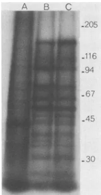
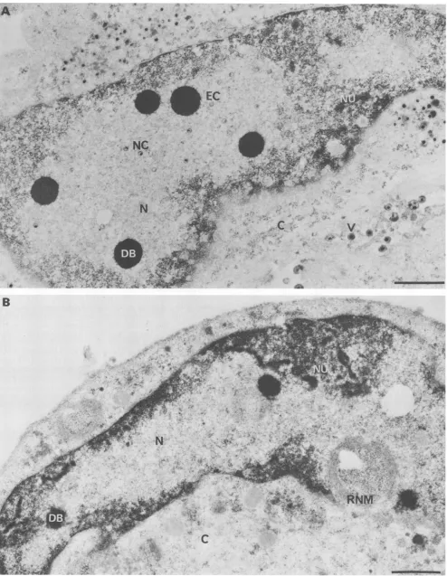
We next examined the course of DNA and protein syn-thesisduringinfection. The kineticsofthymidine incorpora-tionduringinfection atthe nonpermissivetemperaturewere the same formutant and parent (data not shown), and CsCl gradient density analysis (Fig. 2) showed that for both the majority (75%) of the labeled thymidine was incorporated into material havingthe buoyant densityof viral DNA (p = 1.728g/cm3). At 37°C,theelectrophoreticprofiles of A44ts2-inducedpolypeptidesatearly (datanotshown) and late(Fig. 3) times after infection were qualitatively indistinguishable from those ofA44ts+; thus,thecomplex regulatoryprogram of gene expression during HSV infection (67) does not appeartobedisruptedin themutant.Although nosignificant alterations in DNA or protein synthesis were apparent, electron microscopic examination of A44ts2-infected cells revealed that even atlate times ininfection, viralcapsidsor capsid-related structuresfail tobe assembled in the nucleus
at the nonpermissive temperature (Fig.4). Several hundred electron micrographsof A44ts2-infected nuclei from numer-ous
experiments
at thenonpermissive
temperature were examined without the detection of such structures. On theother hand. all other alterations of the nucleus commonly seen during productive infections with wild-type HSV-1 (Fig. 4A) (13, 22, 51, 55. 59) were also seen in mutant-infected nuclei(Fig. 4B). suchasmarginated hostchromatin, extensive electron-translucent nucleoplasm, disaggregated nucleoli, the presenceof electron-dense bodies, and nuclear membrane duplication. Taken together, the above results indicate that the scope of the growth defect of A44ts2 is
limited to events required forcapsid assembly.
Several studies with bacteriophages (reviewed in refer-ences 19and20)and herpesvirus (35) have led to the notion
that in those cases in which cutting of unit-length genomes fromconcatemersaccompaniestheformation of viablevirus particles, capsid assembly isa necessaryprerequisite tothe cutting event. We (10)have previously describedtheuse of
aselective extraction procedure (32)tostudythe
processing
VOL. 63. 1989
on November 10, 2019 by guest
http://jvi.asm.org/
E
U-aL.
0~ -j -J w
n >D
O 100 0
'I
< 10
-JllJ0 w
0O 1 D
1-b
0
0
0'11~
*o'- \o~00
0
1 5 10 15 20 25 30
HOURS POSTINlFECTION
HOURS POSTINFECTION
FIG. 1. Production ofinfectious virus by A44ts+ and A44ts2 at permissive and nonpermissive temperatures. (a) Single-cycle growth
kinetics. Cellswereinfected at30°C(----) or38°C( ), andvirusyieldsweredeterminedatthe indicatedtimesasdescribedinMaterials andMethods.Symbols:0,0,A44ts+;A, A,A44ts2.(b) Effect oftemperatureshift-uponA44ts2growth.Cultures infectedat30°C(0)were
transferredto38°C(0,O,A) andassayedasindicated in Materials andMethods.Temperature shiftswere at8h(0),13h(O), and18 h(A)
postinfection.
oflarge genomes (Mr = 85 x 106) from intracellular, pre-sumably concatemeric, DNA precursors during the infec-tious cycle of a herpesvirus. Applyingthe technique tothe A44ts+ and A44ts2 infections, wefoundthat, in contrast to
1
cm
N
0
0)
-0 U -.
-Q -0 0
I
Ctf)
11
1
1
0
20 30 40 50
[image:4.612.138.496.72.305.2]FRACTION
NUMBER
FIG. 2. Cesium chloride density gradients of DNA from cells infectedatthenonpermissivetemperature.Cells infectedat38°Cwith A44ts+ (upper panel) or A44ts2 (lower panel) were labeled with [3H]thymidine (0) from 6 to 9 h postinfection as described in Materials and Methods.
['4C]thymidine-labeled
cellular DNA(0)wasaddedas an internaldensity reference. The measured densities (in grams percubic centimeter)of selected fractionsare indicated (O).
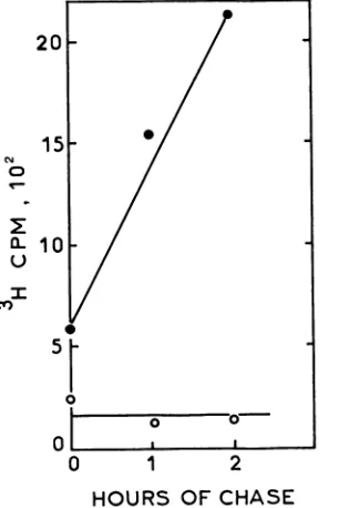
thesituation withA44ts+, labeled A44ts2 DNA couldnotbe chased intothe Hirt supernatant (Fig. 5) atthe nonpermis-sive temperature, indicating a block in the production of unit-length DNA. This was supported by the finding that restrictionfragments representingthephysical endsof unit-length genomes were absent in intracellular A44ts2 DNA replicated at the nonpermissive temperature (data not
shown).
AB C
-205
116 94
-67 -45
-30
FIG. 3. Electrophoretic profiles of [35S]methionine-containing
polypeptides induced in cells infectedatthenonpermissive
temper-ature. Mock-infected (lane A), A44ts'-infected (lane B), and A44ts2-infected(laneC)cellsat37°C werelabeled from 22 to24 h postinfection, and extracts were electrophoresed as described in Materials and Methods. Thepositionsof molecularweight standards
(x103)runin the same gel are indicated to the right.
-1-
CO\_X1.80
1.80
O
I0^
j1S
1.70
5
\1.60O
r"
I.
9
on November 10, 2019 by guest
http://jvi.asm.org/
[image:4.612.92.288.427.660.2] [image:4.612.395.494.466.656.2]B
.44
o yefls2* 2K#siS.
Rht'JM
R.~~~~~~~~~~~~~~~~1
[image:5.612.57.551.51.688.2]I~~~~~~~~J
IFIG. 4. Electronmicrographs of infected cells late in the growth cycle at the nonpermissive temperature. Cells infected (10 PFU per cell) with A44ts+ (A) or A44ts2 (B) at 37°C were processed for electron microscopy at 17 h postinfection by Epon embedding and uranyl-lead acetate staining as previously described (55). EC, Empty capsids; NC, nucleocapsids; V, enveloped virions; N, electron-translucent nucleoplasm; DB, dense bodies; NU, disaggregated nucleoli; RNM, reduplicated nuclearmembrane; C, cytoplasm. Bars, 1 p.m.
2173
on November 10, 2019 by guest
http://jvi.asm.org/
2174 PERTUISET ET AL.
0
I O
1
2
HOURS OF CHASE
FIG. 5. Analysis by Hirt extraction of unit-length viral DNA produced at the nonpermissivetemperature.Cells infected (10 PFU per cell) with A44ts+ (0) or A44ts2 (0) at 37°C and labeled with [3H]thymidine (5 pCi/mI) 8 to 10 h postinfection were extracted either immediately after the labeling period or after the indicated periods ofchase as previously described (10). The data points refer to total radioactivity in two successive Hirtsupernatants. At 30°C, radioactivity entered the Hirt supernatant during infection with either virus (data not shown).
Marker rescue of thets2 mutation andnucleotide sequence
of therescuingregion. Tolocate the ts2 mutationin the viral
genome,we used calcium phosphate coprecipitates of intact A44ts2 DNA and linearized plasmids containing strain KOS EcoRI fragments (28) to transfect cell monolayers at the nonpermissive temperature (37°C) as described in Materials and Methods. Of 12 plasmids from thelibrary. 8 were tested
(Table 1; Fig. 6a), and only the plasmid carryingthe EcoRI Afragment (genome coordinates0.493 to 0.636) gave rise to ts+ plaques in this assay. Subfragments of E(oRI-A were
N I
INI SmAp
A S. A S. HNI H ApN APIS,4H HNI
II II
l1@bp
NI
SIHNI Hi Nl K
1 I l
A;K:
Ap:
H _ _ _
4_
4-4----Hi:
N :
NI: -*-.
S.:
-FIG. 7. Restriction map and DNA-sequencing strategy of the regionbetween mapunits 0.551 and 0.565. Subfragments from the Accl (0.551)-Kpnl (0.565) fragment of Fig. 6 produced by the indicated enzymes were subclonedintoM13 vectorsandsequenced by thedideoxynucleotide method.A, Accl;Ap,ApaI;H,HaeIl; Hi, HincII;K,KpnI; N, Ncol; NI, NlallI; S,Sacl; Sm,SmaI.
inserted into pBR327 and used in successive marker rescue experiments to further delimit the region bearing the lesion.
Inone series ofexperiments (Table 1; Fig.6b, c, and d), the smallest fragment allowing rescue showed this region to
include 3.8 kilobase pairs (kbp) between a SaII site at
coordinate 0.545 and aBamHI siteat coordinate 0.570. An
additional series of rescueexperiments(Table 1; Fig. 6e)was
done with a 3.1-kbp A44ts+ SalI-KpnI fragment (0.545 to
0.565) and its subfragments cloned in M13mpl8 and M13mpl9.Here, the minimal subfragmentgivingrescue was
the 1,837-base-pair(bp)
Asdc
I-KpnI fragment betweencoor-dinates 0.553 and 0.565.
Thelarger of thetwoA44ts+AccI-KpnI fragmentsshown inFig. 6e,Ac(cI(0.551)-KpnI(0.565), waschosenfor further study. The complete nucleotide sequencewasdeterminedby thedideoxy-chain termination method (58) accordingto the strategy outlined in Fig. 7. During the sequencing, we
encountered various degrees of band compression in the gels,aphenomenon sometimes observed with thehigh-G+C
j d g
I'
moI-i
__I___ II_
0.493
T
a i
I
e ikh kh o
Ir
0.636c)
d)
e)
0.521
1
T
7
0.5700.545
1
0.565. EcoR I
?
.BamH I
T
. SalI
I
. Kpn I7
. Acc I TFIG. 6. Summary of marker rescue experiments todetermine the location ofthe A44ts2 mutation. (a)EcoRI map of the strain KOS
genome (49, 64). EcoRI restriction fragment profiles of strain A44ts+ and strain KOS DNAsare identical, and A44ts2 DNAcontains an
additionalEcoRIsite in fragment I(notshown).(atoe)Restriction fragments used in markerrescueexperiments (solid lines). Thoserescuing the A44ts2mutationare hatched, and those givingno rescue are stippled. Coordinates withinEcoRI fragmentAare basedon0.636as the
genomic position of the right-hand site (70), and theBainHIfragmentsinthisregion (71)areindicated.
a)
b)
I
mmmgim.
1---- VW
--I
-4
on November 10, 2019 by guest
http://jvi.asm.org/
[image:6.612.326.564.71.240.2] [image:6.612.117.270.77.306.2] [image:6.612.137.505.522.679.2]AccI
GTAGACCRACGACGRGACCGGGCGGGAATGACTGTCGTGCGCTGTAGGGRGCGGCGAATTRTCGATCCCCCGCGG 75
CCCTCCRGGAACCCCGCAGGCGTTGCGAGTACCCCGCGTCTTCGCGGGGTGTTATACGGCCACTTAAGTCCCGGC 150
ATCCCGTTCGCGGACCCAGGCCCGCGGGCRTTGTCCGGATGTGCGGGCRGCCCGGACGGCGTGGGTTGCGGACTTT 225
Accl
CTGCGGGGCGGCCCARATGGCCCTTTARACGTGTGTATACGGACGCGCCGGGCCAGTCGGCCARCACRACCCACC 300
GGAGGCGGTRGCCGCGTGTGGCTGTGGGGTGGGTGGTTCCCCCTTGCGTGAGTGTCCTTTCGACCCCCCCCTCCC 375
CCGGGTCTTGCTAGGTCGCGATCTGTGGTCGCARTGAAGACCRRTCCGCTACCCGCRACCCCTTCCGTGTGGGGC 450
M K T N P L P A T P S V W G 14
GCGAGTACCGTGGARACTCCCCCCCACCACRCGCGATRCCGCGGGGCAGGGCCTGCTTCGGCGCGTCCTGCGCCCC 525
G S T V E L P P T T R D T A G 0 G L L R R V L R P 39
CCGATCTCTCGCCGCGACGGCCCAGTGCTCCCCAGGGGGTCGGGACCCCGGAGGGCGGCCAGCRCGCTGTGGTTG 600
P I S R R D G P V L P R C S G P R R A A S T L W L 64
CTTGGCCTGGACGGCACAGACGCGCCCCCTGGCGCCCTGACCCCCAACGACGATRCCCRACRCCCCCTCGACAAG 675
L G L D C T D A P P G A L T P H D D T E 0 R L D K 89
ATCCTGCCGCGCACCATCCGCGGGGGGGCGCCCCTCRTCCCCTCCCCGCGCCATCRTCTAACCCGCCAACTCRTC 750
I L R G T M R G G A A L I G S P R H H L T R 0 V 1 114
CTGACCCGATCTGTGCCAACCCAACCCGGATCGTCCCCCCACGCTCCTTCTGCCGCTCCCCCACCCCCCCGACCTC 825
L T D L C 0 P H A D R A C T L L L A L R H P A D L 139
CCTCACCTGGCCCACCRGCCGCCCCGCCAGGCCCGCACCACCGCCCGCCTCGCGACCCCTCCCGCCACCTCATC 900
P H L A H 0 R A P P G R 0 T E R L G E A W G 0 L M 164
CGAGCCACCGCCCTGCGGTCGGGCCCACCCCGCACCGCTGCRCCCGCCCCCGGCCTCGTGTCCTTTARCTTCCTC 975
E A T A L C S C R A E S C C T R A C L V S F H F L 189
GTGGCGCCCTTGCCCCCCTCCTRCCACCCCCCCCACCCCCCCARTCCGGTACCGCCCCACGTCRCGCCARCTRC 1050
V A A C A A S Y D A R D A A D A V R A H V T A N Y 214
CCCCCCCGCCCGGTCCGCCCGCGCCTCCATCGTTTTTCCCRCTGTCTGCGCCCCRTGGTTCRCACGCACCTCTTC 1125
R C T R V C A R L D R F S E C L R A M v H T H V F 239
CCCCRCCGCATCATGCCGTTTTTCCGGCCGGCTCGTGTCGTGCGTCACCCAGGRCGAGCTRGCGAGCGTCACCGCC 1200
P H E V M R F F C C L V S W V T Q D E L A S V T A 264
GTCTCCCGCCCCCCCACCRCGGGCGGCGCCRCCGGCCACCCGGGCCGGCCCCGCTCCGCCCCTGRTCCTCCCGGCG 1275
V C A C P 0 E A A H T G H P C R P R S R V I L P A 289
TGTCCGTTCGTGCACCTCGRCGCCCRGCTGCGGCTGGGGCGCCCGCGCGCGGCGTTTCTGTRCCTGGTACTCACT 1350
C A F V D L D A E L C L G C P G A A F L Y L V L T 314
TACCCCAGCCCCCCCGACCACGGACTGTCTTGTGTGTRCCTGATCARCACCCRGCTCCCCCCGCCCGGGTTGCAC 1425
Y R 0 R R D 0 E L C C V Y V I K S 0 L P P R C L E 339
CCGCCCCTGCAGCGGCTGTTTGGCGCCTCCGGATCCCRARCRCGATTCRCGGCACCGRAGRCRTGACGCCCCCG 1500
P A L E R L F C R L R I T H T I H G T E D M T P P 364
GCCCCRAACCGRARCCCCGACTTCCCCCTCGCGGGCCTGGCCCCCRATCCCCRRACCCCGCGTTCCTCCCCTCGC 1575
A P H R H P D F P L A G L A A N P 0 T P R C S A C 389
CACGTCACGAACCCCCAGTTCGCCGRCAGGCTGTRCCGCTGGCAGCCGGACCTGCGGGGGCRCCCCGRCCCRCGC 1650
0 V T H P 0 F A D R L Y R W Q P D L R G R P T A R 414
ACCTGTACCTACGCCCCCTTTCCGRCACTCCCACTCATCCCCGRAGRTAGTCCCCGCTGCCTGCRCCGCACCCGRG 1725
T C T Y R A F A E L G M M P E D S P R C L H R T E 439
CGCTTTCGGCGGTCACGCTCCCCGTTCTCRTCCTGGCAAGCGTGGTGTGGTGCCCCGGCGRGTGGCGGGCATGC 1800
R F G A V T V P V V I L E C V V W C P G E W R A C 464
GCGTGCCCCGTAGCAAACCCCCGCCCCACACAACGCTCCGCCCCCRACCCCTTCCCCGCTGTCRCTCGTTGTTCG 1875
A *** 465
TTGRCCCGGACGTCCGCCARRTARRGCCACTRAARCCCGRARCGCGRGTGTTGTAACGTCCTTTGGGCGGGAGGA 1950
RGCCRCARRATGCARATCGGATACATGGAAGGRACACACCCCCGTGACTCAGGACRTCCCCGTGGCCTTTTGGGT 2025
-KpnlI
TTCACTGAAACTCGCCCGCCGCCCCACCCCTGCGCGRTGTGGATRRARAGCCAGCGCGGGTGGTTTAGGGTRCc 2098
FIG. 8. Nucleotide sequence ofA44ts+ DNAin thegenomicinterval 0.551 to0.565. Theamino acid sequenceofthepredicted product of ORF.553is shown in one-letter code. Also shownareputativeCAAT(single underline)and TATA(doubleunderline)transcription signals forthe mRNAsmapped(2, 3) to thisregion(seetext),aswellasthelocations(arrowheads)of 5' termini for the 3.6-kb(position116) and 1.9-and 7-kb (position 278)transcripts (M. Flanagan and E. K. Wagner, personalcommunication)and the cap site(position 2095)ofthe5-kb transcript specifying the ribonucleotide reductase large subunit (23). Putative polyadenylationsignals (47, 54)forthe 1.9-kb transcript are
indicated by filled circles.
2175
on November 10, 2019 by guest
http://jvi.asm.org/
[image:7.612.110.496.31.674.2]2176 PERTUISET ET AL. (a)
I NGSQPTNSt{FTLNEQTLCGTNISLLGNNRFIQIGCNGLIM4TYAPGFFGNWSRDLTIGPRFGGLNKQPIHVPPKRTETASIQVTPRSIVINRMNNIQINPTSIGNPQVTIRLPLNNFKSTTQLIQWV
114 ILTDLCQPNADRAGTLLLALRHPADLPHLAHQRAPPCRQTERLGEAWCQI.M4EATALGSCRAESGCTRAGLVSFNFLVAACAASYDARDAADAVRAHVTANYRGTRVGARLDRFSECLRAMVHiTtIV
* *0* a 0
126
SLTDFFR*PDIEHMACGSIVLILRHPS*DMI
GE*ANTLTQAGRDPDV*LLEGLRN*LFNACTAPWTVG
GGGL*RAYVT*SLS
IAACR*AEE*YTDKQAADANRTAIVSAYGCSRIIETRL
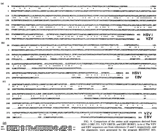
IRFSECLRA1VQCIfN 239 FPHEVMRFFGGLVSWVTQDELASVTAVCAGPQEAAHiTGHPGRPRSAVILPACAFVDLDAELGLGGPCAAFLYLVLTYRQRRDQELCCVYVIKSQLPPRGLEPALERLFGRLRITNTIHiGTE.DMT 251 FPHRFISFFGSLLEYTIQDNLCN ITAVAKGPQEAARTDKTSTRRVTANIPACVFWDVDKDLNILSADGLKNfVFLVFWYTQRRQREGVRLHLALSQLNEQCFCRGIGFLLGRIRAENAAWGTEGVAN363 PPASPNRNPDFPLAGLAANPQTPRCSAG....QVTNPQFADRLYRWQPDLRGRPTARTCTYAAFAELGMMPEDSPRCLHnTERFGAVTVPVVILEGVWCPGEWCA 465 HSV I
* to * * *--- * *--0 - *0 --to 0*-- * * * * ---- *-
*---376 THQPYNTRALPLVQLSNDPTSPRCSIGCEITGVNWNLARQRLYQWTGDFRGLPTQLSCMYAAYTLIGCTIPSESVRYTRRMERFGGYNVPTI%rLEGVVWGTNTWNEC 481 VZV 26 DTAGQGLLRRVLRPPISRRDGPVLPRGSGPRRAASTLWLLGLDGTDAPPGALTPNDDTEQALDKILRGTMRGGAALICSPRHILTRQVILTD...LCQPNADRAGTLL...LAL
*-- * * * * ****0 **0so*- * **0
1 MKVQGSVDRRRLQRRIAGLLPPPARRLNISRGSEFTRDVRGLVEEIAQASSLSAAAWRAGLLAPGEVAVAGGGSGGGSFSWSGWRPPVFGDFLIHASSFNNAEATGTPLFQFKQSDPFSGYDAV 134 RHPADLPHLAHQRAPPGRQTERLGEAWGQLMEATALGSGRAESGCTRAGLV.SFNFLVAACAASYDARDAADAVRAHVTANYRGTRVGARLDRFSECLRAMMITHVFPHEVKRFFGGLVSWTQD
126 FTPLSLFIL.... MHGRGVAARVEACGCL...TRMANLLYDSPATLADLVPDFGRLVA... DR RFHNFITPV....GPLVENIKST
258 ELASVTAVCAGPQEAAHTGHPGRPRS..AVILPACAFVDLDAEL..GLGGPGAAFLYLVLTYR,QRRDQELCCVYVIKSQLPPRGLEPALFRLFGRLRITNTIHGTEDNTPPAPNRNPDFPLAGLA
* *- *- ~~*-- * * **--o** - to* *- ** *
199 YLNKITTVVHGP...VVSKAIPRSTVKVTVPQEWFVDLDAWLSGGAGGGGGVCFVGGL... GLQ...CPADARLYVALTYEEA
379 ANPQTPRCSAGQVTNPQFADRLYRWQPDLRGRPTARTCTYAAFAELGMPEDSPRCLHIRTERFGAVTVPVILE GVVWCPG...EWRACA 465 HSVI
274 GPRFTFFQSSRGHCQIMNILRIY... YSPSIMiR....YAVVQP,LHIEELTFGAVACLGTFS&TDGVRRSA 337 EBV
(C)
2 CSQPTNSHFTLNEQTLCGTNISLLGNNRFIQIGNCLHMTYAPGFFnN...WSRDLTIGPRFGGLNKQPIHVPPKRTETASIQVTPRSIVINRMNNIQINPTSIGNPQVTIRLPLNNFKSTTQLIQ
32 GSEFTRDVRGLVEEHAQASSLSAAAVVRAGLLAPGEVAVAGGGSGGGSFSWS...GWRPPVFGDFLIH...ASSF.NNAEATGTPLF
124 QVSLTDFFRPDIEHAGSIVLILRHP.SDMIEANTLTQAGRDPDVLLEGLRNLFNACTAPrTVGEGGGLRAYVTSLSFIACRAEEYTDKQAADANRTAIVSAYGCSRKETRLIRFSECLRAMVQ
* * 0*0* - ~ 0*** * 0*
-112 QFKQSDPF....SGVDAVFTPLSLFILCSRHGRGVAAR...VEAGGLTRLALLYDSPATLADLVPDFGRLVADR...RF... 248 CHVFPHRFISFFGSLLEYTIQDNLCNITAVAKGPQEAARTDKTSTRRVTANIPACVFWDVDKDLHLSADGLKHVFLVFWTQRRQREGVRLHLALSQLNEQCFGRGIGFLLGRIRAENAAWGTEG
182 ...HNFITPVCPLVENIKSTYLNKITTVVHGP.VVSKAIPRSTVKVT ..VPQEAFVDLDAWLSGGAGGGGGVCFVGGLGLQPCPADARLYVAL.. TYEEAGPRFTFFQSSR...GHCQ
373 VANTHQPYNTRALPLVQLSNDPTSPRCSIVEITZVNWNLARQRLYQTGDFVCLPTQLSCMYAYTLIGTIPSESVRYTRRMERFGGYNVPTIWLEGVWGGTNTWN479 VZV
289 IMNILRIY.... YSP..SIMHRYAVVQPL[IIEEL... TFGAVACLGTFSATDGWRRSAFNYRGSSLPVVEIDSFYSNVSDWEVIL 364 E BV
(d)
V-/
'l
S I w G 243HSVVI
A L AGjAp//3//0VZV R v _ _ N I K H D 317
(68%) sequences ofHSV (44). Although most ambiguities arising from this effect were resolvable, one recalcitrant sequencefrom nucleotide 1057to nucleotide 1064 (see Fig. 8)wasonly decipherablewith datafrom strain 17
communi-cated to us by M. A. Dalrymple and D. J. McGeoch. The three amino acids(Thr-Arg-Val)derived from this sequence
therefore may not be strictly representativeof strainA44. Thesequenceof the2,098-bpAccI-Kpnl fragment (Fig. 8) was searched for probable ORFs with the aid of the codon preferenceprogram of Staden and McLachlan(68)and data
from 12 HSVprotein-coding regionsfrom the Genbank data base.Among several possibilities indicatedbythe positions
of initiation-termination codons in the six reading phases, onlyoneextendedORF,whichwerefertohereasORF.553,
adhered welltoHSV codonusage (datanotshown). The last 100bp oftheAccI-KpnIfragmentagreewell withpreviously published sequencesin thevicinity ofthe KpnI site(23, 46).
The sequence ATAAAAA at position 2067 is thought to
belong to the promoter for a 5-kilobase (kb) RNA
tran-scribed rightward from the middle of the KpnI site (2, 23, 46). This RNA has been identified as the message for the large subunit of the viral ribonucleotide reductase (21, 53).
FIG. 9. Comparison of the amino acid sequencesderived from
HiSV-1ORF.553, VZV ORF20, and EBVBORF1. (atoc) The VZV andEBVsequencesarefrom references 15 and5, respectively,and
the alignments wQre generated by the program BESTFIT (65). Asterisks denote conservedresidues, and dotsaregapsintroduced by theprogramtoachieve thehighest scoring alignment. (d) Region of maximum sharedsequencehomologyinthepredicted productsof
the three ORFs. Hatched boxes designate identical residues, And openboxesidentifyconservative substitutions(A = I = L= V,D = E). The numbertotherightofeachsequenceshows theposition of the last residue in thecorresponding totalsequence.
Three additional transcripts have also been assigned to the region including and immediately surrounding the 0.551-to-0.565 interval (2), all of which are initiated upstream of ORF.553. One, a 3.6-kb early species, is transcribed left-ward,and theremainingtwo,5'coterminal late RNAs of 1.9 and 7 kb,are transcribed rightward.Thepositions ofthe5' ends of the three species havebeen determined (M.
Flana-gan and E. K. Wagner, personal communication) and are
indicated in Fig. 8. Several putative transcriptional control
signals (12) occur in this region; for the 3.6-kb RNA,
sequences corresponding to TATA and CAAT boxes are
present at positions -25 and -76, respectively, and two possible TATA boxes at -17 and -29, aswell as aCAAT box at -39, are associated with the 1.9- and 7-kb RNAs.
Downstreamfrom ORF.553aretwopolyadenylation signals, acanonical AATAAA sequence (54) 91 bpfrom the
termi-(b)
9 ...LTRQV
* "
on November 10, 2019 by guest
http://jvi.asm.org/
[image:8.612.66.566.69.488.2]HSV-1 GENE REQUIRED FOR CAPSID ASSEMBLY 2177 nation codon, and 28 bp farther along, the frequently
asso-ciated consensus sequence YGTGTTY (47). Transcriptional termination in this region would give rise to an RNA of 1.6 kb, in good agreement with the 1.7 kb obtained from Si nuclease mappingexperiments (2).
ORF.553 itself extends from the ATG triplet at position 409, the context of whichconforms to the preferred initiator sequences of Kozak (34), to the TGAterminator at position 1804. It thereby has the capacity to code for a 465-amino-acid protein with a predicted molecular weight of 50,175, thus fitting well with the 54,000-molecular-weight in vitro translation product of the 1.9- and 7-kb mRNAs (3). In the context of typical HSV codon usage, the in-phase triplets exhibit the position-dependent bias toward elevated G+C content already noted for various ORFs of HSV (33, 44, 45) and Epstein-Barr virus (EBV) (24); third positions are high-est(86%), first positions areintermediate (73%), andsecond positions, which select amino acids of differing physico-chemical properties, are lowest (56%).
A computer-implemented search of the EMBL and Gen-bank data bases failed to find nucleotide sequences with significant homology to the sequence of Fig. 8. However, in a survey of the NBRF protein data base for identical heptameric peptides, only one example was found, and this belonged to the predicted 364-amino-acid product (Fig. 9b) ofthe EBVBORF1 reading frame (5). As in the present case, BORF1 is located just upstream of two reading frames attributed to the viralribonucleotide reductase (5, 24). While this work was in progress, the total nucleotide sequence of varicella-zoster virus (VZV) became available (15), and we compared the ORF.553 polypeptide with the predicted prod-uct ofVZV ORF20, which also lies directly 5' to the large subunit of VZV ribonucleotide reductase. The comparison revealed a considerably higher level of similarity than is found between the products of ORF.553 and EBV BORF1 even though theNH2-terminal 25% of the proteins appear to have diverged to a point at which the presence of matching residues is likely to befortuitous (Fig. 9a). A comparison of all three sequences shows that maximum sequence conser-vation occurs in a stretch of about 25 amino acids corre-sponding to the region originally detected as homologous to EBV BORF1 (Fig. 9d).
DISCUSSION
The results presented here indicate that the product of ORF.553 (UL38 [43]) functions in capsid assembly. In addi-tion to itsinability to assemble capsids at thenonpermissive temperature (Fig. 4), the mutant A44ts2 is also defective in theproduction of unit-lengthmolecules from replicated viral DNA (Fig. 5). That thesedefects should occur together was not unexpected in view of previous findings with DNA-positive capsid-negative mutants of bacteriophages (19, 20) and theherpesvirus PrV (35) that concatemeric DNA cannot be cut to unit-length molecules in the absence of capsid formation. However, for PrV (35) and HSV-1 (62), the assembly of capsids is not asufficient condition for cutting to take place, since some ts mutants which do produce capsids are nevertheless deficient in the production of unit-length DNA. Clearly, capsid assembly is only one of several requirements for DNA maturation in these viruses.
Although
we have not yet identified the product of ORF.553, it is worth considering whether it could be one of the prominent structural proteins of the viral capsid. There are probably at least six of these (reviewed in references 67 and 69), ofwhich the155,000-molecular-weight major capsidprotein
(VP5[26];
p155 [31]; NC-1 [11]) isreadily ruledout on the basis of both its molecular weight and the genomic coordinates(0.2to0.3[40, 50])of itscoding sequences. Themost serious candidate on the basis of molecular
weight
would seem to be the 50,000-molecular-weight capsid
pro-tein (VP19C [31]; p5O [30]; NC-2 [11]), but studies on intertypicrecombinants haveplaced its gene to theopposite side of theribonucleotidereductase locus, in the coordinate region0.58to0.60(8). Lower-molecular-weight capsid com-ponents, which one could conceive ofas arising from the ORF.553productby proteolysis,forexample,arethe40,000 (VP22a [26]; p40 [30]; NC-3.4 [11])-, the 32,000 (VP23 [26]; p32 [30]; NC-5 [11])-, the 25,000 (VP24 [26]; p25 [30]; NC-6 [11])-, and the 12,000 (p12 [30]; NC-7
[11])-molecular
weight species. However, the genes for the 40,000- and 32,000-molecular-weight species have been mapped to thecoordi-nate regions at 0.327 to 0.332 (52) and 0.66 to 0.76 (38), respectively. Moreover, the 12,000-molecular-weight
spe-cies isahighly basic,histonelikeprotein (11), andnodomain of the ORF.553productappearstohave suchaproperty. By eliminiation, then,theonlycandidateremainingamongthese proteins would be the 25,000-molecular-weight species. Al-ternatively, the ORF.553 product could represent a previ-ously unrecognized capsid component or a nonstructural protein required in the pathway of capsid assembly.
At-tempts are in progress to identify the ORF.553 product and to elucidate its role incapsid assembly.
Comparable ORFs are found in the published nucleotide sequencesof EBV(5) and VZV (15), and it will be of interest to determine whether their products also have a role in capsid assembly. An estimate of the relatedness of these products to the ORF.553 product wasobtained by computer-generated alignments of amino acid sequences (Fig. 9a and b) and by the use of an algorithm developed by Davison and Taylor (17), which takes into account the way in which identical residues are distributed in compared sequences. Their algorithm, HOMREG, scores only those sequence intervals containing at least x matching residues bounded on both sides by at least y unmatched residues, thus giving particular significance to grouped identities. It calculates both the average percentage of sequence identity within the scoredintervals and the percentage of one or the other total sequences given over to these intervals. They applied the algorithm to acomparison of the VZV and EBV ORFs and suggested that the corresponding proteins of VZV ORF20 and EBV BORF1 (Fig. 9c) should be considered as "weak" homologs (i.e., 37% identity over 23% of the VZV sequence). Using their criteria (x = 12, y = 5), we found that the product of ORF.553 is a "moderate" homolog of the EBV BORF1 product (42% identity over 34% of the HSV sequence) and a "strong" homolog of the VZV ORF20 product (51% identity over 60% of the HSV sequence). The relative degree of sequence conservation between the prod-ucts of ORF.553 and the VZV and EBV homologs therefore agrees with previous estimates ofprotein relatedness among these viruses (14-17, 25, 41-43).
In acomparison of our sequencefor strain A44 with that of McGeoch et al. (42) for strain 17, there appear a number of nucleotide changes which we believe can be attributed to strain differences. There are 16 changes in the noncoding sequences-7 substitutions, 1 insertion, and a cluster of 8 deleted bases between positions 371 and 378 of the A44 sequence; none of these changes fall in the putative
tran-scriptional control signals mentioned earlier. Within the ORF there are 12 substitutions, 8 of which occur in silent third positions and 4 of which lead to the amino acid VOL. 63, 1989
on November 10, 2019 by guest
http://jvi.asm.org/
2178 ET AL.
replacements (for A44) V48G, L313F,
T445S,
andC457F.
A knowledge of permissible strain-dependent amino acid changes should prove useful in future attempts to define functionally important regions in this protein.ACKNOWLEDGMENTS
We are indebted to M. Laithier and M. L. Ryhiner for expert technical assistance and toR. Sandri-Goldin andM. Levineforthe gift of the KOS genomic library.We aregratefultoD. J.McGeoch, P. A.Schaffer, M. Flanagan, and E. K.Wagnerforcommunicating data prior to publication. We also thank 0. Croissant for useful discussions and G. Hamon, M. Kress, A.Sarasin, andmembersof his laboratory for access to their computing facilities and helpful advice.
This workwassupported bygrantsfromthe CentreNationalde la RechercheScientifiqueandtheAssociationpourla Recherche sur le Cancer, Villejuif.B.P. was afellow oftheMinisterede la Recherche etde laTechnologie.
LITERATURECITED
1. Addison, C., F. J. Rixon, J. W. Palfreyman, M. O'Hara, and V. G. Preston. 1984. Characterization ofaherpes simplexvirus type 1 mutant which has a temperature-sensitive defect in penetration of cells and assembly of capsids. Virology 138: 246-259.
2. Anderson, K. P., R. J. Frink, G. B. Devi, B.H.Gaylord, R. H. Costa,and E. K.Wagner. 1981.Detailed characterization ofthe mRNAmappingintheHindlIl fragmentKregion ofthe herpes simplex virus type 1 genome. J. Virol. 37:1011-1027.
3. Anderson, K. P., L. E. Holland, B. H. Gaylord, and E. K. Wagner.1980. Isolation and translation ofmRNAencodedbya
specific region of the herpes simplex virus type 1 genome. J. Virol. 33:749-759.
4. Atkinson, M. A., S. Barr, and M. C. Timbury. 1978. The fine structureof cells infected withtemperature-sensitivemutantsof herpes simplex virus type 2. J.Gen. Virol. 40:103-119. 5. Baer, R., A. T. Bankier, M. D. Biggin, P. L. Deininger, P.J.
Farrell,T. J.Gibson,G.Hatfull, G. S.Hudson,S. C.Satchwell,
C.Seguin,P.S.Tuffnel,and B. G. Barrell.1984.DNA sequence andexpression of the B95-8Epstein-Barrvirusgenome. Nature (London) 310:207-211.
6. Batterson, W., D. Furlong, and B. Roizman. 1983. Molecular genetics of herpes simplex virus.VIII. Further characterization ofatemperature-sensitive mutant defective in release of viral DNA and in other stages of the viral reproductive cycle. J. Virol.45:397-407.
7. Ben-Porat, T., and A. S. Kaplan. 1985. Molecular biology of
pseudorabies virus, p. 105-173. In B. Roizman (ed.), The herpesviruses, vol.3. PlenumPublishing Corp.,NewYork. 8. Braun, D. K.,W. Batterson,andB.Roizman. 1984.
Identifica-tion and genetic mapping of a herpes simplex virus capsid
protein that bindsDNA.J. Virol. 50:645-648.
9. Cabral,G. A., and P. A.Schaffer. 1976. Electron microscopic studies of temperature-sensitive mutants of herpes simplex
virustype 2. J. Virol. 18:727-737.
10. Cebrian, J., D. Bucchini, and P. Sheldrick. 1983. "Endless" viralDNAin cellsinfected with channel catfish virus.J. Virol. 46:405-412.
11. Cohen, G. H., M. Ponce de Leon, H. Diggelmann, W. C. Lawrence,S. K.Vernon, and R.J. Eisenberg. 1980. Structural analysis of the capsid polypeptides of herpes simplex virus types 1 and 2. J.Virol.34:521-531.
12. Corden, J., B. Wasylyk, A. Buchwalder, P. Sassone-Corsi, C. Kedinger, andP. Chambon. 1980. Promoter sequences of
eu-karyotic protein-codinggenes. Science209:1406-1414. 13. Dargan, D., and J. H. Subak-Sharpe. 1983. Ultrastructural
characterization ofherpes simplextype 1 (strain 17)
tempera-turesensitivemutants.J. Gen. Virol. 64:1311-1326.
14. Davison, A. J., and D. J. McGeoch.1986.Evolutionary compar-isonsoftheSsegmentsinthe genomesofherpessimplexvirus type 1andvaricella-zostervirus. J. Gen. Virol.67:597-611. 15. Davison, A. J., and J. E. Scott. 1986. The complete DNA
sequenceof varicella-zostervirus. J. Gen. Virol.67:1759-1816. 16. Davison, A. J., andJ. E. Scott. 1986. DNA sequence ofthe major capsid protein geneof herpes simplex virus type 1. J. Gen. Virol. 67:2279-2286.
17. Davison,A.J., and P.Taylor. 1987.Genetic relations between varicella-zoster and Epstein-Barr virus. J. Gen. Virol. 68: 1067-1079.
18. Dixon, R. A.F.,and P.Schaffer. 1980. Fine structuremapping
andfunctionalanalysisoftemperature-sensitivemutantsin the gene encoding the herpes simplex type 1 immediate early proteinVP175.J. Virol.36:189-203.
19. Earnshaw,W. C.,andS. R.Casjens. 1980. DNApackaging by thedouble-strandedDNAbacteriophages. Cell21:319-331. 20. Feiss, M.,andA. Becker. 1983. DNApackagingandcutting,p.
305-330. In R. W. Hendrix, J. W. Roberts, F. W. Stahl, and R. A. Weisberg (ed.), Lambda II. Cold Spring Harbor
mono-graphseries 13. Cold Spring HarborLaboratory, Cold Spring Harbor,N.Y.
21. Frame, M. C., H. S. Marsden, and B. M. Dutia. 1985. The ribonucleotide reductase inducedby herpessimplexvirus type1 involves minimally a complex oftwopolypeptides (136Kand
38K).J. Gen. Virol. 66:1581-1587.
22. Friedman, A.,J.E.Coward,H.S.Rosenkranz,andC.Morgan. 1975. Electron microscopic studies on assembly of herpes simplex virus upon removal of hydroxyurea block. J. Gen. Virol. 26:171-181.
23. Frink,R.J.,K.G.Draper,and E. K.Wagner.1981.Uninfected cell polymeraseefficientlytranscribesearlybutnotlateherpes simplex virus type 1 mRNA. Proc. Natl. Acad. Sci. USA 78:6139-6143.
24. Gibson, T. J., B. G. Barrell, and P.J. Farrell. 1986. Coding
contentandexpressionof theEBVB95-8genome in theregion from base62,248tobase82,920. Virology 152:136-148. 25. Gibson, T. J., P. Stockwell, M. Ginsburg, and B.G. Barrell.
1984. Homology between two EBV early genes and HSV ribonucleotide reductase and 38K genes. Nucleic Acids Res. 12:5087-5098.
26. Gibson, W.,and B.Roizman.1972.Proteinsspecified by herpes simplexvirus. VIII.Characterization andcomposition of mul-tiple capsidforms ofsubtypes1and2. J. Virol. 10:1044-1052. 27. Goad, W. B., and M. I. Kanehisa. 1982. Patternrecognitionin
nucleic acid sequences. I. A general methodfor
finding
localhomologiesandsymmetries. Nucleic AcidsRes. 10:247-263. 28. Goldin, A. L., R. M. Sandri-Goldin, M. Levine, and J. C.
Glorioso. 1981. Cloning of herpes simplex virus type 1 se-quencesrepresentingthe wholegenome. J. Virol.38:50-58. 29. Graham,F.L.,andA.J.VanDer Eb.1973. Anew
technique
forthe assayofinfectivityofhumanadenovirus5 DNA. Virology 52:456-467.
30. Heilman,C.J., Jr.,M.Zweig,J.R.Stephenson,and B.Hampar. 1979.Isolation ofanucleocapsid polypeptideofherpes
simplex
types 1 and 2 possessing immunologically
type-specific
and cross-reactive determinants. J. Virol. 29:34-42.31. Heine,J. W., R. W.Honess, E. Cassai,and B. Roizman. 1974. Proteins specified by herpes simplex virus. XII. The virion polypeptidesof type1 strains.J. Virol. 14:640-651.
32. Hirt, B. 1967. Selective extraction of polyoma DNA from infectedmousecell cultures. J. Mol.Biol. 26:365-369. 33. Honess,R. W.1984.Herpessimplexand "theherpescomplex":
diverseobservations and aunifying
hypothesis.
J.Gen. Virol. 65:2077-2107.34. Kozak,M. 1986. Pointmutations defineasequence
flanking
the AUG initiatorcodon that modulates translation byeukaryotic
ribosomes. Cell 44:283-292.
35. Ladin, B. F., M. L. Blankenship, and T. Ben-Porat. 1980. Replication of herpesvirus DNA. V. Maturation of
concate-mericDNAofpseudorabiesvirustogenomelengthis relatedto
capsidformation. J.Virol. 33:1151-1164.
36. Ladin, B. F., S. Ihara, H. Hampl, and T. Ben-Porat. 1982. Pathway ofassemblyofherpesvirus capsids: an
analysis using
DNA' temperature-sensitive mutants of
pseudorabies
virus.Virology 116:544-561.
37. Laemmli,U.K.1970.Cleavageof structuralproteins duringthe
on November 10, 2019 by guest
http://jvi.asm.org/
HSV-1 GENE REQUIRED FOR CAPSID ASSEMBLY 2179
assembly of the head of bacteriophage T4. Nature (London) 227:680-685.
38. Lemaster, S., and B. Roizman. 1980. Herpes simplex virus phosphoproteins. II. Characterization of the virion protein kinase and of the polypeptides phosphorylated in the virion. J. Virol. 35:798-811.
39. Maniatis, T., E. F. Fritsch, and J. Sambrook. 1982. Molecular cloning, alaboratory manual. Cold Spring HarborLaboratory. Cold Spring Harbor, N.Y.
40. Marsden, H. S., N. D. Stow, V. G.Preston, M. C. Timbury, and N. M.Wilkie. 1978. Physical mapping of herpes simplex virus-induced polypeptides. J. Virol. 28:624-642.
41. McGeoch, D. J. 1987. The genome of herpes simplex virus: structure, replication and evolution. J. Cell Sci. 7(Suppl.): 67-94.
42. McGeoch, D. J., M. A. Dalrymple, A. J. Davison, A. Dolan, M.C.Frame, D. McNab, L. J. Perry, J. E. Scott, and P. Taylor. 1988. Thecomplete DNA sequence of the long unique region in the genome of herpes simplex type 1. J. Gen. Virol. 69: 1531-1574.
43. McGeoch, D. J., and A. J.Davison. 1986. DNA sequenceof the herpessimplex virus type 1 gene encoding glycoproteingH. and identification of homologues in the genomes of varicella-zoster virus andEpstein-Barr virus. Nucleic Acids Res. 14:4281-4292. 44. McGeoch, D. J., A. Dolan, S. Donald, and D. H. K. Brauer. 1986. Complete DNA sequence of the short repeat region of herpes simplex virus type 1. Nucleic Acids Res. 14:1727-1745. 45. McGeoch, D. J., H. W. Moss, D. McNab, and M. C. Frame. 1987. DNA sequenceandgeneticcontentof theHindIlIl region in the shortunique componentoftheherpessimplexvirustype 2 genome: identification ofthe gene encoding glycoprotein G. andevolutionary comparisons.J. Gen. Virol. 68:19-38. 46. McLauchlan, J., and J. B.Clements. 1983. Organization of the
herpes simplex virus type 1 transcription unit encoding two early proteinswith molecularweight of 140,000 and 40.000. J. Gen. Virol.64:997-1006.
47. McLauchlan, J., D. Gaffney, J.L.Whitton, and J.B. Clements. 1985.The consensus sequenceYGTGTTYlocated downstream from the AATAAA signal isrequired for efficient formation of mRNA3' termini. NucleicAcids Res. 13:1347-1368.
48. Mocarski, E.S., L.E. Post, andB. Roizman. 1980. Molecular engineeringof theherpes simplexvirus genome: insertion ofa
second L-S junction into the genomecausesadditional genome inversions. Cell 22:243-255.
49. Morse, L. S., T. G. Buchman,B. Roizman,and P. A.Schaffer. 1977. Anatomy ofherpes simplex virus DNA. IX. Apparent exclusionofsomeparentalDNAarrangementsin thegeneration of intertypic (HSV-1 x HSV-2) recombinants. J. Virol. 24:
231-248.
50. Morse, L.S.,L.Pereira, B. Roizman,and P. A. Schaffer. 1978. Anatomy ofherpes simplex virus DNA. XI. Mapping of viral genes by analysis of polypeptides and functions specified by HSV-1 x HSV-2 recombinants. J. Virol. 26:389-410.
51. Nii, S., H. S. Rosenkranz, C. Morgan, and H. M. Rose. 1968. Electron microscopy of herpes simplex virus. III. Effect of hydroxyurea. J. Virol. 2:1163-1171.
52. Preston, V.G., J.A. V.Coates,and F.J. Rixon.1983. Identifi-cation and characterization of a herpes simplex virus gene product required for encapsidation of virus DNA. J. Virol. 45:1056-1064.
53. Preston, V. G., J. W. Palfreyman, and B. M. Dutia. 1984.
Identification ofa herpes virus type 1 polypeptide which is a
component of the virus-induced ribonucleotide reductase. J. Gen. Virol. 65:1457-1466.
54. Proudfoot, N. J., and G. G. Brownlee. 1976. 3' Noncoding region sequencesin eucaryotic mRNA. Nature (London) 263:211-214. 55. Puvion-Dutilleul, F., J. Pedron, M. Laithier, and P. Sheldrick. 1982. Ultrastructural studies on the nucleus of herpes simplex type 1-infected cells. Biol. Cell 44:249-260.
56. Reissig, M., and A. S. Kaplan. 1962. The morphology of noninfective pseudorabies virus produced by cells treated with 5-fluorouracil. Virology 16:1-8.
57. Robb, J. A., and. R. G.Martin. 1970.Genetic analysis of simian virus 40. 1. Description of microtitration and replicating tech-niques for virus. Virology 41:751-760.
58. Sanger, F., S.Nicklen, and A. R. Coulson. 1977. DNA sequenc-ing with chain-terminating inhibitors. Proc. Natl. Acad. Sci. USA 74:5463-5467.
59. Schaffer, P. A., J. P. Brunschwig, R. M. McCombs, and M. Benyesh-Melnick.1974.Electronmicroscopic studiesof temper-ature-sensitive mutants of herpessimplex virus type 1. Virology 62:444-457.
60. Schaffer, P. A., V. C. Carter, and M. C. Timbury. 1978. Collaborative complementation study oftemperature-sensitive mutantsofherpessimplexvirus types 1 and 2. J.Virol. 27:490-504.
61. Sheldrick,P., M. Laithier, D. Lando,and M. L. Ryhiner. 1973. Infectious DNA from herpes simplex virus: infectivity of dou-ble-stranded and single-stranded molecules. Proc. Natl. Acad. Sci. USA70:3621-3625.
62. Sherman, G., and S. L. Bachenheimer. 1987. DNAprocessing in temperature-sensitive morphogenic mutants of HSV-1. Virol-ogy 158:427-430.
63. Sherman, G., and S.L.Bachenheimer. 1988.Characterizationof intranuclearcapsids madeby tsmorphogenic mutantsof
HSV-1. Virology 163:471-480.
64. Skare, J., andW. C.Summers. 1977. Structureand function of herpesvirusgenomes. II.EcoRI,Xbal, andHindlIl
endonucle-ase cleavage sites on herpes simplex type 1 DNA. Virology 76:581-595.
65. Smith, T. F., and M. S. Waterman. 1981. Comparison of biosequences. Adv. AppI. Math. 2:482-489.
66. Soberon, X.,L.Covarrubias, and F.Bolivar.1980.Construction and characterization of new cloning vehicles. IV. Deletion derivatives of pBR322andpBR325. Gene 9:287-305.
67. Spear,P.G., andB. Roizman. 1980.Herpessimplexviruses,p. 615-745. In J. Tooze (ed.), The molecular biology of tumor
viruses. 2nded.. part 2. ColdSpring Harbormonograph series lOB, Cold Spring Harbor Laboratory, Cold Spring Harbor, N.Y.
68. Staden, R.,and A. D. McLachlan. 1982.Codonpreferenceand
its use in identifying protein coding regions in long DNA sequences. Nucleic Acids Res. 10:141-156.
69. Wagner, E.K. 1985. Individual HSVtranscripts. Characteriza-tion of specific genes. p. 45-104. In B. Roizman (ed.). The herpesviruses. vol. 3. PlenumPublishing Corp., NewYork.
70. Weller, S. K.,E. P.Carmichael,D. P.Aschman,D.J.Goldstein,
and P. A.Schaffer. 1987.Genetic andphenotypic characteriza-tion ofmutantsinfour essential genes that maptotheleft half of HSV-1 UL DNA. Virology 161:198-210.
71. Wilkie, N. M., A. Davison, P. Chartrand, N. D. Stow, V. G.
Preston, and M.C. Timbury. 1978. Recombination in herpes simplex virus: mappingof mutations andanalysisofintertypic recombinants. Cold Spring Harbor Symp. Quant. Biol. 43:
827-840.
72. Yanisch-Perron, C., J. Vieira,andJ. Messing. 1985. Improved M13 phage cloning vectors and host strains: nucleotide
se-quencesof the M13mpl8andpUC19vectors.Gene 33:103-119.
VOL. 63,1989