0022-538X/89/020730-09$02.00/0
Copyright©3 1989,American Society forMicrobiology
Identification of
mar
Mutations
in
Herpes Simplex
Virus
Type 1
Glycoprotein
B
Which Alter
Antigenic
Structure and Function
in
Virus
Penetration
STEVEN L. HIGHLANDER,' D. J. DORNEY,2tPHILIPJ. GAGE,3 THOMAS C. HOLLAND,2t WEIZHONG CAI,4§ STANLEY PERSON,4MYRON LEVINE,5 ANDJOSEPH C. GLORIOSO2,3*
Programin Cellular and Molecular Biology,' UnitforLaboratoryAnimalMedicine,2 Department of Microbiology and Immunology,3 and Department ofHuman Genetics,5 The University of MichiganMedicalSchool, AnnArbor, Michigan
48109, and Department ofMolecular and CellBiology, Pennsylvania State University, University Park, Pennsylvania
168024
Received5July 1988/Accepted 19 October 1988
Analysis ofsix monoclonalantibody-resistant (mar)mutantsin herpes simplexvirus type1 glycoprotein B identified twotype-common(IIand III)and twotype-specific (IandIV) antigenic sitesonthis molecule.To derive additional information on the location of these sites, mar mutations were mapped and nucleotide alterationswereidentifiedbyDNAsequencing.Eachmutantcarriedasingleaminoacidsubstitutionresulting from a G-to-Abase transition. Alterations affecting antibody neutralization wereidentified at residues 473,
594, 305, and 85 formutantsin sites I through IV,respectively. TwoclonallydistinctsiteIIantibodies each
selectedmarmutants(GlytoArgatresidue594)that exhibited areduction in therateofentry(roe)intohost cells. AsiteIImarrevertantthatregainedsensitivitytoneutralizationbysiteIIantibodiesalso showed normal entry kinetics. DNAsequencing of this virus identified a single base reversion of the site II marmutation,
resulting in restorationofthewild-typesequence(ArgtoGly).This finding demonstratedthat themarandroe
phenotypes were the result ofa single mutation. To further define structuresthat contributed to antibody
recognition, monoclonalantibodies specific forallfour sitesweretestedfortheirabilitytoimmune precipitate
a panel of linker-insertion mutant glycoprotein B molecules. Individual polypeptides that contained single insertionsof2to28 amino acids throughout the external domainwerenotrecognizedor wererecognized poorly
by antibodies specific for sitesIIandIII, whereasnoinsertionaffectedantibody recognitionof sites Iand IV.
marmutationsaffecting either site IIor IIIwere previously showntocausetemperature-sensitive defects in
glycoprotein B glycosylation, and variants altered in both these sites were temperature sensitive for virus
production. Taken together, the data indicate thatantigenic sites II and III are composed ofhigher-order
structures whoseintegrity is linked with theabilityofglycoproteinBtofunctionin virusinfectivity.
GlycoproteinB (gB)of herpes simplex virus type 1 (HSV-1) was the first viral glycoprotein shown to be required for virus infectivity (31). This conclusion was based on analyses ofconditionallethal mutations that mapped to the gB gene (10, 17, 23, 31). Such temperature-sensitive
(ts)
mutants replicate normally at 34°C, but virus produced at higher temperature (39°C) is noninfectious due to a block in viruspenetration(31). Two tsmutations have now been identified
by DNA sequencing; both result in a single amino acid
substitution in the external domain of gB (3). Two other mutant phenotypes have been shown to result from single
amino acid changes in the gB coding sequence. These
includearate-of-entry (roe) mutation located in the external
domain (4, 10) andasyncytial (syn) mutation residing in the cytoplasmic anchor which induces fusion of infected-cell membranes (4, 10).
Recently, plasmids carryingHpaIlinker-insertion mutants in gB have been tested for the ability to complement a gB-virus in transient expression assays (5). Most mutant poly-peptides lacked terminal carbohydrate modifications and were unable tosupport virus replication, furtherillustrating
* Correspondingauthor.
tPresentaddress: Difco Laboratories, AnnArbor, MI 48108. tPresentaddress: WayneState University, Detroit, MI48201. § Present address: Dana-Farber Cancer Institute, Boston, MA 02115.
the requirement of this molecule for virus infectivity (5).
Two insertions resulted in the production of gB molecules that failed to complement a gB- virusat high temperature and were located near reported ts mutations (5). Taken together, these data indicate that gB is essential to virus replication atthe levelof virus penetration.
Previously,wedescribed theuseofapanelofgB-specific monoclonal antibodies (MAbs) to select aseries of neutral-ization escape variants referred to as MAb-resistant (mar)
mutants (24). These mutants grew normally at 37°C and produced a gB protein that was functionally indistinguish-able from wild-type gB. Each mutant was tested for resis-tance to complement-dependent neutralization by 12 clon-ally distinct MAbs. Five unique reactivity patterns were seen,indicatingthepresence ofatleastfive distinct epitopes onthis antigen (24). Usinga series oftransiently expressed chain-terminating gB mutants in radioimmune precipitation assays, antibody recognition sites have been physically mapped (13). The results showed that residues required for recognition of three sites (I, III, and IV) were contained in theamino-terminal halfofthegB molecule, whereas recog-nition of site II involved residues more proximal to the transmembranedomain.
Because of theexistence of ts defects resulting from point mutationsingB (3),mar Bmutants were each tested for their abilityto process gBnormally and support virus replication at the nonpermissive temperature (39°C). Although mar
730
on November 10, 2019 by guest
http://jvi.asm.org/
mar MUTATIONS IN HSV-1 gB 731 mutantsalteredin a single site grew normally at an elevated
temperature, radioimmune precipitation of infected-cell ex-tracts demonstrated that single mar mutants altered in sites II or III had reduced carbohydrate processing of the major gB precursor (pgB) to its mature form at high temperature (24). Furthermore, multiple mar mutants, possessing both
site II and III alterations, produced no mature gB at 39°C (24) and showed reduced titers when grown at 390C. With
these mutants, and ts mutants in gB isolated from other HSV-1 strains, it was shown that site IIand III antibodies
failed to recognize pgB produced by these mutants at high
temperature. Thus, it appeared that some gB-specific MAbs
could recognize and select for changes in thermolabile
structuresinvolved in thefunction ofgB invirus replication
(24).
In agreementwiththeaboveprediction,it has been shown that thebinding ofsomeMAbs canaffectvirusreplication. A
site III-specific MAb neutralized virus in the absence of complement (24). This antibody had no effect on virus
adsorption,indicatingthat neutralizationoccurredby
block-ing ofviruspenetration (13). Although antibodies specific for sites I andIV required complement for neutralization, they also reducedthe rate ofviruspenetration in the absence of complement(13). In contrast, antibodies specific for site II
hadnoeffectonentry, suggestingthat this sitewas function-ally distinct from other sites.
Thisreportextendsouranalysis oftheantigenicstructure
ofgB andfurther definestheextenttowhich these domains contribute to gB function. Data on the location of mar
mutations that cause functional defects in gB identified regions involvedin viruspenetration. In addition,
examina-tion of thenature ofantigenic determinants associated with ts defects in replication suggests that their integrity is
dependent on the maintenance of higher-order structures
that are required for the proper function of gB in virus
infection.
MATERIALSAND METHODS
Cells and virus strains. Vero cellswere cultured in
Eagle
minimum essential medium (GIBCO Laboratories, Grand
Island,N.Y.) supplementedwithnon-essentialamino
acids,
10 mM
N-2-hydroxyethylpiperazine-N'-2-ethanesulfonic
acid, and 5% fetal calfserum(GIBCO). Wild-type
HSV-1(KOS-321)andantigenic variantsweregrownandtiterswere determinedon Vero cellsas described earlier(16).
Neutral-ization resistant mutants were isolated from KOS-321 as
described previously (16) and were
designated
monoclonal antibody resistant (mar).Multiple
mar mutants were gener-atedbymixed infection andsequentialantibody
selectionin each epitope (24). A revertantvirus,
R3 mar B4.1, wasisolated from the
temperature-sensitive
double mutant marB2/4.1by growthin Verocellsat39°Candwasshown tobe sensitive to neutralization by site II antibodies
(24).
Thegeneration ofthegB- virus K082and its
propagation
onthegB-expressing cell lineD6hasbeendescribedelsewhere(6).
MAbs. The production of
HSV-specific
MAbs has been described in detail elsewhere (16). MAbswereproduced
as mouseascites fluid, clarifiedby
ammonium sulfate precipi-tation, and suspended inphosphate-buffered
saline. Inim-munoprecipitation experiments, the anti-gB pool contained
equal amounts of MAbs B3, B4, B5, and
B6,
whereas theanti-gC pool contained MAbs
Cl,
C3, C7,C8,
Cll,
C13,
C15, C16,and C17(25).Marker rescue of marB mutants. Cotransfection ofmar
mutantDNAandclonedKOS-321 sequences
corresponding
to gB gene fragments was performed as described by
Gra-ham andvanderEbb(12)andmodifiedbyHomaetal. (19). The blackplaqueassayfor detection ofmarkerrescuedvirus was performedas described earlier by Holland et al. (17). Recombinant viruses
forming
blackplaques
were detectedby immunostaining
with theselecting
MAb andsecondary
goat anti-mouse horseradish
peroxidase-conjugated
immu-noglobulin (Sigma
ChemicalCo.,
St.Louis,
Mo.)(17).
The percent rescue of the marphenotype
was determinedby
dividing
the number ofrecombinant blackplaques
by
thetotal numberof
plaques formed, multiplied by
100.Sequencing
of HSV-1 gBantigenic
variants. Bothchemicalcleavage (26)
anddideoxy (30) sequencing techniques
wereemployed.
Insequencing
mar mutants, the entireregion
shown to marker rescue each mutant was
sequenced
with theexception
of marB4.1,
for which the entirecoding
sequence was determined. All sequences were
compared
with thewild-type gB
sequenceof KOS-321(29).
Sequencing
of the revertant, R3marB4.
1,
wasperformed by
thedideoxy
method,
substituting genomic
viral DNA for yeastgenomic
DNA as atemplate (21;
J. M.Huibregste,
D. R.Engelke,
and D. J.Thiele,
Proc. Natl. Acad. Sci.USA,
inpress)
by
using
the modified T7polymerase (33)
Sequenase
(United
StatesBiochemicals, Cleveland, Ohio).
Afterelectrophore-sis,
sequencing gels
wereexposed
to Kodak XAR5 film(Eastman
KodakCo., Rochester, N.Y.)
for 72 h at-70°C
withintensifying
screens. Revertantsequencing
was limitedto the
regions
encoding
themutations found in theparental
single
mutants marB2.1 and B4.1.marmutant rate ofentry. Therate of entry for KOS-321 was determined
(14)
by
using
anacidrestrictedentry assayadapted
fromHuang
andWagner
(20).
Briefly,
virus was adsorbedto Vero cellmonolayers
at4°C,
atwhich temper-aturepenetration
isprevented (18).
Monolayers
werewashed with
phosphate-buffered
saline-MgCl2,
overlaidwith
medium,
andincubatedat37°C.
At various timesafterthe temperature
shift,
monolayers
were treated for 1 min withalow-pH
citratebuffer(40
mMcitricacid,
10 mMKCl,
135 mM NaCl[pH 3.0])
and then washedpromptly
withphosphate-buffered
saline-MgCl2. Monolayers
wereover-laid with
methylcellulose
(2%
fetalcalfserum;GIBCO),
andplaque
formation was scored after 3days.
Treatment ofmonolayers
with thelow-pH
buffercausestheinactivationof allextracellular virus.Therefore,
virus that entered cellswas resistant to such treatment and formedplaques
on the infectedmonolayer.
At selectedintervals,
thepercentage of intracellular viruswascalculatedasthe numberofsurviving
PFU oncitrate-treatedmonolayers
dividedby
the numberofplaques
produced
on untreatedmonolayers,
multiplied by
100.Radioimmune
precipitation
of insertionmutants inHSV-1gB. The
generation
ofinsertionmutantsinHSV-1gB
(KOS)
hasbeenpublished
elsewhereby
Caietal.(5).
Transfectionofmutant
gB
plasmids
andtheirexpression
in Vero cellsby
stimulation with a
gB-
virus(K082)
was describedprevi-ously (5, 13).
Each mutantplasmid
wasindividually
trans-fected into cells and intrans-fected with K082 48 h later. Cultures were labeled with[35S]methionine
for 1h, solubilized,
andprecipitated
withantibody
pools
or individual MAbs. After recovery withprotein-A Sepharose
beads,
polypeptides
were
subjected
toelectrophoresis
on sodiumdodecyl
sul-fate-polyacrylamide gels
andfluorographed
as describedpreviously (16).
Western
blotting.
Infected celllysates
weresubjected
toelectrophoresis
on 10%polyacrylamide
slabgels
asde-scribed
previously (16).
Transfer to BA85 nitrocelluloseVOL.63,1989
on November 10, 2019 by guest
http://jvi.asm.org/
732 HIGHLANDER ET AL.
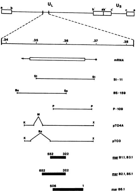
Us
b is C c a
., _,I.
.34 .35
~~~~~~~~
.36 .37~mRNA
.381
[image:3.612.66.305.70.405.2]St
TABLE 1. Markerrescuefrequencies of gBmarmutantsa RescuefrequencyofgBplasmids mar Mutant
BS-159 St-11 P-109 pTO3 pTO4A
Bli 1.0 2.2 <0.1 0.9 NTb
B2.1 2.2 3.5 <0.1 <0.1 2.0
B3.1 1.1 1.5 <0.1 4.1 NT
B4.1 <0.1 <0.1 <0.1 <0.1 <0.1
B5.1 4.8 8.5 <0.1 <0.1 3.1
B6.1 <0.1 1.0 3.5 NT NT
Rescuefrequencyasdetermined bypercentblack-plaque recombinants
(17).
bNT,Nottested.
St-11
as s.
p p
i
K~f
Be
K
x
x
552 322
ma 81.1,B3.1
052 322
marB2.1, B.1
506 1
mar96.1
FIG. 1. gB markerrescue fragments. Five different cloned gB fragments wereusedtomarkerrescuemarB mutations in cotrans-fectionassays.Allwerederived fromtheEcoRI-Ffragment of strain KOS (15). The locations of the gB message (*-*) and the coding region (ILi) in relation to the rescuing fragments (H ) are indi-cated. The regions shown to contain mar mutations (_) are
derived fromthe results in Table 1. Key: Ba, BamHI; Bs, BstEII; K, KpnI; M, MluI;P, PstI;Sa, Sall; St, SstI; X, XhoI.
(Schleicher & Schuell Co., Keene, N.H.) was performed
overnightat10 V/cminblotting buffer (15mMTris, 125 mM glycine, 20% methanol) at 15°C. The transferred filter was
blocked with 5% bovine serum albumin in
phosphate-buffered salinefor 1 hat37°C, followed by incubation with primary and secondary antibody for 1 h each at 37°C. Between incubations, filters were washed with
phosphate-buffered saline at 37°C. Detection of specifically bound antibody with 4-chloro-1-napthol (Sigma)was performed as
describedpreviously (17).
RESULTS
MappingofgBmarmutations. Todefine discrete regions of
thegBgeneto be sequenced foreach ofthe six singlemar
mutants, marker rescue experiments were performed with
the gB containing plasmids diagrammed in Fig. 1. St-11, BS-159, and P-109wereall subcloned from the KOS EcoRI
F fragment as described previously (15). These plasmids contain sequences of the HSV-1 KOSgB gene
correspond-ingto amino acid residues 1 through 816, 321 through904, and1 through 506, respectively. pTO3 and pTO4A are also
EcoRI-F subclones that have internaldeletions
correspond-ingtoregionsin theexternal, transmembrane, and cytoplas-mic domainsofgB. They containcoding sequencesfor the entire gB protein exclusive of amino acid residues 653 through824 inpTO4A and 553through 875 inpTO3.
Cotransfection experiments with genomic mar mutant DNA and linearized plasmid DNA were performed as de-scribedpreviously (19).Markerrescueof themarphenotype
was determined on the basis of blackplaques inan
immu-noperoxidaseplaqueassay (17). AllmarB mutants failed to
be recognizedby the antibodies towhich they areresistant (24). Therefore, plaques formed by recombinants in which themarmutationwasreplaced by wild-typeKOSsequences
could be identifiedbytheirreactivitywith theselectingMAb (16).
Site Imutants marB1.1 and B3.1wererescuedby St-11, BS-159, and pTO3 (Table 1), indicating the presence ofa
mutation in the region coding for residues 322through 552. Similarly,site IImutantsmarB2.1and B5.1wererescuedby
St-11, BS-159,andpTO4A (Table 1), localizingthe mutation tothesequencescorrespondingtoresidues322through652.
mar B6.1 was rescued with St-11 and P-109, indicating an
alteration in the region encoding residues 1 through 506 (Table 1). These resultsare depictedatthe bottom ofFig.1. The site III mutant, mar B4.1, was not successfully rescued withanyof thefragmentsshown inFig.1.However, itwasrescued(4.0%)with theentire EcoRI Ffragment (map
coordinates 0.315to0.421), placingthe mutation in thesame
general regionof thegenomeasthegBgene.Thus,we were
unable to confirm that the mutation causing the mar B4.1
phenotype lies solely within the coding sequence for gB. However, gB alone is specifically precipitated from deter-gent-solubilized infected-cell extracts by antibody B4 (13), making it unlikely thatanother virus- or host-encoded
mol-ecule is involved in maintenance ofantigenicsite III. DNAsequencingofmarmutantsingB.Bothenzymatic(30)
and chemical cleavage (26) sequencing strategies were
em-ployed in mar mutant sequencing. All coding sequences implicated in marker rescue experiments were sequenced.
Because we were unsuccessful in attempts to map themar
B4.1mutation,the entiregB coding sequenceof this mutant
was determined.
Abase transition of GtoAatnucleotide 2207 resulted in
a Ser-to-Asn changeat residue 473 for both site I mutants,
marB1.1 andB3.1. A transitionofGtoAatnucleotide2569 caused theGlyat residue594tobecomeanArginbothmar
B2.1 and B5.1 (site II). Two G-to-A nucleotide transitions
were found in the site III mutant mar B4.1. The first
occurred at nucleotide 1702 and resulted in a Glu-to-Lys
substitutionat residue 305. Thesecond, at nucleotide 2419, didnotresult inachange intheamino acidsequenceof the
translated product. Inantigenic site IV, atransition of Gto
UL
St
J. VIROL.
on November 10, 2019 by guest
http://jvi.asm.org/
[image:3.612.318.559.81.170.2]mar MUTATIONS IN HSV-1 gB 733
MAX B1.1, 3.1
maX B2.1, 5.1
mar B4.1
SITE I
469 477
Asn
Leu Arg Glu Gln Ser Arg Lys Pro Pro
CTC CGA GAG CAG AGC CGC AAG CCC CCA A
2194 2220
SITE II
590 598
Arg
Ser Ser Arg Pro Gly Ala Cys Tyr Ser AGC TCG CGG CCC GGG GCC TGC TAC AGC
A
2557 2583
SITE III
301 309
Lys
Tyr Gly Tyr Arg Glu Gly Ser His Thr TAC GGC TAC CGG GAG GGG TCG CAC ACC
A
1690 1716
SITE IV
081 089
Asp
msU B6.1 Pro Arg Pro Ala Gly Asp Asn Ala Thr
CCA CGC CCC GCC GGC GAC AAC GCG ACC
A
1030 1056
FIG. 2. Nucleotide and aminoacid substitutions in mar B mu-tants. The regions depicted at the bottom of Fig. 1 and the entire coding sequence for mar B4.1 were sequenced as described by Maxam and Gilbert (26) or Sanger et al. (30). Individual base changesandthepredicted amino acid changesareshowninboldface typeforeach mutantcomparedwithwild-type KOS-321 sequences (29).SubstitutionsinmarB1.1and3.1andin mar B2.1and5.1were identical, although thesemutants wereisolatedwithclonally distinct antibodies. The numbering scheme forthe DNA sequence assigns the number1tothefirstbaseoftheXhoI siteat0.372 mapunitson theviralgenome(15; S. Person,unpublished data). Numbering for amino acids designatestheinitiation methionineas 1.
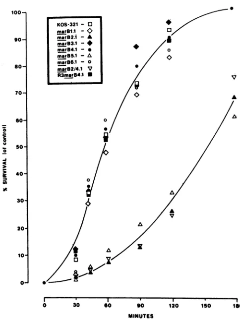
100-
90-
So-
70-I
0 I
-
S0-SO
40-ItOS321- a3
marB1.1 -0
marsB2.1- A
msrB3.1-* marB4.1
-marB5.1 -A mar86.1 - 0
msrB2/4.1 V
R3marB4.1 N
0
V
AN
30/ A
20I
10 *
0
I I *
0 30 60 90 120 150 ISO MINUTES
FIG. 3. Rate ofentry kinetics formar B mutants. The rate of penetration forKOS-321 andantigenic variants wasdeterminedby usingacitric acid buffer inactivationassay asdescribed previously (13).Wild-type andmutantviruseswereadsorbedtocell monolay-ers at4°C for2 hand then shiftedto370C. Atthe indicatedtimes, monolayers were treated with citratebuffer(pH 3.0)for1min. The percentsurvivalrepresents thefraction ofinput virusthatentered cellsat agiven time,compared withacontrolsamplethatreceived noacidtreatment.
Aat nucleotide 1043, changed Gly at residue 85 to Asp in marB6.1. Therefore, one amino acid substitution was pre-dictedto occurinthesequenced region of eachmarmutant (Fig. 2).
Effect ofantigenic variation on rate of virus entry.
Previ-ously, we showed thatantigenic variation in HSV-1gBcan result in impaired glycoprotein processing and function at
high temperature (24). The gBrate of entry phenotype (11) was attributed to a mutation at residue 553 in the ts B5
variant, which mapped within the limits of the external
domain of gB (4, 10).Therefore,therateof entry for eachmar B mutant wasassayedtodetermine whetherantigenic varia-tion in gBcanalsocausealterations in therateof virus entry.
Virus was adsorbed to cellmonolayers at4°C to prevent
penetration(18) and then shiftedto37°C toinitiate entry.At various times after the shift, monolayers were washed briefly with a low-pH citrate buffer, resulting in inactivation of all extracellular virus (14). Internalized virus was resistant to such treatment and replicated to form plaques. The wild-type virus, KOS-321 (Fig. 3), entered cells slowly up to 30 minafter shift to 37°C but began to enterrapidlyat 45 min after shift. This rate was maintained until most virus had entered cells(2hpostshift),with allinfectiousvirus
becom-ing resistant to citrate treatment by 3 h postshift. Similar
entrykinetics wereobserved with mar mutantsB1.1, B3.1, B4.1, and B6.1 (Fig. 3). However, marB2.1 and B5.1 (Fig.
3) entered cells more slowlythan did thewild type and did notachievecompleteentryuntil 5to6 hpostshift.Thesetwo mutantswereboth selectedby antibodiesrecognizing site II, suggesting that the mar mutation was responsible for the altered entry phenotype.
In a previous study (24) a ts mutant, mar B2/4.1, was
generated bymixed infection with themutantsmarB2.1and marB4.1followedby selectionwith MAbs B2 and B4. Then a revertantof this doublemutant wasselectedonthebasisof growth at hightemperature. Thismutant had also partially regained the ability to process gB and simultaneously
re-gained sensitivity to neutralization with antibody B2,
indi-catingthat themarandtsphenotypeswerethe resultof the same genetic alteration
(24).
However, the revertant re-mained resistant to antibody B4 and was thereforedesig-natedR3 marB4.1.
Todetermine whether themarand slow entryphenotypes
werethe resultof thesamemutation, the entry kinetics for thedoublemar mutantandrevertantweretested in the assay described above.marB2/4.1entered cellsat a ratesimilarto the parent,marB2.1 (Fig. 3).The
finding
that the combina-tion of the B2and B4 mutations inasingle
gBgene hadno VOL. 63,1989on November 10, 2019 by guest
http://jvi.asm.org/
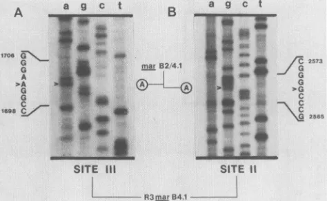
[image:4.612.314.556.66.389.2] [image:4.612.58.298.73.364.2]A
314
2573 9C
D-0o
pgc1
B
245
C
077
m* |pgB
M1 2 3 45 6 M 1 2 3 4 5 6 Ml 2 3 4 5 6
i2565
R3m..arB4.1
FIG. 4. Sequence of R3marB4.1 withgenomic viral DNAas a
template. A 32P-end-labeled 16-base-pair oligonucleotide primerwas
annealed to 1 ,ug of CsCl-purified revertant DNA template and sequenced essentiallyasdescribedpreviously (21; Huibregtseetal., inpress). After ethanol precipitation, samplesweresuspended in 4
RI of loading dye and subjected to electrophoresis on an 8% denaturing acylamide gel. Panel A showssequencessurrounding the site IIImutation, and panel B showssequencessurrounding the site
IImutation. Arrows indicate the location of altered nucleotides and circled bases depict the relevant nucleotides in the parental mutant
marB2/4.1.
additional effect on rate ofentry was expected, since only
theparental mutantmarB2.1, andnotthemarB4.1parent, exhibited theslowentrykinetics. Asanticipated, the
rever-tant R3 mar B4.1 entered cells at a rate similar to that of wild-type virus and of the marB4.1 parent (Fig. 3). There-fore, reversion of the B2 antibody resistancewas accompa-nied by reversion of the slowentrykinetics displayed bymar
B2.1. This result strongly suggested that, like the tempera-ture-dependent defect in gB processingseeninmarB2.1,the
reduced rate of virus entry was caused by the site II mar
mutation.
Identification of nucleotideandpredictedamino acid
rever-sion inR3 marB4.1. As described above, the revertant R3 mar B4.1 was used to demonstrate that the ts and roe
phenotypes wereattributabletothemarmutation in site II. To determine theexactgeneticnatureof the reversionevent and to confirm the association of the mar, ts, and roe
phenotypes, regions of the gB gene from R3 mar B4.1
corresponding to the mar B4.1 and mar B2.1 lesions were
sequenced. This analysis was performed by using a novel
sequencing technique where the intact viral genome was
used as atemplate for double-stranded dideoxy sequencing
(seeMaterials and Methods).
The parental mutant marB 2/4.1 had mutations identical
tothose that resulted in amino acidsubstitutionsin thesingle
marmutants,B2. 1 and B4.1,fromwhichitwasderived(data
notshown). TheG-to-A transition causing site III neutrali-zation resistance located at nucleotide 1702 (Fig. 1) was
present in the revertant (Fig. 4A). However, the site II G-to-Atransitionseenatnucleotide 2569 inmarB2.1(Fig. 1)
was no longer present, and the revertant contained the wild-type sequence at this location (Fig. 4B). Therefore, precise reversion of amino acid594ispredicted and confirms thatthe site II mar, ts, and roe phenotypes result fromthe
samebase substitution.
Immuneprecipitation ofHSV-1 gB insertionmutants. Pre-viously, it was shown that selecting antibodies failed to immunoprecipitate gB produced by mar mutants (24). In addition, antibodies representingtwoantigenic sites (II and
2 Xl.u-thf) at 313 6XQ(eu-thr)at 437 6X(leu-thr) at 590
FIG. 5. RadioimmuneprecipitationofHpaIlinker-insertion
mu-tants of HSV-1 gB. Vero cells transfected with insertion mutant
plasmid DNA were infected 48 h posttransfection with the gB-mutantK082. After infection for 7h, cellswerelabeled for1h with 50 xCi of [35S]methionine per ml. Panels A, B, and C show detergent-solubilizedextractsofcellstransfected with mutants314, 245, and 077, respectively. Lane M is an extract of
mock-trans-fected, K082-infected Verocells precipitated with apool of
anti-HSVglycoprotein C MAbs (25). Lanenumbers identify precipita-tion with the indicatedantibody (1 through 6).
III)reacted with thegBprecursor(pgB)fromanumberofts
mutants grown at 34°C but failed to recognize the same
molecule when it was produced by infection at 39°C (24). This resultsuggestedthat antibodiesspecificfor sitesII and IIIrecognized discontinuous epitopes whose integrityrelied
on ahigher-order structure.
Tofurtheridentify regions ofgB involved in the
mainte-nance of these structures, a series ofplasmids containing
HpaI linker-insertion mutations in the gB gene (5) were
examined. These linkers varied in number and resulted in 2 to 28 in-frame amino acid insertions at different locations throughout the gB product. The effect of insertions on molecularstructurewasmeasuredbyalteration in theability of MAbs to recognizemutant polypeptides inradioimmune precipitation assays. All insertions in the external domain resulted in altered gB processingand the production ofgB molecules that were unable to complement a gB- virus,
indicatingalethal defect in virus replication (5). Thesingle insertion in the cytoplasmic domain had noapparent effect
ongB synthesis orvirus infectivity.
Experimentswereperformed by transfectingmutant plas-mid DNA intoVerocells,followedby superinfectionwitha
gB-virus(K082)toactivate theexpressionof themutantgB gene (5, 13). Infected cells were pulsed for 1 h with
[35S]methionineat7 hpostinfectionandimmunoprecipitated with MAbs Bi through B6. Figure 5 shows the results for three different insertion mutants. Figure SA shows precipi-tations fromcells transfected with mutant 314, which
con-tains a four-amino-acid insertion after residue 313. Lane 4
lacked the bandpresentinotherlanes, demonstratingaloss ofrecognition by antibody B4 (site III).Thisresultwasnot unexpected, since the marmutation atresidue 305 (Fig. 2) caused a similar loss ofreactivity (24), indicating that this
regionofgB isimportantforrecognition bythismonoclonal antibody. Of interest was the observation that this same
insertion affectedprecipitation ofgB by twoother antibod-ies, B2 andB5 (Fig. SA, lanes 2 and 5, respectively), both specificforantigenicsiteIIandfor which thecorresponding
mar mutation maps some distance away (Fig. 2). This suggeststhatsitesIIand IIImaysharea commonstructural
entityeventhough residuesinvolvedintheir recognition by
antibodies lie far apart on the primary sequence. Fig. SB shows another insertion, 245, which adds 12 amino acids after residue 437. Antibodies B2, B4, and B5 all failed to precipitatethismolecule(Fig. SB, lanes 2, 4, and 5,
respec-A a
17060
G
G
A
A
G
G
169086 ,
on November 10, 2019 by guest
http://jvi.asm.org/
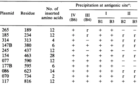
[image:5.612.322.559.68.172.2] [image:5.612.68.305.74.220.2]mar MUTATIONS IN HSV-1 gB 735 TABLE 2. Radioimmune precipitation of gB HpaI
linker-insertion mutants
No.of Precipitation at antigenic sitea:
Plasmid Residue inserted IV III I II
amino acids
(B6)
(B4) B1 B3 B2 B5265 189 12 + r + + -
-185 234 12 + r + + r r
314 313 4 + - + + r r
147B 380 6 + + + + r r
245 437 12 + - + + -
-154 463 28 + + + + r r
077 590 12 + + + + -
-177B 595 6 + + + + -
-086 623 14 + + + + r r
070 734 2 + + + + r r
117 816 12 + + + + + +
a+,Precipitationofmutantpolypeptide;r,significantly reduced precipita-tionwhencomparedwith wild-type gB;-,complete absenceofprecipitation.
tively). Thisinsertion lies between residuespredicted tobe
critical for recognition by these antibodies (Fig. 2). Figure SC shows the results for mutant 077, which carries an
insertion of 12amino acids after residue 590. Reactivity of
siteII antibodies (B2, B5) (Fig. SC, lanes 2 and 5,
respec-tively)wasabolishedbythis insertion.Again,this result was notunexpected, sincethe site IImarmutation is located at
residue 594 (Fig. 2). In addition, this mutant also demon-strated that loss ofsite II recognition does not necessarily
correlate with reduction or loss ofsite III recognition, as would be suggested by Fig. 5A and B.
Table 2 shows thereactivity ofall six antibodies with the
entire panel of insertionmutants. Severalgeneraltrendsare apparent. Site II antibodyrecognition was inhibited, either
partially orcompletely, by eachofthe 10insertions in the external domain ofgB. Neither the size northelocation of the insertion appeared to correlate with reduction versus
complete loss of reactivity. Site III antibody recognition appeared to be somewhat less affected but was altered by insertions throughout the amino-terminal half of the mole-cule.Site I and site IVantibody recognitionwasnotaffected byanyinsertion, suggestingthat thesedeterminants areless
dependent than the others on tertiary structure. In fact,
mutant 154 had 28 amino acids inserted 11 residues away
fromtheresidueaffected bysite Imarmutations(Fig. 2),yet this alteration had no effect on precipitation of pgB by antibodies
Bi
and B3. Finally, the single insertion in thecytoplasmic domain (mutant 117) did not affect
antibody
recognition of any site, suggestingthat this region haslittle or noeffectonthe structureofthe external domain.
Determination of antibody recognition in Western blots.
Recognition of antigenic sites in gB molecules
containing
insertion mutations varied widely, suggesting differingcon-tributions ofhigher-orderstructure to
antibody binding.
Tofurther examine the requirements for
antibody
recognition,
the entire panel of antibodies was tested for the ability to reactwith denaturedgB in Western blots. KOS-321-infected celllysates
were subjectedtoelectrophoresis,
blotted, andincubatedwithMAbs
Bi
through B9.OnlyantibodyB6(site IV)recognizedthe denaturedgBmolecule(datanotshown), suggesting that antibody recognition of this site relies onrelativelylocalizedstructures. LikeB6, siteIantibodies
(Bi
andB3) weretype specific (24) andinhibited virus
penetra-tion (13), and their
reactivity
with gBwas notinhibitedby
insertional mutagenesis (Table 2). However, unlike
B6,
these MAbs did not react with denatured gB. Therefore, maintenance of antigenic sites I, II, and III must be depen-dent on structures in gB that are lost upon denaturation.
DISCUSSION
Our studies of the immunobiology and function of HSV glycoproteins have been greatly facilitated by the use of virus-neutralizing glycoprotein-specific MAbs and mar mu-tants (14, 24, 25). In some instances, antibody binding interfered with infectivity, suggesting that antigenically im-portant structures overlap with regions that directly contrib-ute to virus penetration (13, 14). Consistent with this pre-diction, some antibody-selected variants were altered in gB function (24) (Fig. 3). Therefore, this type ofanalysis serves two purposes. First, antigenic sites on HSV glycoproteins canbeoperationallyidentified and characterized on the basis ofantibody-mutant reactivity patterns, thusproviding infor-mation on the antigenic structure of the molecule. Second, with MAbs and antibody-selected mutants as complemen-tary molecularprobes, large domains, and specific residues can be examined for functional importance. Below, we summarize the results of ourimmunologic analysis of gB. In addition, mutations that affect the function of gB are dis-cussed in the context of antigenic sites and theirrespective contributions to virus infectivity.
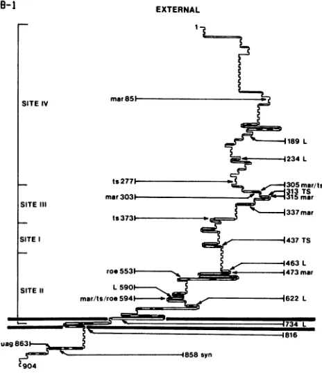
Antigenic site IV.Antigenic site IV appears to residenear the amino terminus of the gB molecule (13) (Fig. 6). Al-though it was notpossible to accurately define the limits of site IV byusing truncation mutants (13), a deletion mutant missing residues 44to233of gBwasshowntolack site IVby radioimmune precipitation (S. Highlander, unpublished data). This agreeswith thelocation ofthesite IV mutationat residue 85. In addition, the single site IV-specific antibody,
B6, recognized denatured gB in a Western blot, suggesting
that site IVconsists ofalinear determinant.
The Gly residue at amino acid 85 is type specific since HSV-2containsanArgatthatsameposition.This correlates with thefinding thatantibody B6 neutralized HSV-1 andnot HSV-2 and that this region ofgB is relatively nonhomolo-gous when compared with other herpesvirus gB homologs
(2, 9, 22, 27, 29, 32). Despitethe lackof conservation in this region, B6 inhibits the rate of virus penetration (13) and is notable as being our only gB-specific MAb that inhibited cell-to-cell spread of virus (13) and prevented zosteriform spread of virus in vivo (J. Mester, J. C. Glorioso, and B. Rouse, unpublished data).
Antigenic siteIII. The single siteIIIantibody,B4,required
sequencesbetweenresidues 283 and 380 for immune
precip-itation of gB (13) (Fig. 6). A base substitution wasfound in mar B4.1 which resulted in a Glu-to-Lys conversion of residue 305. Amino acid 305wasshowntobeaGluin HSV-2 as well, correlating with the ability of antibody B4 to
neutralize both serotypes (24). In the absence of
comple-ment, neutralization of both HSV-1 and HSV-2 by B4 resulted from prevention of virus penetration (13). The substitution at residue 305 also affected terminal
glycosyl-ation in the mutant mar B4.1 at a high temperature (24).
These findings indicate that structures represented by anti-genic siteIIIdataare importanttothefunction ofgB. Intwo strain KOS mutants, ts J20 and ts J12, changes at residues 277and 373, respectively, havebeen identified which inhibit gB processing at high temperature and render mutantvirus incapable ofpenetratingcell surfaces(3)(Fig. 6),
supporting
the notion that this region can affect
gB
processing and, by
extension, virusinfectivity. Unlike marB4.1, these mutants VOL. 63, 1989on November 10, 2019 by guest
http://jvi.asm.org/
GB-I
IY
SITEIV
SITEIII
SITE
SITE 11
189L
.-e -4234L
ts2771 - ,.-305mar/ts
313TS
mar3031 215mar
v 337mar
ts3731-,
1437TS
463 L
roe553 , - . 1473mar
L590
mar/ts/roe 5941--_ 622L
1816
uag863
1858syn
Z904
CYTOPLASMIC
FIG. 6. Predicted secondary structure of HSV-1 KOS-321 gB andlocationof knownmutations.Mutations inHSV-1KOS-321gB that have been identified by DNA sequencing are depicted on a
schematic representationof the moleculegenerated bythe protein structure analysis program MSEQ (1) with Chou and Fassman
parameters (8). Alphahelices(_),betasheets(=),randomcoil (-),andbetaturns(r- )aredepicted.Otherpublishedmutations
are shown as well (4, 6, 7, 20a, 28). Uppercase letters denote insertion mutations, and lowercase lettersdenote aminoacid sub-stitutions. The amino acid number indicates the altered residue (substitution, termination) ortheresiduepreceding insertion. Anti-genic sitesareshownas predicted by Highlanderetal. (13).
areneutralizedbyantibody B4, identifyingasecondtypeof
ts mutation in this region that does not alter antigenic recognitionof site III (S. Highlander, unpublished data).
As reported earlier, antibody B4was ableto immunopre-cipitate pgBproduced byanumberoftsmutants at34°Cbut
failed to recognize thesame molecule at the nonpermissive temperature (24). This finding indicated that site III was
dependent on gB conformation, a prediction confirmed by
thefact that insertionsover abroad region (residues 189to 437) reduced or blocked recognition ofgB by B4 (Table 2, Fig.6). Moreover,allof theseinsertionmutationsresulted in
theproductionofinactivegBmoleculesasmeasuredby their
failure to complement a gB- virus in transient expression assays (5) (Fig. 6). Taken together, these observations suggestthatahigher-orderstructureisrequiredtomaintain antigenic site III and necessary for gB function in virus penetration.
Pelletetal. reportedthe locationofmutations thatresult
in neutralizationresistanceinHSV-1strain FgB(28).Ofthe
three mutants examined, all had changes between residues
303and 335,close tooursiteIII mutationat residue305. In
additiontobeingahighly antigenicareaof the molecule(Fig.
6), it is almostidentical in HSV-1 and HSV-2(2, 32) and is highly homologous among all herpesvirus gB genes
exam-ined (9, 22, 27, 29). This conservation of the
primary
sequence suggests atertiary homology
aswell and indicatesthat the structureofthisregion isimportantforgBfunction. This is supported by the demonstration that
antigenic
vari-ation in this site can result in temperature-dependent
insta-bility that leads to defects in
glycosylation
and function athightemperature (5, 24).
Antigenic site I. With truncated gB
molecules,
siteI-specific antibodies were predicted to
recognize
a structure betweenresidues381 and 441(13)
(Fig. 6). However,
thesiteImutation inmar B1.1 and B3.1waslocated atresidue 473. The Ser-to-Asn change at this residue was unexpected in that site I antibodies
immunoprecipitate
a gB moleculecontaining
only
the amino-terminal 441 aminoacids,
suggest-ing that an interaction with residue 473 is not required for antibody binding. This could be explained by the introduc-tion of the larger, charged side chain, which prevents anti-body contact with residues contained within the first 441 residuesof the gB molecule. Alternatively, residue 473 may be required for neutralization, but its loss may not besufficient toeliminate
antibody
binding. Thefirsthypothesis
is supported by the finding that gB produced by mar B1.1 and B3.1 is notprecipitated byasite I antibody (24). These molecules contain residues sufficient for antibody
recogni-tion (1 through 441) but are not precipitated. Sequence variation in this region in otherherpesvirus gB genes (9, 22,
27, 29) suggests little conservation of function. However,
site I-specific antibodies did reduce the rate at which the virus entered cells (13), indicating that this general region
may play a rolein virus infection.
Antigenic site II. In addition to causing ts defects in carbohydrate processing at high temperature (24), mar mu-tants selected by site II-specific antibodies also have a reduced rate of virus penetration (Fig. 3). Theidentification
of the site II mar mutation at residue 594 not only agreed
with the predicted location of site II antibody recognition (13) but also mapped 41 amino acids away from the rate of entry mutation in tsB5 at residue 553 (4) (Fig. 6). Because antibodies specific for site II are reactive with both sero-types, thefinding that Gly at residue 594is conserved in both HSV-1 and HSV-2 was not surprising because site II anti-bodies can neutralize HSV-2. This residue is located in a region (residues 500 through 700) of gB that is highly homologous with other herpesviruses (2, 9, 22, 27, 29, 32), suggesting a conservation of higher-order structure and function.
Like site III antibodies, site II antibodies fail torecognize
the pgB produced by ts mutants at the nonpermissive temperature (24) anddid not bind denatured gB in a Western blot. However, site II was even more susceptible to
desta-bilization than site III since every insertion tested in the external domain either reduced or destroyed antibody rec-ognition of site II (Table 2). Some insertions affected both site II and III recognition, but the corresponding mar muta-tionsfor these sites lie 289 residues apart (Fig. 2). Although these data are difficult to interpret without detailed informa-tion on the tertiary structure of gB, we speculate that these nonoverlapping epitopes are contained within a common higher-order structure created by folding of the molecule. Alternatively, sites II and III could represent structures that are spatially distinct yet sensitive to denaturation by inser-tions throughout the external domain of gB. Regardless, the finding that at least two of the four sites we have defined are comprised of widely separated sequences is not unexpected. Chapsal and Pereira demonstrated that over half of the
a .l
L-- IG ==j 'v. 1734L
on November 10, 2019 by guest
http://jvi.asm.org/
[image:7.612.70.302.76.353.2]mar MUTATIONS IN HSV-1 gB 737 gB-specific MAbs tested were directed against discontinuous
epitopes (7).
Conclusion. In considering the data presented here and previously, it seems clear that antigenic sites II and III contain structures important for gB function in virus infec-tion. This conclusion is based on the following evidence. First, mutations that alter the ability of gB to function in viruspenetration occur within sequences defined as neces-saryfor the recognition of these sites (13). These mutations can occurnaturally (3, 4) or may be selected on the basis of resistance to neutralizing antibodies (Fig. 2). Second, both sitesaretype common, and the primary sequences that make up these regions are very conserved between many gB
homologs. This suggests that their secondary and tertiary
structures are also conserved and likely reflect structural
constraints imposedbygBfunction. Third, only these sites are lost from the replication-defective ts and insertion mu-tants. It may be that the structures recognized by these
antibodiesarerequired for gB functionandarelost in mutant
gB molecules. However, only antibodies to site III can
neutralize virus infectivity without complement. Antigenic sites III, I, and IV lie in the amino-terminal half of the gB molecule, and antibodies specific for each of these sites
inhibitvirusinfectivity. Antibody bindingtothis regionmay
sterically inhibit an interaction that is required for
penetra-tion. Conversely, antibodies specific for antigenic site II do notinhibit penetrationyet select mutantswhichshow altered entry kinetics. Although this determinant is affected by
much ofthe structure oftheexternal domain, it apparently
does notcontributetogBfunction in the same manner as the sequencescomprisingsite III. On the basisofthelocation of siteII marmutations, itappears to be veryimportanttothe entryprocessnonetheless.
Itremains tobeseen whether theseregions havediscrete
functions and to what degree antigenic sites II and III
interact. Using smaller, clonedfragments ofthegB gene to express portions ofthe polypeptide, we hope to determine whether a fraction of the mature molecule can specifically interactwithanother. If so, it may bepossible to use intra-or intermolecular complementation between different mu-tants to identify distinct functional domains. These
experi-ments, coupled with an extensive mutational analysis of these regions, may lead to a more precise definition of
residuesand structures thatcontribute togB function.
ACKNOWLEDGMENTS
This work was supported by Public Health Service grants RROO100, GM34534, A118228, A111513, and GM27819 from the National Institutes of Health and by grant MV-167B from the AmericanCancerSociety. S.L.H. was supported bythe Graduate Program in Cellular and Molecular Biology (National Research Service Award training grant 2T32GM07315 from the National Institutes ofHealth) and byaHoraceRackhampredoctoral fellow-ship, and P.J.G. was supported by an F. G. Novy predoctoral fellowship, allattheUniversity ofMichigan. Wealsoacknowledge the supportofT.C.H. by the Dental Research Institute Associate Program fromtheOffice of the Vice Presidentfor Research atthe UniversityofMichigan.
We thank Elizabeth Smiley and Linda Harper for excellent technicalwork with monoclonal antibodies.Wearedeeply indebted
toFredHoma,PatVenta, DavidEngelke,and JohnHuibregtsefor experttechnical advice.
LITERATURECITED
1. Black,S.D., and J. C. Glorioso. 1986.MSEQ:a microcomputer-based approach to the analysis, display, and prediction of proteinstructure. Biotechniques4:448-460.
2. Bzik,D.J.,C.DeBroy,B. A.Fox,N. E.Pederson,andS. Person. 1986. The nucleotidesequenceof thegB glycoproteingene of HSV-2andcomparisonwith thecorrespondinggeneofHSV-1. Virology155:322-333.
3. Bzik, D. J., B. A. Fox, N. A. DeLuca, and S. Person. 1984. Nucleotide sequence specifying theglycoproteingene, gB, of herpes simplexvirus type 1.Virology 133:301-314.
4. Bzik, D. J., B. A. Fox, N. A. DeLuca, and S. Person. 1984. Nucleotide sequence of a region of the herpes simplex virus type 1 gBglycoprotein gene: mutations affecting rateof virus entryandcellfusion. Virology137:185-190.
5. Cai, W., S. Person, C. DeBroy, and B. Gu. 1988. Functional regionsandstructural features ofthegBglycoproteinofherpes simplexvirustype 1:ananalysisof linkerinsertionmutants.J. Mol.Biol. 201:575-588.
6. Cai, W.,S.Person,S. C.Warner, J.Zhou,and N.DeLuca.1987. Linker-insertion nonsense and restriction-site deletion
muta-tions ofthegBglycoproteingeneofherpessimplexvirustype 1. J. Virol. 61:714-721.
7. Chapsal, J. M., and L. Pereira. 1988. Characterization of epitopesonnativeanddenaturedformsofherpessimplexvirus glycoproteinB. Virology164:427-434.
8. Chou,P.Y.,andG. D. Fasman. 1978. Empiricalpredictionsof proteinconformation. Annu. Rev. Biochem. 47:251-276. 9. Cranage,M.P.,T.Kouzarides,A.T.Bankier,S.Satchwell,K.
Weston, P. Tomlinson, B. Barrell, H. Hart, S. E. Bell, A. C. Minson, and G. L. Smith. 1986. Identification of the human cytomegalovirus glycoproteinB gene andinduction of neutral-izing antibodies via its expression in recombinant vaccinia virus. Eur.Mol. Biol. Org. 5:3057-3063.
10. DeLuca,N.D.,D.J. Bzik,V.C.Bond,S.Person,and W.Snipes. 1982. Nucleotide sequences of herpes simplex virus type 1 (HSV-1) affecting virus entry, cell fusion, and production of glycoproteingB (VP7). Virology 122:411-432.
11. DeLuca,N.D.,D.J. Bzik,S.Person,and W.Snipes.1981.Early
events in herpes simplex type 1 infection: photosensitivity of fluorescein isothiocyanate-treated virions. Proc. Natl. Acad. Sci. USA78:912-916.
12. Graham,F. L.,andA.J.vanderEbb. 1973. Anew technique for the assay of infectivity of human adenovirus. Virology 52:456-467.
13. Highlander, S. L., W. Cai, S. Person, M. Levine, and J. C. Glorioso. 1988. Monoclonal antibodies define a domain on herpes simplexvirusglycoproteinB involved in virus penetra-tion.J. Virol. 62:1881-1888.
14. Highlander,S.L.,S. L.Sutherland,P.J. Gage,D.C.Johnson, M. Levine, andJ. C. Glorioso. 1987. Neutralizingmonoclonal antibodies specific for herpes simplex glycoprotein D inhibit viruspenetration.J. Virol. 61:3356-3364.
15. Holland, L. E., R. M. Sandri-Goldin, A. L. Goldin, J. C. Glorioso, and M. Levine. 1984. Transcriptional and genetic
analysesoftheherpessimplexvirustype 1 genome:coordinates 0.29to0.45. J. Virol.49:947-959.
16. Holland, T. C., S. D. Marlin, M. Levine, and J. C. Glorioso. 1983. Antigenic variants ofherpes simplex virusselected with
glycoprotein-specific monoclonal antibodies. J. Virol. 45:672-682.
17. Holland, T. C., R. M. Sandri-Goldin, L. E. Holland, S. D. Marlin,M.Levine, and J.C. Glorioso. 1983. Physicalmapping ofthe mutation inan antigenicvariantofherpes simplexvirus type 1 by use of an immunoreactive plaque assay. J. Virol. 46:649-652.
18. Holmes,J. H.,andD. H. Watson.1961. An electronmicroscope study of the attachment and penetration of herpes virus in BHK21 cells.Virology21:112-123.
19. Homa,F.L.,T. M. Otal,J.C. Glorioso, andM. Levine.1986. Transcriptionalcontrolsignalsofaherpessimplexvirustype1 late(gamma2)genelie within bases -34to +124relativetothe 5' terminus of the mRNA. Mol.Cell. Biol. 6:3652-3666. 20. Huang, A. S., and R.R. Wagner. 1964. Penetration ofherpes
simplex virus into human epidermoid cells. Proc. Soc. Exp.
Biol. Med. 116:863-869.
20a.Huff, V., W. Cai, J. C. Glorioso, and M. Levine. 1988. The VOL.63, 1989
on November 10, 2019 by guest
http://jvi.asm.org/
carboxy-terminal 41 amino acids of herpes simplex type 1 glycoprotein B are not essential for production of infectious virus particles. J. Virol. 62:4403-4406.
21. Huibregste, J. M., and D. R. Engelke. 1986. Directidentification of small sequence changes in chromosomal DNA. Gene 44: 151-158.
22. Keller, P. M., A. J. Davidson, R. S. Lowe, C. D. Bennet, and R. W. Ellis. 1986. Identification and structure of the gene encoding gpII, a major glycoprotein of varicella-zoster virus. Virology 152:181-191.
23. Little, S. P., J. T. Jofre, R. J. Courtney, and P. A. Schaffer. 1981. Avirion-associated glycoproteinessential forinfectivity ofherpes simplex virustype 1.Virology 115:149-410. 24. Marlin, S. D., S. L. Highlander, T. C. Holland, M. Levine, and
J. C. Glorioso. 1986. Antigenic variation (mar mutations) in herpes simplex virus glycoprotein B can induce temperature-dependent alterationsin gBprocessing and virus production.J. Virol. 59:142-153.
25. Marlin, S. D., T. C. Holland, M. Levine, and J. C. Glorioso. 1985. Epitopes of herpes simplex virus 1 glycoprotein C are clustered intwodistinct antigenic sites.J. Virol.53:128-136. 26. Maxam, A. M., and W. Gilbert. 1977. A new method for
sequencingDNA. Proc. Natl. Acad. Sci. USA74:560-564. 27. Pellet, P. E., M. D. Biggin, B. Barrell, and R. Roizman. 1985.
Epstein-Barr virus genome may encode a protein showing significantaminoacid and predicted secondarystructure
homol-ogy with glycoprotein B ofherpes simplex virus 1. J. Virol. 56:807-813.
28. Pellet, P. E., K. G. Kousoulas, L. Pereira, and B. Roizman. 1985. Anatomyoftheherpes simplexvirus 1 strain FglycoproteinB gene: primary sequenceand predictedproteinstructureof the wild type and of monoclonal antibody-resistant mutants. J. Virol.53:243-253.
29. Robbins, A. K., D.J.Dorney, M. W.Wathen, M. E.Whealy,C. Gold,R.J. Watson, L. E. Holland, S. D. Weed, M. Levine, J. C. Glorioso,andL. W.Enquist. 1987. The pseudorabiesvirusgll geneis closely relatedto the gB glycoprotein gene ofherpes simplexvirus. J. Virol. 61:2691-2701.
30. Sanger, F., S. Nicklen, and A. R. Coulson. 1977. DNA sequenc-ing with chain-terminatsequenc-ing inhibitors. Proc. Natl. Acad. Sci. USA 74:5463-5467.
31. Sarmiento, M., M. Haffey, and P. G. Spear. 1979. Membrane proteins specified by herpes simplex viruses. III. Role of glycoprotein VP7(B2) in virion infectivity. J. Virol. 29:1149-1158.
32. Stuve,L.L.,S. Brown-Shimer, C. Pachl, R. Najarian, D.Dina, and R. L.Burke. 1987. Structure and expression of the herpes simplex virus type 2 glycoprotein gB gene. J. Virol. 61:326-335. 33. Tabor,S.,and C. C.Richardson. 1987. DNA sequenceanalysis with a modifiedbacteriophage T7 DNA polymerase. Proc. Natl. Acad. Sci. USA 84:4767-4771.