The Phenotype and Function of Hapten specific T-Cell isolated from Hypersensitive patients and healthy human donors
Thesis submitted in accordance with the requirement of the University of Liverpool for the degree of Doctor in Philosophy by
Eryi Wang
August 2015
1 Declaration
I declare that the work presented in this thesis is my own work and has not been submitted previously.
2
Acknowledge ment
Firstly, I will thank my parents. I am so indebted to the love they have given me. Besides huge financial support, they have missed me every day for five years. No language can express my gratitude to them.
I would also like to thank my supervisors, Professor Kevin Park and Dr Dean Naisbitt. It is their guidance and patience that allowed me to complete this thesis. Next I would like to thank my dear members in our group, who are Lee Faulkner, Moha mmed Amali, Andrew Gibson, Andrew Sullivan, Monday Ogese and Arun Tailor and also my dear friend Eunice Zhang, for giving me valuable instructions and helping me improve English. I will remember that for life. I will also not forget my friends and colleagues accompanying me during my memorable stay at Liverpool.
3
Abbreviations
APC Allophycocyanin
APC Antigen presenting cells
CCR Chemokine receptor (C-C) motif CD Cluster of differentiation
cpm Counts per minute CSA Cyclosporin
CTL Cytotoxic T lymphocyte CYP Cytochrome P450 enzyme DC Dendritic cell
DHR Drug hypersensitivity reaction DILI Drug- induced liver injury DMSO Dimethyl sulfoxide DNA Dioxyribonucleic acid DNCB Dinitrochlorobenzene
DRESS Drug reaction with eosinophilia and systemic symptoms EBV Epstein-Barr virus
EDTA Ethylenediaminetetraacetic acid ELISpot Enzyme- linked immunospot FACS Fluorescence activated cell sorting FBS Foetal bovine serum
FITC Fluorescein isothiocyanate
GM-CSF Granulocyte- macrophage colony-stimulating factor GSH Reduced glutathione
HBSS Hanks balanced salt solution
HEPES Hydroxyethyl piperazineethanesulfonic acid HLA Human leukocyte antigen
4 IgE Immunoglobulin E
IL Interleukin
ITAM Immunoreceptor tyrosine-based activation motifs
LAT Transmembrane adapter protein linker for the activation of T-cells LPS Lipopolysaccharide
LTT Lymphocyte transformation test MHC Major Histocompatibility complex Mins Minutes
Mo-DC Monocyte-derived dendritic cells NHS National Health Service
NK Natural killer
PAMP Pathogen associated molecular pattern PBMC Peripheral blood mononuclear cell PBS Phosphate buffered saline
PE Phycoerythrin pH Power of hydrogen
pi Pharmacological interaction RIPA Radioimmunoprecipitation assay RPMI Roswell Park Memorial Institute SFC Spot forming cell
SI Stimulation index
SJS Stevens-Johnson syndrome SMX Sulfamethoxazole
SMX-NOH Sulfamethoxazole hydroxylamine SMX-NO Nitroso sulfamethoxazole
STAT Signal Transducer and Activator of Transcription TAP Transporter associated with antigen processing TCR T-cell receptor
5 Th1 T helper type 1 cell
Th2 T helper type 2 cell Th17 T helper type 17 cell Th22 T helper type 22 cell TNF-α Tumor necrosis factor-α Tregs Regulatory T-cells TT Tetanus toxoid UK United Kingdom w/v weight/volume
WHO World Health Organization
Publication
6
Abstract
Drug hypersensitivity reactions are an important problem for pharmaceutical industry, especially when reactions are observed in late phase clinical trials. Furthermore, management of patients with reactions leads to personal suffering and financial burden on the NHS. React ions are almost impossible to predict as there is no simple relationship between the dose of drug administered and the development of hypersensitivity. In recent years, pharmacogenetic studies identified strong associations between the expression of specific HLA alleles and susceptibility to different forms of hypersensitivity, which would explain why only a small number of drug-exposed patients develop hypersensitivity. Studies utilizing peripheral blood mononuclear cells have detected drug-specific T-cells in patients with hypersensitivity, but not drug-exposed tolerant controls, suggesting that the adaptive immune system plays an important role in the disease pathogenesis. Despite this, there remains a need to further understand mechanisms as more detailed knowledge will assist the development of diagnostic and predictive assays.
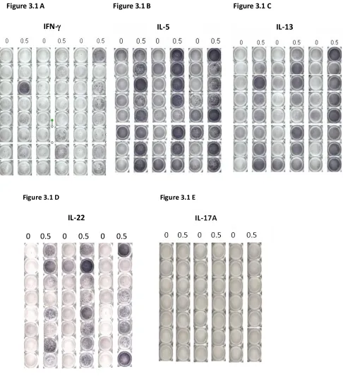
7 with secretion of Th1, Th2 cytokines and IL-22, in the absence of IL-17. Finally, CD4+ nitroso sulfamethoxazole (SMX-NO)-responsive clones were isolated from sulfamethoxazole hypersensitive patients and a similar cytokine secretion profile was observed, which suggests that IL-22 secretion might be a common feature of drug hypersensitivity.
Evolution of T-cell culture methods means it is now possible to prime naïve T-cells from healthy donors to antigens, including drugs, which they have not previously been exposed to. Piperacillin and SMX-NO were found to prime naïve CD4+ and CD8+ T-cells from healthy donors, when the derived antigens were presented in the co ntext of autologous dendritic cells. Cloned drug-specific T-cells secreted a similar panel of cytokines to that observed with patient cells. Of particular importance was the detection of IL-22 in the absence of IL-17.
The final component of the project utilized cloned T-cells with specificity for SMX-NO, piperacillin and flucloxacillin to explore mechanisms of drug-specific T-cell activation and potential cross-reactivity. Clones responsive against all 3 drugs were activated via a hapten mechanism involving (1) formation of protein adducts, (2) antigen processing and (3) presentation of derived peptides in an MHC-restricted manner. No cross-reactivity was observed with the 3 drugs.
8
Contents
Title
Declaration………..1
Acknowledge ments……….2
Abbreviations………..3
Publications……….5
Abstract ………..6
Chapter 1: General introduction……….………...9
Chapter 2: Materials and Methods……….58
Chapter 3: Characterization of T-cells in piperacillin hypersensitivity…...74
Chapter 4: The role of IL-17 and IL-22 producing cells in the skin from piperacillin hypersensitive patients with Cystic fibrosis……….95
Chapter 5: Characterization of the functionality of drug-specific T-cells primed from normal volunteer lymphocytes……….…. …………113
Chapter 6: Priming naïve T-cells to drug haptens and the characterization of drug-specific antigen presentation……..……….…130
Chapter 7: Final discussion ………..159
Appendix………168
9
Chapter 1: General introduction
1.1 Adverse drug reactions Type A (augmented) Type B (bizarre) Type C (chemical) Type D (delayed)
Type E (end of treatment reactions) 1.2 Drug Hypersensitivity
1.2.1 Epidemiological investigation of drug hypersensitivity 1.2.2 The symptoms of drug hypersensitivity
1.3 The types of hypersensitivity Type I hypersensitivity Type II hypersensitivity Type III hypersensitivity: Type IV hypersensitivity 1.4 Immune response
1.4.1 The mechanisms of antigen presentation. 1.4.2 T-cell subsets
1.4.2.1 CD8+ T-cell
1.4.2.2 CD4+ T-cell and phenotypes Th1 cells
Th2 cells Th17 cells Th22 cells
The involvement of Th17 and Th22 cells in drug hypersensitivity
1.4.4 Tissue homing
1.5 HLA associations with drug hypersensitivity 1.6 Mechanisms of drug hypersensitivity 1.6.1 Hapten and Prohapten theory
1.6.2 Pharmacological interaction of drugs with immune receptors: p.i. concept. 1.6.3 Altered self-peptide repertoire model
1.6.4 Drug metabolism and the activation of T-cells 1.6.5 SMX and SMX-NO specific T-cell clones 1.7 Drugs and drug antigens
10
1.1 Adve rse drug reactions
Edwards and Aronson, (2000) define adverse drug reactions (ADRs) as: “An appreciably harmful or unpleasant reaction, resulting from an intervention related to the use of a medicinal product, which predicts hazard from future administration and warrants prevention or specific treatment, or alteration of the dosage regimen, or withdrawal of the product.’’
Several studies have estimated the proportion of patients hospitalized due to the development of ADRs. A study by Lazarou et al (1998) in the USA used four electronic databases and showed that between 1966 and 1976 the total percentage of ADRs was 15.5%. The percentage of the patients with severe ADRs was 6.7% with 0.37% of reactions resulting in fatality. According to a study by Pirmohamed et al (2004) which looked at 18,829 patients hospitalized in the two large hospitals in the UK, 1225 of these were classified as ADRs according to the Edwards and Aronson definition. The total percentage of ADRs was 6.25% with fatality rates of 0.15%. ADRs also extend the length of stay in hospital and enhancing the costs of hospitalization (Suh et al, 2000). ADRs can be classified in terms of clinical and chemical characteristics (Park et al., 1998).
Type A (augme nted): The vast majority (95%) of adverse drug reactions are classified as this type. They are dose-dependent and predictable from the knowledge of pharmacology of the drug, thus they can be prevented. When administratio n of the drug stops the reaction disappears. Examples of type A reactions include (1) gastrointestinal bleeding induced by the treatment of drug combinations of aspirin and warfarin; (2) gastrointestinal bleeding induced by NSAIDS; (3) diuretic- induced renal impairment; and (4) -blocker-induced heart block or hypertension (Pirmohamed et al., 2004).
11 pharmacology. They display individual susceptibility and are non-dose-dependent, although this has been questioned (Uetrecht 2001). Such reactions are also referred to as idiosyncratic reactions as mechanisms have not been clearly defined. The reactions are believed to be related to drug metabolite and immunological components that maybe key to individual susceptibility. Because of this, reactions occur in a small percentage of patients. Despite this, type B reactions are extremely important because they are often severe and can lead to death (Pirmohamed et al., 2004). Type B reactions can involve any organ, and may cause anaphylaxis, and severe skin inflammation such as hypersensitivity-syndromes or drug induced lupus. Exemplar drugs that can induce this type of reaction include antibiotics, such as amoxicillin and flucloxacillin; sulfonamides, such as sulfamethoxazole; non-steroidal anti- inflammatory drugs and anticonvulsants.
Type C (che mical): These reactions can be explained by the chemical structure of the drug or drug metabolite. Paracetamol- induced hepatotoxicity is a well-defined example. The mechanism if tissue injury involves the conversion of the drug by metabolizing enzymes to a reactive qunioneimune intermediate, which (1) induces oxidative stress and (2) binds covalently to proteins. Eventually both of these processes lead to hepatotoxicity.
Type D (delayed): These reactions include delayed effects such as carcinogenicity and teratogenicity after drug administration.
12 withdrawal of anxiolytics.
1.2 Drug Hypersensitivity
Type B ADRs are mostly dominated by antigen-specific immune responses induced following drug exposure. This form of reaction is often termed drug hypersensitivity.
Drug hypersensitivity reactions involve the drug initiating an immune reaction that causes tissue damage in the patient. These reactions can be defined simply as “a serious adverse drug reaction with an immunological aetiology, to an otherwise safe and effective therapeutic agent”.
Alternatively, drug hypersensitivity has been defined by the World Allergy Organization as "an immunologically mediated drug adverse reaction of which the mechanism is IgE or non-IgG mediated and with T-cell mediated reaction largely presented in the latter (Johansson et al., 2004).
1.2.1 Epidemiological investigation of drug hypersensitivity
13 Singapore (Thong et al., 2003) and in Korea (Park et al., 2008). In these studies, 4.2 and 20 per 1000 hospitalized patients, respectively, developed hypersensitivity reactions.
Risk factors of drug hypersensitivity include: gender; female: male ratio of drug hypersensitivity has been estimated to be approximately 2:1 (Impicciatore et al., 2001), age; the rate of drug hypersensitivity is more frequent in elderly people than in children. Concomitant disease may predispose individuals to drug hypersensitivity through altering metabolic pat hways and the immune response to the suspect drug. For example, patients with HIV develop an increased number of drug hypersensitivity reactions when compared with control subjects exposed to the same treatment regime. In particular, reactions to the drug sulfomethoxazole are 10 times more common in patients with HIV infection. Environmental factors such as disease, alcohol consumption, smoking and diet may also be important in an individuals’ susceptibility to ADRs. Furthermore, environment factors may interact with genetic factors and either increase or decrease the risk of an ADR (Pirmohamed et al., 2001). Patients infected with virus such as human herpes virus (HHV) 6 have an increased likelihood of developing hypersensitivity reactions and it has been reported that the pathogenesis of certain drug hypersensitivity syndrome actually involve drug-specific reactivation of the latent virus infection (Ozcan et al., 2010). Patients with cystic fibrosis have ten times higher rates of piperacillin hypersensitivity compared with normal people (Whitaker et al., 2012).
The study of medical genetics has been focused on the associations between HLA genotypes and drug hypersensitivity. This part will be discussed in detail in the following section.
1.2.2 The symptoms of drug hypersensitivity
14 (DRESS), Stevens–Johnson syndrome (SJS) and toxic epidermal necrolysis (TEN). Hypersensitivity reactions also develop in other organs, with the most common being drug-induced liver injury (DILI) (Pavlos et al., 2012).
Skin rashes are the most common manifest of drug hypersensitivity. This may be because skin injury is more visible than other manifestations such as liver injury. Thus, even mild skin conditions are often reported. Furthermore, the skin is a very immunologically active organ (Uetrecht and Naisbitt, 2013). Skin expresses specialized antigen presenting cells such as Langerhans cells and cutaneous dendritic cells that are constantly surveying skin for new and potentially dangerous antigenic determinants. Finally, skin is rich in resident T-cells, which following priming will respond rapidly to antigen encounter. The following skin details the most common cutaneous manifestations of drug hypersensitivity.
Maculopapular exanthema (MPE). MPE are the most common type of skin rash that develops following drug exposure. The reactions account for more than 90% of drug- induced immune-mediated skin rashes (Hunziker et al., 1997). The time to onset is typically 1-2 weeks after treatment (Valeyrie-Allanore et al., 2007). This manifestation alone is not severe and patients commonly recover fullyafter drug withdrawal. Furthermore, it is also relatively safe to
15 molecules, CD8+ T-cells display significantly higher levels of cytotoxicity when activated by the same antigen.
Uriticaria is the second most common manifestation of drug- induced skin rash (Hunziker et al., 1997). It is an IgE- mediated reactions. Penicillins are the most common inducers of urticarial reactions. Urticaria is characterized by relatively large, raised, pruritic skin lesions, which will last for several days. As with other types of drug- induced skin reactions, uriticaria reaction appear very quickly after inadvertent rechallenge. Clinical signs of a reaction often appear minutes to hours after drug exposure.
A fixed drug eruption is a type of drug- induced skin reaction with lesions that always develop at the same site every time when a drug is administered. When the drug is removed, the lesion usually recovers with a fade hyperpigmentation, which make it easy to define the affected area. This reaction is thought to be mediated by cutaneous CD8+ T-cells that are limited to the site of inflammation (Shiohara, 2009). Fixed drug eruptions are commonly mild, but it can be more serious when combined with other systemic manifestations such as fever and arthralgia.
Drug reaction with eosinophilia and systemic symptoms (DRESS) and drug- induced hypersensitivity syndrome (DIHS). Although these two nomenclature are not in totally
agreement, they are used to describe drug- induced symptoms with characteristics including an acute onset of rash, fever, and involvement of at least one of the following: lymphadenopathy, hepatitis, nephritis, pneumonitis, carditis, thyroiditis, and hematologic abnormalities
16 as abacavir and nevirapine. The onset of symptoms is commonly associated with herpes virus reactivation (Descamps et al., 1997). The reason for the virus association is not known but DRESS is associated with some specific HLA genotypes which will be discussed in detail later.
Acute generalized exanthematous pustulosis (AGEP). AGEP is a type of drug- induced skin reaction with characteristics including an acute onset of a non- infectious pustular skin reaction, commonly affecting the face, neck, groin and axillae and manifestations of fever and
neutrophilia (Roujeau et al., 1991; Choi et al., 2010). The main drugs that initiate AGEP are antibotics. AGEP is thought to be mediated by Th17 cells that have been found in the PBMCs and inflamed skin (Fili et al., 2014).
Stevens-Johnson syndrome (SJS) and toxic epidermal necrolysis (TEN). TEN is the most severe type of skin rash with a fatality rate of 30% (Pereira et al., 2007; Downey et al., 2012). SJS is a severe skin rash that is milder than TEN with a fatality rate of 10%. Overlap of SJS/TEN has the fatality rate between 10% and 30%. SJS and TEN are characterized by large scale of skin
detachment between epidermal layer and dermal layer. On histological examination, widespread apoptosis of keratinocytes between dermis and epidermis and mononuclear infiltration can be seen. The time to onset is usually 1-2 weeks, but the time is decreased if the patient is re-exposed to the drug (Roujeau, 2005). SJS-TEN are clearly immune mediated reactions as drug-specific T-cells have been detected in skin in the acute phase of reactions (Nassif et al., 2002). Furthermore, HLA allele associations have been described for certain drugs. However, the lymphocyte
17 and keratinocytes apoptosis is mediated by the release of Fas ligand (Downey et al., 2012) and tumor necrosis factor related apoptosis- inducing ligand (TRAIL) (de Araujo et al., 2011). Recent studies have identified granulysin as another important cytolytic mediator in patients with TEN (Chung et al., 2008).
Drug induced liver injury
Liver injury is of another major manifestation of drug hypersensitivity and is the major cause of drug withdrawal or black box warnings. Drug induced liver injury (DILI) can be divided into two types, hepatocellular and cholestatic. Specifically, if the ratio of alanine transaminase/alkaline phosphatase is less than two, it is considered cholestatic liver injury; if the ratio is greater than 5, it is considered hepatocellular liver injury; if the ratio is between 2 to 5, it is considered an overlap of the two types of liver injury (Danan G, Benichou C, 1993).
Hepatocellular liver injury is characterized by death of hepatocytes. Histologically, infiltratio n of mononuclear cells and eosinophils can be seen (Zimmerman, 1999). Cholestatic liver injury is characterized by a great increase of alkaline phosphatase and bilirubin relative to alanine transaminase. Although hepatocellular liver injury more commonly leads to liver failure
(Chalasani et al., 2008), cholestatic liver injury requires more time to recover, usually more than a month (Hussaini and Farrington, 2007).
The time to onset of DILI is usually 1-3 months; however sometimes the time between drug administration and appearance of DILI can be up to a year (Bjornsson, 2010). In certain
18 The association between HLA genotype and susceptibility of DILI has been reported for several drugs, e.g., flucloxacillin [B*57:01](Daly et al., 2009), ximelagatran [DRB1*07:01 and HLA-DQA1*02] (Kindmark et al., 2008), lapatinib
[HLA-DRB1*0701-DQA1*0202/DQB1*0203](Spraggs et al., 2011), lumiracoxib [DRB1*15:01] (Kindmark et al., 2008), anti-tuberculosis drugs DQB1*0502] (Chen et al., 2015), and isoniazid [HLA-DRB1*03], rifampin [HLA-DQA1*0102], and ethambutol [HLA-DQB1*0201] (Sharma et al., 2002).
1.3 The types of hypersensitivity
The adaptive immune response is an important component of host defense against infection. However, sometimes the adaptive immune system over-reacts to innocuous agents such as pollen, food and drugs. This type of reaction is named hypersens itivity and is harmful and may cause serious tissue damage. Hypersensitivity reactions have been classified into 4 broad types by Gell and Coombs as shown in Table 1.1 (Gell and Coombs., 1963).
19
Figure 1.1A The mec hanism of Type I hypersensiti vity. Activated B cells secreted large a mount of Ig G antibody, which binds to the FcR e xpressed on the surface of mast cells. When an allergen binds it cross -links two Ig E on the cell surface, the mast cell degranulates and releases histamine and leukotrienes, thereby inducing infla mmation.
II. Type II hypersensitivity is induced by the binding of IgM or IgG antibodies to antigen on the surface of cells and thereafter activating the complement cascade, which causes the massive death of the cells. For example, IgG or IgM antibody- induced destruction of red blood cells or platelets can be induced by some drugs, such as penicillin and cephalosporin. Drugs bind to cell surfaces and become a target of anti-drug IgG antibodies, which in turn activate the complement cascade and lead to cell damage.
III. Type III hypersensitivity: reactions are caused by soluble antigens. The reaction occurs when the complex of IgM or IgG and antigen accumulate in the circulation or in the tissue and activates complement system. The large immune complexes can be removed by monocyte-phagocyte system, whereas small complexes can escape and deposit in blood vessel walls, where they can ligate Fc receptors on leukocytes, generating local inflammatory response and increase vascular permeability. Fluid and cells then enter the site of inflammation from local blood vessels. IgG forms immune complexes by binding FcgRIII, triggering the complement cascade by activating complement fragment C5a and in turn causing tissue damage.
Mast cell
Activation
Activated B cell
Inflammation
Histamine Leukotrienes
20
Figure 1.1B The mechanism of type II and type III hypersensitivity. Type II and type III hypersensitivity are induced by IgG and IgM antibodies. In type II, Ig antibodies bind to the surface of innocent cells. Then the comple ment system act ivates and induces cytotoxic ity. Type III hypersensitivity is due to the accumu lation of antigen-comple x in blood vessels or tissue. The comple xes activate the comple ment system which then causes massive tissue damage.
IV. Type IV hypersensitivity is mediated by antigen-specific effector T-cells. The response can take many weeks to develop but symptoms appear rapidly, after the re-challenge with the antigen. This type of hypersensitivity is also called delayed-type hypersensitivity. When activated, T-cells secrete cytokines which induce infiltration of inflammatory cells and the release of cytokines which in turn causes local tissue damage. Type IV hypersensitivity as further classified into four subsets depending on the different cytokines secreted by T-cells.
complement
TYPE III REACTIONS
Soluble antigen
antibodies bind to cell surface proteins
complement
IgG
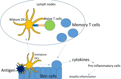
21 Figure 1.1C The mechanism of type IV hypersensitivity. Antigen penetrates the skin and is confronted with dendritic cells (DCs). DCs uptake the antigen, travel to lymph nodes where they mature and efficiently prime naïve T-cells. The naïve T-cells then proliferate and turn into memory T-cells which then travel into the blood and back to inflammatory site. There they secrete cytokines, promoting the recruitment of other inflammatory cells, thereby amplifying skin inflammation.
Type IVa hypersensitivity is induced by Th1-type T-cell responses. Th1-type T-cells secrete IFN-γ and TNF-α. These cytokines stimulate the expression of adhesion molecules on endothelial cells and increase the vessel permeability which allows plasma and inflammatory cells to the inflammatory site. Moreover, IFN-γ promotes macrophages to secrete TNF-α and IL-12, which stimulates NK cells. As macrophages contain TNF-α receptors on self- surface, they can be auto-activated by TNF-α and then generate more TNF- and IL-12, forming a positive feedback chain and thereby amplifying the inflammatory response. An example of type IVa hypersensitivity is tuberculin skin test (Sinigaglia et al., 1985). Type IVb correlates to Th2 type immune responses. Th2 secreting T-cells secrete IL-4, IL-5 and IL-13 cytokines, which stimulate B-cells to secrete
Antigen
Immature DCs
Lymph nodes
Mature DCs Naïve T cells
Memory T cells
cytokines
Skin cells
Amplify inflammation [image:22.612.77.472.74.334.2]22 antibodies such as IgE and IgG4. IgE in turn activate mast cells and IgG4 is associated with allergy, which suggest a link with type I hypersensitivity. IL-5 secretion leads to an eosinophilic inflammation, which is the characteristic of many drug hypersensitivity reactions (Pichler et al., 2003, Yawalkar et al., 2000).
Type IVc reactions involve cytotoxic T-cell migration into the tissue and direct cytotoxicity to tissue cells by release of cytolytic molecules such as perforin/ granzyme B. Alternatively, T-cells may secrete FasL and induce apoptosis through triggering the Fas receptor. Cytotoxic T-cells play a role in the drug hypersensitivity reactions such as maculopapular or bullous skin diseases, neutrophilic inflammation and in contact dermatitis. Type IVc reactions appear to be predominant in severe drug hypersensitivity reactions especially SJS/TEN syndrome, in which keratinocytes are killed by CD8 cytotoxic T-cells (Yawalkar et al., 2000, Schnyder et al., 1998). In general, CD8 T cell- mediated cytotoxic skin reactions are more severe tha n CD4 T-cell mediated cytotoxicity, as MHC I is ubiquitously expressed in keratinocytes as well as other skin cells, leading them being potential targets of CD8+ cytotoxic T-cells. In contrast, CD4 cytotoxic T-cells only recognize fewer cells expressing MHC II molecules, mainly antigen presenting cells. Hence, the massive presence of CD8 cytotoxic T-cells found in the blister fluid of SJS/TEN is a characteristic of the symptom.
23 the recruitment of neutrophils. Cytokines play a fundamental role in pathogenesis of drug hypersensitivity and their function is summarized in table 1.2.
Table 1.1Extended Coombs and Gell Classification Adapted from (Pichler 2003)
Classification Type of Immune responses
Pathol ogic char acteristics
Clinical systems Cell types
Type I IgE Mast-cell
degranulation
Urtica ria anaphylaxis
B ce lls/Ig Type II Ig G and IgM FcR-dependent
cell destruction
Blood cell dyscrasia
B ce lls/Ig Type III Ig G, IgM and
comple ment
Immune co mple x deposition
Vasculit is B ce lls/Ig Type Iva Th1 (IFN-,
TNF-)
Monocyte activation
Ec ze ma Th1 cells
Type IVb Th2
(IL-5 and IL-4)
Eosinophilic infla mmat ion
Maculopapular e xanthema,
bullous e xanthema
Th2 cells
Type IVc CTL (pe rforin and granzy me B and
fasl)
CD4-or CD8-med iated killing
of cells
Maculopapular e xanthema, ecze ma, bullous,
e xanthema, pustular e xanthema
Cytotoxic T-ce lls
Type IVd T-cells (IL-8) Neutrophil recruit ment and
activation
Pustular e xanthema
24
Table 1.2 Brief summar y of cytokines and their func tions (Adopted from Janeway 2012 Immunobiology)
Family Cytokine Functi on Producer cells
Interleukins IL-1 Fever, T-cell activation, macrophage activation
Macrophages, epithelial cells
IL-1 Th17 differentiation, Fever, T-ce ll activation, mac rophage activation
Macrophages, epithelial cells
IL-2 T-cell proliferation Th1
IL-3 Synergistic action in early hae matopoiesis Th1, Th2 IL-4 B ce ll act ivation, Ig E switch, induce
differentiat ion into Th2 ce lls
T-cells, mast cells IL-5 Eosinophil gro wth, differentiation T-cells, mast cells IL-6 T- and B-ce ll growth and differentiation,
Th17 d ifferentiation, fever
T-cells, macrophages IL-8 Che mo attractant for neutrophils and T-cells Macrophages IL-10 Potent suppressant of macrophage functions Monocytes IL-12 Activates NK ce lls, induces CD4 T-cell
differentiat ion into Th1 ce lls
Macrophages, dendritic cells
IL-13 B-ce ll growth and differentiation, inhibits Th1 cells
T-cells IL-17A Induces cytokine production by epithelia,
endothelia, and fibroblasts, proinfla mmatory
Th17 ce lls IL-17F Induces cytokine production by epithelia,
endothelia, and fibroblasts, proinfla mmatory
Th17 ce lls, monocytes IL-21 Induces proliferation of B, T and NK ce lls,
promotes Th17 d iffe rentiation
Th2
IL-22 Ep ithelia l barrie r, pro-infla mmatory agents NK cells, Th17 ce lls, Th22 ce lls
IL-23 Induces prolife ration of Th 17 Dendritic cells IFN- Macrophage activation, increased expression
of MHC mo lecules and antigen processing components, Ig class switching, suppresses Th2
T-cells, NK ce lls
Colony-stimulat ing factors
G-CSF Stimu lates neutrophil development and differentiat ion
Fibroblasts and monocytes
GM -CSF Stimu lates growth and differentiation of dendritic cells
Macrophages, T-cells TNF fa mily TNF- Pro motes infla mmat ion, promotes
differentiat ion into Th22 cells
Macrophages, NK cells, T-cells
Fas Ligand Apoptosis T-cells
Transforming growth factor beta (TGF-β) superfa mily
TGF-1 Anti-infla mmat ion, induces Th17 and Tregs differentiat ion
T-cells, monocytes
TGF-3 Pro motes Th17 d iffe rentiation, pro motes infla mmat ion
[image:25.612.87.530.133.656.2]25
1.4 Immune response
The immune system is designed to help the body to remove harmful microorganisms or viruses that enter it. Antigen presenting cells (APCs)can interact with other cells of the immune system, to achieve this.
Dendritic cells (DCs) are the most powerful antigen presenting cell as they are the only type of APCs that can stimulate naïve T-cells. When they are activated, they present peptide antigens on the cell surface using MHC class I and MHC Class II molecules (See Figure 1.2.1). They travel out of the inflammatory site to the local lymph nodes where they meet naïve T-cells and prime them through an interaction of the MHC-peptide complex with the T-cell receptor (TCR) and by interactions between the costimulatory molecules. Thereafter, primed T-cells become antigen-specific effecter T-cells that circulate the body and can return to inflammatory sites to help resolve the infection via cytokine secretion and recruitment of pro-inflammatory cells.
1.4.1 The mechanis ms of antigen presentation.
26
Figure 1.2.1 Class I MHC molecules display peptides to CD8 T-cells. Endogenous protein is cut into small peptides by the proteasome. These peptides are then carried by TAP (The transporter associated with antigen processing) and transported into endoplasmic reticulum (ER), where they will meet up with MHC I molecules. MHCI molecules then bind with the peptides, forming MHC I-peptide complexes. The complexes are then transported to surface of the cell and displayed to CD8 T-cells.
proteasome TAP
MHC I-peptide complex
endogenous protein
ER
27
Figure 1.2.2 Class II MHC molecules display peptides derived from non-self-proteins, especially bacterial proteins. When pathogen is swallowed by the antigen presenting cells, they are trapped into the phagosome. They meet chemicals and enzymes from lysosomes and in turn are digested into small peptides. This process is termed phagocytosis. MHC II molecules do not load peptides in the ER. Rather, in the ER their binding grooves are occupied by invar iant chain peptides, protected from the binding of endogenous proteins. MHC II - invar iant chain complexes are transported from ER and enter endosomes, where the chain peptides are replaced with pathogen peptides digested through phagocytosis. The MHC II-peptide complexes are then transported to the cell surface and displayed to CD4 T-cells.
MHC
II-Invariant chain complex
Phagosome
ER
Class II MHC- peptide-complex
Invariant chain
28 Figure 1.3 After contacting of TCR with antigenic -peptide-MHC complex that displayed on the surface of dendritic cells, naïve T-cells are activated and expanded. During priming, micro-milieu cytokines determine the fate of the antigen specific T-cells and consequently the role they play in the immune response.
1.4.2 T-cell subsets
29 signals determine the nature of the T-cells that are activated and their subsequent phenotype. T-cells are subdivided into CD4 and CD8 T-cells according to the expression of the co-receptors CD4 and CD8, respectively. These co-receptors interact and stabilize MHC-peptide-T cell receptor complex when T-cell receptors recognize antigen. After drug priming, of the cytokine micro-environment determines the nature of the T-cell response. CD4+ T helper cells can differentiate into various subsets such as Th1, Th2, Th17, Th22, Th9 secreting cells. Each subset is dominant in a specific type of inflammation. In the context of drug hypersensitivity, Th1 and Th2 secreting T-cells are known to be involved in d ifferent forms of tissue injury. However, the newer subsets have not been investigated.
1.4.2.1 CD8+ T-cell
CD8+ T-cells have been found in SJS/TEN skin lesions and these cells are also the dominant in T-cell population in blister fluid (Nassif et al., 2004). Moreover, CD8+ T-cells have been shown induce to drug antigen-specific cytotoxicity both in vivo and in vitro (Wu et al., 2006; Rozieres et al., 2010). In carbamazepine- induced drug hypersensitivity, drug specific CD8+ T-cells have been cloned and these clones show a strong toxicity against target cells in vitro. CD8+ T-cells isolated from patients with abacavir- induced hypersensitivity reactions also kill target cells by recognizing drug-peptide-MHC I complexes displayed on surface of antigen presenting cells (Chessman et al., 2008).
1.4.2.2 CD4+ T-cell and phenotypes
As described above, CD4+ T-cells play an important role in delayed type drug hypersensitivity and reactions have been classified into 4 sub-types based on the effects of different CD4+ T-cell subsets (cytokine secretion and subsequently inflammatory cell recruitment).
30 ability to differentiate into specialized subsets. These effector T-cells are defined by the expression of a restricted panel of cytokines and the expression of specific master regulator transcription factors (Szabo et al., 2003). Initially, the understanding of distinctive populations of differentiated CD4+ T-cells came from analysis of T-cell clones isolated from mice (Mosmann, Coffman 1989). Subsequently, (Bottomly et al., 1989) identified the key cytokines of each T-cell subset and the T-cell populations were named Th1 and Th2 secreting cells, in which T helper 1 (Th1) cells express T-bet and selectively produce interferon (IFN)-, while Th2 cells express Gata3 and produce cytokines such as IL-4 and IL-13 (Nakayamada et al., 2012). To understand the mechanisms of drug hypersensitivity, it is important to define the T-cell subsets involved. The picture is complicated by the fact that in certain circumstances, IFN- secreting T-cells also secrete Th2 cytokines. However, T-cell subset classification holds its value in certain circumstances: first, stable Th1 and Th2 lineage with typical cytokine expression can be obtained by cytokine polarization during naïve T-cells expansion. Second, in terms of host defense, each T-cell subset plays a particular role in pathogen eradication, e.g. intracellular bacteria for Th1 cells and helminths for Th2 cells. Third, these subsets express stable key regulators, T-bet for Th1 and GATA3 for Th2. Finally, the classification of T helper cell subsets renders a great therapeutic value in allergic and autoimmune diseases, generally Th2 cells induce allergic diseases, whereas Th1 and Th17 (discussed in de tail below) cells induce autoimmune diseases.
Th1 cells
31 expression and promote antigen presenting cell function. IL-2 functions as a growth factor promoting T-cell proliferation. Th1 cells may also mediate local inflammation at sites of infection. Th1 cells express the CCR5 homing receptor as a character of this T-cell subset. T-bet is a major transcription factor involved in the inducing the release of IFN - and Th1 cell differentiation (Szabo et al., 2000). IFN- responses to Leishmania major are significantly decreased in T-bet knockout mouse (Szabo et al., 2002).
Th2 cells
Previously, it was thought that a main function of Th2 secreting T-cells was helping B cell antibody generation. However, recent research found that follicular B helper T-cells (Tfh) are the main subsets that mediate this process and Th2 cell just play a regulatory role in the antibody generation. Th2 cells produce IL-4, IL-5, and IL-13 which are important in the elimination of parasites, such as helminthes. IL-4 induces Th2 differentiation and inhibits Th1 differentiation together with IL-13. Th2 cells may affect eosinophils, mast cells, and basophils by cytokine release i.e. IL-4, IL-5, and IL-13. Th2 cells express the CCR3 homing receptor. Th2 polarization requires the addition of IL-4 (Le Gros et al., 1990). GATA3 is the master regulator of Th2 (Zheng et al., 1997). GATA3 is also critical for the development of CD4+ T-cell responses (Ho et al., 2009). GATA3 expression is up-regulated or down-regulated during Th2 and Th1 polarization, respectively (Zhang et al., 1997). Moreover, Th2 differentiation is completely blocked in vivo and in vitro in the absence of GATA3 (Zhu et al., 2004).
32 Dysregulation of Th1 responses has been associated with an autoimmune response. For example, IFN-expression in the target tissue is associated with clinical signs of experimental
autoimmune encephalomyelitis (EAE). Furthermore, based on the evidence of blocking one of the heterodimers of 12p40, EAE got alleviated. However, blockage of the other c hain of IL-12 did not protect from EAE, but made the condition more severe (Krakowski et al., 1996; Tran et al., 2000; Gran et al., 2002; Zhang et al., 2003; Gutcher et al., 2006). This confusion did not get resolved until the discovery of IL-23, an important inducer of Th17 secreting cells. These cells share a p40 chain with IL-12 and have a unique heterodimer chain of p19 (Oppmann et al., 2000). This data implies blocking p40 blocks both Th1 and Th17 signaling and thus, the previous conclusion that EAE is solely Th1 induced autoimmune disorders has to be questioned.
Depletion of p19 of IL-23 but not p35 of IL-12 was shown to block EAE, which confirms that EAE is dominated by Th17 but not Th1 cells (Cua et al., 2003; Langrish et al., 2005).
33 2007; Yang et al., 2007).
Th17 cells are critical in host defense. They also play important roles in immune disorders such as psoriasis (Krueger et al., 2007), rheumatoid arthritis (Kirkham et al., 2006), multiple sclerosis (Matusevicius et al., 1999), inflammatory bowel disease (Sarra et al., 2006), asthma (Molet et al., 2001; Barczyk et al., 2003) and EAE (as discussed above).
In host defense, Th17 cells can be induced by pathogen-associated molecular patterns (PAMP) which are external stimuli derived from extracellu lar pathogens such as bacteria (Chtanova et al., 2004) and fungi.
In psoriasis, Th17 cells are the major subset of T-cells isolated from skin lesions (Pane et al., 2008) and with CCL20/CCR signaling being important for infiltration of inflammatory cell to the skin. Moreover, the blockage of p40 has been shown to reduce the psoriatic skin area (Krueger et al., 2007).
In patients with rheumatoid arthritis, IL-17, IL-1 and TNF have been shown to play a direct role in joint destruction (Kirkham et al., 2006). F urthermore, the molecule of RANKL expressed on the surface of Th17 induces cartilage and bone destruction directly (Kotake et al., 1999; Miranda-Carus et al., 2006; Sato et al., 2006).
34
Th22 cells
The phenomenon that some T helper cells secrete more than one signature cytokine is called plasticity. A T-cell population was found as they did not secrete signature cytokines such as IFN-, IL-4 and IL-17, but IL-22 solely (Eyerich et al., 2009). Therefore, this population is named after the cytokine IL-22, Th22. The specific microenvironment for differentiating Th22 cells is composed of TNF- and IL-6. Skin DCs have also been shown to pla y a critical role in Th22 differentiation (Duhen et al., 2009). Once differentiated, the Th22 phenotype remains stable and does not convert to other cell types (Eyerich et al., 2009). The aryl- hydrocarbon-receptor (AHR) is thought to be the master transcriptional regulator for Th22 cells (Trifari et al., 2009). Th22 express characteristic chemokine receptors such as CCR4 and CCR10 (Duhen et al., 2009), which suggest that they reside in normal skin and inflammatory skin, migrating following exposure to CCL27, a ligand of CCR10. The IL-22 receptor is expressed exclusively on tissue cells, mostly in epithelial cells. In contrast to many cytokine receptors, the receptor for IL-22 is not expressed on immune cells (Wolk et al., 2004). Therefore, tissue cells are t he major target of IL-22. Accordingly, IL-22 secreting lymphocytes are strongly enriched in peripheral tissue (Eyerich et al., 2009; Anmmziato et al., 2007). In terms of host defense, both Th17 cells and Th22 cells are involved in protection from bacterial infection. Th17 cells play a role in bacteria eradication (Lin et al., 2009), whereas Th22 have a protective effect on epithelial cells, especially in epithelial cell regeneration, proliferation and enhancement of migration (Nograles et al., 2008; Wolk et al., 2006).
35 differentiation that leads to considerable thickening and scaling of the epidermis (Nestle et al., 2009; Perera et al., 2012). Together with IL-17 and TNF-, IL-22 induces hyper-proliferation of keratinocytes, leading to the maintenance of acanthosis which is a hallmark of psoriasis (Wolk et al., 2009; Boniface et al., 2005; Delle et al., 2007).
Th22 cells also play a pathological role in inflammatory bowel disease and rheumatoid arthritis (Zenewicz et al., 2008; Kim et al., 2012). Moreover, serum IL-22 was reported to be elevated in patients with Crohn’s disease. 22 elevation was thought to be induced by the activation of IL-23/Th17 signaling (Schmechel et al., 2008).
The involve ment of Th17 and Th22 cells in drug hypersensitivity.
Similar to Th17 cells, there is very limited evidence to support a role for Th22 cells in drug hypersensitivity. Th17 cells might play a role in AGEP and SJS/TEN, whereas IL-22 secretion has only been detected in patients with AGEP.
36 In SJS/TEN, it has also been shown that the proportion of circulating Th17 cells is elevated among the CD4+ T-cell population in blisters and this proportion is decreased following improvement of the disease. In contrast, this phenomenon is not observed with Th1 or Th2 cells (Watanabe et al., 2011). It is not hard to understand the involvement of Th17 cells since Caproni et al (2006) has reported that neutrophils play a pathologic role in SJS/TEN by releasing reactive oxygen species and lysosomal enzymes and Th17 cells promote neutrophils recruitment.
1.4.3 TCR signaling and V receptor
The TCR is a multiprotein transmembrane complex comprising TCR (or TCR ), CD3, and CD3 dimers (Alarcon et al. 2003; K uhns and Davis 2012).
TCR are similar to immunoglobulin, in terms of both structure and genes. As for immunoglobulins, the TCR dimers comprise both variable (V) and constant (C) regions which form domains that interact with antigen presented by MHC molecules on the surface of APCs. This interaction forms 3 complementary determining regions (CDR1, 2, and 3) on V regions. CD3 molecules are tightly associated with TCR on T-cell surface. However, CD3 molecules do not bind to antigen. When antigen binds to ab chain of TCR, cell signaling will be transmitted into the cell by phosphorylation of immune-receptor tyrosine-based activation motif (ITAM) that is located on the cytoplasmic tail of CD3. Phosphorylation is the critical event in initiating downstream signaling cascades, during which, phosphorylation of phospholipase C (PLC) and consequently induce calcium influx into cytosol, resulting in the activation of T-cells.
37 (D), and joining (J) segments of the b-chain (Hughes et al., 2003), among which, V has 25 genes, D, J, V, and J have gene numbers of 2, 12, 70, 50, respectively. One particular gene from each group enables the generation of 1015-20 variable TCRs that allow for recognition of almost all the antigens in the universe (Davis, Bjorkman 1988). TCR diversity is often associated with potency of antigen and is often tested by V receptor distributions (Kimber et al., 2012a, b). The activated TCR repertoire can be determined by dependent on the pathway of T-cell activation (Currier et al., 1996). PBMCs stimulated by mitogen (PHA), super-antigen (TSST-1), or normal antigen (tetanus toxoid) show diverse TCR repertoires, in which both fresh blood and PHA stimulated PBMCs showed a normal spread distribution, whereas tetanus toxoid stimulated PBMCs showed a restricted profile. Finally, super-antigen stimulation resulted in a unique pattern of diversity (Currier et al., 1996). To explain the relationship between TCR diversity and potency of antigen, Moon and colleagues suggest that high TCR repertoire diversity could be induced by strong antigen with multiple antigen determinants and verse vice (Moon et al., 2007).
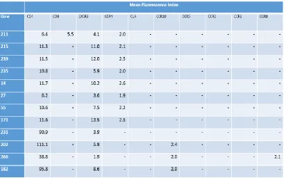
In drug hypersensitivity, V receptors have been found to be expressed in a restricted panel. For example, some V receptors are expressed in a wide distribution panel. In the study by Ko et al., (2011), patient with genotype of HLA-B*15:02 and with carbamazepine induced SJS/TEN were recruited to analyze the drug-specific T-cell receptor repertoire. 16 out of 19 patients with carbamazepine induced SJS/TEN expressed a single clonotype of VB-11-1SGSY and this clonotype was not expressed in all drug-tolerant patients. Carbamazepine specific CD8+ T-cells with this clonotype showed a strong cytotoxicity and this cytotoxicity was inhibited by the addition of anti VB-11-1SGSY antibodies.
38 responses and is restricted by a single HLA allele association (HL1-B*57:01), drug-specific T-cells displayed a random distribution of V receptors (Illing et al., 2012). These findings are in stark contrast to the work of Ko et al exploring carbamazepine- induced SJS.
In my thesis, piperacillin specific T-cells displayed a random distribution of V receptor repertoire. In our lab, nitroso-sulfamethoxazole (SMX-NO) T-cell clones isolated from patients with sulfamethoxazole hypersensitivity also displayed a random TCR V repertoire (unpublished data). It is known that SMX-NO is a strong hapten that is capable of binding to both intracellular protein and the proteins in the serum, and wide V receptor distribution suggests that SMX-NO protein binding generates a large number of peptide epitopes, which subsequently interact with multiple TCRs.
1.4.4 Tissue homing
Cellular tissue homing was dominanted by the interaction between chemokines and their receptor (Campbell & Butcher 2000). As reviewed by Charo & Ransohoff (2006), chemokines are divided into two groups, CC chemokines and CXC chemokines. CC chemokines have two adjacent cysteine residues near the amino terminus whereas in CXC chemokines, two cysteine residues are separated by a single amino acid. CC chemokines tend to induce the migration of monocytes and CXC cytokines tend to induce the migration of neutrophils. For example, CCL2 is a chemokine that stimulates monocytes to migrate from the bloodstream to the tissue. Another example, CXCL8, however, attracts neutrophils out of the blood and migrates into the peripheral tissue.
39 endothelial cells, via changing the conformation of adhesion molecules on lymphocytes and thereafter binding to their ligands on endothelial cells. Next the lymphocytes squeeze between the endothelial cells, going out of the vessel to the tissue. Second ly, chemokines direct the migration of lymphocytes along a gradient of chemokine molecules bound to the extracellular matrix and the surface of endothelial cells. This gradient increases in concentration toward the site of infection.
T-cell priming occurs in lymphoid tissue but effector cells are needed in peripheral tissue at the original site of infection. T-cells express specific chemokine receptors i.e., CCR4, so that release of CCL17 (or TARC) and it’s binding to the receptor causes T-cells to migrate; following a chemokine diffusion gradient and accumulation in the inflamed site.
In immunological conditions targeting skin, antigens are transported to the draining lymph node by dendritic cells. Antigen specific T-cells must then be transported from the lymph node back to skin. When T-cells encounter antigen in the skin, they become activated and may release effector molecules i.e., cytolytic molecules. These cause damage to skin cells, including keratinocytes. Migrating immune cells reach the inflamed sk in initially through a series of selectin and integrin contacts, including cutaneous lymphocyte antigen (CLA). CLAs function as an integrin, which binds to the ligand E-selectin expressed in the endothelium thereby, inducing T-cells to move through the endothelium into the tissue (Rossiter et al., 1994).
40 important role for CCR10 in the skin inflammation. CCR4 was also demonstrated to be important in skin inflammation. CCR4+ cells were only observed in memory skin homing T-cells and not in naïve T-cells and intestine homing T-cells. In chronic cutaneous disease, CLA+CCR4+ T-cells migrate following exposure to the ligands of CCR4, TARC and MDC (Campbell et al., 1999).
Several studies have investigated the role of chemokines and chemokine receptors in drug hypersensitivity. CLA expression on T-cells, isolated from skin and blood of hypersensitive patients, correlated with disease severity (Leyva et al., 2000). Increased expression of CCR4, CCR8, and CCR10 has been implicated in allergic reactions in the skin, such as contact dermatitis, atopic dermatitis and psoriasis (Hudak et al., 2002, Moed et al., 2004, Vestergaard et al., 1999). In patients with dermatitis, CCR4 and CCR10 are important in T-cell migration to the inflamed skin whereas CCR8 was important in homing of memory T-cells to healthy skin (Vestergaard et al., 2003, Schaerli et al., 2004).
In patients with delayed-type drug hypersensitivity, drug-specific CCR6+ T-cells initiate reactions by secreting TNF- and IFN-. This leads to an acute phase response and induction of inflammatory chemokines such as CXCL8 or CCL20, the ligands of CCR6 in keratinocytes (Schaerli et al., 2004).
41
1.5 HLA associations with drug hypersensitivity
42 with lapatinib-induced liver injury indicates MHC I and II molecules can be involved in different forms of hypersensitivity (Spraggs et al., 2011).
1.6 Mechanisms of drug hype rsensitivity
There are three main theories as to how pharmaceutical drugs can act as antigens and generate an immune response (See Figure 1.2).
1.6.1 Hapten and Prohapten theory
The basis of hapten theory was built up by the early research from Landsteiner and Jacobs (1935). They sensitized guinea pigs to the low molecular we ight, chemically reactive compound, dinitrochlorobenezene (DNCB). The authors showed that the immune reaction resulted from the formation of a covalent protein adduct in skin through modification of specific nucleophilic residues. This leads to the hypothesis that chemicals and drugs are too small to function as antigens and activate an immune response directly. The hypothesis states that they bind to proteins forming an intact antigen to trigger an adaptive immune response; β-lactams are common causes of both type I and type IV hypersensitivity.
drug-43 protein conjugation. Furthermore, certain penicillin- specific T-cells are activated specifically with drug-protein adducts. Penicillins contain a 6-aminopenicillanic acid nucleus which includes a -lactam ring and a five numbered ring. When drug-protein conjugates form, the -lactam ring opens up spontaneously and acylates with lysine residue of the binding protein. A similar profile of binding happens with a range of antibiotics, including piperac illin and flucloxacillin, which are discussed in detail in the sections below. In contrast to the penicillins, other compounds only bind to protein after drug metabolism and the liberation of reactive species. For example,
urushiol, an allergen contained in ivy and poison oak induces a severe contact dermatitis in both human and mouse models. Urushiols are oxidased into reactive quinone species and the reactive species binds covalently to amino acid residues on protein (Kalergis et al., 1997). Some of drugs might also be metabolized into reactive intermediates prior binding to proteins. This indirect process is known as the pro-hapten hypothesis. A classical example is the drug halothane, which induces immunological hepatotoxicity in a small percentage of the population. Halothane is metabolized by P4502E1 (Kharasch et al., 1996) into a reactive trifluoroacetyl chloride
intermediate, which binds covalently to lysine residues in proteins forming an antigen (Kenna et al., 1988). Tienilic acid an urisuric diuretic used in the treatment of hypertension was withdrawn from the market in 1980 due to its hepatotoxic potential. Tienilic acid is metabolized by human P4502C9 enzymes to yield a reactive metabolite S-oxide which binds covalently with
nucleophilic groups on the enzyme forming an antigen. Anti-P450 antibodies have been detected in the serum of patients with hepatotoxicity. The most well characterized pro-hapten drug is sulfamethoxazole which will be discussed in the following secretion.
1.6.2 Pharmacological inte raction of drugs with immune receptors: p.i. concept.
44 MHC molecules and TCRs. The theory is based on evidence that drugs can stimulate drug specific T-cells in an MHC processing independent way when APCs were fixed with glutaraldehyde (Pichler et al., 2002).
When a drug stimulates T-cells immediately, as shown by intracellular calcium release, then this response is too fast to require antigen processing (Pichler et al., 2002). Pulsing experiments show that T-cell responses can be inhibited by washing the antigen presenting cells, which removes the non-covalently bound drug. This shows that the drug is binding directly to immune receptors and activating T-cells by a pharmacological mechanism. Drugs that activate T-cells via a p.i. mechanism include carbamazepine, lidocaine, lamotrigine, and sulfamethoxazole (Farrell et al., 2003, Zanni et al., 1998, Schnyder et al., 1998).
1.6.3 Altered self-peptide repe rtoire model
45 altered self-peptide-MHC complex only displayed on the surface of APC in the presence of the drug activates CD8+ T-cells which cause tissue damage (Figure 1.4).
Figure 1.4 Different pathways of T-cell activation in drug hype rsensitivity.
Three mechanism of drug hypersensitivity have been proposed, which are hapten/prohapten theory, p.i. concept and peptide alteration theory.
46
47
48
1.6.4 Drug metabolis m and the activation of T-cells
SMX has been used as a model to study the role of metabolism in drug hypersensitivity for several reasons. Firstly its metabolism is well-defined. Secondly, stable and readily metabolites have been synthesized and are available for functional studies. Thirdly, patient samples are readily available for functional studies. Most of SMX is detoxificated in the liver. The drug is metabolized by hepatic N-acetyltransferase enzymes to an acetylated derivative which is easily eliminated from the body. However, a small amount of SMX is converted to a hydroxylamine intermediate. A reaction is catalyzed by CYP2C9 (Cribb et al., 1995). SMX hydroxylamine is stable and eliminated in urine (Gill et al., 1999). This suggests that most tissues are exposed to the hydroxylamine after SMX administration. The hydroxylamine either undergoes reduction back to SMX, a reaction by catalyzing by NADH cytochrome b5 reductase and CYP3A4 or is spontaneously oxidized to nitroso SMX (SMX-NO). SMX-NO has been shown to bind and modify selective cysteine residues expressed on both cellular and protein serum (Naisbitt et al., 1999, Naisbitt et al., 2001). Modification of cell surface proteins occurs quickly and then these protein conjugates are internalized through caveolae-dependent endocytosis (Elsheikh et al., 2010). Therefore, it is possible for the transport of intermediates (i.e. the hydroxylamine) out of the liver, which is rich in detoxification, to remote areas (i.e. the skin) where it is converted to SMX-NO, which generates protein adducts and ultimately hypersensitivity. The high number of SMX hypersensitivity reactions in patients with HIV and cystic fibrosis might be related to the fact that the redox balance is tipped in favor of a pro-oxidative environment by the disease process (van der Ven et al., 1997, Walmsley et al., 1997).
49 proteins. In rodent models, SMX-NO primed naïve CD4 and CD8 T-cells and the T-cell activation was antigen processing dependent (Farrell et al., 2003, Naisbitt et al., 2001, Naisbitt et al., 2002, Castrejon et al., 2010). On the contrary, administration of SMX does not activate immune cells. In vitro studies using PBMCs from drug naïve volunteers showed that SMX-NO generates T-cell responses in almost 100% of the volunteers (Engler et al., 2004). Application of a DC T-cell priming assay (Faulkner et al., 2012), using naïve T-cells from healthy volunteers who have never exposed to SMX, demonstrated that SMX-NO readily activates naïve T-cells. The newly generated memory T-cells were drug antigen specific. SMX-NO treatment resulted in proliferative responses and cytokine release. Several studies have shown that T-cells from blood and skin of all SMX hypersensitive patients are activated by SMX-NO, which suggests that SMX metabolites and the SMX-NO modified proteins are involved in the development of clinical symptoms in hypersensitive patients (Elsheikh et al., 2010, Schnyder et al., 2000, Burkhart et al., 2001, Nassif et al., 2004). Moreover, it has been shown that SMX-NO stimulates the majority of the drug responsive T-cell clones generated from patients with SMX hypersensitivity (Castrejon et al., 2010).
50 antigen (Naisbitt et al., 2001). A study of SMX hypersensitive patients shows that SMX-NO selectively binds to a single amino acid residue of human albumin (HSA), cysteine 34. It has also been shown that (1) SMX-NO and SMX-NO modification of protein can induce cell death when the levels of binding exceeds a threshold; and 2) SMX-NO metabolite modified necrotic cells provide a strong maturation signal to DCs (Naisbitt et al., 2002, El-Ghaiesh et al., 2012). Based on this discussion, it is clear that, SMX metabolites are generated in the skin and may provide activation signals to DCs.
1.6.5 SMX and SMX-NO specific T-cell clones
In addition to the above discussion, SMX specific T-cell clones have been isolated from b lood of patients with different types of skin eruptions (Schnyder et al., 1998, Brander et al., 1995). T-cells express CD4 or CD8 or both and upon stimulation by drugs, they secrete high levels of IL-5 and perforin which induces the killing of keratinocytes (Schnyder et al., 1998). Furthermore, drug-specific CD8 T-cells have been isolated from patients with TEN, the most severe form of cutaneous hypersensitivity, and fully characterized (Yawalkar et al., 2000, Nassif et al., 2004). Stimulation of T-cells with SMX follows the p.i. concept. T-cells from hypersensitive patients stimulated by SMX bound directly to MHC and T-cell receptor in a non-covalent fashion (Schnyder et al., 1998, Schnyder et al., 2000, Burkhart et al., 2001, Nassif et al., 2004). The threshold period of drug incubation for a T-cell clone response varies from 0.1 to 4 h (Zanni et al., 1998) which is incompatible with the time that is required for antigen presentation. The response is MHC restricted, although not all of the response is restr icted to a specific HLA allele (Sanderson et al., 2007).
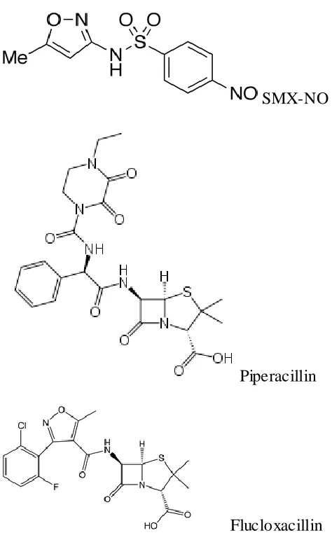
51 In this thesis I have investigated drug hypersensitivity reactions to the pro-hapten to sulfamethoxazole (SMX) and its metabolites nitroso-sulfamethoxazole (SMX-NO) and the β-lactam antibiotics: piperacillin and flucloxacillin.
1.7.1 Sulfonamides
In 1940, Woods showed that sulfonamides prevented bacteria from growing. Sulfonamides prevent bacteria using para-aminobenzoic acid for folate biosynthesis, which is crucial fo r the synthesis of thymidine, purines and bacterial DNA (Woods, 1940). In 1960, the combination of sulfonamides and trimethoprim was developed based on the recognition that they both targeted the same pathway and double inhibition of this pathway is more e ffective than using a single drug (Masters, 2003).
52
Figure 1.5 Metabolis m of SMX and protein conjugate format ion (Su llivan et al, 2015)
1.7.2 β-lactam antibiotics
Piperacillin was developed at the end of the 1970s. It is a wide-spectrum antibacterial agent developed for microbes resistant to other β-lactams, such as Klebsiella pneumonia and
Pseudomonas aeruginosa (Jones et al., 1977). Combined with tazobactam, a B- lactamase, the anti-bacterial effect of piperacillin has been improved especially against B- lactamase producing bacteria (e.g. staphylococci, Escherichia coli, Haemophilus influenza) (Speich et al., 1998). Piperacillin is administered via intramuscular or intravenous injection as a sodium salt because it is poorly absorbed by the intestine.
53 general population (Brock &Roach, 1984; Moss et al., 1984).
For the formation of protein conjugates, the lactam ring is broken by nucleophilic lysine residues, leading to binding of the penicilloyl group (Batchelor et al., 1965). The penicillyol antigen can also be formed by binding of the reactive degradation product penicillenic acid (Levine et al., 1960). Recent advances in mass spectrometry has allowed researchers to use piperacillin as a model to precisely characterize the nature of the piperacillin binding interaction with protein (Figure 1.4.1). As for all β-lactam antibiotics, piperacillin selectively interacts with specific lysine residues on serum proteins such as human serum albumin (Figure 1.4.2). The binding interactions are dependent on the dose and incubation time (Meng et al., 2011, Whitaker et al., 2011). Moreover, as T-cells from hypersensitive patients are activated with piperacillin HSA adducts, it was possible to investigate the minimum level of modification of the drug that can stimulate drug-specific T-cells. At low drug concentration, only Lys541 modification was observed, whereas at higher concentration, up to 13 lysine modifications were detected, four of which (Lys 190, 195, 432, and 541) were detected in patients’ plasma (El-Ghaiesh et al., 2012, Whitaker et al., 2011). A synthetic β- lactam-protein conjugate mimicking the drug antigen found in the patients was found to stimulate PBMCs and 100% β-lactam specific T-cell clones. Whereas T-cell response to drug conjugates is dampened when antigen processing is inhibited, which suggests that the antigenic peptides are derived from drug-protein conjugates.
54 every 100,000 first time users of the drug. (Andrews et al., 2008). The liver injury is predominantly cholestatic in nature. It has now been discovered that flucloxacillin- induced liver injury is associated with the expression of HLA-B*57:01 (Daly AK., et al 2009) and CD8 T-cells play a role in liver injury (Monshi M et al., 2013). The risk of flucloxacillin induced liver injury is increased in females, with a high daily dosage and with increasing age (Elise Andrews et al.,
55
Figure 1.6.1 For mation of pi per acillin-protein conjugate d antigen (adapt fr om Whitaker, Meng e t al., 2011).
56
Figure 1.6.2 Binding site of pi per acillin to HSA protein, whic h are the modifications on lysine residues identifie d by mass spectrometr y. Adapte d fr om Whitaker, Me ng et al., (2011)
Model of albu min showing piperacillin b inding sites at positions Lys190, Lys195, Lys199, Lys212, Lys351, Lys432, Lys525 and Lys541.
1.8 Aim of the thesis
The primary aim of this study is to characterize the phenotype and function of piperacillin specific T-cells isolated from hypersensitive patients’ PBMC (chapter 3) and skin (chapter 4). The involvement of Th17 and Th22 cells in piperacillin hypersensitivity was also studied using a recently established in vitro DC T-cell priming assay (chapter 5). Finally, the priming assay was applied to study the mechanism(s) of drug antigen presentation and T-cell cross reactivity using three haptenic drugs, SMX-NO, piperacillin and flucloxacillin.
57 important because Th17 and Th22-secreting T-cells are known to play a critical role in several resilient and chronic immune- mediated diseases, such as psoriasis, Crohn’s disease, asthma, multiple sclerosis, and inflammatory bowel disease.
The data presented in this thesis aims to answer the following questions:
1. Are Th17 and Th22 involved in the drug hypersensitivity? And if so, which drug-specific cytokines do they secrete?
2. Are there phenotypic and/or functional differences between drug-specific T-cells isolated from skin and PBMC?
3. Is it possible to generate drug specific Th17 cells or Th22 cells under Th17 and Th22 under polarization conditions in vitro?