This is a repository copy of
Electronic conduction in shock-compressed water
.
White Rose Research Online URL for this paper:
http://eprints.whiterose.ac.uk/65661/
Version: Published Version
Article:
Celliers, P. M., Collins, G. W., Hicks, D. G. et al. (22 more authors) (2004) Electronic
conduction in shock-compressed water. Physics of Plasmas. L41-L44. ISSN 1089-7674
https://doi.org/10.1063/1.1758944
[email protected] https://eprints.whiterose.ac.uk/ Reuse
Items deposited in White Rose Research Online are protected by copyright, with all rights reserved unless indicated otherwise. They may be downloaded and/or printed for private study, or other acts as permitted by national copyright laws. The publisher or other rights holders may allow further reproduction and re-use of the full text version. This is indicated by the licence information on the White Rose Research Online record for the item.
Takedown
If you consider content in White Rose Research Online to be in breach of UK law, please notify us by
Electronic conduction in shock-compressed water
P. M. Celliers,1G. W. Collins,1D. G. Hicks,1M. Koenig,2E. Henry,2,3A. Benuzzi-Mounaix,2 D. Batani,3D. K. Bradley,1L. B. Da Silva,1R. J. Wallace,1S. J. Moon,1J. H. Eggert,1 K. K. M. Lee,4L. R. Benedetti,4R. Jeanloz,4I. Masclet,5N. Dague,5B. Marchet,5M. Rabec Le Gloahec,5Ch. Reverdin,5J. Pasley,6O. Willi,6D. Neely,7and C. Danson7
1Lawrence Livermore National Laboratory, Livermore, California 94551
2Laboratoire pour l’Utilisation des Lasers Intenses (LULI), Unite´ Mixte No. 7605, CNRS-CEA-Ecole
Polytechnique-Universite´ Pierre et Marie Curie, 91128 Palaiseau, France
3Dipartimento di Fisica`, G. Occhialini Universita degli Studi di Milano-Bicocca and INFM,
20126 Milan, Italy
4University of California, Berkeley, California 94720 5CEA/DAM Ile de France, 91680 Bruye`res-le-Chaˆtel, France 6Imperial College, London, United Kingdom
7Central Laser Facility, Rutherford Appleton Laboratory, Oxfordshire OX11 0QX, United Kingdom
~Received 5 January 2004; accepted 8 April 2004; published online 25 June 2004!
The optical reflectance of a strong shock front in water increases continuously with pressure above
100 GPa and saturates at;45% reflectance above 250 GPa. This is the first evidence of electronic
conduction in high pressure water. In addition, the water Hugoniot equation of state up to 790 GPa
~7.9 Mbar!is determined from shock velocity measurements made by detecting the Doppler shift of
reflected light. From a fit to the reflectance data we find that an electronic mobility gap ;2.5 eV controls thermal activation of electronic carriers at pressures in the range of 100–150 GPa. This suggests that electronic conduction contributes significantly to the total conductivity along the Neptune isentrope above 150 GPa. © 2004 American Institute of Physics.
@DOI: 10.1063/1.1758944#
Water is one of the most abundant molecules in the solar system, ubiquitous in biology, and a fundamental constituent of the giant planets Neptune and Uranus. In the center re-gions of the outer planets water exists at conditions distrib-uted along an isentrope, at temperatures ranging from 2000 to 6000 K, and pressures ranging from 10 to 800 GPa.1 Elec-trical conductivity at these conditions is important for under-standing magnetic field generation in these planets.2,3 Low temperature ~295 K! water is insulating: the liquid at 0.1 MPa is an amorphous semiconductor with 6.5 eV gap
energy;4 solid phases are expected to remain insulating to
pressures beyond 700 GPa with gap energies larger than 10
eV.5Shock compressed water becomes electrically
conduct-ing at rather low pressures: measurements of the dc conduc-tivity, sdc, along the principal Hugoniot revealed an
expo-nentially increasing trend up to 10 GPa,6,7 followed by a
much slower increase to a saturation level of;20 (Vcm)21 between 35 and 60 GPa.8This conductivity was attributed to dissociation into ionic species, possibly the bimolecular
re-action 2H2O→OH21H
3O1. The result motivated a single
shock Raman scattering study which revealed that the con-centration of intermolecular hydrogen bonds begins to de-crease at 12 GPa and vanishes at 26 GPa,9and inferred that
the conduction mechanism may involve free protons, H2O
→OH21H1. Recent reverberating shock experiments
achieved up to 180 GPa and 5400 K and found that sdc
increased slowly to 200 (Vcm)21,10,11 and remains ionic.
Concurrent ab initio molecular dynamics investigations
elu-cidated details of the ionic conduction mechanisms.12,13 Un-der strong shock compression one expects thermally acti-vated electronic carriers to begin to dominate, however, no theoretical or experimental work to date has focused on elec-tronically conducting phases of high pressure, high tempera-ture water.
In recent years large lasers have allowed access to pres-sures close to 1 TPa. Here we report on the equation of state
~EOS! and optical reflectance of water compressed by a
single strong shock wave spanning the pressure range of 100–790 GPa, a range for which no previous measurements exist. Several large lasers around the world were used, in-cluding the Phebus laser14 and the LULI facility in France, the Omega laser in Rochester, NY,15 and the Vulcan laser in
the UK.16 Previous dynamic measurements of the EOS of
water have been carried out with explosive techniques17–19
and with a two-stage light gas gun8,20to determine the prin-cipal Hugoniot accurately to about 100 GPa. A single datum at 1.4 TPa~Ref. 21!from an underground nuclear experiment has never been repeated. Within experimental uncertainty the new laser-shock data are consistent with existing data, and
also with a tabular EOS from the SESAME database.22,23
More important, we found a strong variation in optical re-flectance along the Hugoniot: below 100 GPa water is opaque and low reflecting~a few %!; above 100 GPa it trans-forms continuously into a metallic-like optical reflector that saturates at reflectivities near 40%–50%. The high
reflectiv-L41
ity is the first unambiguous evidence of electronic conduc-tion in high pressure water.
Cylindrical 6 mm diam stainless steel containers held samples of de-ionized, distilled 99.9% pure H2O. One end of
the container was sealed with a 500mm thick sapphire
win-dow which allowed optical access to the water and water– aluminum interface. The opposite end of the container was sealed with a stepped Al plate~pusher!with the step facing the water. The Al pusher was fabricated from rolled 99.999% pure Al stock by diamond machining to produce step heights
between 15 and 25mm, measured to within 100 nm accuracy
with a white light phase stepping interferometer. A thin poly-styrene film, typically 15mm overcoated with 100 nm of Al, was attached to the flat side of the aluminum plate and served as the ablator. Irradiation of the ablator with one or
several smoothed24,25 laser beams launched a strong shock
which was transmitted into the Al plate and then into the
water. Focal spot sizes 800 mm in diameter were used for
some experiments and 400 mm for other experiments. For
EOS measurements we used a 3.7 ns pulse to produce a steady shock wave, and for some reflectivity measurements we used a shorter 1 ns pulse to load the specimen impul-sively and produce an attenuating shock wave to allow prob-ing over a wide range of pressures.
A line-imaging velocity interferometer system for any
reflector ~VISAR!26,27 recorded light reflected from the
sample cell. This instrument works by reflecting an injection-seeded, Q-switched Nd:YAG laser probe beam from the rear of the target, and relaying an image of the target through a velocity interferometer onto a streak camera slit. For strong
shocks in water (.100 GPa) the probe light was reflected
directly from the shock front.28The Doppler shift of reflected light is manifested as a shift in fringe phase at the output of the velocity interferometer. In most cases we used two
inter-ferometers operating at wavelength l5532 nm with
differ-ent velocity sensitivities to resolve fringe shift ambiguities. For some experiments we used a 1064 nm wavelength oper-ating in one interferometer simultaneously with a 532 nm probe in the other interferometer.
An example recording ~inset in Fig. 1! shows initially stationary fringes produced by the reflection of the probe beam from the Al pusher. The shock emerged first out of the thin Al step, later out of the thick step and was then trans-mitted to the water. The shock front in the water is reflecting and imparts a Doppler shift to the reflected probe, manifested as a fringe shift in the data recorded. We extracted three observables from the VISAR recordings for each shot: the shock velocity versus time, given by the fringe shifts; the shock reflectivity versus time, given by the reflected inten-sity; and the average shock velocity in the Al pusher, given by the break-out times from the top and bottom steps. Sta-tistical uncertainties in the shock velocity determined from fringe shifts are typically 0.3%–1%. Typical uncertainties for the average shock speed in Al were 1.5%–3% and they dominate errors in EOS determination.
To determine EOS points we used the
impedance-matching technique29 which yields the pressure P and
par-ticle speed up at the interface between the Al pusher and the water sample. Used in this analysis are the measured shock
velocities us for water and Al, and the known Hugoniot and
release isentrope for the Al pusher.22The shock compression data shown in Fig. 1 include the early lower pressure experiments,17,18 more recent higher pressure data,8,19 and the single ultrahigh pressure datum.21 Our laser shock data span the unexplored range between 100 and 800 GPa. The Hugoniot calculated from a tabulated EOS for water
gener-ated by Ree,23 available in the SESAME database,22 agrees
well with the new data within experimental uncertainty. To compare these data with SESAME, a linear fit to the usvs up data determined here and the datum reported by Podurets
et al.21 was made. Over this limited pressure range, one in which no phase transitions are expected, a linear form for us vs upis quite good. This fit was converted to the P2r plane using the Hugoniot relations. The difference in density be-tween this fit and SESAME at 100, 500, and 1000 GPa is 0%, 6%, and 4%, respectively. While these values are within the density uncertainties estimated in this work, the data are systematically shifted toward lower density compared to SESAME at pressures between 200 and 1400 GPa.
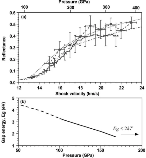
We measured the reflectance of the shock by comparing the probe intensity reflected from the shock to that from the bare Al surface which has a known reflectivity of 0.85
[image:3.612.319.556.52.217.2]60.05. These data are shown in Fig. 2. The systematic error incurred in this process could be up to 10%. Relative uncer-tainties in the reflectance are typically about 20%. For some experiments we observed that an attenuating shock in the sample produces a continuous record of reflectance as a function of the shock velocity; the attenuating shock was generated by driving the Al pusher with a short ~1 ns!high pressure pulse, which allowed rarefaction to overtake the shock propagating in the Al pusher before it reached the sample. In this case simultaneous recording of the Doppler shift~fringe phase!and intensity allowed us to extract shock reflectance over a wide range of shock states. Since the shock was not steady the compressed material behind it con-tained spatial density gradients along the propagation direc-tion; however the gradient scale length is much larger than FIG. 1. Measurements of the principal Hugoniot of water: closed circles
~Ref. 17!, closed squares~Ref. 18!, closed triangles~Ref. 19!, closed dia-monds~Ref. 8!, inverted closed triangles~Ref. 21!, open circles this work. The solid curve is the principal Hugoniot of water calculated from the SESAME database~Refs. 22 and 23!. The inset shows typical data recorded that are described in the text.
the skin depth of the reflected light,;0.1mm.
Temperatures (T) predicted from the SESAME EOS model agree to better than 10% with measurements at lower pressures,20,30so we expect the EOS model to be reasonably
accurate. For 100 GPa,P,300 GPa the model predicts
7000 K<T<30 000 K, and compression 2.7,r/r0,3.5.
While one would expect some increase in reflectivity from a compression-driven increase in the refractive index,30 this can account for at most about 4% reflectivity assuming that the fluid remains an insulator; this is much smaller than the observed saturation levels of 40%–50%. Therefore we at-tribute rapidly increasing reflectivity above 100 GPa to free carriers generated by thermal activation across a mobility gap.
To model the reflectivity we use a standard
semiconduc-tor formalism to estimate the carrier density,31 Ne
52(mekT/2p\2)3/2F(2Eg/2kT) where me is the effective
mass, k is the Boltzmann constant, Eg is the mobility gap
energy in the electronic density of states, and F(h)
5(2/
A
p)*0`A
x/@11exp(x2h)#dx. The dielectric function isgiven by a Drude-like expression, e5eb2vp
2
/v2(1
1i/vt), where eb is the contribution due to bound
elec-trons,vis the angular frequency of the probe beam, andtis
the electron relaxation time. The plasma frequency is vp2
54pNee2/m*, where e is the electron charge, and m*
5me/2 is the reduced mass. Consistent with the treatment of intrinsic semiconductors, the chemical potential is placed
midway within the gap and the mass of the holes and
elec-trons is assumed equal. The relaxation time is taken as t
5gtmin where tmin5l/ve is the minimum scattering time
~Ioffe–Regel limit32!andg*1. Here l52(3/4pNi)1/3 is the interparticle distance, Ni is the total number of particles per unit volume (H2O or H3O1and OH2 and others!andve is
the electron velocity computed by integrating over the Fermi distribution at a given temperature. Estimating the bound electron contributionebis problematic because of the disrup-tion of chemical bonding that occurs above 25 GPa.9,12,13In the absence of data or models we usedeb51. ~Variations in
eb affect the calculated reflectivity mainly below 100 GPa,
where we have no data with which to constrain a fit.! We
calculate the reflectivity from the complex index of refrac-tion, n5
A
e, and the Fresnel formula, R5u(n2n0)/(n 1n0)u2, where n051.33 is the index of unshocked water.Using this model to calculate the reflectivity we have fit the observed reflectance along the compression curve assum-ing a linear variation of Eg along the Hugoniot with respect to the density and temperature: Eg (eV)56.52a(r/r021) 2b(T/T021), withr050.998 g cm23and T05295 K. This
form is consistent with the known gap energy of 6.5 eV at the initial state,4and takes into account an expected variation
in density and temperature of the gap energy.12 The
three-parameter best fit, a51.32, b50.043 andg51.05, produces a varying gap energy ranging from 3.3 to 2 eV within the
range of 100–150 GPa, respectively.33 The variation of Eg
along the Hugoniot is shown in Fig. 2~b!. The predicted re-flectivities compare well with the observations at both 532 and 1064 nm. Increasing Eg tends to shift the predicted ris-ing edge of the reflectance toward higher velocities~higher P and T), while the collisionality factor g controls the reflec-tivity in the saturation limit at high P and T ~largergleads to larger conductivity and higher reflectivity!. The relaxation time is close to the Ioffe–Regel limit32~i.e., g;1), indica-tive of strong scattering. This behavior has been found in shock compressed D2,34 as well as in LiF and Al2O3.35 When Eg/2kT<1 the gap is effectively closed through tem-perature smearing of the Fermi distribution, and the fluid is
better characterized as a dense plasma. For
shock-compressed water this transition occurs for P>170 GPa, and
T>15 000 K.
While this simple model does match the initial increase in shock reflectance well, it does not reproduce the reflec-tance saturation observed in the data. The observed satura-tion can be accounted for by limiting the carrier density near
1022 cm23 at about 2300 K ~250 GPa!. This amounts to
about 1 in 10 initial molecules contributing a free carrier, suggesting that even at these extreme temperatures and pres-sures, the chemistry is quite complex.
It is interesting to compare electrical conductivities esti-mated from this model with earlier measurements of
sdc, 8,10,11
which all point to an electronically insulating ionic conduction mechanism; in particular, observations of the
gal-vanic potential between dissimilar electrodes10 confirmed
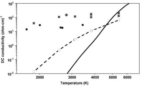
this. Using the reflectivity fit to determine Ne(Eg), we esti-mate the electronic contribution to the dc conductivity using a Drude model,se5Nee2gtmin/m*. Figure 3 shows a
[image:4.612.55.293.55.311.2]com-parison of the estimated se that corresponds to the states
FIG. 2.~a!Optical reflectance of the shock front as a function of the shock velocity and pressure along the principal Hugoniot at 532 nm~solid line with error bars!and at 1064 nm~dashed line with closed cirles and error bars!. Fits to these data are shown for 532 nm~chain-dashed!and 1064 nm
observed by Chau et al. In this comparison we find that se
!sdc except at the highest pressure observed, 180 GPa,
wherese/sdcis about 1/3. Thus Eg is high enough to pre-vent significant electronic conduction for the sample condi-tions observed in Refs. 10 and 11 consistent with the conclu-sions of those studies.
Above 5000 K ~where the Chau et al. measurements
stop! we estimate that se will begin to dominate. This has implications for conductivity estimates in the interior of
Neptune and Uranus. We have computedsealong the
Nep-tune isentrope, also shown in Fig. 3, and find that se con-tributes at least as much as the ionic contribution at tempera-tures above 5000 K. This may have significant bearing on planetary models that rely on conductivity data to understand generation of the magnetic field. Temperatures between 4500 and 6000 K correspond to a broad range of pressures, from 150 to 800 GPa along this isentrope; furthermore, details of this isentrope are model dependent and rather uncertain. Since the thermally activated nature ofsedepends
exponen-tially on both T and Eg, the electronic conductivity will
depend strongly on precise details of how both Eg and T
vary along the isentrope. By comparing a variety of models that match the reflectance data, we estimate that the electron conductivity model presented here is accurate to within about a factor 4 at temperatures between 1000 and 23000 K and pressures between 15 and 250 GPa.
The authors thank the operations staff at the Phebus, LULI, Omega and Vulcan facilities, and W. Unites for their effort during these experiments.
This work was performed under the auspices of the U.S.
DOE by Lawrence Livermore National Laboratory ~LLNL!
under Contract No. W-7405-ENG-48, and also supported un-der EU Training and Mobility Research Contract Nos.
ERBFMGECT 950016 ~Phebus! and
ERBFMGE-CT95-0044 ~LULI!, as well as LULI ACCESS HPRI-1999-CT
00052.
1W. B. Hubbard, Science 275, 1279
~1997!.
2
W. J. Nellis et al., Science 240, 779~1988!.
3N. F. Ness et al., Science 246, 1473~1989!.
4F. Williams, S. Varma, and S. Hillenius, J. Chem. Phys. 64, 1549
~1976!.
5
M. Benoit et al., Phys. Rev. Lett. 76, 2934~1996!.
6
H. David and S. Hamann, Trans. Faraday Soc. 55, 72~1959!.
7S. Hamann and M. Linton, Trans. Faraday Soc. 62, 2234~1966!. 8A. C. Mitchell and W. J. Nellis, J. Chem. Phys. 76, 6273~1982!. 9N. C. Holmes et al., Phys. Rev. Lett. 55, 2433
~1985!.
10
V. V. Yakushev et al., JETP 90, 617~2000!.
11R. Chau et al., J. Chem. Phys. 114, 1361~2001!, temperatures listed here
are incorrect, Fig. 3 uses correct values.
12C. Cavazzoni et al., Science 283,~1999!. 13
E. Schwegler et al., Phys. Rev. Lett. 87, 265501~2001!.
14
G. Thiell et al., Laser Part. Beams 6, 93~1988!.
15T. R. Boehly et al., Opt. Commun. 133, 495~1997!.
16I. N. Ross et al., IEEE J. Quantum Electron. QE-17, 1653~1981!. 17J. M. Walsh and M. H. Rice, J. Chem. Phys. 26, 815
~1957!.
18
L. V. Al’tshuler, A. A. Bakanova, and R. F. Trunin, Sov. Phys. Dokl. 3, 761
~1959!.
19L. P. Volkov et al., JETP Lett. 31, 513~1980!.
20G. A. Lyzenga, T. J. Ahrens, W. J. Nellis, and A. C. Mitchell, J. Chem.
Phys. 76, 6282~1982!.
21
M. A. Podurets et al., Sov. Phys. JETP 35, 375~1972!.
22See National Technical Information Service Document No. DE94-011699.
~J. D. Johnson, SESAME Tables, 1994!. Copies may be ordered from the National Technical Information Service, Springfield, VA 22161. For the aluminum standard we used SESAME table 3719. The water table used here was SESAME table 7150.
23F. H. Ree, J. Chem. Phys. 12, 6287~1982!. 24T. H. Bett et al., Appl. Opt. 34, 4025~1995!. 25S. N. Dixit et al., Opt. Lett. 19, 417
~1994!.
26
L. Barker and R. Hollenbach, J. Appl. Phys. 43, 4669~1972!.
27P. M. Celliers et al., Appl. Phys. Lett. 73, 1320~1998!.
28To check this we tracked propagation of the shock across a gap formed by
placing a quartz plate a known distance from the lower Al step. The gap distance found by integrating the VISAR signal between breakout and impact matched the known gap distance to within measurement accuracy (;1%).
29
W. J. Nellis and A. C. Mitchell, J. Chem. Phys. 73, 6137~1980!.
30S. Kormer, Sov. Phys. Usp. 11, 229~1968!.
31C. Kittel and H. Kroemer, Thermal Physics, 2nd ed.~Freeman, San
Fran-cisco, 1980!.
32
A. Ioffe and A. Regel, Prog. Semicond. 4, 237~1969!.
33Fits to E
g5const, and Eg56.52a(r/r021) result ing;1.4 and produce
very similar reflectivity curves. The reflectance data do not provide a strong constraint on the functional dependence of Eg, and all fits lead to
Eg;2 – 3 eV andg;1 in the pressure range of 100–200 GPa. 34P. M. Celliers et al., Phys. Rev. Lett. 84, 5564~2000!. 35D. Hicks et al., Phys. Rev. Lett. 91, 035502
[image:5.612.57.294.52.197.2]~2003!. FIG. 3. Comparison ofsdcfrom Ref. 8~closed circles!, from Ref. 11~closed
squares!, from Ref. 10~closed diamond!andse~open squares and dashed
curve! as a function of that temperature. Each of the open squares was computed for the states observed in Ref. 11~closed squares at same T). The solid curve shows the estimatedsefor water at states distributed along the
Neptune isentrope.