0022-538X/96/$04.0010
Copyrightq1996, American Society for Microbiology
Separate Functional Domains of the Herpes Simplex Virus
Type 1 Protease: Evidence for Cleavage inside Capsids
BARBARA J. ROBERTSON,1PATRICK J. M
CCANN III,1LINDA MATUSICK-KUMAR,1†
WILLIAM W. NEWCOMB,2JAY C. BROWN,2RICHARD J. COLONNO,1
ANDMIN GAO1* Department of Virology, Bristol-Myers Squibb Pharmaceutical Research Institute, Wallingford, Connecticut
06492-7660,1and Department of Microbiology and Cancer Center, University of Virginia
Health Science Center, Charlottesville, Virginia 229082
Received 30 January 1996/Accepted 12 April 1996
The herpes simplex virus type 1 (HSV-1) protease (Pra) and related proteins are involved in the assembly of viral capsids and virion maturation. Pra is a serine protease, and the active-site residue has been mapped to amino acid (aa) 129 (Ser). This 635-aa protease, encoded by the UL26 gene, is autoproteolytically processed at two sites, the release (R) site between amino acid residues 247 and 248 and the maturation (M) site between residues 610 and 611. When the protease cleaves itself at both sites, it releases Nb, the catalytic domain (N0),
and the C-terminal 25 aa. ICP35, a substrate of the HSV-1 protease, is the product of the UL26.5 gene. As it is translated from a Met codon within the UL26 gene, ICP35 c,d are identical to the C-terminal 329-aa sequence of the protease and aretranscleaved at an identical C-terminal site to generate ICP35 e,f and a 25-aa peptide. Only fully processed Pra (N0and Nb) and ICP35 (ICP35 e,f) are present in B capsids, which are believed to
be precursors of mature virions. Using an R-site mutantA247Svirus, we have recently shown that this mutant protease retains enzymatic activity but fails to support viral growth, suggesting that the release of N0 is
required for viral replication. Here we report that another mutant protease, with an amino acid substitution (Ser to Cys) at the active site, can complement theA247Smutant but not a protease deletion mutant. Cell lines expressing the active-site mutant protease were isolated and shown to complement theA247Smutant at the levels of capsid assembly, DNA packaging, and viral growth. Therefore, the complementation between the R-site mutant and the active-site mutant reconstituted wild-type Pra function. One feature of this intragenic complementation is that following sedimentation of infected-cell lysates on sucrose gradients, both N-termi-nally unprocessed and processed proteases were isolated from the fractions where normal B capsids sediment, suggesting that proteolytic processing occurs inside capsids. Our results demonstrate that the HSV-1 protease has distinct functional domains and some of these functions can complement intrans.
Herpesvirus capsids are icosahedral protein shells approxi-mately 15 nm in thickness and 125 nm in diameter. Three types of capsids, termed A, B, and C, can be isolated by sucrose gradient centrifugation (15). C capsids contain the entire viral genome and are able to mature into infectious virus. A and B capsids lack viral DNA and are found in the infected-cell nucleus. A capsids are thought to result from failed attempts at packaging viral DNA, while B capsids are thought to be inter-mediates in capsid assembly, because they differ from A and C capsids by the presence of large amounts of the viral assembly protein ICP35 (VP22a) (3, 15, 35, 36, 41). There is a family of ICP35 proteins designated ICP35 a through f (3). Although all forms of ICP35 can be detected on Western blots (immunob-lots) of infected cells, only forms ICP35 e,f are present in B capsids (1, 7, 15, 35, 41, 46). Because ICP35 e,f are present in B capsids and absent from the mature virions, it has been suggested that the role of ICP35 e,f in capsid assembly is analogous to that of the scaffold proteins of double-stranded bacteriophage (4, 14, 16).
In addition to ICP35 e,f, B capsids are composed of at least seven other proteins: VP5, VP19C, Nb (VP21), VP23, N0
(VP24), VP26, and the recently reported UL6 gene product (1,
16, 37, 40, 43, 52). Three capsid proteins are encoded by a single open reading frame (ORF) designated UL26, and two transcripts have been mapped to this ORF (20, 25, 33). One transcript, designated UL26, encodes the 635-amino-acid (aa) protease. The other more abundant shorter transcript, desig-nated UL26.5, encodes the 329-aa protein ICP35 (26, 45). Since residue 307 (Met) of Pra is also the start codon for ICP35, the two transcripts are in frame and are 39coterminal (19, 25, 26).
The full-length herpes simplex virus type 1 (HSV-1) pro-tease (Pra) undergoes autoproteolytic processing at two sites, the release (R) site and the maturation (M) site, which lie between Ala and Ser at residues 247 and 248 and residues 610 and 611, respectively (9, 42). Cleavages at these sites generate N0, Nb, and a 25-aa peptide (Fig. 1A). Identification of N0(the
N-terminal 247 aa of Pra) as the catalytic domain of the pro-tease suggests that the HSV-1 propro-tease has multiple domains (27, 28, 53). Since ICP35 overlaps and is in frame with the C-terminal half of Pra, ICP35 c,d can be trans cleaved at its C terminus by the protease to generate ICP35 e,f and a 25-aa peptide (Fig. 1A).
Another herpesvirus protease, the product of the cytomeg-alovirus (CMV) UL80 gene, in addition to autoproteolysis at R and M sites (56), has a third internal (I) cleavage site located in the middle of the catalytic domain (2, 21, 39, 54). The CMV protease can trans cleave the HSV-1 substrates, but the HSV-1 protease cannot cleave the CMV substrates (55).
The HSV-1 protease is a serine protease, and its active site has been mapped to Ser-129 (11, 27). Its requirement for * Corresponding author. Mailing address: Department of Virology,
Bristol-Myers Squibb Pharmaceutical Research Institute, 5 Research Pkwy., Wallingford, CT 06492-7660. Phone: (203) 284-6692. Fax: (203) 284-6088. Electronic mail address: [email protected].
† Present address: American Cyanamid, Department of Plant Bio-technology, Princeton, NJ 08543.
4317
on November 9, 2019 by guest
http://jvi.asm.org/
proper capsid assembly confirmed that the protease is essential for the production of infectious virus (13, 44). Although capsid structures are observed in the absence of functional protease, the major capsid protein VP5 does not adopt the correct
con-formation and viral DNA is not encapsidated (44). Genetic and functional analyses revealed that the full-length protease (Pra), the catalytic domain (N0), and the R and M cleavage site
mutant proteases are proteolytically active (26, 32, 53). How-FIG. 1. (A) Polypeptide products of UL26 and UL26.5 ORFs. The HSV-1 protease (Pra), substrate (ICP35 c,d), and cleavage products Prb, N0(VP24), Na, Nb (VP21), and ICP35 e,f (VP22a) are described in the text. The cleavage sites (R and M sites) of Pra and ICP35 c,d are indicated by arrowheads. The UL26 amino acid numbers of the N and C termini of each protein are indicated. (B) The ORFs of HSV-1 wt and mutant protease plasmids, cell lines, and viruses. Arrows indicate cleavage sites. A slash through an arrow denotes an uncleavable site due to a point mutation at P1. The dashed line indicates a frameshift in the ORF of the catalytic domain of the protease, rendering it inactive, while leaving the ICP35 ORF unaffected. An X through the protease ORF indicates a point mutation at the active-site serine, rendering it inactive. M over L indicates that the methionine at 307 (start codon for ICP35) is changed to leucine. The wavy line denotes the HCMV protease. Construction of these mutants is described in Materials and Methods.
on November 9, 2019 by guest
http://jvi.asm.org/
[image:2.612.106.498.81.599.2]ever, the catalytic domain of the protease, N0, alone is
insuf-ficient to support viral growth, suggesting that Na may be required to direct the catalytic domain to the site of capsid assembly (13). These results also indicate that in addition to enzymatic activity, the HSV-1 protease may have additional functions required for viral replication.
Three products of proteolytic processing, N0, Nb, and ICP35
e,f, are found in B capsids (15, 37, 52, 53), but the fate of the fourth product, the C-terminal 25-aa peptide of both Pra and ICP35 c,d, is unknown. We and others recently reported that the C-terminal 25-aa peptide of Pra and ICP35 are involved in the formation of sealed capsids and may directly interact with VP5 (8, 13, 22, 31, 38, 50).
In this study, we report that two lethal mutant proteases can complement each other in trans. This intragenic complemen-tation was observed at the levels of capsid assembly, DNA packaging, and viral growth. Our results demonstrate that the HSV-1 protease is a multifunctional protein and some of its functional domains can be genetically separated. One feature of the complementation between these two mutants is that following sedimentation of infected-cell lysates, Prb was iso-lated from the fractions where normal B capsids sediment. Thus, the precursor of N0and Nb can be incorporated into
capsids, suggesting that proteolytic processing occurs inside capsids.
MATERIALS AND METHODS
Cells and viruses.Vero cells were grown and maintained as described previ-ously (23). The growth medium for the neomycin-resistant cell lines BMS-MG22 and BMS-S129C (see below) included 250mg of the antibiotic G418 per ml. BMS-MG22 cells (Fig. 1B) express full-length HSV-1 protease with a Met-to-Leu change at residue 307, which can fully support the growth of protease mutant viruses (13).
The wild-type (wt) HSV-1 strain KOS1.1 was propagated and assayed as described previously (23, 24). The mutant protease viruses m100 and A247S were grown in BMS-MG22 cells.
Plasmids.Construction of plasmids pM307L, pSVPra, pSVN0, and pUCICP35 has been described previously (13, 29, 31). Plasmid pSV/HCMVN0/HSVNa was constructed as follows: a 492-bp PstI-ApoI fragment containing the human CMV (HCMV) catalytic domain gene and the R-site junction of HCMV and HSV-1 protease gene was generated by standard PCR technique by using pT7CMVProA as a template (12) and two oligonucleotides (59-GGGTCTTT TTTGCCTGGGCTGCGTCACTTCGCCAGG-39and 59-AAGGCCGAATTTT TCGCTCGCCTTGACG-39) as primers. The sequences which are underlined and in bold type represent the coding region at the R site of the HCMV catalytic domain and HSV-1 Na, respectively. Plasmid pHCMVN0/HSVNa was con-structed by three-way ligation of the PCR product, a 3,438-bp PstI-Asp718I fragment of pT7HCMVN0C and a 1,180-bp ApoI-Asp718I fragment of pT7635A. Plasmid pSV/HCMVN0/HSVNa was constructed by ligation of the XbaI-Asp718I fragment of pHCMVN0/HSVNa into pJ3V(34). The coding region at the junc-tion between HCMVN0and HSVNa was confirmed by DNA sequencing. Plas-mid pSV/HCMVN0was constructed by ligation of the XbaI-AspI fragment of pT7HCMVN0C into pJ3V. Plasmid pGSTN0S129C was constructed by standard PCR techniques, and the mutation was confirmed by elimination of the MscI site, with the generation of Bpu11021 site. Plasmid pSV/S129C/M307L was con-structed by three-way ligation of the 364-bp EagI-HpaI fragment of pGSTN0S129C and the 1,274-bp HpaI-Asp718I fragment of pM307L into the 3,788-bp EagI-Asp718I fragment of the vector pSVN0. Thus, pSV/S129C/M307L encodes Pra with a Ser-to-Cys change at residue 129 and a Met-to-Leu change at residue 307. Plasmid pUC/S129C/M307L was constructed by replacement of a 1,628-bp Eco47III-Asp718I fragment of pRB4057 (26) with the same fragment derived from pSV/S129C/M307L.
Cleavage assay inEscherichia coli.Procedures for cleavage assays in the E. coli system were described previously (6, 32).
Isolation of the S129C protease-expressing cell lines.Vero cells were trans-formed with the plasmid pUC/S129C/M307L and pSVneo as described previ-ously (13). G418-resistant colonies were grown into cultures and screened for the expression of the mutant protease and the ability to complement the growth of A247S virus. The cell clone BMS-S129C was used for experiments in this study (Fig. 1B).
Analysis of viral DNA and proteins.For slot blot analysis, DNA was prepared and analyzed essentially as described by Weinheimer et al. (53). Cells were infected with virus at a multiplicity of infection (MOI) of 10 PFU per cell unless otherwise specified.
For Western blot analysis, infected-cell lysates were analyzed by sodium
do-decyl sulfate-polyacrylamide gel electrophoresis (SDS-PAGE) (13, 53). The pro-cedure for Western blot analysis was described previously (13, 53). The anti-ICP35 monoclonal antibody (MAb) MCA406 (18) was used to detect anti-ICP35 and protease-related products Pra, Prb, Na, and Nb (1:1,000 dilution; Serotec, Ox-ford, England).
Electron microscopy.The procedure for examination of infected cells by electron microscopy were was described previously (11).
trans-complementation assays.The procedure for trans complementation has been described previously (13, 31). Vero cells were transfected with 3mg of wt or mutant plasmids. At 20 h posttransfection, the cells were infected with 3 PFU of either m100 or A247S virus per cell and allowed to undergo a single cycle of infection. Virus yield was measured by plaque assay on BMS-MG22 and Vero cells.
Indirect immunofluorescence.Indirect immunofluorescence was performed was described previously (13). Primary antisera, specifically, anti-HCMV cata-lytic domain (1:50 dilution) or anti-HSV-1 N0(1:100 dilution), were detected by using fluorescein-conjugated goat anti-rabbit antibody (1:100 dilution).
RESULTS
Genetic evidence for distinct functions of the HSV-1 pro-tease.The N-terminal 247-aa sequence of the HSV-1 protease (N0) is enzymatically active but is insufficient to support viral
growth (13, 30). This suggests that the C-terminal 388-aa se-quence (Na) of the protease may perform a function required to support viral growth, perhaps by providing the specific signal required to localize N0to the site of capsid assembly (30). If
localization is the only function of Na, then a chimeric protease consisting of the HCMV catalytic domain (HCMVN0) and
HSV-1 Na should retain functionality, since the CMV catalytic domain has the ability to cleave HSV substrates (55). To test this hypothesis, we constructed a plasmid containing a chimeric gene product consisting of the HCMV catalytic domain and HSV-1 Na (Fig. 1B) and examined expression and subcellular distribution of the gene product in transfected Vero cells by immunofluorescence. HCMV N0, like HSV-1 N0(13), showed
approximately equal intensities of cytoplasmic and nuclear staining (Fig. 2A). However, the chimeric gene product with the HCMV catalytic domain fused to HSV-1 Na, like HSV-1 wt Pra (13), demonstrated predominantly nuclear staining (Fig. 2B). These results suggest that when HSV-1 Na was fused to the HCMV catalytic domain, it restored the ability of the HCMV protease to localize in the nucleus.
We then examined whether the chimeric gene product re-tained the ability to autoprocess using the E. coli expression system. wt virus-infected Vero cells (Fig. 3A, lane 14) and the full-length wt HSV-1 protease (Pra) expressed in E. coli (Fig. 3A, lane 27) as well as the catalytic domain of the protease, N0,
coexpressed with ICP35 in E. coli (Fig. 3A, lanes 9 to 12) were used as positive controls. As expected, HSV-1 Na and Nb were produced when the chimeric protein was expressed alone or coexpressed with HSV-1 N0(Fig. 3A, lanes 15 to 18 and lanes
19 to 22). Bands between Prb and Na in these lanes may represent cleavage at the I site of the HCMV catalytic domain (2, 54). These results indicate that the chimeric protease has the ability to cleave itself at an R site composed of HCMV P and HSV-1 P9 residues. To determine whether the chimeric protease retains the ability to trans cleave ICP35, the full-length HSV-1 protease or the chimeric protease was coex-pressed with ICP35 and examined by Western blotting (Fig. 3B). As expected, ICP35 c,d were trans cleaved into ICP35 e,f either by the HSV-1 protease (Fig. 3B, lanes 1 to 4) or by the chimeric protease (Fig. 3B, lanes 5 to 8).
To determine whether the HCMV-HSV chimeric protease could support the growth of the HSV-1 protease mutant vi-ruses m100 and A247S, trans-complementation experiments were performed. The mutation in the m100 virus eliminates synthesis of the functional protease and, therefore, ICP35 can-not be cleaved (13). Although the mutation in A247S abolishes
on November 9, 2019 by guest
http://jvi.asm.org/
the release of N0, this mutant protease retains the ability to
autoprocess at the M site and trans cleave ICP35 (30, 53). Neither of these mutant viruses grow on Vero cells but require HSV-1 protease-expressing BMS-MG22 cells for their propa-gation (13, 30). As shown in Table 1, transfection with the wt protease gene (Pra) complemented the growth of both mu-tants, while the HCMV-HSV chimeric protease (HCMVN0/
HSVNa) was not able to support the growth of these mutant viruses. Therefore, substitution of HSV-1 N0with the HCMV
N0did not restore the complete functionality of the protease.
These results suggest that HSV-1 N0, in addition to its
enzy-matic activity, may have another function(s) required for viral replication.
Intragenic complementation of HSV-1 protease mutants.To further investigate the multiple functions of the HSV-1 pro-tease, another protease mutant, S129C, was constructed. Plas-mids pSV/S129C/M307L and pUC/S129C/M307L were derived from the plasmid pM307L (12), which expresses a protease with a Met-to-Leu change at residue 307 under the control of the simian virus 40 early promoter and its own promoter, respectively. The M307L mutation in the protease gene elim-inates the synthesis of ICP35 but does not affect the protease
functions required to support virus growth (13). Therefore, mutations in the protease gene should affect only the protease and not ICP35. The plasmid pSV/S129C/M307L encodes the mutant Pra with a Ser-to-Cys change at residue 129 and a Met-to-Leu change at residue 307 (Fig. 1B). Since the HSV-1 protease is a serine protease and the active site has been mapped to Ser-129 (11), neither the full-length S129C protease nor the mutant N0demonstrates enzymatic activity in the E.
coli expression system (Fig. 3A, lanes 6 to 8 and lanes 3 to 5,
respectively). However, the S129C protease is still a substrate for cleavage when coexpressed with wt N0(Fig. 3A, lanes 23 to
26). In addition, the mutant S129C protease, like wt Pra and the A247S protease (12, 13), localized to the nucleus (Fig. 2C). Therefore, we examined whether the defect of the noncleav-able R site, but enzymatically active A247S protease could be complemented by providing the inactive, but cleavable mutant S129C protease in trans.
[image:4.612.154.457.73.200.2](i) Intragenic complementation between mutant virus A247Sand mutant plasmid S129C.Complementation experi-ments were performed to determine whether expression of the S129C protease from a plasmid could complement the growth of m100 and A247S mutant viruses. Plasmids encoding wt pro-FIG. 2. Subcellular locations of mutant proteases. Vero cells were transfected with pSV/HCMVN0(A), pSV/HCMVN0/HSVNa (B), or pSV/S129C (C). At 20 h posttransfection, cells were processed for an indirect immunofluorescence assay using polyclonal antisera specific for either HCMVN0(A and B) or HSV-1 N0(C).
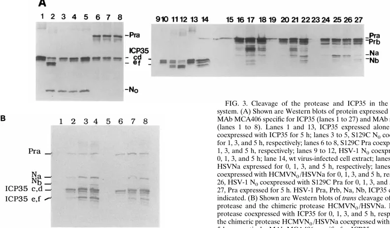
FIG. 3. Cleavage of the protease and ICP35 in the E. coli coexpression system. (A) Shown are Western blots of protein expressed in E. coli probed with MAb MCA406 specific for ICP35 (lanes 1 to 27) and MAb specific for HSV-1 N0 (lanes 1 to 8). Lanes 1 and 13, ICP35 expressed alone for 5 h; lane 2, N0 coexpressed with ICP35 for 5 h; lanes 3 to 5, S129C N0coexpressed with ICP35 for 1, 3, and 5 h, respectively; lanes 6 to 8, S129C Pra coexpressed with ICP35 for 1, 3, and 5 h, respectively; lanes 9 to 12, HSV-1 N0coexpressed with ICP35 for 0, 1, 3, and 5 h; lane 14, wt virus-infected cell extract; lanes 15 to 18, HCMVN0/ HSVNa expressed for 0, 1, 3, and 5 h, respectively; lanes 19 to 22, HSV-1 N0 coexpressed with HCMVN0/HSVNa for 0, 1, 3, and 5 h, respectively; lanes 23 to 26, HSV-1 N0coexpressed with S129C Pra for 0, 1, 3, and 5 h, respectively; lane 27, Pra expressed for 5 h. HSV-1 Pra, Prb, Na, Nb, ICP35 c,d and e,f and N0are indicated. (B) Shown are Western blots of trans cleavage of ICP35 by the HSV-1 protease and the chimeric protease HCMVN0/HSVNa. Lanes 1 to 4, HSV-1 protease coexpressed with ICP35 for 0, 1, 3, and 5 h, respectively; lanes 5 to 8, the chimeric protease HCMVN0/HSVNa coexpressed with ICP35 for 0, 1, 3, and 5 h, respectively. MAb MCA406 specific for ICP35 was used.
on November 9, 2019 by guest
http://jvi.asm.org/
[image:4.612.95.491.492.723.2]tease Pra and HCMVN0/HSVNa were also used in these
ex-periments. Neither the S129C/M307L protease nor the chi-meric protease was able to complement the growth of m100 (Table 1). However, the S129C/M307L protease, but not the HCMVN0/HSVNa protease, could fully complement the
growth of A247S virus (Table 1). These results suggest that the growth of A247S virus was complemented by a function pro-vided by the N0domain of the S129C protease.
(ii) Phenotype of the mutant S129C.To determine the basis of intragenic complementation between HSV-1 protease mu-tants, we isolated a cell line, named BMS-S129C, which ex-presses the mutant S129C/M307L protease. The enzymatic activity of the mutant protease from infected-cell extracts was examined. In these assays, ICP35 was provided by mutant viruses, either m100 or A247S. Cell extracts were prepared at 10 h postinfection (p.i.), separated by SDS-PAGE, and ana-lyzed by Western blotting (Fig. 4). Since the amino acid se-quence of ICP35 is identical to the C-terminal portion of the protease (26, 33), the MAb we used, MCA406, reacted with ICP35 and several of the autoproteolytic products of the pro-tease. In wt virus-infected cells, ICP35 c,d was processed to ICP35 e,f (Fig. 4, lanes 1, 2, and 3). However, in m100-infected BMS-S129C cells (Fig. 4, lane 6), like m100-infected Vero cells (Fig. 4, lane 4), normal amounts of ICP35 c,d were produced but not processed to the ICP35 e,f forms. This result again demonstrates that the mutant S129C protease does not have the ability to cleave ICP35. As we reported recently (30), the mutant A247S protease retains enzymatic activity, but cleavage of ICP35 c,d to ICP35 e,f is less than in wt virus-infected Vero cells (Fig. 4, compare lanes 1 and 7). This enzymatic activity is restored to wt level in A247S-infected BMS-S129C cells, as evidenced by the increased processing of ICP35 (Fig. 4, com-pare lanes 1, 7, and 9).
(iii) Evidence that the proteolytic processing events relevant to capsid assembly occur inside capsids.Although fully pro-cessed protease (N0 and Nb) and ICP35 (ICP35 e,f) are
present in B capsids (15, 37, 52, 53), it is unknown whether it is necessary for the cleavages to take place inside capsids in order to form infectious particles. The intragenic complemen-tation between our two mutants may provide a tool to address this question. We postulated that if the cleavages occur prior to
capsid assembly, one would expect that only fully processed protease (N0and Nb derived from S129C protease) and ICP35
e,f derived from A247S virus-infected cells would be present in B capsids; if the cleavages occur during or after capsid assem-bly, one would expect that not only fully processed protease but also the partially processed protease Prb, derived from
A247S virus-infected cells could be detected in B capsids. We,
therefore, examined whether the enzymatically active, but non-cleavable R-site A247S protease is present in capsids isolated from A247S-infected BMS-S129C cells.
wt or A247S virus-infected BMS-S129C cell extracts were subjected to centrifugation through 20 to 50% sucrose gradi-ents. Extracts from m100-infected BMS-S129C cells and
A247S-infected Vero cells were used as negative controls.
Af-ter sedimentation of infected-cell lysates, capsid bands were visualized only in extracts of wt virus- and A247S-infected S129C cells, but not in extracts of m100-infected BMS-S129C cells or A247S-infected Vero cells (results not shown). Fractions were collected, subjected to SDS-PAGE, and ana-lyzed by Western blotting with MAb MCA406 (Fig. 5). As expected, in wt virus-infected BMS-S129C cell extracts, Nb and ICP35 e,f were observed in fractions 10 to 12 corresponding to B capsids (Fig. 5A). In contrast, no apparent Nb and ICP35 peak was present in similar fractions from the m100-infected BMS-S129C cell extracts (Fig. 5B) or A247S-infected Vero cells (results not shown). Most of the unprocessed ICP35 c,d appeared to accumulate in the first few fractions (the top) of the gradient in m100-infected BMS-S129C cell extracts (Fig. 5B). In mutant A247S-infected BMS-S129C cells, Prb, appar-ently derived from the A247S protease (Pra), was observed in addition to Nb and ICP35 e,f in the fractions where wt B capsids sediment (Fig. 5C). To ensure that bands in samples of fractions 10 to 12 in Fig. 5C were Prb and Nb, not Pra and ICP35 c,d, we reexamined fractions 10 and 11 shown in Fig. 5A and C with controls side by side. Both Pra and Prb were present in the control sample (Fig. 5D, lane 1), and as ex-pected, neither Pra nor Prb was present in these B-capsid fractions of wt virus-infected BMS-S129C cells (Fig. 5D, lanes 4 and 5). In contrast, Prb is present in these fractions of
A247S-infected BMS-S129C cells (Fig. 5D, lanes 2 and 3).
When the same samples from fractions 10 and 11 of Fig. 5A and C were examined with a polyclonal anti-N0 serum, the
catalytic domain of the HSV-1 protease (N0) was detected in
wt- and A247S virus-infected BMS-S129C cell extracts (Fig. 5D, lanes 6 to 10). In order to generate N0, the full-length
[image:5.612.57.298.92.235.2]protease (wt Pra or S129C Pra) must be cleaved. Therefore, bands in fractions 10 and 11 of Fig. 5A and C must be Nb, not ICP35 c,d. This conclusion was further confirmed by the fact that these bands did not react with an antiserum specific for the
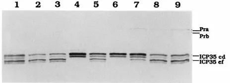
[image:5.612.319.551.590.674.2]FIG. 4. Western blot analysis of HSV-1 protease-related proteins in infected cells. Vero (lanes 1, 4, and 7), BMS-MG22 (lanes 2, 5, and 8), and BMS-S129C (lanes 3, 6, and 9) cells were infected with wt (lanes 1 to 3), m100 (lanes 4 to 6), or A247S (lanes 7 to 9) virus. Total proteins were prepared at 10 h p.i., separated by SDS-PAGE, and transferred to a nitrocellulose filter. The filter was probed with the MAb MCA406.
TABLE 1. Intragenic complementation between HSV-1 protease mutantsa
Gene transfected Virus super-infected
Titer (PFU/ml)b
Complemen-tation indexc
Expt 1 Expt 2 Expt 1 Expt 2
pUC18 m100 2.43103 5.23102 1 1
A247S 1.03102 1.83102 1 1 Pra m100 5.43105 1.43105 225 265
A247S 6.03104 2.43104 600 133 S129C/M307L m100 2.43103 7.63102 1 1.5
A247S 8.03104 5.43104 800 300 HCMVN0/HSVNa m100 3.0310
3 7.83102 1.3 1.5
A247S 2.83102 8.03102 2.8 4.4
aVero cells were transfected with the plasmids indicated. At 20 h
posttrans-fection, cells were infected with either m100 or A247S at 3 PFU per cell and incubated for an additional 20 h before being harvested.
b
Viral yield was determined by plaque assays on BMS-MG22 cells at 2 days p.i.
c
Expressed as viral yield relative to that for cells transfected with pUC18 DNA.
on November 9, 2019 by guest
http://jvi.asm.org/
C-terminal 25-aa peptide (results not shown). In addition, Prb, not Pra, was also detected by this anti-N0serum only in
frac-tions 10 and 11 of A247S-infected S129C cells, not in those of wt virus-infected cells (Fig. 5D, compare lanes 7 and 8 with lanes 9 and 10). Taken together, these results demonstrate that
the precursor protease can be incorporated into capsids. This is consistent with the hypothesis that the capsids are the rele-vant site of proteolytic processing during the formation of infectious virus (30).
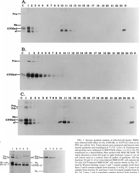
[image:6.612.74.522.73.629.2](iv) Encapsidation of viral DNA.We then examined whether FIG. 5. Sucrose gradient analysis of infected-cell lysates. BMS-S129C cells were infected with either wt (A), m100 (B), or A247S (C) virus at an MOI of 10 PFU per cell for 16 h. Total extracts were prepared and layered onto 20 to 50% sucrose gradients and centrifuged at 77,1753g for 1 h. Fractions were collected, and proteins were subjected to SDS-PAGE (lanes 1 to 23 or 25). Proteins were transferred to a nitrocellulose filter probed with MAb MCA406. The sedimen-tation direction is from left (top) to right (bottom). Lanes C, wt virus-infected cell extract used as a control; lanes B, pellets of gradients. (D) Samples from fractions 10 and 11 of wt virus-infected BMS-S129C cell extracts shown in Fig. 5A and A247S-infected BMS-S129C cell extracts shown in Fig. 5C were exam-ined by Western blotting. Lanes 2 and 7 contain samples from fraction 10, and lanes 3 and 8 contain samples from fraction 11 of Fig. 5C. Lanes 4 and 9 contain samples from fraction 10, and lanes 5 and 10 contain samples from fraction 11 of Fig. 5A. Lanes 1 and 6 containing samples derived from fraction 1 of A247S-infected BMS-S129C cell extracts were used as Pra, Prb, ICP35 c,d and ICP35 e,f controls. MAb MCA406 was used for lanes 1 to 5, and a polyclonal anti-N0serum was used for lanes 6 to 10.
on November 9, 2019 by guest
http://jvi.asm.org/
viral DNA is packaged in the capsids formed in A247S-infected BMS-S129C cells. Aliquots from each fraction of the sucrose gradients shown in Fig. 5 were prepared and hybridized to a
32P-labeled plasmid, pUCICP35. As shown in Fig. 6A, the
B-capsid fractions containing Nb and ICP35 e,f did not contain significant amounts of viral DNA (fractions 10 to 12); the wt viral DNA localized in faster sedimenting fractions, peaking at fractions 13 and 14. Similarly, the mutant viral DNA also localized in faster sedimenting fractions but appeared to sed-iment from the C-capsid fractions (fraction 13) to the bottom of the gradient of the A247S-infected BMS-S129C cell extracts (compare Fig. 6A and C). The nature of this observation is unknown at this time. No significant amount of viral DNA was detected throughout the gradient of m100-infected BMS-S129C cell extract (Fig. 6B). We, therefore, conclude that the intragenetic complementation between the two mutant
pro-teases restored the wt protease functions required for encap-sidation of viral DNA.
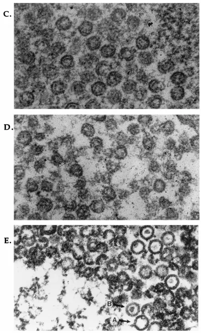
(v) Electron microscopic study. Thin sections of virus-in-fected cells were examined to determine whether capsid struc-tures are formed. As expected, cells infected with wt virus were found to contain three types of capsids, A, B, and C capsids, as well as virions (Fig. 7A and results not shown). Although capsid structures were observed in A247S-infected Vero cells (Fig. 7C) and m100-infected BMS-S129C cells (Fig. 7D), all were the electron-transparent (DNA2) capsids. In contrast, in
A247S-infected BMS-S129C cells, three types of wt-like
cap-sids as well as virions were observed (Fig. 7B and E and results not shown). The size of B capsids in A247S-infected S129C cells was very similar to that of wt virus-infected BMS-S129C cells. However, significant numbers of capsids, espe-cially in the nuclear section (Fig. 7E), were aberrant.
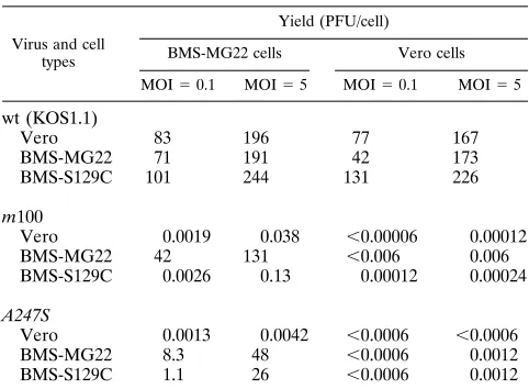
(vi) Growth of A247S in BMS-S129C cells.To determine whether A247S grows on BMS-S129C cells, plaque assays were performed. The mutant A247S virus formed slightly smaller plaques than wt virus on BMS-S129C cells (results not shown). Since the plaques were smaller, we performed a single-cycle growth experiment to examine the growth property of A247S virus on S129C cells. The yield of wt virus on BMS-S129C cells was comparable to those on Vero cells at MOIs of 5 and 0.1, indicating that the mutant S129C protease did not exhibit a negative trans-dominant phenotype (Table 2). In agreement with trans-complementation experiments, the m100 virus did not grow on either Vero or BMS-S129C cells. As we reported recently, the A247S virus exhibits a slightly trans-dominant phenotype (30), and the yield of this mutant on the protease-expressing BMS-MG22 cell line was approximately three- to fivefold lower at MOIs of 5 and 0.1 than that of m100 virus. Although A247S virus failed to grow on Vero cells, the yield of A247S virus on BMS-S129C cells was close to that of the mutant on BMS-MG22 cells at an MOI of 5, but the yields were sevenfold lower at an MOI of 0.1. Since recombination does occur at very low frequency between homologous DNA sequences present in the mutant genomes and in the chromo-somes of the transformed cell lines, the samples from A247S-infected cells were examined for wt recombination. Table 2 shows that wt recombination in A247S virus-infected BMS-MG22 and BMS-S129C cells was almost undetectable at the dilutions we examined. Because BMS-S129C cells support the growth of only A247S, and not of m100, we conclude that the complementation between two mutant proteases reconstitutes the wt protease activity required to support viral growth.
DISCUSSION
In this study, two mutant proteases were used to examine functional domains of the HSV-1 protease. Neither of these mutant proteases was able to support the growth of the pro-tease deletion mutant virus. One mutant propro-tease is expressed from the virus A247S, which contains an Ala-to-Ser change at residue 247. This mutant protease fails to autoprocess at the R site but retains the ability to cleave at the M site and also the ability to cleave its substrate ICP35 (30). The other mutant protease, expressed from a transformed cell line, contains a Ser-to-Cys mutation at its active site, Ser-129. The mutant S129C protease, unlike the A247S protease, lacks enzymatic activity but is a substrate for an active protease. When these two mutant proteases are provided in trans, they are able to restore viral growth, suggesting that an essential function is being supplied by the S129C protease. The possibility that the Nb domain of the S129C protease was supplying the essential function was ruled out, since the HCMVN0/HSVNa chimera FIG. 6. Encapsidation of viral DNA. Aliquots from each fraction of the
sucrose gradients shown in Fig. 5 were prepared and hybridized to a32P-labeled plasmid, pUCICP35. BMS-S129C cells infected with wt virus (A), m100 (B), and A247S (C).
on November 9, 2019 by guest
http://jvi.asm.org/
did not support the growth of either the m100 or A247S virus. Our results suggest that HSV-1 protease is composed of mul-tiple functional domains and some of these domains can be genetically separated.
The catalytic domain (N0) and Nb of the protease as well as
ICP35 e,f are all components of B capsids (15, 37, 52, 53). Intrageneic complementation between two mutant proteases
suggests that N0 may have another function(s) besides
enzy-matic activity required for viral growth. There are several pos-sible roles that N0may play inside B capsids. First, following its
release from Pra inside capsids, N0 may be responsible for
trans cleaving ICP35. In this case, trans processing of ICP35 by
N0must be an extremely fast event, or the capsids containing
unprocessed ICP35 are unstable, since capsids containing N0 FIG. 7. Electron micrographs of thin sections of virus-infected cells. Cells were infected with different viruses at an MOI of 10 PFU per cell. Cells were fixed and prepared at 16 h p.i. as described in Materials and Methods. (A) wt virus-infected BMS-S129C cells; (B) A247S-infected BMS-S129C cells; (C) A247S-infected Vero cells; (D) m100-infected BMS-S129C cells; (E) A247S-infected BMS-S129C cells. Magnifications,332,500 (for panels A and B) and3107,250 (for panels C, D, and E).
on November 9, 2019 by guest
http://jvi.asm.org/
FIG. 7—Continued.
4325
on November 9, 2019 by guest
and unprocessed ICP35 have not been observed. A second possible role for N0, like the bacteriophage T4 prohead
pro-tease (T4PPase), may be to digest itself, Nb, and ICP35 e,f during the encapsidation of viral DNA (17, 48). In this case, either the specificity of the protease must be changed or the specificity of the protease is dependent on the substrate con-formation rather than its primary sequence, as a consensus cleavage recognition site has not been identified elsewhere in these proteins. Third, N0, like ICP35 e,f, may play a structural
role in the formation of B capsids. This hypothesis is consistent with several observations: (i) the HCMV catalytic domain can provide enzymatic activity but cannot support viral growth; (ii) processing of ICP35 in A247S-infected Vero cells is insufficient to support viral growth (29), suggesting that the protease has a function which is distinct from its enzymatic activity; and (iii) N0 is a component of B capsids. Although capsid structures
were observed from HSV-1 protease mutant-infected cells by electron microscopy, such capsids have not been isolated by sucrose gradient centrifugation (13, 29, 44), suggesting that release of N0is required for capsid stability. The exact role of
N0, whether enzymatic, structural or undefined, has yet to be
determined.
HSV-1 protease undergoes autoprocessing at the M and R sites, releasing N0, Nb, and a 25-aa peptide; however, whether
this autoprocessing occurs in cis or in trans is unknown. Al-though our intragenic complementation experiments did not exclude the possibility of cis cleavage, it clearly demonstrated that trans cleavage of the mutant protease S129C by the A247S protease can lead to functional capsid assembly and produce infectious virus.
Since processing of ICP35 is restored to wt level in A247S-infected BMS-S129C cells, we carefully examined the S129C protease for residual enzymatic activity. The inability of the S129C protease to trans cleave ICP35 in the E. coli coexpres-sion system or in m100 virus-infected BMS-S129C cells sug-gests that the mutant protease lacks enzymatic activity. Our experiments also excluded the possibility that the elevated pro-cessing of ICP35 in A247S-infected BMS-S129C cells is due to the restoration of enzymatic activity following the release of S129C N0from full-length S129C protease, since S129C N0is
not proteolytically active. The restored processing of ICP35
occur? During T4 phage head assembly, gp21 is incorporated into the prohead core as an inactive zymogen which is activated only after the assembly of the shell (48), and no cleavage takes place in mutants in which assembly is blocked. In contrast, the enzymatic activity of the HSV-1 protease is not linked to the assembly of capsids, since the protease can undergo autopro-cessing and trans cleave ICP35 in transfected cells (26–28), an
E. coli coexpression system (6, 32), the baculovirus system (49,
51), and assembly-negative mutant virus-infected Vero cells (8). In the latter case, no capsid structures were observed by either sucrose gradient analysis or electron microscopy (7). However, these cleavage events may not reflect the exact role of the protease and ICP35 during the virion maturation. Tem-perature shift experiments with HSV-1 protease mutant ts1201 virus-infected cells indicated that cleavages may occur inside of the capsids, and only these capsids can mature to package viral DNA (44). However, these results cannot rule out the possi-bility that aberrant capsids may be dissociated and reassembled or that free, preexisting capsid proteins were assembled into normal capsids when the temperature was shifted from non-permissive to non-permissive. On the basis of the baculovirus sys-tem, Thomsen et al. (50) also proposed a model where the uncleaved protease, Pra, and its substrate ICP35 are used to assemble B capsids. An interesting feature of the intragenic complementation between the two mutant proteases from our experiments is that Prb as well as Nb and N0is present in the
B capsids of A247S-infected BMS-S129C cells. These capsids retain the ability to package viral DNA, as demonstrated by the presence of viral DNA in gradient fractions corresponding to C capsids, and mature into infectious virus. In m100-infected BMS-S129C cells and in A247S-infected Vero cells, B capsids could not be isolated and viral DNA was not encapsidated. These results strongly suggest that Prb, N0, Nb, and ICP35 e,f
must coexist in the same B capsids in A247S-infected BMS-S129C cells. Our results and the results of others (6, 7, 10, 27–29) support the hypothesis that when the protease and its substrate ICP35 interact, the specific cleavage occurs; however, only those cleavages that occur during or after capsid assembly are able to form functional capsids capable of packaging of viral DNA.
It was surprising to observe that the processing of ICP35 in
A247S-infected BMS-S129C cells was restored to wt levels. It
has been recently reported that in an in vitro assay, HSV-1 protease activity is stimulated over 100-fold by water structure-forming cosolvents, such as antichaotrophic salts (18, 57). This stimulation effect is most likely due to changes in the confor-mation of the substrate as well as the protease, since both the susceptibility of the substrate to proteolysis by trypsin and the protein fluorescence spectra of the protease are altered in the presence of solvents (57). These cosolvents may be mimicking the conformation of the protease under optimum conditions, i.e., inside the capsid. It is therefore conceivable that the re-stored processing of ICP35 in A247S-infected BMS-S129C
Vero 0.0019 0.038 ,0.00006 0.00012
BMS-MG22 42 131 ,0.006 0.006
BMS-S129C 0.0026 0.13 0.00012 0.00024
A247S
Vero 0.0013 0.0042 ,0.0006 ,0.0006
BMS-MG22 8.3 48 ,0.0006 0.0012
BMS-S129C 1.1 26 ,0.0006 0.0012
aCells were infected with viruses at the indicated MOIs, incubated at 378C for
18 h, and harvested. Titers of progeny viruses were determined in BMS-MG22 and Vero cells.
on November 9, 2019 by guest
http://jvi.asm.org/
[image:10.612.57.298.90.267.2]cells is completely due to the conditions of the capsid environ-ment.
The formation of heterodimers and multimers of two genet-ically discrete mutant protein molecules provides a common mechanism for intragenic complementation (47, 58). While this article was being revised, Darke et al. (5) reported that the HCMV protease forms a dimer. A simple model for our in-tragenic complementation results is that the A247S and S129C proteases form a functionally active heterodimer. The full-length A247S protease provides the enzymatic activity in order to trans cleave ICP35 and release N0from the S129C protease.
The N0 domain of S129C protease then supplies another
es-sential function for the formation of functional capsids which can mature into infectious virus. Further experiments will di-rectly test whether these two mutant proteases form a het-erodimer during or after capsid assembly.
ACKNOWLEDGMENTS
We thank Bernard Roizman for providing plasmid pRB4057. We are grateful to Steve Weinheimer for providing polyclonal antiserum against the HCMV catalytic domain. We thank Steve Weinheimer, Carolyn Diianni, Greg Yamanaka, and Laurence Tiley for helpful discussions.
This work was supported in part by a grant from the National Science Foundation (MCB-9119056) to J.C.B.
REFERENCES
1. Baker, T. S., W. W. Newcomb, F. O. Booy, J. C. Brown, and A. C. Steven. 1990. Three-dimensional structures of maturable and abortive capsids of equine herpesvirus 1 from cryoelectron microscopy. J. Virol. 64:563–573. 2. Baum, E. Z., G. A. Bebernitz, J. D. Hulmes, V. P. Muzithras, T. R. Jones, and
Y. Gluzman.1992. Expression and analysis of the human cytomegalovirus UL80-encoded protease: identification of autoproteolytic sites. J. Virol. 67: 497–506.
3. Braun, D. K., B. Roizman, and L. Pereira. 1984. Characterization of post-translational products of herpes simplex virus gene 35 proteins binding to the surfaces of full capsids but not empty capsids. J. Virol. 49:142–153. 4. Casjens, S., and J. King. 1975. Virus assembly. Annu. Rev. Biochem. 44:
555–611.
5. Darke, P. L., J. L. Cole, L. Waxman, D. L. Hall, M. K. Sardana, and L. C. Kuo.1996. Active human cytomegalovirus protease is a dimer. J. Biol. Chem. 271:7445–7449.
6. Deckman, I. C., M. Hagen, and P. J. McCann III. 1992. Herpes simplex virus type 1 protease expressed in Escherichia coli exhibits autoprocessing and specific cleavage of the ICP35 assembly protein. J. Virol. 66:7362– 7367.
7. Desai, P., N. A. DeLuca, J. C. Glorioso, and S. Person. 1993. Mutations in herpes simplex virus type 1 genes encoding VP5 and VP23 abrogate capsid formation and cleavage of replicated DNA. J. Virol. 67:1357–1364. 8. Desai, P., S. C. Watkins, and S. Person. 1994. The size and symmetry of B
capsids of herpes simplex virus type 1 are determined by the gene products of the UL26 open reading frame. J. Virol. 68:5365–5374.
9. DiIanni, C. L., D. A. Drier, I. C. Deckman, P. J. McCann III, F. Liu, B. Roizman, R. J. Colonno, and M. G. Cordingley.1993. Identification of the herpes simplex virus-1 protease cleavage sites by direct sequence analysis of autoproteolytic cleavage products. J. Biol. Chem. 268:2048–2051. 10. DiIanni, C. L., C. Mapelli, D. A. Drier, J. Tsao, S. Natarajan, D. Riexinger,
S. M. Festin, M. Bolgar, G. Yamanaka, S. Weinheimer, C. A. Meyers, R. J. Colonno, and M. G. Cordingley.1993. In vitro activity of the herpes simplex virus-1 protease with peptide substrates. J. Biol. Chem. 268:25449–25454. 11. DiIanni, C. L., J. T. Stevens, M. Bolgar, D. R. O’Boyle II, S. W. Weinheimer,
and R. J. Colonno.1994. Identification of the serine residue at the active site of the herpes simplex virus type 1 protease. J. Biol. Chem. 269:12672–12676. 12. Gao, M., et al. Unpublished data.
13. Gao, M., L. Matusick-Kumar, W. Hurlburt, S. F. DiTusa, W. W. Newcomb, J. C. Brown, P. J. McCann III, I. C. Deckman, and R. J. Colonno.1994. The protease of herpes simplex virus type 1 is essential for functional capsid formation and viral growth. J. Virol. 68:3702–3712.
14. Gibson, W., A. I. Marcy, J. C. Comoli, and J. Lee. 1990. Identification of precursor to cytomegalovirus capsid assembly protein and evidence that processing results in loss of its carboxy-terminal end. J. Virol. 64:1241–1249. 15. Gibson, W., and B. Roizman. 1972. Proteins specified by herpes simplex virus. VIII. Characterization and composition of multiple capsid forms of subtypes 1 and 2. J. Virol. 10:1044–1052.
16. Gibson, W., and B. Roizman. 1974. Proteins specified by herpes simplex virus. X. Staining and radiolabeling properties of B capsids and virion
pro-teins in polyacrylamide gels. J. Virol. 13:155–165.
17. Goldstein, J., and S. P. Champe. 1974. T4-induced activity required for specific cleavage of bacteriophage protein in vitro. J. Virol. 13:419–427. 18. Hall, L. D., and P. L. Darke. 1995. Activation of the herpes simplex virus type
1 protease. J. Biol. Chem. 270:22697–22700.
19. Heilman, C. J., M. Zweig, J. R. Stephenson, and B. Hampar. 1979. Isolation of nucleocapsid proteins of herpes simplex virus types 1 and 2 possessing immunologically type-specific and cross-reactive determinants. J. Virol. 29: 34–42.
20. Holland, L. E., R. M. Sandri-Goldin, A. L. Goldin, J. C. Glorioso, and M. Levin.1984. Transcriptional and genetic analyses of the herpes simplex virus type 1 genome: coordinates 0.29 to 0.45. J. Virol. 49:947–959.
21. Holwerda, B. B., A. J. Wittwer, K. L. Duffin, C. Smith, M. V. Toth, L. S. Carr, R. C. Wiegand, and M. L. Bryant.1994. Activity of two-chain recombinant human cytomegalovirus protease. J. Biol. Chem. 269:25911–25915. 22. Hong, Z., M. Beaudet-Miller, J. Durkin, R. Zhang, and A. D. Kwong. 1996.
Identification of a minimal hydrophobic domain in the herpes simplex virus type 1 scaffolding protein which is required for interaction with the major capsid protein. J. Virol. 70:533–540.
23. Knipe, D. M., M. P. Quinlan, and A. E. Spang. 1982. Characterization of two conformational forms of the major DNA-binding protein encoded by herpes simplex virus 1. J. Virol. 44:736–741.
24. Knipe, D. M., and A. E. Spang. 1982. Definition of a series of stages in the association of two herpesvirus proteins with the cell nucleus. J. Virol. 43: 314–324.
25. Liu, F., and B. Roizman. 1991. The promoter, transcriptional unit, and coding sequences of the herpes simplex virus 1 family 35 proteins are con-tained within and in frame with the UL26 open reading frame. J. Virol. 65:206–212.
26. Liu, F., and B. Roizman. 1991. The herpes simplex virus 1 gene encoding a protease also contains within its coding domain the gene encoding the more abundant substrate. J. Virol. 65:5149–5156.
27. Liu, F., and B. Roizman. 1992. Differentiation of multiple domains in the herpes simplex virus 1 protease encoded by the UL26 gene. Proc. Natl. Acad. Sci. USA 89:2076–2080.
28. Liu, F., and B. Roizman. 1993. Characterization of the protease and other products of amino-terminus-proximal cleavage of the herpes simplex virus 1 UL26 protein. J. Virol. 67:1300–1309.
29. Matusick-Kumar, L., W. Hurlburt, S. W. Weinheimer, W. W. Newcomb, J. C. Brown, and M. Gao.1994. Phenotype of the herpes simplex virus type 1 protease substrate ICP35 mutant virus. J. Virol. 68:5384–5394.
30. Matusick-Kumar, L., P. J. McCann III, B. J. Robertson, W. W. Newcomb, J. C. Brown, and M. Gao.1995. Release of the catalytic domain N0from the herpes simplex virus type 1 protease is required for viral growth. J. Virol. 69:7113–7121.
31. Matusick-Kumar, L., W. W. Newcomb, J. C. Brown, P. J. McCann III, W. Hurlburt, S. P. Weinheimer, and M. Gao.1995. The C-terminal 25 amino acids of the protease and its substrate ICP35 of herpes simplex virus type 1 are involved in the formation of sealed capsids. J. Virol. 69:4347–4356. 32. McCann, P. J., III, D. R. O’Boyle II, and I. C. Deckman. 1994. Investigation
of the specificity of the herpes simplex virus type 1 protease by point mu-tagenesis of the autoproteolysis sites. J. Virol. 68:526–529.
33. McGeoch, D. J., M. A. Dalrymple, A. J. Davison, A. Dolan, M. C. Frame, D. McNab, L. J. Perry, J. E. Scott, and P. Taylor.1988. The complete DNA sequence of the long unique region in the genome of herpes simplex virus type 1. J. Gen. Virol. 69:1531–1574.
34. Morgenatern, J. P., and H. Lane. 1990. A series of mammalian expression vectors and characterization of their expression of a reporter gene in stably and transient transformed cells. Nucleic Acids Res. 18:1068.
35. Newcomb, W. W., and J. C. Brown. 1989. Use of Ar1plasma etching to localize structural proteins in the capsid of herpes simplex virus type 1. J. Virol. 63:4697–4702.
36. Newcomb, W. W., and J. C. Brown. 1991. Structure of the herpes simplex virus capsid: effects of extraction with guanidine hydrochloride and partial reconstitution of extracted capsids. J. Virol. 65:613–620.
37. Newcomb, W. W., B. L. Trus, F. P. Booy, A. C. Steven, J. S. Wall, and J. C. Brown.1993. Structure of the herpes simplex virus capsid: molecular com-position of the pentons and the triplexes. J. Mol. Biol. 232:499–511. 38. Nicholson, P., C. Addison, A. M. Cross, J. Kennard, V. G. Preston, and F. J.
Rixon.1994. Localization of the herpes simplex virus type 1 major capsid protein VP5 to the cell nucleus requires the abundant scaffolding protein VP22a. J. Gen. Virol. 75:1091–1099.
39. O’Boyle, D. R., II, K. Wager-Smith, J. T. Stevens III, and S. P. Weinheimer. 1995. The effect of internal autocleavage on kinetic properties of the human cytomegalovirus protease catalytic domain. J. Biol. Chem. 270:4753–4758. 40. Patel, A. H., and J. B. Maclean. 1995. The product of the UL6 gene of herpes
simplex virus type 1 is associated with virus capsids. Virology 206:465–478. 41. Perdue, M. L., J. C. Cohen, C. C. Randall, and D. J. O’Callaghan. 1976. Biochemical studies on the maturation of herpesvirus nucleocapsid species. Virology 74:194–208.
42. Person, S., S. Laquerre, D. Prashant, and J. Hempel. 1993. Herpes simplex virus type 1 capsid protein, VP21, originates within the UL26 open reading
on November 9, 2019 by guest
http://jvi.asm.org/
J. Gen. Virol. 69:2879–2891.
47. Shepard, A. A., and N. A. Deluca. 1989. Intragenic complementation of herpes simplex virus regulatory protein ICP4. J. Virol. 63:1203–1211. 48. Showe, M. K., E. Isobe, and L. Onorato. 1976. Bacteriophage T4 prohead
proteinase. II. Its cleavage from the product of gene 21 and regulation in phage-infected cells. J. Mol. Biol. 107:55–69.
49. Tatman, J. D., V. G. Preston, P. Nicholson, R. M. Elliott, and F. J. Rixon. 1994. Assembly of herpes simplex virus type 1 capsids using a panel of recombinant baculovirus. J. Gen. Virol. 75:1101–1113.
50. Thomsen, D. R., W. W. Newcomb, J. C. Brown, and F. L. Homa. 1995. Assembly of the herpes simplex virus capsids: requirement for the carboxyl-terminal twenty-five amino acids of the proteins encoded by the UL26 and UL26.5 genes. J. Virol. 69:3690–3703.
51. Thomsen, D. R., L. L. Roof, and F. L. Homa. 1994. Assembly of herpes simplex virus (HSV) intermediate capsids in insect cells infected with
re-55. Welch, A. R., E. C. Villarreal, and W. Gibson. 1995. Cytomegalovirus protein substrates are not cleaved by the herpes simplex virus type 1 proteinase. J. Virol. 69:341–347.
56. Welch, A. R., A. S. Wood, L. M. McNally, R. J. Cotter, and W. Gibson. 1991. A herpes maturational proteinase, assemblin: identification of its gene, pu-tative active site domain, and cleavage site. Proc. Natl. Acad. Sci. USA 88:10792–10796.
57. Yamanaka, G., C. L. DiIanni, D. R. O’Boyle II, J. Stevens, S. P. Weinheimer, I. C. Deckman, L. Matusick-Kumar, and R. Colonno.1995. Stimulation of the herpes simplex virus type 1 protease by anticheaotrophic salts. J. Biol. Chem. 270:30168–30172.
58. Zabin, I., and A. V. Fowler. 1978.b-Galactosidase, the lactose permease protein and thiogalactoside transacetylase, p. 89–121. In J. H. Miller and W. S. Reznikoff (ed.), The operon. Cold Spring Harbor Laboratory, Cold Spring Harbor, N.Y.