0022-538X/97/$04.0010
Copyrightq1997, American Society for Microbiology
Toward Highly Efficient Cell-Type-Specific Gene Transfer with
Retroviral Vectors Displaying Single-Chain Antibodies
TE-HUA TEARINA CHU†ANDRALPH DORNBURG*
Department of Molecular Genetics and Microbiology, Robert Wood Johnson Medical School, University of Medicine and Dentistry of New Jersey, Piscataway, New Jersey 08854
Received 15 July 1996/Accepted 10 September 1996
Recently, we constructed retroviral vector particles derived from spleen necrosis virus (SNV) that display a single-chain antibody (scA) on the viral surface. By transient transfection protocols, we showed that such particles are competent for infection and cell type specific. Efficient infection was dependent on the presence of wild-type envelope, although wild-type SNV was not infectious on target cells (T.-H. T. Chu and R. Dornburg, J. Virol. 69:2659–2663, 1995; T.-H. T. Chu, I. Martinez, W. C. Sheay, and R. Dornburg, Gene Ther. 1:292–299, 1994). In this study, stable packaging lines were constructed and detailed biological and biochemical studies were performed. Chimeric scA-envelope fusion proteins were expressed as efficiently as wild-type envelope and were stable over a period of at least 6 h. Only a fully functional wild-type envelope could act as a helper for efficient virus penetration. The ratio of wild-type envelope protein to chimeric envelope protein appears to determine the efficiency of infection. Virus titers of targeting vectors obtained from stable packaging lines were
as high as 104
CFU/ml. A 25-fold concentration of vector virus stocks resulted in a 200-fold increase in virus
titers (up to 106
CFU/ml). These data indicate that an inhibitor of infection was (at least partially) removed by the concentration protocol. Our data show that this technology has several variables for further improve-ments and, therefore, has the potential to become a powerful tool for cell-type-specific in vivo human gene therapy.
Human gene therapy has the potential to become a major new clinical practice to cure not only inborn errors of metab-olism but also a large variety of acquired diseases, like cancer, diabetes, and AIDS (5, 8, 20, 26). However, the lack of a cell-type-specific gene delivery system forces most current gene transfer protocols to be performed in tissue culture. Such ex vivo protocols are too expensive and technically problematic to be used on a widespread basis.
Several attempts have been made to construct cell-type-specific gene delivery tools. For example, retroviral particles were constructed that display the CD4 molecule to target hu-man immunodeficiency virus-infected huhu-man cells (25). In an-other approach, two different antibodies, one directed against a cell surface protein and the other directed against the viral envelope (Env) protein, were linked by streptavidin at their carboxy termini to direct a retroviral vector to a specific target cell (9, 21). More recently, a peptide hormone was fused to the Env protein to specifically infect cells that express the corre-sponding hormone receptor (12). However, no or only very low levels of infectivity were observed.
During the past 6 years, we have been developing retroviral vector particles, derived from spleen necrosis virus (SNV) (7), that display the antigen binding site of an antibody on the viral surface. This has been achieved by fusing a single-chain anti-body (scA) to the Env protein. First, by using a dinitrophenol hapten model system, we showed that such particles were com-petent for infection (4). Similar results have been obtained by Russell et al. with a different hapten model system and murine
leukemia virus (MLV)-derived retroviral vector particles (22). To further investigate the versatility of this system for gene transfer into human cells, we next used an scA (termed B6.2) directed against a surface antigen expressed on human colon cells and various carcinomas. By transient transfection assays, we showed that SNV vector particles that displayed the B6.2 scA on the viral surface were competent for infection as well. The efficiency of infection was strongly enhanced by the pres-ence of the SNV wild-type envelope, which does not confer infectivity on human cells. The infectivity was antigen and cell type specific (2–4). Most recently, similar findings have been obtained with an scA directed against the low-density lipopro-tein receptor (24). In this paper, we describe the construction of stable packaging lines producing such targeting vectors. Detailed biochemical and biological analyses were performed. Our data show that such engineered particles have the poten-tial to become a powerful tool for cell-type-specific in vivo gene delivery.
Establishment of stable cell lines.To test the biological and
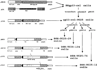
biochemical properties of retroviral targeting vectors, stable cell lines were established from single selected cell colonies transfected (13) with the construct of interest. All stable cell lines were derived from DSgp13-cxl cells, which express SNV Gag-Pol proteins and the retroviral vector pCXL (Fig. 1). The retroviral vector pCXL transduces the bacterial b -galactosi-dase gene (19). Transfection of an envelope expression plas-mid into this cell line creates a complete retroviral packaging system.
First, a stable packaging cell line was made that expresses a chimeric scA-Env fusion protein by introducing the plasmid pTC25 into DSgp13-cxl cells. The resulting cell line was termed gp13-cxl-TC25 (Fig. 1). In pTC25, the scA gene is fused to the complete transmembrane (TM) coding region of the SNV env gene. Since the conserved SU/TM cleavage motif has not been retained in this construct, this chimeric scA-env gene product * Corresponding author. Present address: Center for Human
Virol-ogy, Division of Infectious Diseases, Thomas Jefferson University, Jefferson Alumni Hall, 1020 Locust St., Suite 329, Philadelphia, PA 19107.
† Present address: Dept. of Immunochemistry, The New York Blood Center, New York, NY 10021.
720
on November 9, 2019 by guest
http://jvi.asm.org/
was not expected to be proteolytically cleaved. Therefore, it could be distinguished from wild-type envelope in polyacryl-amide gels after immunoprecipitation with an TM anti-body.
Earlier, we found that the efficiency of infection was en-hanced when wild-type envelope was coexpressed on retroviral particles containing the B6.2 scA-Env fusion protein (2). To test this further, gp13-cxl-TC25 cells were used for the con-struction of cell lines that express chimeric plus wild-type or mutant SNV envelope genes (Fig. 1). Thus, all cell lines de-rived from such clones were expected to express equal levels of Gag-Pol and the retroviral vector pCXL. In fact, although the complete loss of reverse transcriptase activity was recurrently observed in some single isolated DSgp13-cxl cell clones, stable cell lines derived from DSgp13-cxl cells used in this study expressed about equal levels of reverse transcriptase (data not shown).
Four different cell lines were established from gp13-cxl-TC25 cells that produce retroviral vector particles displaying both chimeric and wild-type Env proteins: two cell lines that express fully functional SNV wild-type Env proteins were made and were termed DSH-TC25-134 and DSH-TC25-29 (Fig. 1). These cell lines contain the envelope expression plas-mids pRD134 and pIM29, respectively. Plasplas-mids pIM29 and pRD134 are identical except that pIM29 does not contain the adenovirus tripartite leader sequence, which enhances
enve-lope expression in D17 cells about 10-fold (16, 23). Thus, DSH-TC25-134 and DSH-TC25-29 cells differ only in the level of expression of wild-type envelope (Fig. 1 and 2). The ratio-nale for using these two env expression vectors was to get a first insight into whether the wild-type/chimeric envelope expres-sion ratio has an effect on protein processing and/or on the efficiency of infection.
To shed light on the role of wild-type envelope in infection of composite targeting vectors, two more cell lines were estab-lished from gp13-cxl-TC25 cells. These cell lines express mu-tant SNV envelopes and were termed DSH-TC25-76 and DSH-TC25-12 (Fig. 1). Cell line DSH-TC25-12 contains plas-mid pTC12, which has two point mutations within the SU/TM cleavage site. These point mutations abolish cleavage of the envelope precursor and, hence, the membrane fusion function (15). However, they do not abolish transport of the protein to the cell surface (14a). Cell line DSH-TC25-76 contains plasmid pTC76, which carries a point mutation in the middle of SU. This mutation does not abolish protein processing or transport to the cell surface (18). However, it reduces the efficiency of wild-type SNV infection by about 4,000-fold. Our data indicate that the latter mutation interferes with receptor binding (15, 18).
The rationale for using these mutant envelope proteins was to determine whether the wild-type envelope helps virus pen-etration only by supplying a functional membrane fusion
do-FIG. 1. Pedigree of retroviral packaging lines for production of vectors that display the B6.2 scA and plasmids used for their construction. In pRD136 (15–17), SNV Gag-Pol proteins are expressed from the MLV U3 promoter (MLV-pro) followed by the adenovirus tripartite leader sequence (AVtl). Plasmid pCXL contains a standard SNV-derived retroviral vector with SNV long terminal repeats (LTRs), which transduces the bacterial lacZ gene (19). In plasmid pTC25, the scA gene B6.2 was fused to TM of the SNV envelope (2, 3). Plasmids pRD134 and pIM29 contain the complete wild-type envelope gene of SNV and have been described recently (15, 16). Plasmids pTC12 and pTC76 contain mutant SNV envelope genes with point mutations at amino acid position 398-399 (SU/TM cleavage site) and at position 192 (Asp to Arg in the middle of SU), respectively. These point mutations abolish membrane fusion and receptor binding, respectively (15, 18). DSgp13-cxl cells have been derived from a single selected dog D17 cell clone and contain plasmids pRD136 and pCXL (2). DSgp13-cxl cells were transfected (13) with plasmid pTC25, which contains the B6.2 scA gene fused to TM of the SNV envelope gene. A cell line was established from a single cell colony and termed gp13-cxl-TC25. gp13-cxl-TC25 cells were transfected with plasmids pIM29, pRD134, pTC76, and pTC12 to give DSH-TC25-29, DSH-TC25-134, DSH-TC25-76, and DSH-Tc25-12 cells, respectively. These cell lines express both the chimeric and the wild-type SNV envelope genes. L, hydrophobic leader sequence of the SNV env gene. Plasmid sequences (derived from pUC19) that abut the above-mentioned vectors are not shown.
VOL. 71, 1997 NOTES 721
on November 9, 2019 by guest
http://jvi.asm.org/
[image:2.612.117.489.70.341.2]main or whether some interaction of SU with a cellular recep-tor is also involved. In the first case, a mutation in SU would not abolish the helper function of wild-type Env, whereas a mutation in TM would. In the second case, more complex interactions may occur; SNV can infect a large variety of spe-cies and cell lines, including dog D17 cells. Thus, it appears that SNV is docking to a housekeeping receptor, like some other retroviruses. The corresponding receptor may also be present on human cells. However, it may carry mutations which reduce the affinity of the SNV envelope below a thresh-old level necessary to mediate infection, similar to what is observed with ecotropic MLV (14). Thus, it may be possible that the targeting scA-envelope restores this affinity by anchor-ing the particle to the cell surface. Once bound to the cell surface, the complete SNV envelope may be able to interact with its cellular receptor to initiate membrane fusion. In this case, a mutation in a receptor-interacting domain of SU would also diminish the helper function of wild-type Env.
For control experiments, a standard retroviral packaging cell line has also been made from DSgp13-cxl cells by transfection of plasmid pRD134. This cell line was termed DSH134E, and it expresses only the wild-type SNV envelope.
Expression and stability of chimeric and wild-type envelope
proteins.Intracellular expression and stability of the chimeric
and wild-type envelopes were examined by pulse-chase analysis and immunoprecipitation.35S-labeled Env proteins were
pre-cipitated by anti-TM monoclonal antibodies (kindly provided by L. Lee, Regional Poultry Research Laboratory, East Lan-sing, Mich.) followed by sodium dodecyl sulfate-polyacryl-amide gel electrophoresis and radioautography (Fig. 2). The monoclonal antibody is very specific for the SNV envelope TM peptide; no bands were detected when immunoprecipitations were performed with control D17 cells or DSgp13-cxl cells (15, 18) (data not shown).
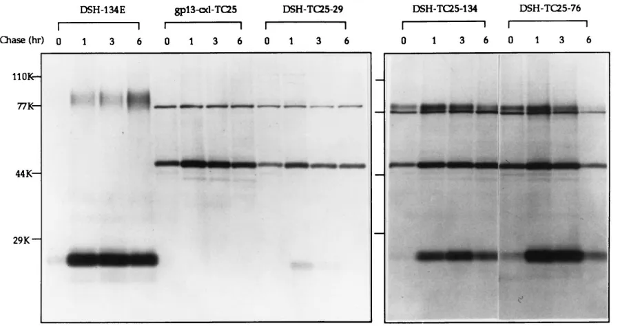
The anti-TM monoclonal antibody precipitated a 24-kDa protein in cells expressing the wild-type SNV envelope protein (Fig. 2, DSH-134E). The size of this band corresponds to the predicted size of the TM peptide. Coinciding with earlier ob-servations (18), only small amounts of the SU peptide were coprecipitated by this antibody. TM was hardly detected im-mediately after labelling and could be immunoprecipitated after 1 h (Fig. 2, DSH-134E). Since the anti-TM antibody does not bind to noncleaved precursor envelope proteins (data not shown), it can be concluded that SU/TM cleavage was not complete after 1 h. The wild-type Env appeared to be stable over a period of at least 6 h.
The anti-TM antibody precipitated a 49-kDa protein from cells expressing the scA-TM fusion protein immediately after labelling (Fig. 2, gp13-cxl-TC25). The molecular weight of this peptide precisely reflects the predicted size of the chimeric envelope. Another protein, with a molecular mass of about 77 kDa, was always coprecipitated from cells that express gpTC25. This protein was not detected in immunoprecipitates from envelope-negative D17 control cells (data not shown) or cells expressing the wild-type envelope alone. Since the molecular weight of this protein is too low for it to be interpreted as a dimeric scA-Env fusion protein, it appears that this protein is of cellular origin. It was probably associated with and copre-cipitated with the chimeric envelope protein. The chimeric envelope was also stable over a period of 6 h.
[image:3.612.85.535.70.307.2]When the wild-type Env was coexpressed with the chimeric protein, both proteins were detected. The ratio of chimeric Env to wild-type Env was approximately 1:1 in cells expressing wild-type Env and the chimeric construct from identical gene expression vectors (Fig. 2, DSH-TC25-134). Neither protein appeared to have a negative effect on the stability of the other peptide. Similar results were obtained with the receptor-bind-ing mutant gpTC76 coexpressed with the chimeric envelope
FIG. 2. Pulse-chase experiment to study expression and stability of envelope proteins. Confluent cell monolayers were labelled with 40mCi of [35S]methionine and [35S]cysteine (Tran35S-label; ICN Biomedicals, Costa Mesa, Calif.) per ml for 40 min; this was followed by incubation with Dulbecco’s minimal essential medium supplemented with 7% fetal bovine serum for 0, 1, 3, or 6 h. Cell lysates were prepared as described previously (10). Incorporated [35S]methionine and [35S]cysteine were determined by scintillation counting. An excess (0.1ml of ascites fluid) of an anti-TM antibody (6) (kindly provided by L. Lee, Regional Poultry Research Laboratory, East Lansing, Mich.) was preabsorbed with protein A-Sepharose beads (Pharmacia-LKB, Piscataway, N.J.). Lysate (33107CPM) from each sample was then incubated with the immunocomplex, precipitated, and subjected to sodium dodecyl sulfate–12% polyacrylamide gel electrophoresis as described previously (10). The gels were fixed and exposed to Kodak X-ray film as described previously (10).
on November 9, 2019 by guest
http://jvi.asm.org/
(Fig. 2, DSH-TC25-76), although this mutant Env appeared to be slightly less stable (see also below). Coinciding with earlier observations (16, 23), considerably less wild-type Env was de-tected in cells expressing the wild-type Env without the ade-novirus tripartite leader sequence (Fig. 2, DSH-TC25-29).
The chimeric envelope was also detected with anti-TM an-tibodies in cells coexpressing the SU/TM cleavage mutant gpTC12 envelope protein (data not shown). Thus, the presence of this mutant envelope did not have a significant negative effect on the expression of the chimeric scA-Env protein. Al-though the SU/TM cleavage mutant protein could be detected on the cell surface with a polyclonal anti-SU antiserum by fluorescence-activated cell sorter analysis, it could not be pre-cipitated with anti-TM antibodies (data not shown). These data suggest that the anti-TM antibody recognizes the TM epitope only if it is properly folded.
Cell surface expression of gpTC25 and wild-type envelopes has been demonstrated earlier (3, 16). Attempts were also made to compare the levels of wild-type and chimeric enve-lopes in virus particles. However, the sensitivity of the assays was not sufficient to detect any of these proteins (including wild-type Env), even in preparations of concentrated virus stocks (data not shown).
Infection studies of cell-type-specific SNV vector particles.
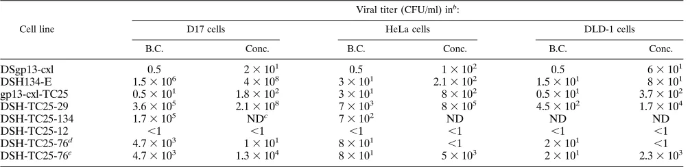
Virus titers on D17, DLD-1, and HeLa cells were determined in serial dilutions with supernatant media harvested from con-fluent cell cultures. The cell line releasing wild-type vector particles (DSH-134E) produced 1.5 3106 CFU/ml of
super-natant medium on D17 cells (Table 1). Minimal infectivity was seen on HeLa and DLD-1 cells. Particles displaying the scA-Env fusion protein alone showed hardly any infectivity on D17 or DLD-1 cells. Consistent with our data from transient trans-fection experiments (2), a low level of infectivity was consis-tently observed on HeLa cells. No infectivity was observed on 293 or 143T cells, which do not express the B6.2 antigen (data not shown).
Particles displaying both wild-type and chimeric envelope proteins infected D17 cells about 10 times less efficiently than those containing the wild-type envelope alone. These particles also infected HeLa cells, with an average titer of 7 3 103
CFU/ml. Interestingly, particles from cells expressing low lev-els of wild-type Env (DSH-TC25-29) were about 10 times more infectious than those from cells expressing high levels of Env (DSH-TC25-134, Table 1 and Fig. 3). Again, no infectivity was observed on human 293 or 143T cells (data not shown).
Particles harvested from cells that express the chimeric en-velope with the wild-type enen-velope containing an impaired membrane fusion function (DSH-TC25-12) could not infect D17, HeLa, or DLD-1 cells at all (Table 1). The low level of infectivity conferred by the chimeric protein alone was com-pletely inhibited by this mutant Env. These data indicate that a membrane fusion function has to be supplied by the wild-type envelope. Mixed particles, containing an SU mutant envelope and the chimeric Env (DSH-TC25-76), were about 200-fold less infectious on D17 cells than wild-type vector particles. The infectivity of such virions on the human cells was reduced about 10- to 100-fold compared with particles coexpressing wild-type envelope. The infectivity on HeLa cells was still slightly higher than that of particles containing the chimeric envelope alone (Table 1). These data indicate that a fully functional wild-type envelope is needed to act as a helper for infectivity.
Infection studies on concentrated viruses.High vector virus
titers are the most important feature of targeting vectors for application in human gene therapy. To test if concentrated virus stocks of targeting vectors can be obtained, ultracentrif-ugation or ultrafiltration was applied.
[image:4.612.58.561.82.204.2]First, virus particles were pelleted by ultracentrifugation. Tissue culture supernatant media were collected and clarified by low-speed centrifugation, and this was followed by centrif-ugation in an SW27 rotor (Beckman Instruments, Palo Alto, Calif.) at 25,000 rpm for 90 min at 48C. The pellets were resuspended in phosphate-buffered saline and either subjected to infection immediately or stored at2208C for later analysis. As observed with other retroviruses (11), the particles contain-ing the wild-type SNV envelope alone had a decreased infec-tivity on D17 cells after ultracentrifugation. Surprisingly, all particles, even with wild-type Env alone, became significantly infectious on human cells (data not shown). These data suggest that the virus particles were modified by ultracentrifugation TABLE 1. Biological activity of retroviral particlesa
Cell line
Viral titer (CFU/ml) inb:
D17 cells HeLa cells DLD-1 cells
B.C. Conc. B.C. Conc. B.C. Conc.
DSgp13-cxl 0.5 23101 0.5 13102 0.5 63101
DSH134-E 1.53106 43108 33101 2.13102 1.53101 83101
gp13-cxl-TC25 0.53101 1.83102 33101 83102 0.53101 3.73102
DSH-TC25-29 3.63105 2.13108 73103 83105 4.53102 1.73104
DSH-TC25-134 1.73105 NDc 73102 ND ND ND
DSH-TC25-12 ,1 ,1 ,1 ,1 ,1 ,1
DSH-TC25-76d 4.73103 13101 83101 ,1 23101 ,1
DSH-TC25-76e 4.73103 1.33104 83101 53103 23101 2.33103
aVirus particles were harvested from various packaging cell lines expressing chimeric and/or wild-type Env proteins (see also Fig. 1) and subjected to infection of
D17, HeLa, or DLD-1 cells in serial dilutions to determine virus titers. Infectivity studies were performed as described recently (2). To determine the number of cells expressing the bacterial lacZ gene, the cells were stained with 5-bromo-4-chloro-3-indoyl-b-D-galactopyranoside as described previously (19). D17 cells (a dog osteosarcoma cell line obtained from the American Type Culture Collection) were grown in Dulbecco’s minimal essential medium (DMEM) containing 6% calf serum. DLD-1 cells (human colon carcinoma) and HeLa cells (human cervical carcinoma) were grown in DMEM containing 10% fetal bovine serum.
bB.C., infectivity of virus particles harvested from confluent cell cultures before concentration; Conc., infectivity of virus particles after a 25-fold concentration by
ultrafiltration. Concentration was performed as follows. A 15-ml volume of supernatant was added to a Centriprep-100 tube containing a cellulose membrane with a 100-kDa molecular mass cutoff (Amicon, Beverly, Mass.). The Centriprep-100 was centrifuged in a tabletop centrifuge (Sorvall RC5B) with swing-out buckets at 3,000 rpm for 30 min at 48C. The filtrate was discarded and spun for another 10 min. The same procedure was repeated until a final volume of approximate 0.6 ml was obtained (usually three spins were performed). The concentrated supernatant was either subjected directly to infection or stored at2208C for further analysis.
cND, not done.
dThe concentration conditions were identical to those used for the other samples indicated above. eGentle concentration conditions were used (see the text).
VOL. 71, 1997 NOTES 723
on November 9, 2019 by guest
http://jvi.asm.org/
and that the increase in infectivity was most certainly nonspe-cific (data not shown).
However, the biological properties of the virus particles did not appear to be altered when the particles were concentrated by ultrafiltration, using Amicon Centriprep-100 tubes. Al-though the virus particles were concentrated only 25-fold in these experiments, the infectivity of wild-type vector particles (RD134) on D17 cells increased about 250-fold (Table 1), producing titers of up to 43108CFU/ml. A similar increase
was seen with all other vector stocks. For example, the virus titer of particles with mixed envelopes increased from 73103
to 8 3 105 CFU/ml on HeLa cells. The infectivity of virus
particles with no viral envelope was also increased by ultrafil-tration. Particles produced from envelope-negative cells may incorporate various cellular proteins which may mediate low levels of nonspecific infection.
Coinciding with our earlier observations, particles displaying the targeting envelopes together with the mutant Env gpTC12 could not infect any of the cells, either before or after concen-tration. These data suggest that the increase of infectivity was not caused by nonspecific effects. It is noteworthy that poorly reproducible results were obtained with particles containing the chimeric protein plus the SU envelope point mutant, gpTC76. In general, the mutation in SU appeared to result in less stable envelope proteins. The usual ultrafiltration protocol applied to the other vector virus solutions almost completely abolished infectivity. However, a gentle concentration (shorter spinning periods) increased infectivity about 10-fold.
Discussion.Previously, we and others have shown that
ret-roviral vectors that display an scA on the viral surface enable cell-type-specific gene delivery (3, 24). However, several ques-tions still needed to be addressed to prove the potential of this gene delivery system for future application in human gene therapy. In this study, we constructed stable packaging cell lines and investigated the stability of targeting vectors and their efficiency of infection.
Chimeric scA-Env fusion proteins were expressed as effi-ciently as wild-type envelope. When the wild-type or mutant SNV env genes were copresent with the chimeric env gene, both proteins were expressed corresponding to the promoter strength. Moreover, both wild-type and chimeric Env proteins were stable, and neither protein had a negative effect on the stability of the other protein.
[image:5.612.65.555.71.394.2]Earlier we reported that the addition of wild-type envelope to particles containing a targeting envelope bestows infectivity on cells that do not contain a suitable receptor for SNV (3, 4). We hypothesized that (i) the chimeric Env protein can act as a targeting molecule to bind the virus to the cell surface of target cells and (ii) wild-type envelope is needed as a helper, probably for efficient membrane fusion. In this study we investigated this phenomenon in more detail. We found that a mutant wild-type envelope defective in membrane fusion did not confer infec-tivity. On the contrary, this mutant Env protein abolished in-fectivity completely. We expected that an envelope protein defective in receptor binding would still be able to act as a viable helper by supplying a fully functional membrane fusion
FIG. 3. Infection of HeLa cells with retroviral particles transducing the bacterial lacZ gene. Top left: HeLa cells infected with 200ml of undiluted vector virus containing the SNV wild-type envelope (harvested from DSH helper cells); bottom left: HeLa cells infected with 200ml of vector virus displaying the B6.2 scA and wild-type envelope expressed from pIM29; top right: HeLa cells infected with 100ml of vector virus containing the B6.2 scA plus the SNV wild-type Env after a 25-fold concentration by ultrafiltration; bottom right: same as the panel above it but at a higher magnification.
on November 9, 2019 by guest
http://jvi.asm.org/
domain. However, this mutant envelope increased the infec-tivity of targeting vectors only minimally.
These data show that a fully functional wild-type Env is needed to act as a helper for virus penetration. We hypothesize that the targeting envelope anchors the virus particle on the cell surface. Once bound to the cell surface, the unmodified SNV wild-type envelope can interact with its corresponding cellular receptor on human cells, to which it normally has insufficient affinity to initiate membrane fusion. However, as a result of the stabilized virus binding via the targeting envelope, all necessary conformational changes can take place to trigger membrane fusion.
We further hypothesize that the ratio of wild-type envelope to targeting envelope plays an important role in efficient infec-tion. Thus, two different packaging lines were constructed which express different amounts of wild-type Env. Earlier, we found that the density of wild-type envelope on the cell surface does not influence the level of infectivity of vector virus parti-cles containing wild-type Env alone (16). However, the level of wild-type Env expression did play a role in the efficiency of infectivity in particles with mixed envelopes. Surprisingly, smaller amounts of wild-type Env resulted in more infectious particles (Table 1). These data indicate that the ratio of wild-type Env to scA-Env protein is important for efficient infectiv-ity. For example, the presence of too much wild-type Env may inhibit efficient binding of the scA to its antigen on the cell surface.
Vector virus titers obtained from stable producer lines were as high as 104CFU/ml. The utility of the cell-type-specific gene
delivery system described here for human gene therapy de-pends upon high virus titers. Thus, experiments were also performed to obtain higher virus titers by concentration. Con-centration of retrovirus particles has proven to be very prob-lematic in the past, and technologies have been developed to bypass such difficulties, e.g., by pseudotyping MLV with the envelope of vesicular stomatitis virus (1). Ultracentrifugation of particles with mixed envelopes led to nonspecific results. However, higher titers for all vectors were obtained by con-centration of the viral particles by ultrafiltration. This proce-dure did not have any negative effects on the stability of vector particles. Surprisingly, the level of infectivity increased up to 250-fold, although the virus solution had been concentrated only about 25-fold. These data indicate that this procedure not only concentrates the amount of infectious virus in solution but also may remove an inhibitor of viral infection or particle maturation or simply create more individual virus particles available for infection. The infection efficiency of the targeting vectors increased to a titer of close to 106CFU/ml.
In summary, data presented in this paper demonstrate that retroviral vectors displaying the antigen binding site of an antibody have the potential for direct in vivo gene therapy, although higher vector virus titers still need to be obtained. We have shown that this cell-type-specific gene delivery system has several variables that can be further optimized.
We thank Paul Olson and R. Pomerantz (Thomas Jefferson Univer-sity, Philadelphia) for comments on the manuscript. We thank L. Lee (Regional Poultry Research Laboratory, East Lansing, Mich.) and S. Oroszlan (National Cancer Institute) for gifts of antibodies against the SNV envelope.
This work was supported by grants from the New Jersey Commission on Cancer Research and the March of Dimes Birth Defect Founda-tion.
REFERENCES
1. Burns, J. C., T. Friedmann, W. Driever, M. Burrascano, and J.-K. Yee. 1993. Vesicular stomatitis virus G glycoprotein pseudotyped retroviral vectors: concentration to very high titer and efficient gene transfer into mammalian and nonmammalian cells. Proc. Natl. Acad. Sci. USA 90:8033–8037. 2. Chu, T.-H. T., and R. Dornburg. 1995. Retroviral vector particles displaying
the antigen-binding site of an antibody enable cell-type-specific gene trans-fer. J. Virol. 69:2659–2663.
3. Chu, T.-H. T., I. Martinez, P. Olson, and R. Dornburg. 1995. Highly efficient eucaryotic gene expression vectors for peptide secretion. BioTechniques 18:890–899.
4. Chu, T.-H. T., I. Martinez, W. C. Sheay, and R. Dornburg. 1994. Cell targeting with retroviral vector particles containing antibody-envelope fusion proteins. Gene Ther. 1:292–299.
5. Cournoyer, D., and C. T. Caskey. 1993. Gene therapy of the immune system. Annu. Rev. Immunol. 11:297–329.
6. Cui, Z.-Z., L. F. Lee, R. F. Silvia, and R. L. Witter. 1986. Monoclonal antibodies against avian reticuloendotheliosis virus: identification of strain-specific and strain-common epitopes. J. Immunol. 136:4237–4242. 7. Dornburg, R. 1995. Reticuloendotheliosis viruses and derived vectors. Gene
Ther. 2:301–310.
8. Dropubic, B., and K.-T. Jeang. 1994. Gene therapy for human immunode-ficiency virus infection: genetic antiviral strategies and targets for interven-tion. Hum. Gene Ther. 5:927–939.
9. Etienne-Julan, M., P. Roux, S. Carillo, P. Jeanteur, and M. Piechaczyk. 1992. The efficiency of cell targeting by recombinant retroviruses depends on the nature of the receptor and the composition of the artificial cell-virus linker. J. Gen. Virol. 73:3251–3255.
10. Gilmore, T. D., and H. M. Temin. 1986. Different localization of the product of the v-rel oncogene in chicken fibroblasts and spleen cells correlates with transformation by REV-T. Cell 44:791–800.
11. Hunter, E., and R. Swanstrom. 1990. Retrovirus envelope glycoproteins. Curr. Top. Microbiol. Immunol. 157:187–253.
12. Kasahara, N., A. M. Dozy, and Y. W. Kan. 1994. Tissue-specific targeting of retroviral vectors through ligand-receptor interactions. Science 266:1373– 1376.
13. Kawai, S., and M. Nishizawa. 1984. New procedure for DNA transfection with polycation and dimethyl sulfoxide. Mol. Cell. Biol. 4:1172–1174. 14. Malhotra, S., A. G. Scott, T. Zavorotinskaya, and L. M. Albritton. 1996.
Analysis of the murine ecotropic leukemia virus receptor reveals a common biochemical determinant on diverse cell surface receptors that is essential to retrovirus entry. J. Virol. 70:321–326.
14a.Martinez, I., and R. Dornburg. Unpublished data.
15. Martinez, I., and R. Dornburg. 1995. Mapping of receptor binding domains in the envelope protein of spleen necrosis virus. J. Virol. 69:4339–4346. 16. Martinez, I., and R. Dornburg. 1995. Improved retroviral packaging lines
derived from spleen necrosis virus. Virology 208:234–241.
17. Martinez, I., and R. Dornburg. 1996. Partial reconstitution of a replication-competent retrovirus in helper cells with partial overlaps between vector and helper cell genomes. Hum. Gene Ther. 7:705–712.
18. Martinez, I., and R. Dornburg. 1996. Mutational analysis of the envelope protein of spleen necrosis virus. J. Virol. 70:6036–6043.
19. Mikawa, T., D. A. Fischman, J. P. Dougherty, and A. M. C. Brown. 1992. In vivo analysis of a new lacZ retrovirus vector suitable for lineage marking in avian and other species. Exp. Cell Res. 195:516–523.
20. Morgan, R. A., and W. F. Anderson. 1993. Human gene therapy. Annu. Rev. Biochem. 62:191–217.
21. Roux, P., P. Jeanteur, and M. Piechaczyk. 1989. A versatile and potentially general approach to the targeting of specific cell types by retroviruses: ap-plication to the infection of human by means of major histocompatibility complex class I and class II antigens by mouse ecotropic murine leukemia virus-derived viruses. Proc. Natl. Acad. Sci. USA 86:9079–9083.
22. Russell, S. J., R. E. Hawkins, and G. Winter. 1993. Retroviral vectors displaying functional antibody fragments. Nucleic Acids Res. 21:1081– 1085.
23. Sheay, W., S. Nelson, I. Martinez, T.-H. T. Chu, and R. Dornburg. 1993. Downstream insertion of the adenovirus tripartite leader sequence enhances expression in universal eucaryotic vectors. BioTechniques 15:856–861. 24. Somia, N. V., M. Zoppe, and I. M. Verma. 1995. Generation of targeted
retroviral vectors by using single-chain variable fragment: an approach to in
vivo gene delivery. Proc. Natl. Acad. Sci. USA 92:7570–7574.
25. Young, J. A. T., P. Bates, K. Willert, and H. E. Varmus. 1990. Efficient incorporation of human CD4 protein into avian leukosis virus particles. Science 250:1421–1423.
26. Yu, M., E. Poeschla, and F. Wong-Staal. 1994. Progress towards gene ther-apy for HIV infection. Gene Ther. 1:13–26.
VOL. 71, 1997 NOTES 725