0022-538X/95/$04.0010
Copyrightq1995, American Society for Microbiology
Cross-Protective Immune Responses Induced in Rhesus Macaques by
Immunization with Attenuated Macrophage-Tropic Simian
Immunodeficiency Virus
JANICE E. CLEMENTS,1,2* RONALD C. MONTELARO,3M. CHRISTINE ZINK,1,4ANGELA MARTIN AMEDEE,5
STEPHANIE MILLER,1ANITA M. TRICHEL,5BETH JAGERSKI,3DEBORAH HAUER,1
LOUIS N. MARTIN,5RUDOLF P. BOHM,5ANDMICHAEL MURPHEY-CORB5
Division of Comparative Medicine,1Department of Molecular Biology and Genetics,2and Department of Pathology,4
Johns Hopkins University School of Medicine, Baltimore, Maryland; Department of Molecular Genetics and
Biochemistry, University of Pittsburgh School of Medicine, Pittsburgh, Pennsylvania3; and
Tulane Regional Primate Research Center, Covington, Louisiana5
Received 21 October 1994/Accepted 17 January 1995
The simian immunodeficiency virus (SIV) macaque model of AIDS has provided a valuable system with which to investigate vaccine approaches for protection against human immunodeficiency virus type 1 (HIV-1) infection. In particular, the ability of macaques persistently infected with attenuated infectious molecular clones of SIV to resist challenge with the pathogenic parental swarm has conclusively demonstrated that protective immunity can be achieved by immunization prior to exposure. The breadth of these protective responses and the immunological correlates of protection, however, have not been identified. In addition, vaccine studies have mainly employed lymphocyte-tropic strains of HIV-1 and SIV. Recent studies have implicated macrophage-tropic strains in the transmission of HIV-1 and have suggested that these virus strains should be examined in vaccine strategies. Macrophage-tropic viruses may confer additional advantages in the induction of protective immunity by replication in antigen-presenting cells. In this study, the immune response of rhesus macaques inoculated with an attenuated macrophage-tropic recombinant of SIVmac239 (SIV/17E-Cl)
was evaluated with respect to protective immunity by heterologous challenge at various times after infection. Vigorous type-specific neutralizing-antibody responses restricted to SIV/17E-Cl were evident by 2 weeks postinfection. By 7 months, however, cross-reactive neutralizing antibodies emerged which neutralized not only SIV/17E-Cl but also the heterologous primary isolate SIV/DeltaB670. Challenge of SIV/17E-Cl-infected mon-keys with SIV/DeltaB670 at various times postinfection demonstrated that protective responses were associated with the appearance of cross-reactive neutralizing antibodies. Furthermore, passive transfer of sera from SIV/17E-Cl-infected animals passively protected two of four naive recipients.
The simian immunodeficiency virus (SIV) macaque model of AIDS provides an excellent system with which to investigate the role of molecularly defined viruses in the pathogenesis of disease and the induction of protective immune responses for vaccine development. In both regards, infectious molecular clones of SIV have provided considerable data on the patho-genesis of diseases caused by lentiviruses (7, 12, 14, 17–19, 34). For example, the molecular clone SIVmac239 causes an
AIDS-like disease in macaques; a deletion in the nef gene results in an infectious virus that is nonpathogenic but replication competent in vivo (14, 15). Studies have shown that monkeys infected with SIVmac239Dnef for more than 2 years are
pro-tected upon intravenous challenge with a primary culture of SIVmac251, the parent isolate from which this clone was
de-rived (7). However, the immune responses responsible for the protection observed in these studies remain unknown.
Vaccines for both human immunodeficiency virus type 1 (HIV-1) and SIV have utilized, for the most part, lymphocyte-tropic strains because they are prevalent during acute infection and late-stage disease (22, 28). SIVmac239 is lymphocyte tropic
and replicates poorly in macrophages; the virus reproducibly causes AIDS in monkeys but only rarely causes infection of or primary pathological lesions in the central nervous system or
lungs of infected animals (15, 34). In contrast, macrophage-tropic viruses derived by in vivo passage of SIVmac239 have
been shown to be involved in primary central nervous system and lung disease (8, 35). Sequences in the gp120 portion of the
env gene of SIV have been shown to confer macrophage
tro-pism (1, 2, 26). The role of macrophage-tropic strains in virus transmission and progression to AIDS has not been clearly determined. However, macrophage-tropic viruses may be pre-ferred for the induction of protective immunity because they may infect and elicit systemic and local immune responses in antigen-presenting cells not only in internal lymphoid organs but also in the skin, gut, and other mucosal tissues, thereby providing protection at the mucosal surface. This may be highly relevant for HIV-1 infection and transmission, since recent studies analyzing the viruses present early after infec-tion with HIV-1 provide strong evidence that the transmitted viruses are genotypically homogeneous, compared with the heterogeneity of viruses isolated later in infection (5, 22, 28), and phenotypically macrophage tropic (regardless of route of transmission), compared with the lymphocyte-tropic viruses that predominate later during disease progression (5, 22, 28, 37–40). Thus, an HIV vaccine directed against monocyte-tropic variants at the mucosal surfaces should offer an advan-tage, given that monocyte-tropic variants are the likely source of transmitted HIV.
In this study, the SIV model was used to examine the infec-tion of rhesus macaques with a molecularly cloned macro-* Corresponding author. Mailing address: Johns Hopkins University
School of Medicine, 720 Rutland Ave., Traylor G-60, Baltimore, MD 21205. Phone: (410) 955-9770. Fax: (410) 955-9823.
2737
on November 9, 2019 by guest
http://jvi.asm.org/
phage-tropic virus and to determine the nature and timing of the protective immune response. A macrophage-tropic recom-binant virus was constructed by inserting the surface glycopro-tein (gp120) and a portion of the transmembrane glycoproglycopro-tein of a macrophage-tropic strain of SIV (SIV/17E-Br) into the SIVmac239 infectious molecular clone (1, 35). This
recombi-nant clone SIV/17E-Cl replicates efficiently in primary rhesus macrophages but poorly in primary rhesus lymphocytes in vitro (1).
Macaques immunized with SIV/17E-Cl rapidly developed antibody responses to this virus, and these responses were monitored for virus neutralization and for antibody avidity and conformation dependence. These antibody responses broad-ened over time, and the immune responses in the macaques protected the animals against challenge with a heterologous strain of SIV. Further, passive immunization of naive ma-caques resulted in protection against heterologous virus chal-lenge. This study provides evidence of cross-protective im-mune responses induced by a macrophage-tropic clone of SIV and demonstrates that protective responses correlate with the broadening of neutralizing antibody.
MATERIALS AND METHODS
Viruses.A macrophage-tropic strain (SIV/17E-Br) was obtained by passage of SIVmac239 in rhesus macaques (35). A recombinant molecular clone that
con-tains the surface glycoprotein (gp120) and a portion of the transmembrane glycoprotein (gp41) of SIV/17E-Br in the SIVmac239 molecular clone was
con-structed (1). Stocks of SIV/17E-Cl were prepared by transfection of DNA from the infectious molecular clone into primary macaque peripheral blood lympho-cytes (PBL). This stock was used to infect fresh PBL, and a stock containing 103
50% tissue culture infective doses (TCID50) of virus was obtained. Virus stocks
used for neutralization assays were prepared in the human T-cell line CEMx174, and all virus stocks were assayed for infectivity as described below.
SIVmac239 was prepared by transfection of the DNA from the infectious clone
(kindly provided by R. Desrosiers) into PBL, and this stock was expanded in CEMx174 cells. Virus stocks from uncloned viruses (SIV/17E-Br and SIV/ DeltaB670) were obtained by infection of CEMx174 cells.
Cells.Primary rhesus macaque lymphocytes and macrophages were obtained from heparinized peripheral blood collected from adult macaques. The blood was centrifuged at 1,3003g for 15 min, plasma was removed, and cells were resuspended in a 23volume with Hanks buffered saline solution. Peripheral blood mononuclear cells (PBMC) were isolated on either Ficoll-Hypaque or Percoll density gradients. The cells were washed three times with Hanks buffered saline solution and resuspended in medium to culture either lymphocytes or macrophages. When indicated, mononuclear cells were isolated from lymph node biopsied tissue.
To culture PBL, cells were resuspended at 106cells per ml in RPMI 1640
containing 10% fetal bovine serum, gentamicin (50mg/ml), glutamine (2 nmol/ ml), 10 mM HEPES (N-2-hydroxyethylpiperazine-N9-2-ethanesulfonic acid) buffer, 100 U of recombinant human interleukin-2 per ml, and 2.0mg of phyto-hemagglutinin per ml and cultured for 2 days. The cells were washed and resuspended in the same medium without phytohemagglutinin.
To culture primary macrophages, cells were resuspended at 23106cells per
ml in RPMI 1640 containing 10% human serum, gentamicin (50mg/ml), glu-tamine (2 nmol/ml), 10 mM HEPES buffer (RPMI, 10% fetal bovine serum), and 20 U of macrophage colony-stimulating factor per ml and 25 U of granulocyte-macrophage colony-stimulating factor per ml (both gifts from Genetics Institute, Cambridge, Mass.) and cultured for 5 days. Nonadherent cells were removed, and the cultures were refed with the same supplemented medium.
Rhesus macaques.Eight rhesus monkeys born at the Tulane Primate Center were inoculated intravenously via the saphenous vein with 10 TCID50 of a
cryopreserved preparation of cell-free SIV/17E-Cl propagated in rhesus lympho-cytes. All monkeys seroconverted and became persistently PCR positive follow-ing infection. Infection was determined by PCR amplification of viral sequences in PBL by using nested primers specific for the viral long terminal repeat (see ‘‘PCR’’ below). Cell-free viremia was assessed by quantitation of viral core antigen in sera by using a commercially available sandwich enzyme-linked im-munosorbent assay (ELISA) (Coulter Electronics, Hialeah, Fla.) (20). Of par-ticular interest was either a reciprocal decline in CD41(helper) and an increase in CD81(suppressor) T lymphocytes or a selective decline in the CD41CD291 helper-inducer T-lymphocyte population. A selective decline in CD41CD291T lymphocytes has been shown to be a reliable early indicator of disease progres-sion in monkeys infected with SIV/DeltaB670 (27). Clinical status was evaluated by a biweekly physical examination which included complete blood counts,
pal-pation of peripheral lymph nodes and spleen, and monitoring for signs of op-portunistic infections.
On day 244 postinfection, two monkeys (L235 and L238) were challenged with 50 animal infectious doses of a cryopreserved preparation of SIV/DeltaB670 grown on rhesus primary phytohemagglutinin blasts. Lymph node biopsies were performed immediately prior to challenge and 14 days postchallenge to evaluate the presence, if any, of sequences specific to the challenge virus at either time point. Lymphocytes derived from these tissues, along with those obtained from the peripheral blood (samples taken up to 6 months postchallenge), were sepa-rated by Ficoll-plaque density gradients and subjected to lysis for DNA isolation and PCR.
Three additional animals (M697, M700, and M462) were challenged similarly, except that inoculation with SIV/DeltaB670 was effected on days 178, 192, and 192, respectively, following infection with SIV/17E-Cl.
Infectivity assays.Fivefold dilutions of plasma or supernatant fluids from cultures were inoculated into the wells of a 96-well tissue culture plate containing RPMI 1640 containing 10% human serum, gentamicin (50mg/ml), glutamine (2 nmol/ml), and 10 mM HEPES buffer, and 106CEMx174 cells were added to each
well. The wells were assessed for virus-specific cell cytopathology (CPE) at 3, 5, and 7 days postinfection, and the 7-day results were used to calculate the TCID50
by the method of Karber (13).
Neutralization assays.Virus neutralizations were done in 96-well tissue cul-ture plates containing RPMI 1640 plus 10% fetal bovine serum. Fivefold serial dilutions of plasma (heat inactivated at 568C and clarified by centrifugation) were added to the well with 10 to 100 TCID50of virus and incubated for 1 h at 378C.
CEMx174 cells (106cells) were added to each well, and the development of CPE
was recorded at 7 days. This was used to calculate the 50% neutralization endpoint by the method of Karber (13).
Neutralization assays were done in primary macaque macrophages as de-scribed above except that primary macaque macrophages were cultured in 96-well plates for 5 days prior to the addition of virus or virus incubated with serial dilutions of plasma. The end point was determined at 14 days by the addition of CEMx174 cells (23106), and CPE was assessed and the 50% neutralization end
point was calculated as described above.
Infectious-center assays.Twenty-four-well tissue culture dishes were used to coculture twofold serial dilutions of primary cells (beginning at 106cells) with
CEMx174 cells (105
), and these cells were assessed for virus-induced CPE at 7 and 10 days.
PCR.PBMC were isolated from heparanized blood on Ficoll-Hypaque density gradients. When indicated, mononuclear cells were isolated from lymph node biopsies by gentle teasing of biopsied tissue. Cells were washed with RPMI culture medium prior to lysis, and the DNA was purified from detergent-dis-rupted cells by solvent extraction followed by spooling onto a glass rod. Identi-fication of viral sequences was performed using a nested PCR with conserved sequences in the viral long terminal repeat. Each PCR mixture contained 10 mM Tris-HCl (pH 9.0 at 258C), 50 mM KCl, 1.75 mM MgCl2, 0.01% (wt/vol) gelatin,
2 mM deoxynucleoside triphosphates, 20 pM 59and 39oligonucleotide primers, and 2.5 U of Taq polymerase (32) (Promega). One microgram of DNA was then amplified by 30 cycles in a DNA Thermocycler (Perkin-Elmer Corp., Norwalk, Conn.). The first cycle consisted of denaturation at 948C for 1 min, annealing at 558C for 1 min, and extension at 728C for 1 min plus 10 s for each of 30 cycles. DNA in a second nested round was denatured at 948C for 1 min, annealed at 458C for 1 min, and extended at 608C for 1 min plus 10 s for each of 30 cycles. Fifteen percent of the amplified product was then electrophoresed through a 2% agarose gel and visualized by ethidium bromide staining. The sequences of the long terminal repeat-specific primer pairs used in the first round were (59) 59-ata-gtt-gca-gta-cat-gtg-gct-agt-g-39and (39) 59 -tct-ctg-cct-ctt-tct-ctg-taa-tag-ac-39, and those for the second round were (59) 59 -ctg-aga-ctg-cag-gga-ctt-tcc-aga-agg-g-39 and (39) 59-agg-cag-aaa-ggg-tcc-tac-aga-cca-ggg-t-39. To confirm the specificity of the amplified product, fragments were Southern blotted after elec-trophoresis to a Magna NT Nylon Transfer Membrane (MSI, Westboro, Mass.) and hybridized to a32P-labeled oligonucleotide complementary to sequences
within the primers used in the amplification reaction. The sequence of the oligonucleotide probe was (59) 59-agc-agg-tag-agc-ctg-ggt-gtt-c-39.
Sequencing of SIV gp120.For sequence analysis, amplification of the entire gp120 envelope sequence was performed by nested PCR with the conditions described above and primers (59) 59ttg-agg-gag-cag-gag-aac-tca-tta-39and (39) 59-cca-ggc-ggc-gac-tag-gag-aga-tgg-gaa-caa-39for the first round and (59) 59 cct-cca-acg-agc-gct-ctt-cat-39 and (39) 59 cct-gct-gtt-gcg-aga-aaa-ccc-aag-aac-cct-agc-39for the second round. Sequences specific for the external domain of gp41 were similarly amplified with primers (59) 59-gaa-cat-aca-ttt-att-ggc-atc-cta-g-39 and (39) 59-aag-cag-aaa-ggg-tcc-taa-cag-acc-agg-gt-39for the first round and (59) 59-cca-ttg-gtc-aaa-cat-ccc-aca-tat-act-gga-39and (39) 59 -cca-ggc-ggc-gac-tag-gag-aga-tgg-gaa-cag-39for the second round. Second-round products were cloned into the TA cloning vector (Invitrogen, San Diego, Calif.). Following transfor-mation, colonies containing appropriately sized inserts were selected, and plas-mid DNA was purified by alkaline lysis. Inserts containing the envelope region were sequenced by dideoxy sequencing with Sequenase (U.S. Biochemical, Cleveland, Ohio). The resulting sequences were analyzed by using the ‘‘dots’’ alignment program provided by the Mullins laboratory at Stanford University assisted by a SUN sparkstation.
on November 9, 2019 by guest
http://jvi.asm.org/
Measurement of SIV envelope glycoprotein-specific antibody conformation dependence.Serum samples from the group of five SIV/17E-Cl-infected monkeys were analyzed for conformation dependence by comparing the serum antibody reactivities with native and denatured viral envelope glycoprotein substrates in a concanavalin A (ConA)-ELISA procedure that maximizes maintenance of pro-tein structure, as described for HIV-1 gp120 immunoassays (31). Preparations of SIV purified by density gradient centrifugation and disrupted with 1% Triton X-100 (TX) were used as a source of native viral envelope glycoproteins. Pre-liminary experiments utilizing different strains of SIV (SIVmac251, SIVmac239,
SIV/DeltaB670, and SIVB7) as sources of viral glycoprotein antigen in the conformation dependence assay (and in the avidity assay described below) indi-cated that the level of strain variation in the envelope glycoprotein sequence did not affect the reactivity patterns observed with the polyclonal macaque immune serum. Therefore, in the experiments whose results are presented here, the SIVB7 strain was used as the standard reference glycoprotein antigen. SIVB7, kindly provided by Edmundo Kraiselburd (University of Puerto Rico Primate Center), is a noninfectious virus produced by a CEMx174 cell line infected with a reverse transcriptase-defective variant of SIVmmH3.
To produce denatured envelope glycoproteins, the purified SIVB7 virus was treated with dithiothreitol to reduce disulfide bonds and then with iodoacetic acid to achieve an irreversible carboxymethylation of the reduced sulfhydryl groups (6). These reaction conditions were chosen because they should quanti-tatively disrupt envelope glycoprotein disulfide bonds and affect tertiary protein structure without extensive alterations in protein secondary structural properties, as would be expected from treatments with chaotropic salts or ionic detergents. Immunolon II Microtiter plates (Dynatech Laboratories) were incubated with 2.5mg of ConA (Vector Laboratories) per well in 50ml of phosphate-buffered saline (PBS) (pH 7.4) for 1 h at room temperature and then washed two times with PBS containing 0.1% TX (PBS-TX). The ConA-coated plates were then used to adsorb the envelope glycoproteins from preparations of native or dena-tured SIVB7 (3mg/50ml in each well). It is estimated that 3mg of SIVB7 contains about 90 ng of envelope glycoprotein. The ConA-adsorbed viral glycoproteins were washed four times with PBS-TX and then blocked with 100ml of 5% nonfat dry milk in PBS (BLOTTO) per well for 1 h at room temperature. After the blocking solution was removed, 50ml of appropriately diluted test monkey serum was added to each well and incubated for 1 h at room temperature. All test sera were diluted in BLOTTO to produce an A450 of about 1.0 in the standard
ConA-ELISA procedure. After the serum incubation, the wells were washed with PBS-TX, and 50ml of a 1:1,000 dilution of peroxidase-conjugated anti-human immunoglobulin G in BLOTTO per well was added for a 1-h incubation at room temperature. The wells were once again washed with PBS-TX, and 200
ml of TMBlue (TSI) substrate was added and left for approximately 15 min before color development was terminated by the addition of 50ml of 1 N sulfuric acid per well. Antibody reactivity to the ConA-anchored native or denatured envelope glycoprotein substrates was then determined by measuring the A450. A
conformation index was then calculated from the ratio of antibody reactivities to native and denatured envelope glycoprotein substrates. Thus, the conformation index is a direct measure of the conformation dependence of a particular anti-body sample, i.e., the larger the conformation index, the greater the requirement for native envelope glycoprotein structure.
Measurement of envelope glycoprotein-specific antibody avidity.The avidity index values of serum antibodies to the viral envelope glycoproteins (10, 11) were determined by measuring the resistance of antibody-envelope glycoprotein com-plexes in the ELISA to 8 M urea. For these avidity assays, the ConA-anchored native viral envelope glycoprotein substrate was prepared from TX-disrupted SIVB7 in microtiter plates as described above for the conformation dependence assays. Test sera, diluted to give an A450of about 1.0, were reacted
with the ConA-glycoprotein substrate as described above, and then the wells were washed with PBS-TX. Triplicate wells containing the glycoprotein immune complexes were then treated in parallel for 5 min with either PBS or a solution of 8 M urea in PBS. Following this treatment, the sample wells were washed thoroughly with PBS-TX, incubated with TMBlue, and monitored at 450 nm, as described above. The avidity index was then calculated from the ratio (A/B3 100%) of the absorbance value obtained with the urea treatment (A) to that observed with the PBS treatment (B). Antibodies with avidity index values of
,30% are designated low-avidity antibodies, those with values of 30 to 50% are designated intermediate-avidity antibodies, and those with values of.50% are designated high-avidity antibodies (11).
RESULTS
Recombinant macrophage-tropic virus SIV/17E-Cl.The re-combinant molecular clone SIV/17E-Cl contains the gp120 sequence and a portion of the gp41 sequence (to amino acid 730) from the macrophage-tropic/neurotropic virus strain SIV/ 17E-Br in the background of the infectious molecular clone SIVmac239 (1, 14, 35). A comparison of the cell tropisms of the
two parental viruses and the recombinant molecular clone (Ta-ble 1) showed that the env sequences from the
macrophage-tropic strain SIV/17E-Br conferred a tropism for macrophages upon SIVmac239. Although the recombinant clone replicates to
a level comparable to that of the parental virus (SIV/17E-Br) in primary rhesus macrophages in vitro, it replicates relatively poorly in primary PBL. Thus, this molecular clone appears to be attenuated in its ability to replicate in primary lymphocytes.
Inoculation of rhesus macaques with SIV/17E-Cl.Eight rhe-sus macaques were inoculated intravenously with 10 TCID50of
SIV/17E-Cl that had been grown in primary rhesus lympho-cytes. All monkeys seroconverted and became persistently PCR positive following inoculation. Five of these monkeys were serially monitored for infectious virus by culture of both PBL and macrophages (Table 2). At early times (7 to 14 days) postinoculation, the virus could be cultured from all animals from both populations; thereafter, the virus could not be tured from lymphocytes and could only periodically be cul-tured from macrophages. However, macrophage cultures were persistently positive by PCR analysis for SIV/17E-Cl envelope sequences for up to 55 weeks after inoculation.
The development of an AIDS-like disease was monitored in all eight SIV/17E-Cl-infected monkeys by detection of viral p26 in serum and by flow cytometric measurement of changes in T-lymphocyte populations in the peripheral blood; CD41 (helper) and CD81(suppressor) T lymphocytes, as well as the CD41 CD291 (helper-inducer) T-lymphocyte population, were monitored in all animals. A selective decline in CD41 CD291T lymphocytes has been shown to be a reliable early indicator of disease progression in monkeys infected with the pathogenic isolate SIV/DeltaB670 (27). Unlike the case for infection of macaques with the lymphocyte-tropic strain SIVmac239 (14, 34), no significant changes were observed in
[image:3.612.315.556.82.146.2]any T-lymphocyte populations, and none of the SIV/17E-Cl-infected animals had detectable SIV p26 antigenemia at any time postinfection (data not shown). This low level of virus
TABLE 1. Cell tropism of SIV strains
Virus TCID50
a
CEMx174 Rhesus PBL Rhesus macrophages
SIVmac239 105 104 101
SIV/17E-Br 105 104 104
SIV/17E-Cl 103 102 104
a
TCID50produced in the indicated cell type and titrated in CEMx174 cells.
TABLE 2. Isolation of virus from inoculated monkeys
Animal Cellsa Isolation of virus on day:
7 21 28 195 218 258
L235 Macrophages 1 2 2 2 2 2
PBL 1 2 2 2 2 2
L238 Macrophages 1 2 2 2 2 2
PBL 1 2 2 2 2 2
L471 Macrophages 1 2 1 2 1 2
PBL 1 2 2 2 2 2
L652 Macrophages 1 2 1 2 1 2
PBL 1 2 2 2 2 2
M118 Macrophages 1 2 1 2 1 2
PBL 1 2 2 2 2 2
a
Cells were cultured from the peripheral blood as described in Materials and Methods. For macrophages, virus was assayed both by titration of supernatants and by cocultivation with CEMx174 cells at 14 days after isolation. For lympho-cytes, virus was assayed both by titration of supernatants and by development of virus-induced CPE at 7 and 14 days after isolation.
on November 9, 2019 by guest
http://jvi.asm.org/
[image:3.612.315.555.559.686.2]replication in animals infected with SIV/17E-Cl probably re-flects the cell tropism of the virus and the pattern of replication of SIV in macrophages in vivo. Five monkeys have been mon-itored for 21 months, and four of them (two challenged and two unchallenged) have become virus negative by culture and intermittently negative by PCR analysis of PBMC.
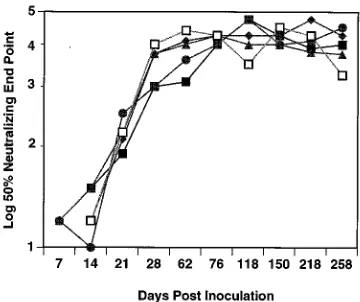
Development of neutralizing-antibody responses. In con-trast to the low level of replication of the recombinant virus, there was rapid development of a humoral immune response in all of the animals. By 14 days postinoculation, all five monkeys monitored serially had neutralizing antibodies directed against the infecting strain, SIV/17E-Cl (Fig. 1). These titers rose rapidly, peaked at 5 months postinfection, and remained con-stant throughout the following year. Neutralization assays were routinely done with a T-cell line (CEMx174); however, when the assays were done with primary rhesus macrophages, an equivalent level of neutralizing antibody was measured. The production of high levels of neutralizing antibodies in monkeys infected with SIV/17E-Cl is in direct contrast to the absence or low levels of neutralizing antibodies made in response to in-fection with the parental strain SIVmac239. Thus, envelope
sequences in SIV/17E-Cl that confer macrophage tropism also appear to be responsible for eliciting a strong neutralizing-antibody response in vivo.
To determine the specificity of the neutralizing antibodies, neutralizations were done at monthly intervals with the follow-ing virus strains: (i) SIVmac239, (ii) the uncloned parental
strain SIV/17E-Br, (iii) an additional recombinant clone (SIV/ 17E-Fr) that contains the entire env gene from SIV/17E-Br, and (iv) a heterologous primary isolate of sooty mangabey monkey origin, SIV/DeltaB670 (Table 3). Within the first 150 days postinoculation, sera obtained from the SIV/17E-Cl-in-fected monkeys, with the exception of that from monkey L652, neutralized only SIV/17E-Cl. In contrast, samples obtained later neutralized not only SIV/17E-Cl but also SIV/17E-Br, the recombinant virus SIV/17E-Fr, and the heterologous isolate SIV/DeltaB670 (Table 3). The neutralizing titer against the heterologous primary isolate, SIV/DeltaB670, was never as high as those observed with the homologous clone, SIV/17E-Cl (Table 4). To examine whether the homologous neutralizing-antibody response to SIV/DeltaB670 in macaques was lower than that observed for SIV/17E-Cl, the neutralizing titers to SIV/DeltaB670 in the sera from monkey C344, a long-term survivor of SIV/DeltaB670 infection (one of only two survivors
of over 400 animals inoculated with this virus), were deter-mined. Indeed, the titers were 2 log units less than those for SIV/17E-Cl, indicating that there appears to be a weaker neu-tralizing-antibody response to SIV/DeltaB670 in infected ani-mals (Table 3). Nevertheless, the pattern of neutralizing activ-ity was highly consistent in multiple samples taken throughout the infection in all of the animals tested and with respect to repeated determinations performed on the same sample. The rapid appearance of broadly reactive neutralizing antibodies in monkey L652 remains unexplained (Table 4). For the majority of these animals, a broadening of neutralizing antibody from type-specific to group-specific activity was observed 6 to 8 months after infection (Table 4).
The ability of antiserum raised against SIV/17E-Cl late in the infection to neutralize SIV/DeltaB670 was surprising given the genetic diversity of the two viruses. Unlike SIV/17E-Cl, which consists of a single genotype of SIVmac251 lineage, SIV/
DeltaB670 is a primary isolate consisting of a swarm of genetic variants cultured from the lymph node of a rhesus monkey infected with SIV from a sooty mangabey monkey. Sequence comparison of the V1 hypervariable regions of env found within the SIV/DeltaB670 genetic quasispecies identified a di-vergence of less than 10%, whereas a similar comparison of these sequences with SIV/17E-Cl showed an average diver-gence of over 25%. To place this difference in perspective, a comparison of SIV/17E-Cl V1 sequences with those obtained from the Los Alamos database for SIVmac251, SIVmne, SIV/
STM, and HIV-2ROD showed divergences of 9.5, 16, 30, and 40%, respectively. Thus, persistent infection with the attenu-ated molecular clone SIV/17E-Cl induces over time neutraliz-ing antibodies against a genetically diverse strain of SIV.
Avidity and conformation dependence of antibody responses to SIV/17E-Cl. In light of the kinetics of the appearance of neutralizing-antibody responses in the macaques infected with SIV/17E-Cl, we sought to examine the evolution of SIV enve-FIG. 1. Neutralizing-antibody titers (log10) that increased over time in the
[image:4.612.87.267.70.221.2]sera from monkeys inoculated with SIV/17E-Cl. Neutralization assays were done against SIV/17E-Cl virus. The results are the averages of at least two indepen-dent assays, as described in Methods and Materials, and are for monkeys L235 (■), L238 (F), M118 (h), L652 (}), and L471 (å).
TABLE 3. Determination of neutralizing-antibody specificity
Monkey Neutralization titer
a
SIV/17E-Cl SIV/17E-Br SIV/17-Fr SIV/DeltaB670
L235 4.3 0.7 0.7 1.0
L238 4.8 0 2.3 1.0
L471 4.5 0 2.3 1.0
L652 3.4 1.8 3.0 1.75
M118 4.5 0.9 2.6 1.2
C344b 4.2 NTc NT 2.1
a
50% neutralization end point (log10) for sera taken 219 days postinoculation. b
Long-term survivor of SIV/DeltaB670 infection.
c
NT, not tested.
TABLE 4. SIV/DeltaB670-specific neutralization titers
Day postinfection
SIV/DeltaB670-specific neutralization titerawith monkey:
L235 L238 L471 L652 M118
90 NTb 0 NT 1.25 NT
118 NT 0 0 1.25 0
150 0 0 0 1.0 0
195 0.7 0 0.7 1.75 0
219 1.0 1.0 1.0 1.75 1.2
244 1.0 1.0 NT NT NT
a
50% neutralization end point (log10). b
NT, not tested.
on November 9, 2019 by guest
http://jvi.asm.org/
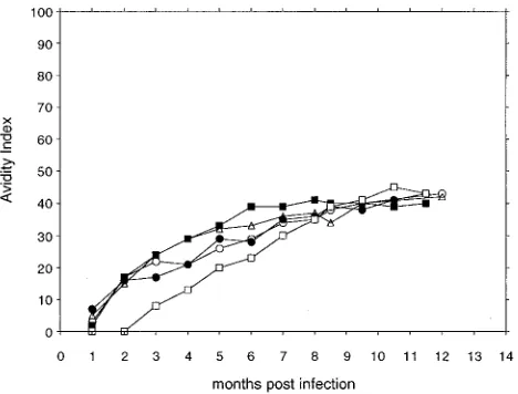
[image:4.612.315.556.84.182.2] [image:4.612.316.555.620.711.2]lope glycoprotein-specific antibody responses with respect to their avidity and conformation dependence, properties which have been frequently used as immune correlates of protection. For these purposes, native viral glycoproteins from purified SIV preparations were anchored onto ConA in microtiter plates for the respective immunoassays. Serum antibody avid-ities were determined by measuring the stabilavid-ities of antibody-antigen complexes to a urea wash, as described previously (11). In this functional assay, avidity index values of below 30% are considered to indicate low-avidity antibodies, values of be-tween 30% and 50% are considered to indicate intermediate-avidity antibodies, and values of above 50% are considered to indicate high-avidity antibodies.
The data in Fig. 2 demonstrate that the envelope glycopro-tein-specific antibody responses in the five SIV/17E-Cl-in-fected monkeys increase in avidity over the first 7 months postinfection and apparently level off at an intermediate avid-ity of about 40% thereafter. The relatively slow evolution of antibody avidity in the glycoprotein-specific antibody responses indicates an ongoing maturation of humoral immune re-sponses to this chronic infection during at least the first 7 months postinfection. At this time, the antibody avidity ap-pears to reach a maximum level that is maintained even after a subsequent virus challenge at 8 months postinfection. The slow increase in the avidity of antibodies to the SIV envelope glycoprotein is in distinct contrast to the relatively rapid in-crease in avidities observed during other viral infections (33), perhaps indicating an important escape mechanism by which SIV eludes immune responses soon after infection.
Previous studies have indicated a correlation between anti-body populations specific for conformation-dependent epi-topes and the presence of broadly neutralizing antibody reac-tivity to HIV-1 (9, 25, 36) and SIV (21). Therefore, the second antibody parameter measured was the conformation depen-dence of the envelope glycoprotein-specific antibody responses in the longitudinal panel of serum samples from the SIV/17E-Cl-infected macaques. For this assay, the reactivity of the se-rum antibodies at a standard dilution was measured in parallel against ConA-anchored native viral glycoprotein and against
denatured viral glycoprotein produced by reductive carboxy-methylation of protein cysteine sulfhydryl groups. Thus, this assay compares the reactivity of serum antibodies with a native viral glycoprotein complex with that with envelope glycopro-teins in which all disulfide bonds have been irreversibly re-duced to alter protein tertiary structure, without deliberate denaturation of the envelope protein secondary structure.
The data in Fig. 3 indicate that the serum antibodies present at all time points tested resulted in conformation index values ranging from about 1.5 to 8, indicating that the serum antibod-ies were on average two- to eightfold more reactive with the native viral glycoprotein substrate than with the denatured viral glycoprotein antigen. It is interesting that the highest levels of antibody conformation dependence were observed at 2 months postinfection (average index55) and that the con-formation index in general decreased at later time points to an average of 2.5 at the time of virus challenge. The conformation index remained unchanged for the several months postchal-lenge. The predominance of conformation-dependent anti-body produced in SIV/17E-Cl-infected monkeys is reminiscent of the case for the serum antibodies produced in HIV-1-fected individuals (25, 36) and in monkeys experimentally in-fected with various strains of SIV (21). To our knowledge, however, this is the first time that this parameter has been reported for serum samples taken over the course of an exper-imental infection in monkeys, thereby revealing the early dom-inance of conformation-dependent antibodies and an unex-pected decline in the relative abundance of conformation-dependent antibodies with the progression of the persistent infection.
Lack of heterogeneity of env sequences in macaques. To examine whether the broadening of the neutralizing-antibody response and increased avidity of the antibody for the viral glycoprotein were due to the development of heterogeneity in
env protein composition, the genetic diversity of gp120
se-quences in virus present in two of the animals that were chal-FIG. 2. Avidity of serum antibodies for SIV envelope glycoproteins. The
[image:5.612.316.553.71.253.2]antibody avidity index was determined by measuring in a ConA-ELISA the resistance of test serum antibody-envelope glycoprotein immune complexes to disruption by treatment with 8 M urea, as described in Materials and Methods. Longitudinal serum samples were obtained from the following five macaques at various times after infection with SIV/17E-Cl: L235 (■), L238 (F), M118 (h), L652 (}), and L471 (å).
FIG. 3. Conformation dependence of serum antibodies to SIV envelope gly-coproteins. The conformation dependence of envelope glycoprotein-specific se-rum antibodies elicited by infection with SIV/17E-Cl was determined by mea-suring in a ConA-ELISA the antibody reactivities against native SIV envelope glycoproteins and against denatured envelope glycoproteins prepared by reduc-tion and carboxymethylareduc-tion of protein sulfhydryl groups, as described in Mate-rials and Methods. Longitudinal serum samples were obtained from the follow-ing five macaques at various times after infection with SIV/17E-Cl: L235 (■), L238 (F), M118 (h), L652 (}), and L471 (å). The conformation index is defined as the ratio of serum reactivity with native envelope glycoprotein to that with denatured envelope glycoprotein.
on November 9, 2019 by guest
http://jvi.asm.org/
[image:5.612.60.294.73.251.2]lenged (L235 and L238) was examined at 9 days after inocu-lation in the peripheral blood mononuclear cells and at 244 days after inoculation in mononuclear cells from both lymph nodes and the peripheral blood. The entire gp120 sequence (nucleotides 6342 to 8222 [30]) was directly amplified from mononuclear cell DNA. Sequences specific for the external domain of gp41 (nucleotides 7682 to 10195 [30]) were similarly amplified, cloned, and sequenced. Very few nucleotide changes were detected in all clones examined. A comparison of early and late samples failed to reveal any significant difference in either the number of mutations or location of these changes, a finding that suggests that the observed changes were likely due to errors introduced by Taq polymerase. This low level of virus heterogeneity is in direct contrast to infection with the lymphocyte-tropic molecular clone SIVmac239, which rapidly
generates genetic diversity within this same time frame (3, 4). This finding is probably due to the low level of replication observed in the SIV/17E-Cl-infected animals relative to that observed for SIVmac239. These data suggest that the
develop-ment of cross-reactive neutralizing antibodies was likely not due to the generation and selection of antigenic variants or to the presence of a widely divergent virus swarm in these mon-keys but rather may reflect the recognition of less-immuno-genic viral epitopes by the immune system as these responses mature.
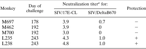
Challenge of SIV/17E-Cl-infected macaques with a heterol-ogous strain of SIV. To determine whether the broadened neutralization response that appeared after 7 months postin-fection in the SIV/17E-Cl-infected macaques could confer broad-spectrum protective immunity against a heterologous virus challenge, infected macaques were inoculated intrave-nously with 50 animal infectious doses of rhesus monkey-grown SIV/DeltaB670 either prior to 7 months (three ma-caques) or after 8 months (two mama-caques) postinfection (Table 5). Lymph node biopsies were performed immediately prior to and 14 days after challenge and, together with PBMC-derived cells, were evaluated for SIV/DeltaB670-specific sequences by PCR. SIV-specific sequences, when present, were further an-alyzed by sequence analysis of cloned PCR products containing the V1 hypervariable domain of gp120 (27). No evidence of infection with SIV/DeltaB670 could be identified in either these samples or PBMC samples obtained during the next 6 months from either of the monkeys (L235 and L238) which had broadly reactive neutralizing responses. In contrast, all three macaques (M697, M462, and M700) challenged earlier in in-fection had SIV/DeltaB670-specific sequences in both PBMC and lymph node-derived mononuclear cells; only one of these animals (monkey M697) had detectable neutralizing antibody to SIV/DeltaB670 at challenge. Multiple SIV/DeltaB670 vari-ants found within the challenge inoculum were identified in monkey M700; this animal died of an AIDS-like disease 95 days postchallenge. The major variant found within the
chal-lenge inoculum was identified in monkey M462, whereas only a minor variant (representing less than 10% of the variants) could be identified in monkey M697. A similar analysis of naive monkeys inoculated with SIV/DeltaB670 has shown that the major variant present in the initial inoculum consistently emerges as the dominant form following in vivo infection (un-published results). Taken together, these data suggest that the clonal emergence of a minor form of the inoculum observed in monkey M697 may represent a variant that has escaped the low level of neutralizing antibody detected in the serum of this animal at challenge. Both M462 and M697 have lived beyond 280 days postchallenge.
Passive protection of naive rhesus macaques. To further define the role of humoral immunity in the protective re-sponses induced by SIV/17E-Cl infection, sera from the two monkeys that were protected from SIV/DeltaB670 challenge were collected, heat inactivated, and evaluated for the ability to passively protect naive recipient monkeys (Table 6). Four monkeys were infused with 10 ml of SIV/17E-Cl antiserum per kg of body weight, with two control monkeys receiving similar amounts of normal monkey serum, and challenged intrave-nously with 50 animal infectious doses of SIV/DeltaB670 7 h later (29). Infection was assessed by PCR of PBMC DNA at weekly intervals postinoculation. As expected, SIV env-specific antibody titers in the monkeys that received immune serum at challenge were four to eight times lower in the recipients than in the donor serum. Both control monkeys were virus PCR positive at 7 days postinoculation. SIV/DeltaB670 infection was evident at 7 days postinoculation in one monkey (N549) that received immune serum; a second animal (M954) was PCR positive at 14 days. The remaining two monkeys remained PCR negative for up to 6 months postinoculation and demon-strated no other signs of virus infection (e.g., seroconversion or clinical disease).
DISCUSSION
The infection of rhesus macaques with an attenuated mac-rophage-tropic clone of SIV induced immune responses that protected against challenge with a highly virulent primary iso-late that differs by 16% overall in gp120 nucleotide sequence and by 25% in the sequence of the V1 hypervariable region of gp120. The induction of this protective response occurred 6 to 8 months postinfection in concert with a plateau in the avidity of gp120-specific antibody responses and a broadening of neu-tralizing-antibody responses. These responses were mainly di-rected toward conformation-dependent epitopes, because binding of antibody could be abrogated by denaturation of viral glycoproteins.
The ability of sera from vaccinated macaques to passively protect two of four naive recipients lends further support to the role of antibody in protection and demonstrates that sterilizing immunity can be induced by an attenuated SIV vaccine. How-TABLE 5. Challenge of SIV/17E-Cl-infected macaques with
a heterologous strain of SIV
Monkey Day of challenge
Neutralization titerafor:
Protection SIV/17E-CL SIV/DeltaB670
M697 178 3.9 0.7 2
M462 192 3.9 0 2
M700 192 3.0 0 2
L235 243 4.3 1.0 1
L238 243 4.8 1.0 1
[image:6.612.315.555.92.168.2]a50% neutralization end point (log 10).
TABLE 6. Glycoprotein-specific antibody titer at challenge in monkeys infused with SIV/17E-Cl antiserum
Monkey gp140 titera PCR postchallenge
N261 1,600 2
N549 800 1
N644 1,600 2
M954 1,600 1
Donor serum 6,400
aReciprocal dilution.
on November 9, 2019 by guest
http://jvi.asm.org/
[image:6.612.58.297.93.174.2]ever, these findings do not address whether these humoral immune responses are the sole mediators of protection. In-deed, the failure to protect all of the monkeys in the passive protection experiment argues that additional responses (e.g., cellular immune responses) may also be employed by an at-tenuated virus vaccine to act in concert with antibody to achieve optimal protection.
High-avidity and conformation-dependent antibodies have been implicated as important correlates for protective anti-body responses to HIV-1 infection (9, 25), suggesting that these properties could be useful in defining the evolution of protective antibody responses in the macaques immunized with the live SIV/17E-Cl. Both the avidity and conformation depen-dence data reveal a maturation of SIV envelope-specific re-sponses that requires at least 6 months postinfection. The requirement for this lengthy maturation process may explain the reported failure of attenuated SIV vaccines at 4 months postinoculation compared with the broad protection observed at 2 years postinoculation with the same attenuated SIV strain (14). In this regard, it has also been reported that broadly protective immune responses to equine infectious anemia virus evolve after 6 to 8 months postinfection in naturally and ex-perimentally infected horses (23). Taken together, these ob-servations suggest that protective immunity to lentiviruses re-quires a complex evolution of immune responses produced during a relatively lengthy maturation process in infected an-imals. The stringent requirements for the production of pro-tective immune responses to lentivirus infections may explain the failure of less-rigorous immunizations with inactivated whole virus and subunit vaccines against SIV (24).
Since previous studies have correlated broadly neutralizing antibody responses to SIV and HIV-1 with conformation-de-pendent antibodies, we sought to examine the conformation dependence of envelope-specific antibody responses in rela-tion to the appearance of type- and group-specific neutralizing antibodies and the establishment of protective immunity in macaques immunized with the SIV/17E-Cl attenuated mac-rophage-tropic strain. The results of these studies revealed a predominance of conformation-dependent serum antibodies at all time points tested postinoculation but also revealed an unexpected decline in the level of conformation-dependent antibodies as the infection progressed. This latter observation suggests that antibodies to linear determinants of the viral envelope glycoprotein become increasingly prevalent over the first 6 months postinfection, when their level appears to reach a stable plateau. One cannot from these data make a definitive correlation between the evolution of protective antibodies to SIV and the generation of linear or conformation-dependent antibody populations. Thus, it will be important to determine further the relationship between type- and group-specific neu-tralizing activities and linear and conformation-dependent an-tibodies to the SIV envelope. Taken together, the antibody avidity and conformation dependence assays indicate a pro-gressive maturation of antibody responses to the attenuated macrophage-tropic SIV vaccine strain for about 7 months postinfection, suggesting that this may be the minimum time required for the evolution of a protective immune response to SIV.
The utilization of viral proteins conferring macrophage tro-pism may offer a selective advantage over the lymphocyte-tropic analogs that have dominated vaccine development thus far. Macrophage-tropic strains of HIV-1 appear to be trans-mitted most efficiently and to establish initial infections in humans (37–40) and are therefore the initial targets for vac-cine-induced protection. The selective presentation of viral proteins by macrophages may also be preferred for the
induc-tion of protective immunity at the mucosal surfaces involved in sexual transmission of HIV-1. The studies described in this report suggest that macrophage-tropic strains may also be bet-ter inducers of protective responses. These responses may be attributable to the different routes of antigen processing and presentation that occur in macrophages. Viral antigens in in-fected macrophages would be expected to present antigen in the context of both major histocompatibility complex classes I and II. The vigorous early induction of neutralizing-antibody responses observed in SIV/17E-Cl-infected monkeys is in strik-ing contrast to the absence of these responses in monkeys infected with SIVmac239, which differs by only seven amino
acid residues in gp120 (1). This may be due to the initial widespread infection of antigen-presenting cells in the periph-eral blood because of the macrophage tropism of the virus. In addition, the macrophage-tropic virus may cause early infec-tion of a broader range of cells in vivo, particularly antigen-presenting cells in the skin, gut, and other lymphoid organs. This could increase the immune response detected in the se-rum but may also elicit important protective local immune responses in the animal. The low level of replication of the macrophage-tropic virus in the CD4 lymphocytes may also contribute to the vigorous immune response. These cells are required for their helper function in the induction of immune responses, and infection by SIV or HIV has been shown to impair these helper functions. The investigation of the pattern of early infection and replication of macrophage-tropic viruses in vivo will be an important aspect in understanding the de-velopment of the protective immune response observed in this study.
This study examined the immune response and the clinical outcome of a strongly macrophage-tropic recombinant of SIV, and results similar to those of Lohman et al. (16) were ob-tained. It provides evidence that this virus elicits a vigorous immune response and that with time this immune response broadens to protect the animal against challenge with a heter-ologous, virulent strain of virus. This macrophage-tropic mo-lecular clone differs from its lymphocyte-tropic parent only in the env gene; thus, these changes have altered the virus cell tropism and host immune response. These results provide a model system in which to examine the development of mac-rophage-tropic-based attenuated and subunit vaccine strate-gies.
ACKNOWLEDGMENTS
We thank Maryann Brooks for the preparation of the manuscript and Calvin Lanclos, Eva Pecunia, Nedra LaCour, Lynn Fresh, Terese Theriot, Gail Plauche, and Kim Burke for technical support.
This work was supported by grants from the NIH to Janice E. Clements (PO1 NS32208), to Michael Murphey-Corb (UO1 AI28243 and RR00164), and to Ronald C. Montelaro (UO1 AI28243).
REFERENCES
1. Anderson, M. G., D. Hauer, D. P. Sharma, S. V. Joag, O. Narayan, M. C. Zink, and J. E. Clements.1993. Analysis of envelope changes acquired by SIVmac239 during neuroadaption in rhesus macaques. Virology 195:616–626. 2. Banapour, B., M. L. Marthas, R. A. Ramos, B. L. Lohman, R. E. Unger, M. B. Gardner, N. C. Pedersen, and P. A. Luciw.1991. Identification of viral determinants of macrophage tropism for simian immunodeficiency virus SIVmac. J. Virol. 65:5798–5805.
3. Burns, D. P. W., C. Collignon, and R. C. Desrosiers. 1993. Simian immuno-deficiency virus mutants resistant to serum neutralization arise during per-sistent infection of rhesus monkeys. J. Virol. 67:4104–4113.
4. Burns, D. P. W., and R. C. Desrosiers. 1991. Selection of genetic variants of simian immunodeficiency virus in persistently infected rhesus monkeys. J. Virol. 65:1843–1854.
5. Cichutek, K., S. Norley, R. Linde, W. Kreuz, M. Gahr, J. Lower, G. von-Wangenheim, and R. Kurth. 1991. Lack of HIV-1 V3 region sequence
on November 9, 2019 by guest
http://jvi.asm.org/
diversity in two haemophiliac patients infected with a putative biologic clone of HIV-1. AIDS 5:1185–1187.
6. Crestfield, A. M., S. Moore, and W. H. Stein. 1963. The preparation and enzymatic hydrolysis of reduced and S-carboxymethylated proteins. J. Biol. Chem. 238:622–626.
7. Daniel, M. D., F. Kirchhoff, C. Czajak, P. K. Sehgal, and R. C. Desrosiers. 1992. Protective effects of a live attenuated SIV vaccine with a deletion in the nef gene. Science 258:1938–1941.
8. Desrosiers, R. C., A. Hansen-Moosa, N. W. King, M. D. Daniel, and D. J. Ringler.1991. Macrophage-tropic variants of SIV are associated with spe-cific AIDS-related lesions but are not essential for the development of AIDS. Am. J. Pathol. 139:29–35.
9. Devash, Y., T. Calvelli, D. Wood, K. Reagan, and A. Rubinstein. 1990. Vertical transmission of HIV-1 is correlated with the absence of high affinity/ avidity maternal antibodies to the gp120 principal neutralizing domain. Proc. Natl. Acad. Sci. USA 87:3445–3449.
10. Hedman, K., J. Heitala, A. Jartikainen-Sorri, K. Raiha, and M. Pietilainen. 1989. Maturation of immunoglobulin G avidity after rubella vaccination studied by ELISA and haemolysis typing. J. Med. Virol. 27:293–298. 11. Hedman, K., and S. A. Rousseau. 1989. Measurement of avidity of specific
IgG for verification of recent primary rubella. J. Med. Virol. 27:288–292. 12. Johnson, P. R., T. E. Hamm, S. Goldstein, S. Kitov, and V. M. Hirsch. 1991.
The genetic fate of molecularly cloned simian immunodeficiency virus in experimentally infected macaques. Virology 185:217–228.
13. Karber, G. 1931. Beitrag zue kollektiven Behandlung pharmakologischer Reihenversuche. Arch. Exp. Pathol. Pharmakol. 162:480–487.
14. Kestler, H., T. Kodama, D. Ringler, M. Marthas, N. Pedersen, A. Lackner, D. Regier, P. Sehgal, M. Daniel, N. King, and R. Desrosiers.1990. Induction of AIDS in rhesus monkeys by molecularly cloned simian immunodeficiency virus. Science 248:1109–1112.
15. Kestler, H. W., D. J. Ringler, K. Mori, D. L. Panicali, P. D. Sehgal, M. D. Daniel, and R. C. Desrosiers.1991. Importance of the nef gene for mainte-nance of high virus loads and for development of AIDS. Cell 65:651–662. 16. Lohman, B. L., M. B. McChesney, C. J. Miller, E. McGowan, S. M. Joye,
K. K. A. Van Rompay, E. Reay, L. Antipa, N. C. Pedersen, and M. L. Marthas.1994. A partially attenuated simian immunodeficiency virus in-duces host immunity that correlates with resistance to pathogenic virus challenge. J. Virol. 68:7021–7029.
17. Luciw, P. A., K. E. S. Shaw, R. E. Unger, V. Planelles, M. W. Stout, J. E. Lackner, E. Pratt-Lowe, N. J. Leung, B. Banapour, and M. L. Marthas.1992. Genetic and biological comparisons of pathogenic and nonpathogenic mo-lecular clones of simian immunodeficiency virus (SIVmac). AIDS Res. Hum. Retroviruses 8:395.
18. Marthas, M. L., B. Banapour, S. Sutjipto, M. E. Siegel, and P. A. Marx. 1989. Rhesus macaques inoculated with molecularly cloned simian immu-nodeficiency virus. J. Med. Primatol. 18:311–319.
19. Marthas, M. L., C. J. Miller, S. Sutjipto, J. Higgins, J. Torter, B. L. Lohman, R. E. Unger, R. A. Ramos, H. Kiyono, J. R. McGhree, P. A. Marx, and N. C. Pedersen.1992. Efficacy of live-attenuated and whole inactivated simian immunodeficiency virus vaccines against vaginal challenge with virulent SIV. J. Med. Primatol. 21:99–107.
20. Martin, L. N., M. Murphey-Corb, K. F. Soike, B. Davison-Fairburn, and G. B. Baskin. 1993. Effects of initiation of 39-azido, 39-deoxythymidine (zidovudine) treatment at different times after infection of rhesus monkeys with simian immunodeficiency virus. J. Infect. Dis. 168:825–835. 21. McBride, B. W., G. Corthals, E. Rud, K. Kent, S. Webster, N. Cook, and
M. P. Cranage.1993. Comparison of serum antibody reactivities to a con-formational and to linear antigenic sites in the external envelope glycopro-tein of SIV induced by infection and vaccination. J. Gen. Virol. 74:1033–1041. 22. McNearney, T., Z. Hornickova, R. Markham, A. Birdwell, M. Arens, A. Saah, and L. Ratner.1992. Relationship of human immunodeficiency virus type 1
sequence heterogeneity to stage of disease. Proc. Natl. Acad. Sci. USA 89: 10247–10251.
23. Montelaro, R., J. Ball, and C. Issel. 1992. Equine retroviruses, p. 257–360. In J. Levy (ed.), The Retroviridae, vol. 2. Plenum Publishing Co., New York. 24. Montelaro, R., and D. Bolognesi. Retrovirus vaccines. In J. Levy (ed.), The
Retroviridae, vol. 4, in press. Plenum Publishing Co., New York. 25. Moore, J. P., and D. D. Ho. 1993. Antibodies to discontinuous or
conforma-tionally sensitive epitopes on the gp120 glycoprotein of human immunode-ficiency virus type 1 are highly prevalent in sera of infected humans. J. Virol. 67:863–875.
26. Mori, K., D. J. Ringler, T. Kodama, and R. C. Desrosiers. 1992. Complex determinants of macrophage tropism in env of simian immunodeficiency virus. J. Virol. 66:2067–2075.
27. Murphey-Corb, M., L. N. Martin, B. Davison-Fairburn, R. C. Montelaro, M. Miller, M. West, S. Ohkawa, G. B. Baskin, J. Y. Zhang, S. B. Putney, A. C. Allison, and D. A. Eppstein.1989. A formalin-inactivated whole SIV vaccine confers protection in macaques. Science 246:1293–1297.
28. Pang, S., Y. Schlesinger, E. S. Daar, T. Moudgil, D. D. Ho, and I. S. Chen. 1992. Rapid generation of sequence variation during primary HIV-1 infec-tion. AIDS 6:453–460.
29. Putkonen, P., R. Thorstensson, L. Ghavamzadeh, J. Albert, K. Hild, P. Biberfeld, and E. Norrby.1991. Prevention of HIV-2 and SIVsm infection by passive immunization in cynomolgus monkeys. Nature (London) 352:436–438. 30. Regier, D. A., and R. C. Desrosiers. 1990. The complete nucleotide sequence of a pathogenic molecular clone of simian immunodeficiency virus. AIDS Res. Hum. Retroviruses 6:1221.
31. Robinson, J. E., D. Holton, J. Lui, H. McMurdo, A. Murciano, and R. Ghod. 1990. A novel enzyme-linked immunosorbent assay (ELISA) for detection of antibodies to HIV-1 envelope glycoproteins based on immobilization of viral glycoproteins in microtiter wells coated with concanavalin A. J. Immunol. Methods 132:63–71.
32. Saiki, R. K., D. H. Gelfand, S. Stoffel, S. J. Scharf, R. Higuchi, G. T. Horn, K. B. Mullis, and H. A. Erlich.1988. Primer-directed enzymatic amplifica-tion of DNA with a thermostable DNA polymerase. Science 239:487–491. 33. Salmi, A. 1991. Antibody affinity and protection in virus infections. Curr.
Opin. Immunol. 3:503–506.
34. Sharma, D. P., M. G. Anderson, M. C. Zink, R. J. Adams, A. D. Donnenberg, J. E. Clements, and O. Narayan.1992. Pathogenesis of acute infection in rhesus macaques with a lymphocyte-tropic strain of SIVmac. J. Infect. Dis. 166:738–746.
35. Sharma, D. P., M. C. Zink, M. G. Anderson, R. Adams, J. E. Clements, S. V. Joag, and O. Narayan.1992. Derivation of neurotropic simian immunode-ficiency virus from exclusively lymphocyte-tropic parental virus: pathogenesis of infection in macaques. J. Virol. 66:3550–3556.
36. Steimer, K. S., C. J. Scandella, P. V. Skiles, and N. L. Haigwood. 1991. Neutralization of divergent HIV-1 isolates by conformation-dependent hu-man antibodies to Gp120. Science 254:105–108.
37. Wolfs, T. F. W., G. Zwart, M. Bakker, and J. Goudsmit. 1992. HIV-1 genomic RNA diversification following sexual and parenteral virus transmis-sion. Virology 189:103–110.
38. Wolinsky, S. M., C. N. Wike, B. T. M. Korber, C. Hutto, W. P. Parks, L. A. Rosenblum, K. J. Kunstman, M. R. Furtado, and J. L. Munoz.1992. Selec-tive transmission of human immunodeficiency virus type-1 variants from mothers to infants. Science 255:1134–1137.
39. Zhang, L. Q., P. MacKenzie, A. Cleland, E. C. Holmes, A. J. L. Brown, and P. Simmonds.1993. Selection of specific sequences in the external envelope protein of human immunodeficiency virus type 1 upon primary infection. J. Virol. 67:3345–3356.
40. Zhu, T., H. Mo, N. Wang, D. S. Nam, Y. Cao, R. A. Koup, and D. D. Ho. 1993. Genotypic and phenotypic characterization of HIV-1 in patients with pri-mary infection. Science 261:1179–1181.