0022-538X/96/$04.0010
Copyrightq1996, American Society for Microbiology
Chimeric Simian/Human Immunodeficiency Virus That Causes
Progressive Loss of CD4
1T Cells and AIDS
in Pig-Tailed Macaques
SANJAY V. JOAG,1ZHUANG LI,1LARRY FORESMAN,2EDWARD B. STEPHENS,1LING-JUN ZHAO,1
ISTVAN ADANY,1DAVID M. PINSON,3HAROLD M. MCCLURE,4ANDO. NARAYAN1*
Marion Merrell Dow Laboratory of Viral Pathogenesis and Department of Microbiology,1Laboratory Animal
Resources2and Department of Pathology and Laboratory Medicine,3University of Kansas
Medical Center, Kansas City, Kansas 66160-7420, and Yerkes Regional Primate Center, Emory University, Atlanta, Georgia 30322
Received 30 August 1995/Accepted 31 January 1996
By animal-to-animal passage of simian/human immunodeficiency virus (SHIV) in pig-tailed macaques, we have developed a macaque model of human immunodeficiency virus type 1 (HIV-1) disease in humans. Passaging was begun with a chimeric virus containing theenvgene of HIV-1 HXBc2 and thegagandpolgenes of simian immunodeficiency virus SIVmac239. SHIV was passaged serially in cohorts of two macaques each, using bone marrow-to-bone marrow transfers at 5, 5, and 16 weeks for passages 2, 3, and 4, respectively. The fifth passage was done by using cell-free virus isolated from cerebrospinal fluid of a passage 4 macaque. The virus became more virulent with each passage. Virus replication was restricted in all three animals in passages 1 and 2 but not in five of the six animals in passages 3, 4, and 5. In these animals, intense virus replication in the lymphoid tissues resulted in almost total elimination of CD41T cells within weeks of inoculation, and three of these animals developed AIDS in less than 1 year. The more uniform virus-host interaction initiated by the cell-free virus in the passage 5 animals contrasted with a more variable pattern of disease initiated by infectious bone marrow cells during earlier passages. The virulent cell-free SHIV can now be used to screen the efficacy of vaccines directed against the envelope of HIV-1.
Human immunodeficiency virus type 1 (HIV-1) can infect nonhuman primates such as chimpanzees but does not cause disease in these animals (16). This is a major impediment to understanding the mechanisms of pathogenesis of HIV-1 in-fection and has also retarded efforts to evaluate the efficacy of anti-HIV drugs and vaccines. At present, simian immunodefi-ciency virus SIVmac infection in macaques is the best-charac-terized model of HIV-1 disease (3). HIV-2 infection in ba-boons represents another useful model (1). SIVmac and HIV-2 are closely related viruses, but both have genetic, anti-genic, and structural features in their env genes that clearly distinguish them from HIV-1.
Chimeric simian/human immunodeficiency viruses (SHIVs) that bear the envelope of HIV-1 and are infectious in ma-caques potentially offer a solution to this problem. However, SHIVs have so far proven avirulent in macaques (2, 9, 13, 15, 17). Since the bone marrow (BM) of SIVmac-infected ma-caques has high virus burdens (7) and since specific pathogenic viral genotypes can be selected by serial passage of infectious BM in animals (14), we hypothesized that similar passaging of SHIV-infected BM cells would also result in selection of pathogenic genotypes of this virus. We report here on deriva-tion of a pathogenic SHIV by using this procedure and the initial characterization of the disease process caused by this virus.
MATERIALS AND METHODS
Passage in macaques.SHIV, obtained as described below, was used to begin passaging in young (7 to 19 months old) rhesus (Macaca mulatta) and pig-tailed (Macaca nemestrina) macaques (Fig. 1). Macaques were housed in American Association for Laboratory Animal Care-accredited facilities and were sedated with ketamine for all procedures. Virus was inoculated either in the BM or by the intravenous route. BM inocula immediately reach the systemic circulation and are thus equivalent to intravenous inocula. The rationale for BM inoculation was to guarantee exposure of precursor cells in the BM to virus in the inoculum.
As shown in Fig. 1, we inoculated 10450% tissue culture infective doses (TCID50) of SHIV into the BM of rhesus macaque 8A. Four weeks later, heparinized BM was obtained from this animal, mononuclear cells were purified over Ficoll-Hypaque gradients, and 5 3107cells were inoculated into the femoral BM of two pig-tailed macaques, PLc and PRc. Five weeks later, BM was aspirated from PLc and PRc, pooled, and inoculated into the BM of macaques PPc and PQc. Sixteen weeks later, bone marrow and splenic biopsies were obtained from macaques PPc and PQc, and mixtures of splenocytes and BM cells from both animals were pooled and inoculated into two new pig-tailed macaques, PFb and PNb. Cell-free virus isolated from macaque PNb was used for inocu-lation of macaques 15A and 15B in passage 5.
Viruses. (i) Original SHIV.We obtained a SHIV DNA encoding the env, tat,
rev, and vpu genes of HIV-1 HXBc2 on a background of SIVmac239 (9) from
Joseph Sodroski, Harvard University. Viral DNA was transfected into CEMx174 cells to produce a virus stock, which had a titer of 13104TCID50/ml. The virus stock was stored at2808C. This virus was inoculated into macaque 8A, whose BM cells were then used to begin passaging as described above.
(ii) Virulent SHIV.Virus was obtained by cocultivation of cerebrospinal fluid cells from macaque PNb with indicator C8166 cells. Supernatant fluid was inoc-ulated into phytohemagglutinin (PHA)-activated peripheral blood mononuclear cells (PBMC) from a healthy pig-tailed macaque. The culture developed syncy-tial cytopathic effects (CPE) by day 5, and supernatant fluid was harvested on day 10. This fluid was clarified at 5,0003g for 10 min, and 1-ml aliquots were stored
in liquid nitrogen. This virus was inoculated intravenously into two pig-tailed macaques, 15A and 15B, for passage 5.
(iii) HIV-1 HXBc3.HIV-1 HXBc3 was obtained from P. Nara (National Cancer Institute), and a stock was prepared in CEMx174 cells. The virus stock was stored at2808C.
Cell cultures.Human T-cell lines CEMx174 and C8166 were used as indicators to measure virus infectivity. Cells were cultured at a concentration of 106
/ml in RPMI supplemented with 10 mM N-2-hydroxyethylpiperazine-N9 -2-ethanesul-fonic acid (HEPES) buffer (pH 7.3), 50mg of gentamicin per ml, 531025
M
* Corresponding author. Mailing address: Marion Merrell Dow Laboratory of Viral Pathogenesis, Department of Microbiology, Uni-versity of Kansas Medical Center, 3901 Rainbow Blvd., Kansas City, KS 66160-7420. Phone: (913) 588-5575. Fax: (913) 588-5599. Elec-tronic mail address: [email protected].
3189
on November 9, 2019 by guest
http://jvi.asm.org/
2-mercaptoethanol, 2 mM glutamine, and 10% fetal bovine serum (supplement-ed RPMI).
Processing of blood samples.Heparinized blood obtained from the femoral vein was centrifuged to separate plasma and buffy coats. Plasma infectivity was assayed immediately, and aliquots of the plasma were stored at2808C. PBMC were separated from buffy coat cells by centrifugation through Ficoll-Hypaque density gradients.
Assessment of virus infectivity.The frequency of infectious cells was measured by inoculation of serial 10-fold dilutions of PBMC or other cells onto indicator cells in 24-well tissue culture plates, which were then observed for development of syncytial CPE during a 7-day period. Then 100ml from each well was trans-ferred to another plate, fresh indicator cells were added, and the plates were observed for a further 7 days. Routinely, 106cells from each animal were used for estimation of infectious cell frequency. Results were expressed as the number of infectious cells per 106PBMC or other cells.
For quantitation of infectivity in plasma or culture supernatant fluids, serial 10-fold dilutions of test material were inoculated into freshly prepared CEMx174 or C8166 cell cultures. The cells were cultivated in 24-well tissue culture plates and examined for CPE for 7 days, after which 100ml from each well was
transferred to another plate, fresh indicator cells were added, and the plates were observed for CPE for a further 7 days.
Virus recovery from PBMC cultured with PHA and IL-2.A total of 23106 PBMC were cultured in medium containing 1mg of PHA-P (Wellcome) for 2 days. Cultures were then rinsed once, the cell pellets were resuspended in medium with 100 U of recombinant human interleukin-2 (IL-2; Cetus) per ml, cultured for 5 days, and then centrifuged again, and the cell-free supernatant fluids were assayed for virus infectivity. In some cases, the supernatant fluids were also assayed for SIV p27 concentration. Virus infectivity was assayed by development of CPE in C8166 cells as described above, and SIV p27 was measured by using a capture enzyme-linked immunosorbent assay (ELISA) kit from Coulter Laboratories (Hialeah, Fla.).
Virus recovery from CD81-depleted PBMC.A total of 23106
PBMC were incubated with a monoclonal antibody to CD8 (Dako-T8; Dako) for 1 h at 48C and washed once, and sheep anti-mouse immunoglobulin G-coated paramag-netic beads (Dynal Laboratories) were added. The beads were removed with a magnet. This procedure routinely resulted in removal of 75 to 80% of the CD81 T cells (6). Aliquots (23106cells each) of CD81-depleted and control
unde-pleted PBMC were cultured in PHA medium for 2 days and in IL-2 medium for 5 days as described above, after which the supernatant fluids were assayed for virus infectivity and SIVmac p27 content.
SIV p27 assay.Plasma and culture fluids were assayed for p27 by using a capture ELISA kit from Coulter. For each assay, a standard curve was prepared according to manufacturer’s instructions, and p27 concentrations were calculated from the optical density at 450 nm, using linear regression analysis. The sensi-tivity of detection of p27 in these assays varied between 20 and 30 pg/ml.
[image:2.612.93.258.72.243.2]PCR amplification of DNA.PCR was performed as follows. For amplification of SIVmac gp120 sequences, the oligonucleotide primers used in the first round were 59-GGCTAAGGCTAATACATCTTCTGCATC-39(sense) and 59-ACCC AAGAACCCTAGCACAAAGACCCC-39(antisense), which are complemen-tary to bases 6565 to 6591 and 8179 and 8205, respectively, of SIVmac239 (12). One microgram of genomic DNA was used in the PCR, which was performed with a mixture containing 2.0 mM MgCl2, 200mM each of the four deoxynucle-otide triphosphates, 100 pM each oligonucledeoxynucle-otide primer, and 2.5 U of Taq polymerase (Perkin-Elmer Cetus, Norwalk, Conn.). The template was denatured at 928C for 3 min, and PCR amplification was performed with an automated DNA Thermal Cycler (Perkin-Elmer Cetus) for 35 cycles, using the following profile: denaturation at 928C for 1 min, annealing at 558C for 1 min, and primer extension at 728C for 3 min. Amplification was completed by incubation of the PCR mixture for 10 min at 728C. One microliter of the PCR product was used in a nested PCR performed as described above. For the second round of amplifi-cation, the nested primers were 59-GTAAGTATGGGATGTCTTGGGAATC AG-39(sense) and 59-GACCCCTCTTTTATTTCTTGAGGTGCC-39(antisense), FIG. 1. Passage of SHIV in macaques.
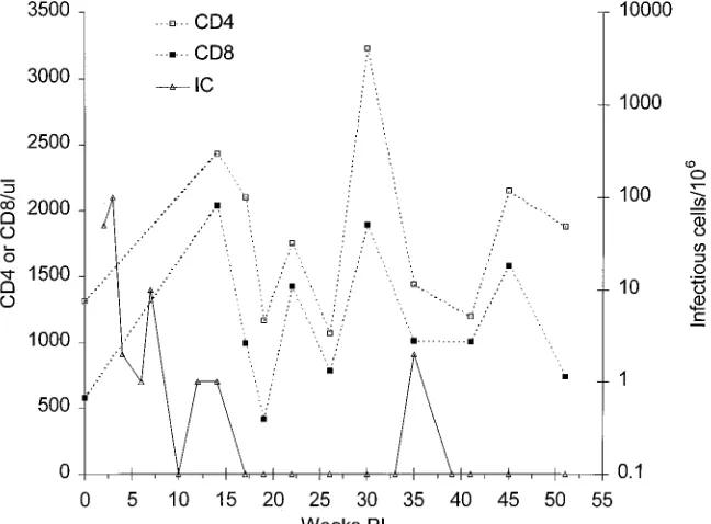
FIG. 2. Frequencies of infectious cells and of CD41and CD81cells in peripheral blood from rhesus macaque 8A. The infectious cell (IC) frequency was measured by inoculation of serial dilutions of PBMC into indicator cells and examining for CPE as described in the text; CD41and CD81frequencies were assessed by FACS analysis.
on November 9, 2019 by guest
http://jvi.asm.org/
[image:2.612.144.468.458.697.2]which are complementary to bases 6598 to 6624 and 8158 to 8184, respectively, of the SIVmac239 genome.
For amplification of HIV-1 HXBc2 gp120 sequences, the oligonucleotide primers used in the first round were 59-CAAAGAAAAATAGACAGGTTAAT TGAT-39(sense) and 59-AGTGCTTCCTGCTGCTCCCAAGAACCC-39 (anti-sense), which are complementary to bases 6166 to 6192 and 7810 to 7784, respectively, of the HXBc2 genome (4). For the second round of amplification, the nested primers were 59-GACTAATAGAAAGAGCAGAAGACAGTGG CA-39(sense) and 59-GAACAAAGCTCCTATTCCCACTGCTCT-39(antisense), which are complementary to bases 6194 to 6223 and 7780 to 7754, respectively, of the HXBc2 genome. The conditions for amplification of the HXBc2 gp120 sequence were the same as described for the SIVmac sequence. A 10-ml aliquot from each of the second-round amplifications was run on a 0.8% agarose gel, and bands were visualized by staining with ethidium bromide. To confirm the spec-ificity of the PCR products, the DNA in the gel was transferred onto nitrocel-lulose and hybridized with32P-labeled gp120 probes generated from the gp120 gene of either SIVmac239 or HIV-1 HXBc2 (data not shown).
PCR/infected cell assay.The assay was performed as described previously (6). In the first round, oligonucleotide primers used were 59-GATGGGCGTGAGA AACTCCGTCTT-39and 59-CCTCCTCTGCCGCTAGATGGTGCTGTTG-39, which are complementary to bases 1052 to 1075 and 1423 to 1450, respectively, of the SIVmac239 gag gene (12). To standardize cell numbers, the fourth exon of
b-actin was amplified with oligonucleotide primers 59-TCATGTTTGAGACCT
TCAACACCCCAG-39and 59-CCAGGAAGGAAGGCTGGAAGAGTGCC-39
(noncoding), complementary to the published sequence (11). The PCR amplifi-cation conditions were as specified above. To increase the sensitivity of the reaction, 1ml of the first PCR product was used as a template for a second amplification performed under the same conditions. The nested SIV primers used were 59-GTTGAAGCATGTAGTATGGGCAGC-39and 59-GCCTCAGG GCAGCGGAACCGCTCA-39, which are complementary to bases 1142 to 1165 and 1356 to 1382, respectively, of SIVmac239. The nestedb-actin primers used were 59-CCCCAGCCATGTACGTTGCTATCC-39and 59-GCCTCAGGGCAG CGGAACCGCTCA-39. Following the second round of amplification, a 10-ml aliquot was removed and run on a 1.5% agarose gel, and bands were visualized by staining with ethidium bromide.
Fluorescence-activated cell sorting (FACS) analysis.Cells were reacted with a monoclonal antibody to CD4 (SIM.4; NIH AIDS Research and Reagents Re-pository) or CD8 (Dako-T8; Dako). After washing, the cells were stained with fluorescein isothiocyanate-conjugated goat anti-mouse immunoglobulin G (Dako), fixed in 1% buffered formalin, and analyzed on an EPICS fluorescence-activated cell counter (Coulter).
Neutralizing antibody assays.We performed the assays as described previ-ously (5). Briefly, serial doubling dilutions of plasma in RPMI were prepared in duplicate or quadruplicate in 96-well plates, 10 to 20 TCID50of the virus was added to each well, plates were incubated for 1 h at 378C, and 104
indicator cells were added to each well. Plates were observed for CPE 7 days later, wells were scored individually, and the 50% neutralization endpoint was calculated by the Ka¨rber method (8).
Raji cell cocultures.Raji is a human B-lymphocyte cell line in which simian
retrovirus type D produces a characteristic CPE (10). Raji cells were maintained at a concentration of 53105
/ml in RPMI containing 5% fetal bovine serum. PBMC or other mononuclear cells (23106
) were cultured with 23106 Raji cells in a 25-cm2
culture flask and examined for CPE for 7 days; 500ml was then transferred to another flask, fresh Raji cells were added, and the flasks were observed for CPE for a further 7 days.
RESULTS
A summary of the passage history is outlined in Fig. 1.
Passage 1.We inoculated 104TCID
50of the chimeric SHIV
into the BM of rhesus macaque 8A. Virus was readily isolated by cocultivation with indicator cells during the first 12 weeks postinoculation (p.i.). Rhesus macaque 8A had infectious cell
frequencies of 10 to 100/106 early in infection, but the
fre-quency declined to,1/106PBMC by week 14 p.i., after which
[image:3.612.59.557.84.291.2]infectious cells were detected only once, at week 35 p.i. (Fig.
[image:3.612.317.557.517.707.2]FIG. 3. Frequencies of infectious cells (IC) and CD41cells in peripheral blood from passage 2 animals. For details, see the legend to Fig. 2.
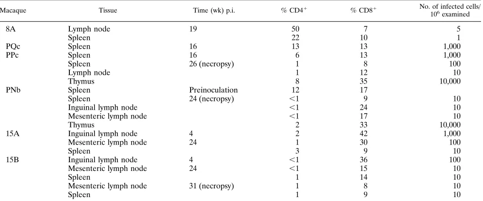
TABLE 1. CD41, CD81, and infectious cell frequencies in lymphoid tissuesa
Macaque Tissue Time (wk) p.i. % CD41 % CD81 No. of infected cells/
106 examined
8A Lymph node 19 50 7 5
Spleen 22 10 1
PQc Spleen 16 13 13 1,000
PPc Spleen 16 6 13 1,000
Spleen 26 (necropsy) 1 8 100
Lymph node 1 12 10
Thymus 8 35 10,000
PNb Spleen Preinoculation 12 17
Spleen 24 (necropsy) ,1 9 10
Inguinal lymph node ,1 24 10
Mesenteric lymph node ,1 17 10
Thymus 2 33 10,000
15A Inguinal lymph node 4 2 42 1,000
Mesenteric lymph node 24 1 30 100
Spleen 3 9 10
15B Inguinal lymph node 4 ,1 36 100
Mesenteric lymph node 24 ,1 15 10
Spleen 1 14 10
Mesenteric lymph node 31 (necropsy) 1 8 10
Spleen 1 9 10
aCD41and CD81frequencies were assessed by FACS analysis. The frequency of infectious cells was assayed by coculture with C8166 cells and examination for
development of CPE.
on November 9, 2019 by guest
http://jvi.asm.org/
2). Plasma infectivity was not detected at any time. Plasma had 325 pg of SIV p27 per ml at week 2, but p27 was not present in later samples up to week 51. Supernatant fluids from PBMC cultured in PHA and then IL-2 yielded p27 at week 2 but not at week 4, 6, or 19 and did not have infectivity. PBMC at weeks
4 and 6 were depleted of CD81cells and cultured in PHA and
IL-2. On both occasions, the supernatant fluids lacked p27 and infectivity.
CD41 and CD81 T-cell frequencies in PBMC remained
normal in this animal during a follow-up period of 1 year p.i.,
and the CD4/CD8 ratio remained .1 at all times (Fig. 2).
Biopsies of mesenteric lymph nodes and spleen were obtained surgically at 19 weeks p.i. Separated lymph node and spleen cells had low infectious cell frequencies (Table 1), although these levels were higher than in PBMC obtained at the same
time (,1/106; Fig. 2). FACS analysis of separated lymph node
cells showed normal CD41 and CD81 frequencies. PBMC
obtained at the same time had 28% CD41 cells and 10%
CD81cells. The remaining mononuclear cells from the lymph
nodes included, among others, B cells, macrophages, and den-dritic cells.
Passage 2.BM cells from rhesus macaque 8A were inocu-lated into two pig-tailed macaques, PLc and PRc, for passage
2, each animal receiving an inoculum of 53 107cells. Like
rhesus macaque 8A, these two animals had infectious cell
fre-quencies of 10 to 100/106early in infection, but the frequency
declined to,1/106PBMC by week 17 p.i., after which
infec-tious cells were not detected (Fig. 3). Plasma infectivity was not detected at any time. SIV p27 was detected in plasma from PRc only at week 3, while plasma from PLc was always nega-tive. Supernatant fluids from PBMC cultured in PHA and then IL-2 yielded infectivity and p27 up to week 23 (PLc) or week 27
(PRc). However, CD81-depleted and control undepleted
PBMC obtained at weeks 31 and 36 from both animals did not yield either infectivity or p27.
In contrast to rhesus macaque 8A, a steep decline in CD41
T-cell counts was seen in both PLc and PRc during the first 3
to 4 weeks p.i. (Fig. 3). Thereafter the counts increased
grad-ually in both animals but remained at 1,000 to 1,500/ml, lower
than the preinoculation CD41 counts. These animals have
[image:4.612.144.469.70.314.2]maintained control of virus replication for more than a year and have remained healthy.
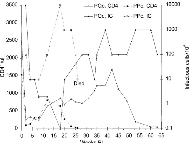
FIG. 4. Frequencies of infectious cells (IC) and CD41cells in peripheral blood from passage 3 animals. For details, see the legend to Fig. 2.
FIG. 5. PCR amplification of DNA from infected macaques. (A) Total cel-lular genomic DNA was extracted from splenic tissue of pig-tailed macaques PPc and PQc and used as a template in a nested PCR to amplify HIV-1 HXBc2 and SIVmac239 gp120 sequences. Lanes 1 to 3, amplification using oligonucleotide primers specific for HIV-1 HXBc2 gp120 sequences from plasmid containing the SHIV genome (lane 1), PPc spleen DNA (lane 2), and PQc spleen DNA (lane 3). Lanes 4 to 6, amplification using oligonucleotide primers specific for SIV-mac239 gp120 sequences from a plasmid containing the complete SIVSIV-mac239 genome DNA (lane 4), PPc spleen DNA (lane 5), and PQc spleen DNA (lane 6). Amplifications of gp120 sequences from uninfected animals, using either set of oligonucleotide primers, were negative (data not shown). (B) A PCR/infected cell assay was used to determine the number of virus-infected cells in PBMC from macaque PPc. Cell suspensions were diluted to 107
, 106 , 105
, 104 , 103
, and 102
cells per ml, and cells were then lysed and digested. Two rounds of PCR amplification were used to detect SIV gag and, as an internal control,b-actin sequences. After the second of amplification, theb-actin yielded a 393-bp band, whereas the amplified SIV gag sequence yielded a 240-bp band. Stds., standards.
on November 9, 2019 by guest
http://jvi.asm.org/
[image:4.612.316.554.437.586.2]Passage 3.Five weeks after PLc and PRc were inoculated, 2 ml of BM was aspirated from both animals, pooled, and inoc-ulated into the BM of two new pig-tailed macaques, PPc and PQc, for passage 3. PQc had a high infectious cell frequency
(104/106PBMC) at week 2, but the frequency declined rapidly
to 10/106by week 4 and further to,1/106at week 16 (Fig. 4).
Thereafter, the infectious cell frequency showed a steady
in-crease, to 102to 103/106PBMC (Fig. 4). Plasma infectivity was
never detected in plasma from PQc, and p27 was present only
at week 2. As in the passage 2 animals, CD41T-cell counts fell
rapidly in PBMC from PQc during the first 2 weeks p.i. and remained low until week 8, after which a steady increase was
seen till week 42, after which the CD41 count fell again to
50/ml by week 63.
The course of infection was more acute in PPc than in PQc. The infectious cell frequency in PBMC from PPc was initially
low (102/106 PBMC) but decreased only slightly, to 10/106
during weeks 4 to 8, after which it increased again to a peak of
104/106PBMC at week 18 and remained at 102to 103/106until
the animal was euthanized (Fig. 4). Plasma infectivity was detected only at week 23. However, p27 was detected in plasma at week 2 and again at weeks 24 and 26. As in PQc and the
passage 2 animals, a steep decline in CD41T-cell counts was
seen in PPc during the first 2 weeks p.i. (Fig. 4). Thereafter, the
CD41count increased till week 12 and then fell to only 28/ml
at week 26, when the animal was euthanized in a moribund condition.
Biopsies of the spleen were obtained surgically from both PPc and PQc at 16 weeks p.i. The infectious cell frequency in
separated spleen cells from both animals was 103/106 cells.
Spleen cells from PPc had 6% CD41 cells and 13% CD81
cells, while PQc spleen cells had 13% CD41cells and 13%
CD81T cells. The remaining mononuclear spleen cells were
mainly B cells, along with some macrophages and dendritic cells. Histological examination showed massive loss of cells in the T-cell-rich areas around the germinal centers. This loss was noted histologically as a depletion of the mantle zone lympho-cytes, with relative sparing of the follicular centers.
The identity of virus isolated from PPc and PQc was con-firmed by amplification of a 1.6-kb DNA fragment from PBMC DNA, using PCR with oligonucleotide primers specific for sequences encoding the gp120 of HIV-1 HXBc2 but not SIV-mac239 (Fig. 5A). Examination of splenocytes from a portion of spleen biopsied from macaques PPc and PQc at 16 weeks
p.i. showed 103infectious cells per 106splenocytes, similar to
the burden in PBMC at that time. Use of a PCR/infected cell
assay confirmed the infected cell frequency of 103to 104cells
per 106splenocytes (Fig. 5B).
Macaque PPc developed progressive anemia and loss of weight and was euthanized at 26 weeks p.i. in a moribund
FIG. 6. (A) Photomicrograph of macaque PPc thymus, showing Hassall’s corpuscles and severe depletion of cortical thymocytes. Hematoxylin-and-eosin stain; bar5253mm. (B) Photomicrograph of a histologically normal thymus, showing clear distinction of cortex and medulla. Hematoxylin-and-eosin stain; bar5253mm. (C) Photomicrograph of PPc spleen biopsy at 16 weeks p.i. Hematoxylin-and-eosin stain; bar563mm. (D) Photomicrograph of PPc stomach. Mycotic hyphae are visible in the glands. The lamina propria contains lymphocytes, plasma cells, and a few macrophages. Hematoxylin-and-eosin stain; bar563mm.
on November 9, 2019 by guest
http://jvi.asm.org/
condition. The CD41count had fallen rapidly during the last 4
weeks, from 164/ml at week 22 to 64/ml at week 23 to 37 at week
25, and was only 28/ml at necropsy. This animal had whipworm
(Trichuris sp.) infestation, which responded to treatment, al-though some parasites were still present at necropsy. Histolog-ical examination of the thymus revealed that the cortex and medulla were indistinguishable, but Hassall’s corpuscles were visible in all areas, suggesting severe depletion of cortical thy-mocytes (Fig. 6A). Examination of the lymph nodes showed that cortices were very cellular with small lymphocytes, with minimal or no follicular development. Mantle zones and ger-minal centers were absent. The spleen contained numerous follicles, but germinal centers were poorly developed and man-tle zones had reduced cellularity (Fig. 6C). The stomach con-tained microscopic mixed mononuclear cell inflammation in the lamina propria and mycotic hyphae in surface debris and in glands (Fig. 6D). The hyphae were morphologically consistent with Candida sp.
Separated lymph node, spleen, and thymus cells were used for FACS analysis and assessed for infectious cell frequency.
Few CD41cells were seen in the thymus, lymph nodes, and
spleen (Table 1). Infectious cell frequencies in the thymus were 10 to 100 times higher than those in the lymph nodes and spleen. Interestingly, both the infectious cell frequency and the
percentage of CD41cells in the spleen were lower at necropsy
than in the biopsy sample obtained at week 16 (Table 1). The B-cell and macrophage frequencies in the lymphoid tissues are not shown.
PBMC, splenocytes, lymph node cells, and BM cells from macaque PPc were cultured with Raji cells for 2 weeks to detect the presence of simian type D retroviruses. None of these cultures developed CPE. All animals were serologically negative for simian retroviruses.
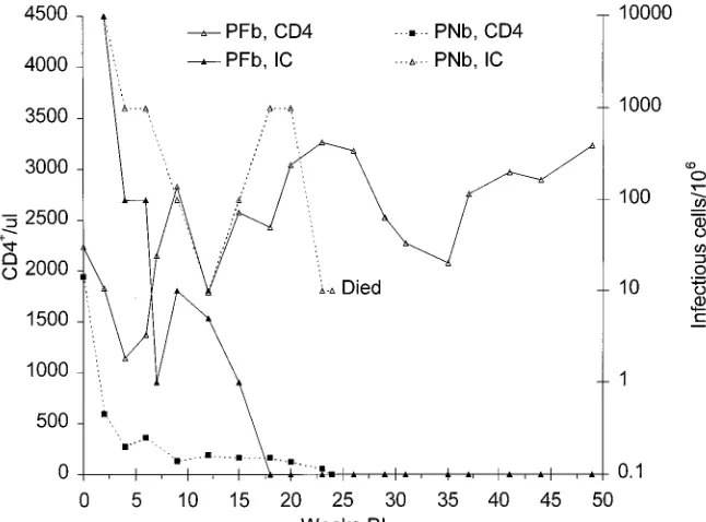
Passage 4.BM cells and spleen cells obtained at 16 weeks p.i. from macaques PPc and PQc were pooled and inoculated into two new pig-tailed macaques, PFb and PNb. Infectious
cell frequencies in PBMC from PFb were 104/106at week 2, but
the virus burden decreased rapidly and by week 18 had fallen
to,1/106, a level that was maintained for the next 30 weeks.
Plasma infectivity was not present at any time, while p27 was detected only at week 2. Supernatant fluids from PBMC cul-tured in PHA and then IL-2 yielded infectivity and p27 during
the first 8 weeks, but neither CD81-depleted nor control
un-depleted PBMC obtained at weeks 18 and 23 yielded either
infectivity or p27. CD41 T-cell counts in blood from PFb
declined during weeks 2 to 4 p.i. and then increased steadily to higher than the preinoculation level (Fig. 6).
In contrast to data from PFb, infectious cell frequencies in PNb remained high during the entire infection (Fig. 6). Plasma infectivity and SIVmac p27 were detected during the first 4
weeks and also during weeks 18 and 20. CD41T-cell counts in
PBMC from PNb fell dramatically during the first 2 weeks and remained low thereafter (Fig. 7). The animal developed severe progressive anemia and weakness and was euthanized at week 24 p.i. to prevent suffering. At necropsy, the blood hemoglobin concentration was only 5.2 g/dl, having declined from 11.4 g/dl at week 12 to 8.9 g/dl at week 20 to 6.2 g/dl at week 23. The
CD41T-cell count in peripheral blood of PNb was less than
10/ml at necropsy and had fallen steadily during the final 6
weeks, from 161/ml at week 18 to 122/ml at week 20 to 56/ml at
week 23 (Fig. 6).
Histopathological lesions in the thymus, lymph nodes, spleen, and stomach of PNb were almost identical to those in PPc. In the thymus, severe depletion of cortical thymocytes was seen and the cortex and medulla were indistinguishable, al-though Hassall’s corpuscles were visible in all areas. Lymph node cortices were very cellular, but with minimal or no fol-licular development, and germinal centers were absent. In the spleen, germinal centers were poorly developed and mantle zones had reduced cellularity in comparison with the normal histological appearance of a biopsy of the organ obtained prior to inoculation (Fig. 8). Histological examination of the gastro-intestinal tract showed severe necrotizing mycotic gastritis, similar to that seen in PPc.
[image:6.612.144.467.72.311.2]Separated lymph node, spleen, and thymus cells were used for FACS analysis and assessed for infectious cell frequency.
FIG. 7. Frequencies of infectious cells (IC) and CD41cells in peripheral blood from passage 4 animals. For details, see the legend to Fig. 2.
on November 9, 2019 by guest
http://jvi.asm.org/
The lymph nodes and spleen had,1% CD41cells, while only
1% of thymocytes were CD41(Table 1). Infectious cell
fre-quencies in the thymus were 1,000 times higher (104/106) than
in the lymph nodes and spleen (10/106).
PBMC, thymocytes, splenocytes, and lymph node cells from macaque PNb were cultured with Raji cells for 3 weeks (pas-saged weekly) to detect the presence of simian type D retro-viruses. None of these cultures developed CPE.
Passage 5.The cell-free virus stock prepared from macaque
PNb had an infectivity titer of 104TCID
50/ml in C8166 cells.
Two 3-year-old pig-tailed macaques, 15A and 15B, were inoc-ulated intravenously with 0.5 ml of this material. The animals had a uniform response, developing high infectious cell
fre-quencies in PBMC and rapid CD41loss (Fig. 9). Plasma
in-fectivity and p27 were detected in both animals (not shown).
The loss of CD41cells was more rapid in both animals than in
PNb. CD41counts in blood from 15A varied between 20/ml
and 60/ml during weeks 4 to 31. In contrast, CD41counts in
blood from 15B were consistently,15/ml from weeks 2 to 31.
Biopsies of inguinal lymph nodes were obtained surgically at 4 weeks, while biopsies of the spleen and mesenteric lymph nodes were obtained at 24 weeks. FACS analysis of separated
lymph node and spleen cells showed less than 2% CD41cells,
establishing that the loss of CD41cells had occurred in
lym-phoid tissues as well as in peripheral blood and that this loss had taken place as early as 4 weeks p.i. The lymph nodes and spleen had minimal or no follicular development, and germinal centers were absent.
Macaque 15B developed uncontrollable diarrhea at week 27 and became progressively cachexic; the animal was euthanized at 31 weeks in an emaciated condition. Histological changes at necropsy in the thymus, lymph nodes, and spleen were similar to those seen in PPc and PNb. Both lungs showed areas of patchy interstitial pneumonia characteristic of Pneumocystis
FIG. 8. Photomicrographs of macaque PNb spleen obtained preinoculation (A) or at necropsy (B). Note the diffuse loss of cells from the mantle zone and marginal zone and the absence of germinal centers in panel B. Hematoxylin-and-eosin stain; bars563mm.
on November 9, 2019 by guest
http://jvi.asm.org/
carinii infection, the presence of which was confirmed by
Go-mori methenamine silver staining.
Neutralizing antibodies.All animals in passages 1 to 4 de-veloped neutralizing antibodies to SHIV and to HIV-1 (Table 2). Antibodies appeared as early as 8 weeks p.i., and neutral-izing antibody titers remained steady for a prolonged period. Plasmas from all animals neutralized not only the original SHIV virus but also SHIVs isolated from macaques PPc and PQc with similar efficiencies (data not shown). Preinoculation plasmas from all animals did not neutralize any of these vi-ruses. None of these plasmas neutralized SIVmac239.
DISCUSSION
We have established a nonhuman primate model of severe
CD41 T-cell loss and AIDS in pig-tailed macaques infected
with animal-passaged SHIV. This is the first demonstration of a lentivirus bearing the HIV-1 envelope that is pathogenic in nonhuman primates. Disease became evident in pig-tailed ma-caques during passage 3, using BM cell inocula. Passage 5, with
cell-free virus, resulted in rapid and severe CD41T-cell loss.
Three animals (PPc, PNb, and 15B) developed AIDS and were
euthanized in a moribund condition with low CD41 T-cell
counts (,50/ml) and opportunistic infections (candidiasis in
PPc and PNb and P. carinii pneumonia in 15B). Two other
animals, PQc and 15A, had CD41T-cell counts of,50/ml at 63
and 33 weeks, respectively, p.i. We have shown that cell-free virus caused disease in macaque 15B. Thus, this virus, which was isolated from infected animals and cultured in vitro, re-produced the disease upon reinoculation into another animal, from which the virus was reisolated.
All animals in this study were screened for antibodies to simian type D retrovirus, and PBMC, lymph node, spleen, BM, and thymus cells from the animals that developed AIDS were cocultured with Raji cells. The absence of antibodies to type D retrovirus and the absence of CPE in Raji cell cocultures rule out the possibility that the disease was due to simian type D retroviruses. Moreover, pathogenic type D retroviruses infect
and deplete T and B cells; in contrast, animals in this study
developed loss of only CD41T cells.
Inoculation of the original SHIV in rhesus macaque 8A resulted in sustained infection but not in disease. The animal
did not develop loss of CD41 T cells, and virus burdens in
peripheral blood and lymphoid tissues subsided within the first 12 weeks to barely detectable levels. This pattern is similar to the results obtained by other investigators, who inoculated rhesus or cynomolgus macaques with various strains of SHIV (2, 9, 13, 15, 17). During passage 2, the pig-tailed macaques PRc and PLc, which were inoculated with BM cells from
rhe-sus macaque 8A, developed initial loss of CD41T cells, but the
cell count recovered to approximately 50% of preinoculation values. This correlated with effective control over virus repli-cation, since by week 25 virus could not be obtained from
PBMC, even after depletion of CD81T cells. The end result
[image:8.612.146.469.70.315.2]was similar to that observed in rhesus macaque 8A. Evidence of increased virulence of SHIV emerged during passage 3, when macaques PPc and PQc were inoculated with BM cells from PRc and PLc. These animals developed a more severe
FIG. 9. Frequencies of infectious cells (IC) and CD41cells in peripheral blood from passage 5 animals. For details, see the legend to Fig. 2.
TABLE 2. Neutralizing antibody titers in SHIV-infected animalsa
Macaque
Neutralizing antibody titer vs:
SHIV HIV-1 HXBc3
8A 64 130
PLc 80 64
PRc 130 320
PPc 64 160
PQc 64 80
PFb 64 130
PNb 32 32
a
Serial dilutions of heat-inactivated plasma from infected animals were incu-bated with 10 to 20 TCID50of stock virus for 1 h, after which indicator cells were added, and plates were observed for development of CPE. Plasma samples from all animals neutralized viruses isolated from PPc and PQc as effectively as the original SHIV. In all macaques, titers against SIVmac239 were,10.
on November 9, 2019 by guest
http://jvi.asm.org/
[image:8.612.315.555.588.685.2]initial loss of CD41T cells. In both animals, the cell count recovered, but in PPc, the cell count dropped precipitously again, ending in development of severe AIDS at 25 weeks. This
second phase of CD41 T-cell loss correlated with increased
virus burdens. PQc developed a similar pattern of disease extended over a longer time period. This animal was still healthy at 63 weeks, but as can be seen in Fig. 4, the prognosis appears to be poor. The virus appeared more virulent in
pas-sage 4. The CD41T-cell count in one of the two macaques,
PNb, fell rapidly during the first 2 weeks and never recovered. Macaque PFb became infected, but this animal appeared to
control the infection and did not develop CD41 T-cell loss,
possibly reflecting variation among individual animals or the virus selected from its BM inoculum.
In contrast to the original cell-free SHIV, which caused no serious pathological effects during two successive passages in macaques, the cell-free virus prepared from passage 4 was extremely virulent. Both animals that were inoculated intrave-nously with this stock, 15A and 15B, developed severe
sus-tained loss of CD41T cells and high virus burdens in the blood
and lymphoid tissues. One of the two, 15B, developed diar-rhea, cachexia, and P. carinii pneumonia at week 29 and had to be euthanized. The other, 15A, was still healthy at 33 weeks, but the prognosis appears to be poor.
Analysis of tissues from the three animals that developed
AIDS showed that the CD41T-cell count seen in the PBMC
was reflected by similar changes in the lymphoid tissues, and this correlated with the intensity of virus replication. The loss
of CD41cells in blood represented a global loss rather than
new regional localization patterns. It is interesting that in both PPc and PNb, thymocytes had a higher frequency of infectious cells than mononuclear cells from the secondary lymphoid tissues (lymph nodes and spleen). Whether this reflects se-quential pattern of infection by this virus in primary and sec-ondary lymphoid tissues or a more prolonged infection in the thymus is not clear. Thymocytes were not obtained from 15B, since this animal had severe thymic atrophy.
A characteristic feature of human AIDS is an increase in virus burden during the terminal phase. This increase was seen in two of the macaques (PPc and PNb) that developed terminal disease. The upsurge in virus replication was manifested as increases in plasma infectivity, plasma p27, and infectious cell frequencies in PBMC.
The development of neutralizing antibodies to HIV-1 in the inoculated animals is similar to observations of HIV infection in humans. Similar to the infection in humans, the presence of neutralizing antibodies did not have any apparent protective effects. Whether such antibodies would have protective effects in passive immunization experiments will be tested in the fu-ture. A number of potential vaccines against HIV-1 infections rely on the induction of a neutralizing antibody response. The SHIV/pig-tailed macaque system thus offers a model in which such vaccines can be evaluated.
In summary, our studies have shown that an SHIV whose tat,
rev, vpu, and env genes were derived from a laboratory strain of
HIV-1 increased in virulence upon passage of infectious BM cells from animal to animal, with the final derivation of a cell-free stock containing pathogenic virus. Increasing virus burdens in PBMC and lymphoid tissues, development of
plasma viremia, progressive loss of CD41 T cells from the
blood, lymph nodes, and spleen, severe infection in the thymus, and development of AIDS have all been documented. The pathogenic SHIV model utilizing a cell-free virus stock, de-scribed here for the first time, now provides the means for studying pathogenesis of HIV-1 infection in so far as the viral envelope contributes to the disease. It also provides a disease-causing challenge virus for evaluating the efficacy of HIV-1 envelope vaccines and anti-HIV-1 drugs.
ACKNOWLEDGMENTS
We thank J. Sodroski for providing SHIV DNA and Peter Nara for providing HIV-1 HXBc3.
This work was supported by grants AI-29382, NS-32203, and RR-06753 from the National Institutes of Health.
REFERENCES
1. Barnett, S. W., K. K. Murthy, B. G. Herndier, and J. A. Levy. 1994. An AIDS-like condition induced in baboons by HIV-2. Science 266:642–646. 2. Cheng-Mayer, C., J. A. Levy, and P. A. Luciw. 1994. Infection of rhesus
macaques with T-cell-line tropic and macrophage-tropic SHIVs. Int. Conf. AIDS 10:61.
3. Desrosiers, R. C. 1990. The simian immunodeficiency viruses. Annu. Rev. Immunol. 8:557–578.
4. Fisher, A. G., E. Collalti, L. Ratner, R. C. Gallo, and F. Wong-Staal. 1985. A molecular clone of HTLV-III with biological activity. Nature (London) 316:262–265.
5. Joag, S. V., M. G. Anderson, J. E. Clements, M. F. McEntee, D. P. Sharma, R. J. Adams, and O. Narayan.1993. Antigenic variation of molecularly cloned SIVmac239 during persistent infection in a rhesus macaque. Virology 195:406–412.
6. Joag, S. V., E. B. Stephens, R. J. Adams, L. Foresman, and O. Narayan. 1994. Pathogenesis of SIVmacinfection in Chinese and Indian rhesus macaques: effects of splenectomy on virus burden. Virology 200:436–446.
7. Joag, S. V., E. B. Stephens, and O. Narayan. 1995. Lentiviruses, p. 1977– 1996. In B. N. Fields, D. M. Knipe, and P. M. Howley (ed.), Virology. Raven Press, New York.
8. Lennette, E. H. 1969. General principles underlying laboratory diagnosis of viral and rickettsial infections, p. 1–65. In E. H. Lennette and N. J. Schmidt (ed.), Diagnostic procedures for viral and rickettsial infections. American Public Health Association, New York.
9. Li, J., C. I. Lord, W. Haseltine, N. L. Letvin, and J. Sodroski. 1992. Infection of cynomolgus monkeys with a chimeric HIV-1/SIVmacX virus that ex-presses the HIV-1 envelope glycoproteins. J. Acquired Immune Defic. Syndr. 5:639–646.
10. Maul, D. H., C. P. Zaiss, M. R. MacKenzie, S. M. Shiigi, P. A. Marx, and M. B. Gardner.1988. Simian retrovirus D serogroup 1 has a broad cellular tropism for lymphoid and nonlymphoid cells. J. Virol. 62:1768–1773. 11. Nakajima-Iijima, S., H. Hamada, P. Reddy, and T. Kakunaga. 1985.
Molec-ular structure of the human cytoplasmic beta-actin gene: interspecies ho-mology of sequences in the intron. Proc. Natl. Acad. Sci. USA 82:6133–6137. 12. Regier, D. A., and R. C. Desrosiers. 1990. The complete nucleotide sequence of a pathogenic molecular clone of simian immunodeficiency virus. AIDS Res. Hum. Retroviruses 6:1221–1231.
13. Sakuragi, S., R. Shibata, R. Mukai, T. Komatsu, M. Fukasawa, H. Sakai, J. Sakuragi, M. Kawamura, K. Ibuki, M. Hayami, et al.1992. Infection of macaque monkeys with a chimeric human and simian immunodeficiency virus. J. Gen. Virol. 73:2983–2987.
14. Sharma, D. P., M. C. Zink, M. Anderson, R. Adams, J. E. Clements, S. V. Joag, and O. Narayan.1992. Derivation of neurotropic simian immunode-ficiency virus from exclusively lymphocytotropic parental virus: pathogenesis of infection in macaques. J. Virol. 66:3550–3556.
15. Shibata, R., and A. Adachi. 1992. SIV/HIV recombinants and their use in studying biological properties. AIDS Res. Hum. Retroviruses 8:403–409. 16. Spertzel, R. O. 1989. Animal models of human immunodeficiency virus
infection. Public Health Service Animal Models Committee. Antiviral Res. 12:223–230.
17. Yamamoto, H., T. Igarashi, Y. Ami, T. Komatsu, R. Shibata, T. Kuwata, A. Adachi, H. Shida, M. Tanaka, and M. Hayami.1994. HIV-1 env specific killer lymphocytes in macaque monkeys infected with SIVmac HIV-1 chi-meric viruses. Int. Conf. AIDS 10:75.