2A-INDUCED RIBOSOME STALLING
Valèrie Odon
A Thesis Submitted for the Degree of PhD
at the
University of St Andrews
2014
Full metadata for this item is available in
Research@StAndrews:FullText
at:
http://research-repository.st-andrews.ac.uk/
Please use this identifier to cite or link to this item:
http://hdl.handle.net/10023/4894
This item is protected by original copyright
2A-induced Ribosome Stalling
A thesis submitted for the Degree of Doctor of Philosopy
By
Valèrie Odon
School of Biology
University of Saint-Andrews
1. Candidate’s declarations:
I, Valerie Odon, hereby certify that this thesis, which is approximately 51 000 words in length, has been written by me, that it is the record of work carried out by me and that it has not been submitted in any previous application for a higher degree.
I was admitted as a research student in May 2009 and as a candidate for the degree of Doctor of Philosophy in Molecular Virology; the higher study for which this is a record was carried out in the University of St Andrews between 2009 and 2012.
Date 10thFeb. 2014 signature of candidate
2. Supervisor’s declaration:
I hereby certify that the candidate has fulfilled the conditions of the Resolution and Regulations appropriate for the degree of Doctor of Philosophy in the University of St Andrews and that the candidate is qualified to submit this thesis in application for that degree.
Date 10thFeb. 2014 signature of supervisor
3. Permission for electronic publication:
In submitting this thesis to the University of St Andrews we understand that we are giving permission for it to be made available for use in accordance with the regulations of the University Library for the time being in force, subject to any copyright vested in the work not being affected thereby. We also understand that the title and the abstract will be published, and that a copy of the work may be made and supplied to any bona fide library or research worker, that my thesis will be electronically accessible for personal or research use unless exempt by award of an embargo as requested below, and that the library has the right to migrate my thesis into new electronic forms as required to ensure continued access to the thesis. We have obtained any third-party copyright permissions that may be required in order to allow such access and migration, or have requested the appropriate embargo below.
The following is an agreed request by candidate and supervisor regarding the electronic publication of this thesis:
(i) Access to printed copy and electronic publication of thesis through the University of St Andrews.
Abstract
Originally 2A was characterised in foot- and -mouth disease virus. Site directed mutagenesis
identified a C-terminus consensus motif [D(V/I)ExNPGP] and it is proposed that 2A interacts with the exit tunnel of the ribosome in a way that a specific peptide bond is skipped between the last glycine of 2A and the proline of 2B, thus providing a discontinuity in translation, resulting in release of discrete proteins from one single ORF. 2A was also identified in other picornaviruses, positive, single and double-stranded RNA insect viruses and mammalian rotaviruses. A motif present at the C-terminus of the 2A oligopeptide [D(V/I)ExNPGP] is very highly, though not completely conserved . The sequence upstream of this motif shows, however, no apparent conservation between 2As of different viruses.
In this study, extensive site-directed mutagenesis were performed on several 2A sequences and a series of ‘hybrid’ 2As comprising different consensus motifs juxtaposed with different upstream contexts were created as part of a detailed analysis of the mechanism of 2A-mediated ribosome stalling. The results demonstrated that a minimal region of twenty to twenty-three amino acids interacts with the exit tunnel of the ribosome to bring about a pause in processivity, alter the peptidyl transferase centre geometry and restrict the ribosome A site via two distinctive stalling mechanisms. Other molecular analyses tested here will require further optimisations or alternative methods: a visual method to explore the dynamics of re-initiation of translation from proline codon, purification of the translation-regulating factors and structural resolution of 2A sequences.
Previously, cellular 2As were identified in non-LTR retrotransposons of trypanosomes. It is reported here as part of two other cellular organisms Saccoglossus kowalevskii (acorn worm) and
Table of contents
Abstract ... 1
Table of contents ... 2
List of abbreviations ... 6
List of tables... 7
List of figures ... 8
Chapter 1 Introduction 1.1 Terminology and numbering of 2A ... 11
1.2 Occurrence of the ribosome-arrest 2A element in viruses ... 11
1.2.1 Occurrence in Picornaviridae ... 12
1.2.1.1 Picornaviruses ... 12
1.2.1.2 The 2A product in Picornaviridae ... 15
1.2.2 Occurrence of the ribosome-stalling 2A sequence in other RNA viruses ... 18
1.3 Discovery of the 2A NPGP sequence ... 26
1.4 Model for 2A Activity ... 30
1.5 The translating ribosome, implications for 2A activity ... 33
1.5.1 Elongation and peptide bond formation ... 33
1.5.1.1 The ribosome ... 33
1.5.1.2 Accommodation and selection of incoming aa-tRNA on the A site. ... 36
1.5.1.3 Peptide bond formation and elongation ... 39
1.5.2 Nascent peptides that influence the elongating ribosome ... 41
1.5.2.1 Antibiotic resistance ... 42
1.5.2.2 SecM regulation of SecA expression ... 43
1.5.2.3 Regulation of tryptophanase expression ... 44
1.5.2.4 Fungal arginine attenuator peptide (AAP) ... 46
1.5.3 Implications for 2A activity ... 46
Chapter 2 Materials and generic methods
2.1 Solutions, bacterial strains and enzymes ... 48
2.1.1 Solutions, media and other reagents ... 48
2.1.2 Bacterial strains and cell lines ... 49
2.1.3 Enzymes, antibodies and kits ... 50
2.2 Protocols... 52
2.2.1 Polymerase chain reaction (PCR) ... 52
2.2.2 DNA gel electrophoresis and gel extraction ... 53
2.2.3 Enzymatic restriction digestions ... 53
2.2.4 TA cloning in pGEM-T-easy ... 54
2.2.5 T4 DNA Ligation ... 55
2.2.6 Transformation by heat shock of competent E. coli strains. ... 55
2.2.7 Plasmid DNA extraction and sequencing ... 55
2.2.8 Protein expression in E. coli. ... 56
2.2.9 Protein expression in eukaryotic cell-free systems ... 57
2.2.10 2A expression in Pichia pastoris ... 57
2.2.11 Mammalian cell culture ... 60
2.2.12 Transient transfections of mammalian cells ... 60
2.2.13 Establishing HeLa stable cell lines with lentiviral vectors. ... 61
2.2.14 Fixing cells for microscopy and imaging ... 61
2.2.15 Mammalian cell lysis for protein analysis ... 62
2.2.16 Protein analysis ... 62
2.2.16.1 SDS-PAGE analysis ... 62
2.2.16.2 Western blot analysis ... 62
2.2.16.3 Total protein quantification by BCA assay ... 63
Chapter 3 Analysis of re-initiation following ribosome stalling 3.1 Introduction ... 64
3.2 Materials and methods ... 67
3.2.1 Plasmids and experimental procedures for a visual method ... 67
3.2.1.1 Plasmids and cloning ... 67
3.2.1.2 Experimental procedures ... 69
3.3 Results ... 72
3.3.1 Visual method exploiting cellular stress to study re-initiation of translation ... 72
3.3.1.1 Transient transfection ... 72
3.3.1.2 Stable transfection ... 75
3.3.2 Bacterial expression of translation-regulating factors ... 81
3.4 Discussion... 82
Chapter 4 Sequence requirements for 2A activity 4.1 Introduction ... 84
4.2 Materials and methods ... 87
4.2.1 General procedures ... 87
4.2.2 Primers for mutations and alterations of 2A consensus motifs ... 89
4.2.2.1 TaV 2A modified with other viral consensus motifs ... 89
4.2.2.2 Hybrid 2A sequences type 1 ... 89
4.2.2.3 Hybrid 2A sequences type 2 ... 91
4.2.2.4 TaV 2A distance between the [GDV] and [NPGP] motifs ... 91
4.2.3 Primers for truncations of 2As and alterations of residues side chains ... 92
4.2.3.1 N-terminal truncation of ADRV, IFV and DHV ... 92
4.2.3.2 Site-directed mutations to TaV 2A ... 94
4.2.3.3 Other selected mutations on the upstream context ... 95
4.2.4 Primers for probing 2A-secondary structure. ... 96
4.3 Results ... 98
4.3.1 Mutations of the consensus sequences ... 98
4.3.1.1 The [DV/IExNPGP] consensus sequences are interchangeable ... 98
4.3.1.2 The consensus motifs have different tolerances ... 100
4.3.1.3 A hierarchy of importance in the consensus amino acids. ... 102
4.3.2 Requirements from the upstream context ... 104
4.3.2.1 The sufficient sequence required ... 104
4.3.2.2 Mutational tolerance of the upstream context ... 105
4.3.3 Probing interactions with the ribosome exit tunnel ... 110
Chapter 5 Inhibition of peptide bond formation
5.1 Introduction ... 125
5.2 Materials and methods ... 127
5.2.1 Puromycin test ... 127
5.2.2 A site proline mutations ... 128
5.2.3 2A expression using the PichiaPink system. ... 130
5.3 Results ... 133
5.3.1 Puromycin test revealed an altered PTC ... 133
5.3.2 2A renders the ribosome A site restrictive ... 135
5.3.3 P. pastoris for resolution of 2A structure required further cloning ... 137
5.4 Discussion... 142
Chapter 6 The 2A collection expands to cellular organisms 6.1 Introduction ... 145
6.2 Materials and methods ... 147
6.2.1 Identification of 2A candidates- search for homologies ... 147
6.2.2 Selection and cloning ... 147
6.3 Results ... 151
6.3.1 2A in B. floridae ... 151
6.3.2 Recently identified 2A in other organisms and viruses. ... 157
6.4 Discussion... 159
Concluding remarks and future work Acknowledgements ... 165
References ... 166
List of abbreviations
aa amino acid
BCA bicinchoninic acid assay
BHK21 baby hamster kidney cells
bp base pair
CherryFP cherry fluorescent protein
DHV duck hepatitis virus
DMEM dulbecco’s modified eagle medium
DMSO dimethyl sulfoxide
DNA deoxyribonucleic acid
eEF2 elongation factor 2
eIF4 initiation factor 4
eRF1 and 3 release factor 1 and 3
FCS fetal calf serum
GFP green fluorescent protein
GUS beta-glucuronidase
HEPES 4-(2-hydroxyethyl)-1-piperazineethanesulfonic acid
IPTG isopropyl β-D-1-thiogalactopyranoside
LB Luria Bertani broth
MES 2-(N-morpholino) ethanesulfonic acid
[35S]-Met radiolabelled [35S]- methionine
MTT 3-(4,5-dimethylthiazol-2-yl)-2,5-diphenyltetrazolium bromide
MW molecular weight
NC negative control
NMR nuclear magnetic resonance
nt(s) nucleotide(s)
OD optic density
o/n overnight
ORF open reading frame
PBS phosphate buffered saline
PC positive control
PCR polymerase chain reaction
PEI polyethileneimine
PTC peptidyl transferase centre
RNA ribonucleic acid
rpm revolutions per minute
SDS-PAGE sodium dodecyl sulfate polyacrylamide gel electrophoresis
TAE tris base, acetic acid and EDTA buffer
TeV tobacco etch virus protease
Tm melting temperature
TnT transcription and translation
U units
uORF upstream open reading frame
UV ultraviolet
v/v volume for volume
w/v weight for volume
Virus species and abbreviations
ABPV Acute bee paralysis virus
ADRV Non A,B, C novel adult diarrhea virus
BmCPV-1 Bombyx mori cypovirus 1
BoRV-C Bovine rotavirus C
CrPV Cricket paralysis virus
DcpCPV-1 Dendrolimus punctatus cypovirus 1
DCV Drosophila C virus
DHV Duck hepatitis virus 1
EeV Euprosterna eleasa virus
EMCV Encephalomyocarditis virus
EoPV Ectropis oblique picorna-like virus
ERAV Equine rhinitis A virus
ERBV Equine rhinitis B virus
FMDV Foot-and-mouth disease virus
HuRV-C Human rotavirus C
IAPV Israeli acute paralysis virus
IFV Infectious flacherie virus
IMNV Penaeid shrimp infectious myonecrosis virus
KBV Kashmir bee virus
LdCPV-1 Lymantria dispar cypovirus 1
LV Ljungan virus
OpbuCPV-18 Operophtera brumata cypovirus 18
PnPV Perina nuda picorna-like virus
PoRV-C Porcine rotavirus C
PrV Providence virus
PTV Porcine teschovirus
SAF-V Saffold virus
TaV Thosea asigna virus
TMEV Theiler’s murine encephalomyelitis virus
List of tables
Table 1.1: The picornaviruses genera, species and serotypes (p. 14) Table 1.2: Viruses employing 2A-induced ribosome stalling (p.17)
Table 1.3: The stalling efficiency of the viral 2A motifs tested to date (p. 25)
Table 1.4: Summative table of relevant prokaryotic and eukaryotic ribosome and translational features (p. 33)
Table 2.1: List of antibodies (p. 51) Table 2.2: List of equipment (p. 51)
Table 3.1: Visual determination of the suitable concentration of geneticin for selection of BHK21 transiently transfected with pSTU1 (p. 73)
Table 4.1 α-helical propensity of the model 2As (p. 116)
Table 5.1: Details of sequences forming the TaV 2A insert for expression in the PichiaPink system (p. 131)
Table 6.1: Novel amphioxus and totivirus/rotavirus 2A sequences tested (p. 148)
Table 6.2: Proteins and nucleotide sequences tested for 2A elements identified in amphioxus non-LTR retrotransposons (p. 149)
List of figures
Figure 1.1: The generic genomic organisation of Picornaviridae (p. 16)
Figure 1.2: Location of 2A coding sequences in the genomes of others ssRNA (+) viruses (p. 20) Figure 1.3: Location of the 2A coding sequence in the genome of dsRNA viruses (p. 22)
Figure 1.4: Phylogenetic tree showing occurrence of 2A in diverse virus groups (p. 24)
Figure 1.5: Picornavirus organisation and schematic representation of the three possible types of activity at the 2A region (p. 26)
Figure 1.6: The bicistronic expression system for analysis of 2A activity (p. 28)
Figure 1.7: C-terminal sequences at the 2A/2B region of cardioviruses (EMCV, TMEV and mengovirus) and aphthovirus (FMDV) (p. 29)
Figure 1.8: Model of the mechanism of 2A-induced ribosome stalling (p. 32) Figure 1.9: Crystal structure of Thermus thermophilus 70S ribosome (p. 34) Figure 1.10: Schematic representation of features of the tRNA molecule (p. 35) Figure 1.11: Summary cartoon depicting activity at the ribosomal PTC (p. 36)
Figure 1.12: Overview of the elongation step on the 70S ribosome, the central role of peptidyl transfer in translocation (p. 38)
Figure 1.13: Activity at the catalytic pocket of the PTC (p. 40) Figure 1.14: Peptide bond formation (p. 40)
Figure 1.15: Expression of ermCL (p. 42)
Figure 1.16: SecM regulation of SecA expression (p. 44)
Figure 1.17: Expression of the TnaA under regulation of TnaC and free Trp (p. 45)
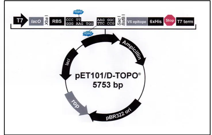
Figure 2.1: Map of pGEM-T vector (p. 54) Figure 2.2: Map of TOPO vector (p. 56)
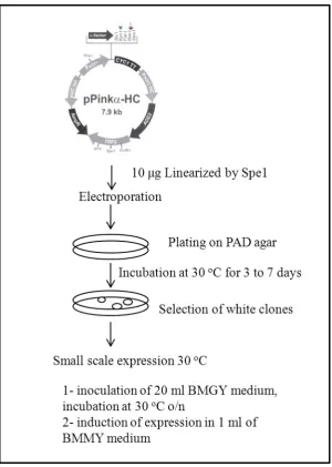
Figure 2.3: Experimental outline for protein expression in Invitrogen PichiaPink system (p. 58)
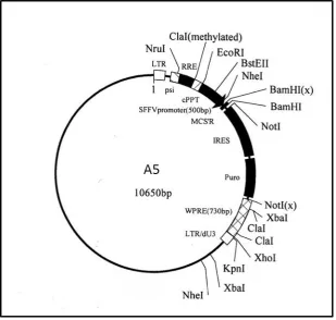
Figure 3.1: Cloning overview for the creation of pSTU1 and lentiviruses A5 for stress study (p. 67) Figure 3.2: Map of A5 lentivirus expression system (p. 68)
Figure 3.3: Molecular weight details of A5V5 and A5ChFP lentiviruses inserts (p. 70)
Figure 3.4: Representation of the TOPOpET101/D-TOPO vector and the molecular weight of the translated proteins (p. 71)
Figure 3.5: Results showing localisation of cherryFP expression by fluorescent microscopy of BHK21 cells transfected with plasmids pSTU1 (A) and pSTU2 (B) (p. 74)
Figure 3.6: Cell-free control translation of pSTU1 and pSTU2 plasmids (p. 74)
Figure 3.7: Effect of different stresses on the viability and cellular concentration of eEF2 in A5V5 HeLa (p. 76)
Figure 3.8: CherryFP-2A with (left) or without V5 (right) did not enter the nucleus (p. 78) Figure 3.9: CherryFP expression in transiently transfected HeLa cell (p. 79)
Figure 3.10: Alignment of lentivirus inserts (bottom line) against expected sequences (top line) (p.80) Figure 3.11: Western blot analyses of expression of translation-regulating factors in BL21 (A) and in Insect cell-free system (B) (p. 81)
Figure 4.1: The ribosome exit tunnel (p. 85)
Figure 4.2: Representative virus 2A sequences aligned by their consensus motifs (p. 88) Figure 4.3: Testing the consensus motif variants (p. 99)
Figure 4.4: The translation of hybrids (p. 100)
Figure 4.5: Translation of TaV and DHV hybrids (p. 101) Figure 4.6: Mutations to TaV 2A consensus sequence (p. 103)
Figure 4.8: Alanine scan of TaV 2A upstream context (p. 105) Figure 4.9: Alanine scan of FMDV 2A upstream context (p. 106) Figure 4.10: Other mutations of the upstream contexts (p. 107)
Figure 4.11: Summary showing importance of residues for efficiency of 2A sequences (p. 108) Figure 4.12: Frequency plot of the residues conservation of FMDV and DHV 2A (p. 109) Figure 4.13: Mutations of TaV Arg 1 and/or 5 (p. 110)
Figure 4.14: Glycine and proline mutations of the upstream context of TaV 2A (p. 111) Figure 4.15: Proline disruption of DHV20, FMDV and TaV 2As (p. 112)
Figure 4.16: Leucine substitutions in the upstream context of DHV, TaV and FMDV 2As (p. 113) Figure 4.17: Subjective grouping of 2A based on sequence similarities of their important region (p. 114)
Figure 4.18: Illustration of the ribosome exit tunnel and interpretative table showing TaV 2A and other stalling peptides (p.122)
Figure 5.1: Details of the dual renilla-luciferase insert for expression in bovine cell line (p. 128) Figure 5.2: Map of pPinkα-HC (p. 130)
Figure 5.3: Creation of the TaV 2A insert for pPinkα vector (p. 131) Figure 5.4: Puromycin test for TaV 2A activity (p. 134)
Figure 5.5: Mutation of Pro20 of pSTA1 TaV 2A (p. 135)
Figure 5.6: Effect of proline synonymous codons on activity of FMDV 2A (p. 136) Figure 5.7: Diagram of the processing of the TaV-cherryFP insert in P.pastoris (p. 138) Figure 5.8: DNA gel electrophoresis of PCR of P. pastoris transformants (p. 139) Figure 5.9: Western blot test of 2A antibody (p. 139)
Figure 5.10: Western blot analysis of the supernatant fractions for four P. pastoris transformants (p. 140)
Figure 5.11: Analysis of cell fractions of P. pastoris transformants, western blot and fluorescence microscopy (p. 141)
Figure 6.1: Summary of 2A –like elements in marine organisms (p. 145) Figure 6.2: Modification of pSTA1 for the cloning of candidate 2As (p. 147) Figure 6.3: SDS-PAGE analysis of test expression for amphioxus 2As (p. 152)
Figure 6.4: Visual representation of key domains in the 2A-containing amphioxus proteins (p. 153)
Figure 6.5: The non-LTR retrotransposons with 2A in amphioxus (p. 154)
Figure 6.6: Visual representation of the non-LTR CR1 organisation in amphioxus (p. 154)
Figure 6.7: Tree showing clustering of Crack non-LTR retrotransposons containing a 2A motif (p. 155)
Figure 6.8: Tree showing clustering of CR1 non-LTR retrotransposons containing a 2A motif (p. 156) Figure 6.9: Activity assay for newly identified 2A in other cellular organisms and viruses (p. 157) Figure 6.10: Details for the selected S. kowalevskii contigs with a 2A sequence tested in vitro (p. 158)
The 2A element is a peptide able to stall the eukaryotic ribosome during the course of its translation and prevent the formation of a peptide bond between a specific glycine and proline pair at the C-terminus. Surprisingly, the ribosome having skipped the formation of this peptide bond is able to resume translation of the remaining RNA sequence past the C-terminal glycine of 2A. This gives rise to two discrete proteins from a single open reading frame (ORF).
A model of action has previously been proposed to explain 2A-induced ribosome stalling and the purpose of this body of work was to further define and elaborate this model.
2A was termed after the genomic region of the viruses where it was first identified; the 2A region of the picornaviruses foot-and-mouth disease virus (FMDV), encephalomyocarditis virus (EMCV) and Theiler’s murine encephalomyelitis virus (TMEV). The C-terminal consensus motif of 2A
[D(V/I)ExNPGP] was later identified encoded by the genomes of several other single-stranded and double-stranded RNA viruses.
1.1 Terminology and numbering of 2A
In this thesis, for simplicity all 2A and 2A-like elements will be referred to as 2A, preceded by the initial of the virus in which it occurs, and if appropriate the length of the 2A sequence tested.
e.g.: DHV20 2A the twenty-residues-long-2A sequence from Duck hepatitis virus (DHV). The numbering of residues follows the N- to C- numbering adopted in the literature for other
ribosome-arresting peptides : SecM, arginine attenuator peptide (AAP), TnaC, (Ito and Nakatogawa, 2002, Gong et al, 2007 and Vasquez-Laslop et al., 2008). In the context of 2A the numbering begins at the first residue of the 2A sequence.
e.g.:
Residue number 1 10 20 30
....|....|....|....|....|....|
FMDV30 2A RHKEDCAPVKQLLNFDLLKLAGDVESNPGP
Based on sequence alignments (Luke et al., 2008) and for the purpose of site-directed mutagenesis (chapter 4 and 5) it has been decided to distinguish two regions in the 2A sequences: the variable region also called the upstream context (underlined in the example above) and the consensus motif, which is more conserved (in blue in the above example).
Throughout this text the ribosome numbering follows E. coli numbering consistent with the ribosome-arresting peptide literature.
1.2 Occurrence of the ribosome-arrest 2A element in viruses
Viruses consist of RNA or DNA genome encapsulated within a virion. They lack the translational machinery necessary to replicate and are obligate parasites of cellular systems. The Baltimore classification divides all viruses into seven categories based upon genome type and mode of
replication (Baltimore, 1971). Positive-stranded RNA viruses, which are the classes of virus relevant to this thesis, have an RNA genome acting like cellular mRNAs. They are translated directly in the cytoplasm of the host cell. Each family of RNA viruses showcase distinctive strategies to express and process their proteins.
1.2.1 Occurrence in
Picornaviridae
1.2.1.1 Picornaviruses
‘Pico’ means small and ‘rna’ refers to the type of genome. Picornaviruses are infectious agents to human and animals. The classification of the Picornaviridae family has been amended extensively in the past four years as newly-identified viruses have been included. The picornavirus study group publishes online (http//www.picornaviridae.com, 2013) the most up-to-date classification. In 2013, to reflect the changes approved by the International Committee on Taxonomy of Viruses (Adams et al., 2013), the Picornaviridae family is organised into the seventeen genera presented in table 1.1.
Their genome is 7500 to 8500 nucleotides (nts) long and organised in a single ORF encoding a polyprotein (figure 1.1). This single precursor is sequentially processed into final-mature proteins, twelve in the case of FMDV. The 5’ end of the picornavirus RNA is the Leader region and comprises 600 to 1100 nts (Palmenberg, 1990). The ORF has three distinct regions called P1, P2 and P3. The underlying rationale for this subdivision lies in the structural or enzymatic functions of the viral proteins encoded. The ORF constitutes 85 to 90 % of the coding capacity, and the remaining 10 to 15 % are shared between the 3’ and the 5’ region (Palmenberg et al., 2009). The 3’ region is between 40
to 120 nts long (Palmenberg, 1990).
The unprocessed polyprotein would be a large protein of about 250 kDa, but is not observed in cultures. It is processed sequentially into precursors and then individual viral proteins by a proteolytic cascade involving a primary and secondary series of events (Palmenberg, 1992). The primary
Table 1.1: The picornaviruses genera, species and serotypes
(From Fernandez- Miragall et al., 2009, Adams et al., 2013 and http//www.picornaviridae.com, 2013)
Picornaviruses using the 2A-induced ribosome stalling mechanism during the course of their polyprotein processing are
highlighted in red showing the serotypes relevant to this thesis.
genus species relevant serotypes
Aphthovirus Foot-and-mouth disease virus
Bovine rhinitis A virus
Bovine rhinitis B virus
Equine rhinitis A virus
Foot-and-mouth disease virus O, A, C, Asia
1, SAT 1, SAT 2 and SAT 3
Bovine rhinitis A virus 1and 2
Bovine rhinitis B virus 1
Equine rhinitis A virus 1
Aquamavirus Aquamavirus A Aquamavirus A
Avihepatovirus Duck hepatitis A virus Duck hepatitis A virus 1 to 3
Cardiovirus Encephalomyocarditis virus
Theilovirus
Encephalomyocarditis virus 1
Encephalomyocarditis virus 2
Theiler’s murine encephalomyelitis virus Vilyuisk human encephalomyelitis virus
Thera virus
Saffold virus 1 to 9
Cosavirus Cosavirus A, B, C and D
Dicipivirus Cadicivirus A
Enterovirus Enterovirus A, B, C, D, E, F, G, H, J
Rhinovirus A, B, C
Erbovirus Equine rhinitis B virus Equine rhinitis B virus 1 to 3
Hepatovirus Hepatitis A virus
Kobuvirus Aichi virus A, B, C
Megrivirus Melegrivirus A
Parechovirus Human parechovirus Human parechovirus 1 to 14
Ljungan virus Ljungan virus 1 to 4
Salivirus Salivirus A
Sapelovirus Porcine sapelovirus
Simian sapelovirus
Avian sapelovirus
Senecavirus Seneca Valley virus Seneca Valley virus 1
Teschovirus Porcine teschovirus Porcine teschovirus 1 to 11
In the 5’ region, the RNA has a VPg protein capping the 5’end (figure 1.1) attached to the 5’ uridylyl nucleotide of the RNA through a tyrosine residue to form a phosphodiester bond (Forss and Schaller, 1982 and Steil et al., 2010). Other key features of the 5’ region are the presence of a cloverleaf structure and an internal ribosome entry site (IRES). This structural element is the key factor controlling the viral-RNA translation by its ability to recruit cellular ribosomes (Kolupaeva et al., 1998).
From the coding region, region P1 yields all the proteins (VP1, VP2, VP3 and VP4) required for the virion formation. The middle part of the picornaviral RNA yields peptides 2A, 2B and 2C. The P3 region yields four final proteins via a series of active intermediates 3AB, 3B VPg, 3CDpro, 3Cpro and the polymerase 3Dpol (Palmenberg, 1990). Picornavirus non-structural proteins are involved in viral genome replication, cellular shut-down and cellular membranes shuffle. 2A protein is highly variable amongst Picornaviridae and is involved in primary processing of the viral polyprotein. 2B protein is a transmembrane protein able to form pores, alter membranes permeability and therefore disturb calcium homeostasis (Sandoval and Carrasco, 1997). In Enterovirus 2B disrupts membrane
permeability in the endoplasmic reticulum and Golgi apparatus (Sanchez-Martinez et al., 2012). It is not established at present if all the picornaviruses 2B functions similarly. 2C has ATPase/GTPase activity and is an essential element of the replication process (Rodriguez and Carrasco, 1993). The 2C and the precursor 2BC bind viral RNA to cellular vesicles. 2C has been shown to participate in the encapsidation step of the viral genome (Vance et al., 1997). 3A disrupts the traffic between endoplasmic reticulum and Golgi apparatus thereby inhibiting host cellular responses such as
production of interferon, interleukins and cytokins mainly by releasing intracellular calcium (Dodd et al., 2001). The host range is defined essentially by 3A (Lama et al., 1998). 3B or VPg is covalently bound to the next viral RNA and is a primer for initiation of replication (Schein et al., 2006). 3Cpro is a chymotrypsin like protease responsible for most of the polyprotein cleavage (Palmenberg, 1992). 3C also takes part outside of the viral protein processing to cellular disruption by cleaving eIF4A, eIF4GI and II, PABP, and degrading p53, thereby controlling cell apoptosis (Blair et al., 1998, Belsham et al., 2000, Barco et al., 2000, Weidman et al., 2001). 3Dpol is a RNA-dependent RNA polymerase, which shows much specificity to each virus type and is the crucial element in viral replication, the enzyme lacks proof-reading and frequent errors induced during replication allow the virus flexible adaptation (Kerkvliet et al., 2010). The cleavage intermediates 2BC, 3AB and 3CDpro have additional functions. 2BC creates small cellular vesicles, 3AB induce 3Dpol activity and 3CDpro participate in polyprotein processing and enhances replication by binding the cloverleaf structure at the 5’UTR (Chase and Semler, 2012).
Figure 1.1: The generic genomic organisation of Picornaviridae
The positive-sense RNA genome has a small protein VPg at the 5’ end (encoded by 3B). The 5’ UTR contains an internal ribosome entry site (IRES). The 3’ end has a poly (A) tail. 3Cpro
carries out most of the proteolytic processing. The genome
is organised in three regions P1, 2 and 3. The schema indicates the peptides encoded by each region (written in white).
1.2.1.2 The 2A product in Picornaviridae
Picornaviral RNA is translated once the IRES has successfully recruited a ribosome. Eukaryotic initiation factor 4 (eIF4) is a principal target for most picornaviruses. The genus Enterovirus uses the 2A protease (2Apro) to cleave eIF4 (Novoa and Carrasco, 1999). Members of the Aphthovirus and
Regions of the picornavirus ORF encodes for proteins with similar functions across genera. The 2A locus however is an exception. For the genus Hepatovirus, the 2A protein is cleaved from VP4 late in the replication. The primary processing is performed by 3Cpro (the only protease this genus encodes) at the 2A/2B junction. Cohen and colleagues (2002) performed serial N- and C-terminal deletions of the 2A sequence. It was found that 40 % of the N-terminal sequence was necessary for infectivity whereas the C-terminal (the remaining 60 %) of the sequence was required for adequate cleavage of VP1/2A by an elusive cellular protease. For this genus, the 2A product is involved in virion assembly and maturation.
Human parechovirus bioinformatics analyses showed that the 2A product is related to the cellular protein H-rev107 involved in cell-growth regulation. The conserved features of this protein are a long hydrophobic domain and an Asn-Cys-Glu motif (Hughes and Stanway, 2000). The 2A from Human parechovirus was thencalled ‘H-box 2A’ and is also identified in Kobuvirus, Megrivirus and
Tremovirus (http//www.picornaviridae.com, 2013). For this category of 2A, 3Cpro acts between the VP1 and 2A.
InEnterovirus, the 2A product is a protease. The primary cleavage has two events, the self-processing cleavage by 2Apro at its C-terminus and the release of the 3ABC precursor by 3Cpro. Apart from performing the primary processing of the picornavirus polyprotein, 2Apro also cleaves eIF4 (inhibition of cap-dependent translation) and nucleoporins (inhibition of mRNA transport to cytoplasm)
(Redondo et al., 2011). Sapelovirus also has this type of 2A protein.
The 2A product for Dicipivirus and Salivirus has not been characterised to date.
Finally, 2A for Aphthovirus and Cardiovirus, as well as other genera (Aquamavirus, Avihepatovirus,
Erbovirus, Senecavirus, Teschovirus and the species Ljungan virus from genus Parechovirus) highlighted in red in the table 1.1, is characterised by the C-terminally conserved motif
Table 1.2: Viruses employing 2A-induced ribosome stalling.
Family Genus Species reference virus for 2A studies
Picornaviridae Aphthovirus Foot-and-mouth disease virus
Bovine rhinitis A virus
Bovine rhinitis B virus
Equine rhinitis A virus
FMDV-O1K (X00871)
Aquamavirus Aquamavirus A
Avihepatovirus Duck hepatitis A virus Duck hepatitis virus 1 (DQ219396)
Cardiovirus Encephalomyocarditis virus
Theilovirus
Encephalomyocarditis virus (Ruckert)
(M81861)
Mengovirus (DQ294633)
Theiler’s murine encephalomyelitis virus
(NGS910_AB)
Erbovirus Equine rhinitis B virus
Parechovirus Ljungan virus
Senecavirus Seneca Valley virus
Teschovirus Porcine teschovirus
Dicistroviridae Aparavirus Acute bee paralysis virus Acute bee paralysis virus (AF150629)
Israeli acute paralysis virus
Kashmir bee virus
Cripavirus Cricket paralysis virus Cricket paralysis virus (AF218039)
Drosophila C virus
Iflaviridae Iflavirus Ectropis oblique picorna-like virus
Infectious flacherie virus Infectious flacherie virus (AB000906)
Perina nuda picorna-like virus
Tetraviridae Permutotetravirus Thosea asigna virus Thosea asigna virus (AF062037)
Euprosterna eleasa virus
Cormotetravirus Providence virus Providence virus (AF548354)
Reoviridae Rotavirus Human rotavirus C
Porcine rotavirus C Porcine rotavirus C (Cowden) (M69115)
Bovine rotavirus C
Non A,B,C novel adult diarrhea
virus
novel adult diarrhea virus (AY632079)
Cypovirus Bombyx mori cypovirus 1 Bombyx mori cypovirus 1(AF433660)
Lymantria disparcypovirus 1
Dendrolimus punctatuscypovirus 1
Operophtera brumata cypovirus 18
Totiviridae Giardavirus Penaeid shrimp infectious
myonecrosis virus
Penaeid shrimp infectious myonecrosis
1.2.2 Occurrence of the ribosome-stalling 2A sequence in other RNA viruses
Worldwide efforts in genome sequencing provided an unprecedented opportunity to compare and align viral genomes. Using local alignment algorithms programmed with the [D(V/I)ExNPGP] consensus motif previously identified in Picornaviridae, revealed that outside aphthoviruses and cardioviruses other mammalian and insect RNA viruses contained the 2A motif (Luke et al., 2008). The classification for these viruses is provided in table 1.2. The 2A elements were cloned and tested in the generic expression-system (figure 1.6). They were able to separate a single ORF into two discrete products with varying efficiencies (summarised in table 1.3) (Luke et al., 2008). A nucleotide sequence coding for 2A was discovered in the genomes of positive single stranded RNA virus
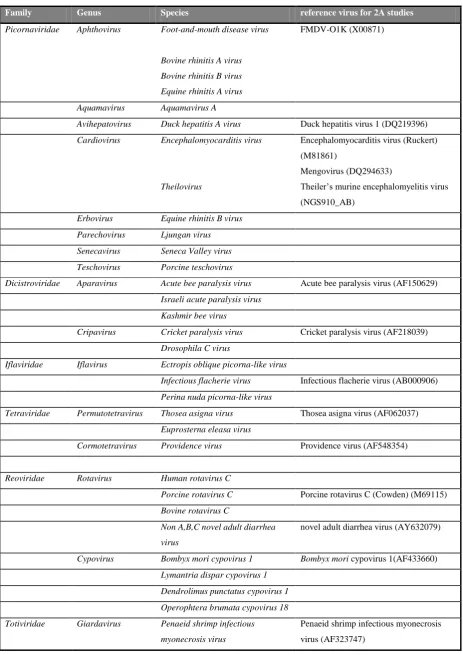
families: Dicistroviridae, Iflaviridae and Tetraviridae. Figure 1.2 provides the position of 2A in relation to these virus genomes.
The virus classifications reported throughout this section follows the latest taxonomy released by the International Committee on Taxonomy of Viruses in 2012 (http//www.ictv.org).
The Dicistroviridae family consists of two genera: Aparavirus and Cripavirus infecting invertebrates such as bees and cricket worldwide (De Miranda et al., 2010).
The generic dicistrovirus genome is organised in two non-overlapping ORFs encoding for two distinct polyproteins. The structural proteins (products of the ORF2) of insect picorna-like viruses have many common features with picornaviruses. However, dicistroviruses are distinguished by the position of their structural protein-coding sequences at the 3’ rather than the 5’ end as for picornaviruses and iflaviruses. Dicistroviruses also have an IRES preceding each ORF (De Miranda et al., 2010). Three out of five species of Aparavirus have a NPGP 2A motif: Acute bee paralysis virus (ABPV), Israeli acute paralysis virus and Kashmir bee virus (KBV). Two species out of nine of Cripavirus have a 2A,
Iflavirus is the only genus in the Iflaviridae family. There are seven species currently classified and 2A was found in the genome of three of these: Infectious flacherie virus (IFV), Peruna nuda picorna-like virus (PnPV), Ectropis oblique picorna-like virus (EoPV). The iflaviruses are the causative agents of flacherie disease for invertebrates such as honey bees and silkworm (Ribiere et al., 2010).
The iflaviruses have a single ORF encoding the capsid protein from the 5’ genomic region and non-structural proteins from the 3’ region (Wu et al., 2002).
Purified EoPV particles were analysed by SDS-PAGE and only two protein masses were detected: 31.5 kDa and 28.8 kDa. Further bioinformatic analyses of the particle proteins revealed homologies to several other ssRNA viruses-virion proteins (Wang et al., 2004). The position of 2A coincides with two junctions. In PnPV and EoPV, 2A is found between VP4 and VP1 (at residues 572- 575) and between the last capsid protein and the helicase of the non-replicative precursor (at residues 1189- 1192). Both 2A sequences were highly efficient in vitro (Luke et al., 2008). It is highly likely that the two virion particles detected correspond to processing events induced by the 2A sequences. The 28.8 kDa protein was detected in twice greater amounts than the 31.5 kDa (Wang et al., 2004). The phylogenetic study published by Wang and colleagues indicates that EoPV, PnPV are closely related to IFV. These three virus species share a common organisation and some sequence similarities. There are several strains of IFV reported and potential new members for the Iflavirus genus, however only one sequence is available to date (GenBank accession number AB000906 (Isawa et al., 1998). IFV has one 2A element at position 1081 to 1084 aa segregating the capsid precursors from non-structural proteins precursor.
The Tetraviridae family taxonomy has been reassessed extensively. The current classification (http://www.ictvonline.org) recognises three genera: Cormotetravirus, Permutotetravirus and
Alphatetravirus. Permutotetravirus consists of two species Europrosterna elaeasa virus (EeV) and
Thosea asigna virus (TaV), both containing a 2A sequence. Cormotetravirus has a single species,
Providence virus (PrV) known to have three 2A sequences.
correspond to the C-terminal segment of the capsid precursor encoded by the ORF2. The N-terminus encodes for a third protein of 17 kDa (p17) of unknown function and not incorporated in the virion. In TaV and EeV, the 2A sequence is found between p17 and the capsid precursor (Zeddam et al., 2010). Providence virus has a monopartite genome but encodes three ORFs (Walter et al., 2010). The first ORF encodes for a protein of 130 kDa (p130) of unknown function. The second ORF encodes for a 40 kDa protein (p104 and in figure 1.2 the replicase polyprotein) in a +1 frame relative to p130. The third ORF begins 4 nts after p104 stop codon. ORF3 encodes for the capsid polyprotein p81 from a read-through event at the second ORF stop codon. PrV has three 2A elements. PrV-2A1 is at the terminus of p130 and produces fragments of 17 and 113 kDa. PrV-2A2 and PrV-2A3 are at the N-terminus of p81. The two 2A elements process p81precursors into fragments of 7, 8 and 68 kDa. The capsid precursor is the 68 kDa which undergoes auto-proteolysis to yield the peptides of 60 and 7.4 kDa composing the virion (Walter et al., 2010). The 7 and 8 kDa segments resulting from PrV-2A2 and 2A3 processing have not been studied.
Figure 1.2: Location of 2A coding sequences in the genomes of other ssRNA (+) viruses
The 2A is marked in red. The key features of the genomes are provided. The proteins sequences associated with virion
formation (VP or CP) are coloured in shades of green, the grey boxes represent the replicative domains, blue and pink
represent other coded proteins. (Graphics are reproduced and adapted from http://www.viralzone.expasy.org, using
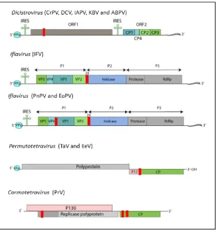
In addition two families of double-stranded RNA viruses, Reoviridae and Totiviridae, have members coding for a 2A sequence.
Reoviruses have segmented genomes. Figure1.3 gives an illustration of the position of the 2A motif in the relevant genomic segment. Reoviruses are organised in two subfamilies: Sedoreovirinae and
Spinareovirinae.
Sedoreovirinae consists of six genera. It includes the genus Rotavirus further organised in five species: Rotavirus A, B, C, D, and E. Rotaviruses have eleven segments encoding six virion forming proteins and five replicative proteins (James et al., 1999). Species of the genus Rotavirus C: Bovine rotavirus C (BoRV), Porcine rotavirus C (PoRV) and Human rotavirus C (HuRV) encode a 2A sequence in their segment 6 (NSp3). In addition, 2A was identified in segment 5 (NSp1) of the rotavirus type non -A,B, C novel adult diarrhea virus (ADRV) (Luke et al., 2008).
Rotaviruses mRNAs lack 3’ poly(A) endings. NSp3 protein enhances rotavirus translation by its ability to circularise the viral mRNAs (Jayaram et al., 2004). When the NSp3 segment of porcine rotavirus C was expressed in COS-1 cells, three products were observed by SDS-PAGE analysis a 45 kDa full-length product, a 38 kDa and a 8kDa product (Langland et al., 1994). Further analysis of the NSp3 aa sequences for type C rotaviruses revealed three regions: a ssRNA binding protein region at the N-terminus, a initiation factor 4G binding region in the mid region, the 2A motif followed by a dsRNA binding protein region (Luke et al., 2008). In other types of rotaviruses, the NSp3 protein is shorter and lacks the 2A-dsRNA binding domain of type C rotavirus (James et al., 1999).
NSp1 is the most variable protein in rotaviruses and targets the host interferon response (Arnold and Patton, 2011). Although NSp1 of ADRV is unrelated to NSp3 of type C rotaviruses, both of these segments contain downstream of 2A a dsRNA binding protein.
The subfamily Spinareovirinae is organised in nine genera and includes the genus Cypovirus, which is further categorised in sixteen species, Cypovirus 1 to 16. Four viruses Bombyx mori cypovirus 1 (BmCPV-1), Lymantria dispar cypovirus 1 (LdCPV-1), Dendrolimus punctatus cypovirus
The family of Totiviridae comprises five genera, amongst these in the genus Giardavirus, the virus
[image:25.595.74.453.317.712.2]Penaeid shrimp infectious myonecrosis virus (IMNV) has two 2A sequences at the N-terminus of ORF1(Luke et al., 2008). Totiviruses have non segmented dsRNA genomes, with two ORFs that overlap (Nibert, 2007). ORF1 encodes the capsid protein and ORF2 the polymerase. The polyprotein processing of ORF1 results in the production of three products of different sizes, 93, 284 and 1228 amino acids (Poulos et al., 2006). The extreme 93 aa N-terminal fragment is a dsRNA binding protein terminated with a 2A sequence (Nibert, 2007). Totiviruses were originally isolated from fungi but in 2006 IMNV was isolated in aquacultures of shrimps in Brazil and Indonesia (Poulos et al., 2006). In IMNV, the capsid protein starts half-way through the ORF1 and is preceded by the dsRNA binding protein, which is an unusual feature for totiviruses. New totiviruses were isolated from drosophila cultures and mosquitoes. These new isolates retain the dsRNA binding protein-2A feature previously observed in IMNV (Isawa et al., 2010).
Figure 1.3: Location of the 2A coding sequence in the genome of dsRNA viruses
2A is marked in red, the rotaviruses, and cypoviruses genomes are segmented and only the relevant segment is shown. The
Sequence alignments proved that the only conserved part was the 2A motif used for identification, as the upstream context is very diverse. Previous analyses of the residues by positions ruled out any regularity or patterns (Luke et al., 2008). The table 1.2 was created with a representative 2A-like sequence for each virus previously identified with a 2A motif in their genome and provides the 2A sequences tested to date and their efficiencies.
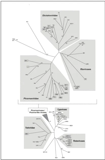
Figure 1.4: Phylogenetic tree showing occurrence of 2A in diverse virus groups
(provided by Prof. Ryan as published in Luke et al., 2008.)
The analysis was based on alignment of the RNA-dependent RNA polymerase domain. The viruses with a 2A
Table 1.3: The stalling efficiency of the viral 2A motifs tested to date
The table features the reported efficiency for viral 2A-sequences previously tested in the in vitro expression system
(Compiled from Luke et al., 2008 and Donnelly et al., 2001b).
virus sequence tested proportion
(%) of processed
proteins
reference
Positive stranded RNA viruses
Picornaviruses
FMDV
FMDV NFDLLKLAGDVESNPG/P 75 Ryan and Drew,
1994 Other picornaviruses
TMEV FREFFKAVRGYHADYYKQRLIHDVEMNPG/P 98 Donnelly et al., 1997 EMCV VFGLYRIFNAHYAGYFADLLIHDIETNPG/P 99 Donnelly et al.,
2001b
ERAV QCTNYALLKLAGDVESNPG/P 99
PTV ATNFSLLKQAGDVEENPG/P 94
SAF-V FTDFFKAVRDYHASYYKQRLQHDVETNPG/P
99
Luke et al., 2008 ERBV EATLSTILSEGATNFSLLKLAGDVELNPG/P
LV YFNIMHSDEMDFAGGKFLNQCGDVETNPG/P Other viruses
Iflaviruses
IFV TRAEIEDELIRAGIESNPG/P 63 Donnelly et al., 2001b
Longer version of IFV
PSIGNVARTLTRAEIEDELIRAGIESNPG/P 99 Luke et al., 2008
EoPV-2A1 GQRTTEQIVTAQGWAPDLTQDGDVESNPG/P PnPV-2A1 GQRTTEQIVTAQGWVPDLTVDGDVESNPG/P EoPV-2A2 TRGGLQRQNIIGGGQRDLTQDGDIESNPG/P PnPV-2A2 TRGGLRRQNIIGGGQKDLTQDGDIESNPG/P
Tetraviruses
TaV RAEGRGSLLTCGDVEENPG/P 99 Donnelly et al., 2001b
EeV RRLPESAQLPQGAGRGSLVTCGDVEENPG/P 99 Luke et al., 2008 PrV-2A1 LEMKESNSGYVVGGRGSLLTCGDVESNPG/P
PrV-2A2 NSDDEEPEYPRGDPIEDLTDDGDIEKNPG/P 94 PrV-2A3 TIMGNIMTLAGSGGRGSLLTAGDVEKNPG/P 99
Dicistroviruses
CrPV LVSSNDECRAFLRKRTQLLMSGDVESNPG/P 88 Luke et al., 2008
ABPV TGFLNKLYHCGSWTDILLLLSGDVETNPG/P 94
Double stranded RNA viruses
Animals rotaviruses
PoRV AKFQIDKILISGDVELNPG/P 31 Donnelly et al.,
2001b
BoRV-C GIGNPLIVANSKFQIDRILISGDIELNPG/P 89 Luke et al., 2008 HuRV-C GAGYPLIVANSKFQIDKILISGDIELNPG/P 82
ADRV-N FFDSVWVYHLANSSWVRDLTRECIESNPG/P 97 Insect cypoviruses
BmCPV-1 RTAFDFQQDVFRSNYDLLKLCGDIESNPG/P 99 Luke et al., 2008 OpbuCPV-18 IHANDYQMAVFKSNYDLLKLCGDVESNPG/P
Totiviruses
1.3 Discovery of the 2A NPGP sequence
The ribosome-stalling hypothesis was derived by a process of elimination. The 2A gene product for poliovirus (belonging to the genus Enterovirus) is 2Apro, an enzyme of 142 residues (Toyoda et al., 1986), distantly related to 3Cpro and which performs a cleavage at its N-terminus. Nicklin and co-workers (1987) constructed various plasmids to demonstrate the proteolytic activity of poliovirus 2Apro. They showed that the cleavage site consisted of a tyrosine-glycine pair. 2Apro catalytic triad was later determined by mutational and structural studies (Hellen et al., 1992, Sommergruber et al., 1997) and consists of Cys106, His18 and Asp35. Compounds such as iodoacetamide and N-ethylmaleimide, inhibitors of thiol proteases, were active against 2Apro (Konig and Rosenwirth, 1988).
The mature 2A protein of Hepatovirus escapes detailed molecular analyses as it so far failed to be purified, suggesting that the protein is either unstable or simply not released. The predicted translated 2A presents no similarities to Aphthovirus, Cardiovirus or Enterovirus counterparts and deletion of 60 % of the C-terminal 2A sequence did not impair replication. For Hepatovirus, cleavage at the 2A/2B junction is carried out by 3Cpro (Martin et al., 1995) whereas the cleavage between VP1/2A occurs in the course of virion formation and is likely mediated by a cellular protease (Martin et al., 1999, Cohen et al., 2002).
Figure 1.5: Picornavirus organisation and schematic representation of the three possible types of activity at the 2A region 3Cpro carries out most of the proteolytic processing. At the 2A/2B junction, 2Apro cleaves at its own N-terminus (example of
protease (Lpro)). 2A is flanked by cleavage sites (example of Hepatovirus) (Derived from information available at http://www.picornaviridae.com).
Theiler’s murine encephalomyelitis virus (TMEV) transcripts lacking 3Cpro
sequence retained the ability to operate the separation at the 2A/2B junction (Roos et al., 1989). Inhibition studies involving encephalomyocarditis virus (EMCV) showed that the 2A/2B processing occurred very quickly and was not influenced by all the inhibitors used (Jackson, 1986). Sequence alignment showed no possible correlation to rhinoviruses and poliovirus 2A. No protease domains were found in the 2A sequences of cardioviruses EMCV and TMEV (Palmenberg, 1990). In a separate study, EMCV 2A could
tolerate large deletions (60 %) at the N-terminal half and still result in the 2A/2B separation. Deletions to the C-terminal of 2B produced a similar result. A sequence alignment showed that the C-terminus of 2A is absolutely conserved amongst all cardioviruses. Subsequent directed mutagenesis identified the cleavage site. For cardioviruses, the cleavage occurred between the last glycine of 2A and the next proline of 2B of the absolutely conserved four amino acid sequence [NPGP] (Palmenberg et al., 1992).
Lpro or 3Cpro do not cleave the 2A/2B junction of FMDV (Ryan et al., 1989, Belsham et al., 1990). Alignment of the 2A regions between FMDV and cardioviruses EMCV and TMEV showed that the C-terminal [NPGP] motif was also conserved. FMDV 2A was later introduced internally in a synthetic reporter system flanked by virally unrelated sequences and translated in rabbit reticulocyte lysates and showed to retain activity. The processing event at the 2A [NPGP] motif did not rely on any other viral elements (Ryan et al., 1991).
This synthetic in vitro approach (Ryan et al., 1991) has been adopted since and consists of two genes encoding for different molecular weight proteins which could then easily be visualised on an
acrylamide gel. The 2A sequence is inserted in-frame between these two genes creating one single ORF on a plasmid harbouring a T7 promoter. The plasmid is then introduced in cell-free systems such as rabbit reticulocyte lysates or wheat germ assays supplemented with [35S]-methionine.
In the current expression system (figure 1.6), 2A is inserted between green fluorescent protein (GFP) and beta-glucuronidase (GUS) coding sequences. Three bands can be visualised. One band
Figure 1.6: The bicistronic expression system for analysis of 2A activity
Cloning involves insertion of the 2A sequence in-frame between GFP (27 kDa) and GUS (70 kDa) coding sequences. The
plasmid is introduced in a cell-free system supplemented with [35S]-methionine and translation is driven by T7 promoter.
The distribution of de novo radiolabeled translation products are analysed on SDS-PAGE gels. Three translation products
can be observed: the full-length non-processed polyprotein (100 kDa) and the two segregated products GFP-2A (~30 kDa)
and GUS (top drawings provided by Prof. Ryan, figure adapted from Donnelly et al., 2001b).
Figure 1.7: C-terminal sequences at the 2A/2B region of cardioviruses (EMCV, TMEV and
mengovirus) and aphthovirus (FMDV)
The violet box highlights the conserved residues, the separation between 2A and 2B occurs between the last glycine of 2A
and the first proline of 2B, shown with a blue arrow (adapted from Donnelly et al., 1997).
It was therefore concluded that the picornavirus 2A region can either encode a proteinase (2Apro) as for enteroviruses, or the translated 2A can be flanked with protease cleavage sites such as for hepatovirusesandparechoviruses, or it can induce a ribosome stalling event characterised by the C-terminal residues [D(V/I)ExNPGP], as is the case for aphthoviruses and cardioviruses (summarised in figure 1.5).
Ryan and Drew (1994) focussed first on understanding how much of the already small peptide (eighteen amino acids) was necessary to maintain 2A activity in an artificial context. It seemed that the critical threshold was thirteen amino acids, although at that length the amount of polyprotein processed was reduced from 75 % to about 65 %. On the other hand adding more amino acids
upstream of 2A altered that activity favourably to the point where almost all the polyproteins (>99 %) were effectively divided into the two expected distinct proteins. Similarly, truncating the EMCV and TMEV cardioviruses 2A to only the last N-terminal eighteen amino acids (Donnelly et al., 1997) resulted in decrease in the proportion of polyproteins processed. It seemed therefore that the upstream context played an important role in increasing 2A efficiency. It was not, however, what explained the mechanism of action.
Based on these observations and inspired by the early work published about Escherichia coli (E. coli) SecM sequence, Ryan and co-workers (1999) proposed that NPGP 2A influences the translating ribosome and affects its ability to perform the formation of a peptide bond between the C-terminal glycine and proline. In E. coli, SecM and SecA form an operon. SecA protein is essential to E. coli
and is a translocase (Sarker and Oliver, 2002). SecM has a C-terminal motif FxxxxWIxxxxGIRAGP able to stall the prokaryotic ribosome (Nakatogawa and Ito, 2002). The intergenic area between SecM and SecA contains a Shine-Dalgarno motif, normally inaccessible, and which becomes available to a ribosome only if the mRNA is re-structured. This change in secondary structure is introduced by the presence nearby of a stalled ribosome on the SecM motif (Butkus et al., 2003). SecM is translated normally in E. coli. When the bacterial cell has an abundance of SecA, SecA interacts with the SecM N-terminal sequence which extends outside the ribosome tunnel. SecA translocase activity acts like a pull and thus un-stalls the ribosome. There is a stop codon following the SecM stalling motif where the ribosome is able to terminate translation. In these conditions the stalled ribosome does not have the time to re-organise the intergenic mRNA structure. When the cellular level of SecA decreases, SecM stalled- ribosomes can re-structure the mRNA, allowing a new ribosome to start translation of SecA downstream of SecM (Yap and Bernstein, 2011).
1.4 Model for 2A Activity
The first model to explain 2A activity was published by Ryan and co-workers (1999) and suggested that translation of the ORF started and progressed normally from a start codon up to the 2A [NPGP] stalling motif. The glycyl-prolyl peptide bond was not being made, the ribosome thus could terminate translation and the imbalance in products suggested that a proportion of the ribosomes stopped while others could carry translation further into the downstream sequence. In this study, computer generated modelling of several C-terminal 2A peptides revealed a tight turn at the [NPG] while the short
eighteen amino acids FMDV 2A was predicted to adopt an α-helix.
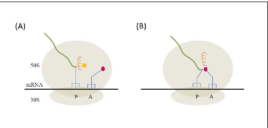
al., 1999). These features are thought to play a critical role in 2A activity. At the peptidyl transferase centre (PTC) within the ribosome, the 2A nascent peptide linked to glycyl-tRNA is located in the P site. When prolyl-tRNA occupies the ribosome A site, the conformation of 2A in the ribosomal exit tunnel prevents peptide bond formation.
Toeprint analyses confirmed that 2A acted upon translating ribosomes, and mapped the activity to be at the Gly-Pro pair (Doronina et al., 2008). Toeprint analysis also confirmed the hypothesis of a pause in translation. In this study, yeasts (strain sup45-2) with reduced release factor 1 (eRF1) activity at 34 o
C, led to an increase in full-length products. In yeasts and yeast extracts with impaired release factor 3 (eRF3) activity, there was a large reduction in the production of downstream and full-length product. This implied a rescue function for factors eRF1 and eRF3 (Doronina et al., 2008).
Doronina and co workers (2008) proposed that the pause in elongation observed is related to dissociation of tRNA from the A site. They argued that the unusual dissociation of prolyl-tRNA from the A site following failure to generate the peptide bond, would leave a structural conformation, similar to a natural stop codon, that can be a substrate for binding of the termination factors.
Figure 1.8: Model of the mechanism of 2A-induced ribosome stalling
The assembled ribosome large and small subunits are represented containing the aminoacyl site (A), peptidyl site (P) and exit site (E), fitted with tRNA deacylated (-OH) or aminoacylated with Pro or Gly. The cartoon shows the possible step by step scenario for when a eukaryotic ribosome encounters the last Gly-Pro residues of the NPGP stalling motif of 2A. (Step a) peptidyl-tRNA is in A site and
(step b) is translocated to the P site, prolyl-tRNA occupies A site. Interaction of the 2A structure within the ribosome exit tunnel and the tight turn at the last NPG residues, precludes peptide bond formation.
(Step c) the prolyl-tRNA exits the ribosomal unit and
(step d) the eRF1/3 complex enters the A sites and hydrolyses the ester bond linking the nascent peptide to tRNA in P site. (Step f) eRF1/3 leaves the A site. The nascent peptide is released.
(Step g) prolyl-tRNA re-enters A site and (step h) is translocated to P site by eEF2.
(step i) the next aa-tRNA enters the A site, peptide bond is formed and the mRNA sequence downstream of 2A is translated. (Figure provided by Prof. Ryan)
1.5 The translating ribosome, implications for 2A activity
1.5.1 Elongation and peptide bond formation
1.5.1.1 The ribosome
The ribosome is a cellular organelle assembled from three (prokaryotes) or four (eukaryotes) molecules of RNA and proteins for the process of translating mRNAs into proteins (summary provided in table 1.4 and the structure of the assembled complex is provided in figure 1.9). It is assembled from two subunits: a large and a small. The number of proteins associated with the ribosome varies. However, the core of the ribosome where peptide bond formation occurs is
conserved across all life and is composed of rRNA only (Nissen et al., 2000). It is thought that peptide bond formation is a conserved mechanism. The reactive centre or the peptidyl transferase centre (PTC) has been located in a deep cleft in domain V, the central loop (and equivalent in eukaryotes) of 23S rRNA (Nissen et al., 2000).
Table 1.4: Summative table of relevant prokaryotic and eukaryotic ribosome and translational
features.
(Adapted from Taylor et al., 2009, Harish and Caetano-Anollés, 2012 and Dever and Green, 2012).
Ribosomal element prokaryotic Eukaryotic
homolog No homologies to prokaryotes Large
subunit
rRNA 23 S (2900 nts) 28 S (4800 nts)
5 S (120 nts) 5 S (120nts)
5.8 S (160nts) Number of
ribosomal proteins
31 50
Proteins associated with exit tunnel
L22 L17
L4 L4
L23 L25
L16 L10e
L38e
Size 50 S 60 S
Small subunit
rRNA 16 S (1540 nts) 18 S (1900 nts)
Number of ribosomal proteins
21 33
size 30 S 40 S
Assembled ribosome size 70 S 80 S
Elongation proteins EF-Tu EF1A
EF-G eEF2
Termination factors Class I
RF1 and 2 eRF1
Termination factors Class II
Figure 1.9: Crystal structure of Thermus thermophilus 70S ribosome
The 30S subunit is on the left, the 50S subunit is on the right. Peptidyl-tRNA is in orange and the mRNA is in green-yellow-red, and is shown wrapped around the neck of the 30S subunit. The cross section also depicts an α-helical nascent peptide
chain (green) in the exit tunnel. 23S is in grey and 16S in cyan (sourced from Noller, 2012 based on his early work Yusupov
et al., 2001).
Figure 1.10: Schematic representation of features of the tRNA molecule
Nucleotides are represented in dots. Of special interest for this study are: the anticodon arm that interacts with specific
mRNA codons and the 3’ end, characterised by the CCA tri-nucleotides (the acceptor arm). The C74C75A76 arm is the site of
aminoacylation by dedicated cellular tRNA synthesases and also plays a critical role in positioning amino acids for peptide
bond formation (taken from Kazantsev and Pace, 2006).
1.5.1.2 Accommodation and selection of incoming aa-tRNA on the A site.
[image:39.595.74.516.198.410.2]Based on data from crystallography and FRET analyses, Rodnina and co-workers (2005) proposed a model for the aa-tRNA selection on the A site. The process involves two steps: the initial selection step and the proofreading step separated by GTP hydrolysis of EF-Tu (in bacteria) or EF1A (in eukaryotes).
Figure 1.11: Summary cartoon depicting activity at the ribosomal PTC
(A)- The A site is selecting for a cognate aa-tRNA specified by an mRNA codon. The nascent peptide (in green) attached to
the P site tRNA is protected (represented by spikes) from hydrolysis by water (orange dot) by a specific configuration of the
PTC.
(B)- When the cognate aa-tRNA is accommodated in the A site, the PTC is re-organised to allow peptide bond formation.
The A and P sites tRNAs are represented in blue, the incoming amino acid in pink. (Created using information from
Schmeing et al., 2005b).
accommodation whereas a correct charged-tRNA will be accommodated to the PTC (Rodnina et al., 2005).
From entering the ribosome coupled to EF-Tu, to adopting the right position for peptide bond
formation, requires that the aa-tRNA CCA arm moves 70Å towards the PTC (Blanchard et al., 2004). A simulated study, aimed at retracing movements of aa-tRNA adaptation to the PTC from the A/T state to the A/A state suggested that the tRNA moved through a corridor made of twenty conserved nucleotides of the 23S rRNA. The authors also argue that a ‘gate’ of 23S rRNA nucleotides:
U2492/C2556/C2573 configures the aa-tRNA CCA arm to facilitate bonding of the tRNA C75 to the 23S rRNA base G2553 (Sanbonmatsu et al., 2005) (depicted in figure 1.10). The accommodation stage of the elongation step is considered the rate limiting step (Rodnina et al., 2005).
Figure 1.12: Overview of the elongation step on the 70S ribosome, the central role of peptidyl
transfer in translocation
The positions of the tRNAs on the ribosome are characterised by the position of their anticodon arms (paired to mRNA
codon on the small subunit) in relation to their acceptor arms located within the large ribosomal subunit). The ribosome large
subunit is in turquoise and the small subunit in gold.
Before peptide bond formation, (I) the ribosome is in the classical state characterised by peptidyl tRNA (in green) in P/P and
exit tRNA (in yellow) in E/E configuration. (II) New aa-tRNAs (in pink) delivered by EF-Tu (in red) are probed on the A
site. The tRNA adopts the A/T state on the A site. Following accommodation, (III) Tu GTPase activity is activated
EF-Tu departs. (IV) The tRNAs adopt the classical A/A and P/P configuration and (V) the peptide is transferred to A site tRNA.
The peptide transfer to the A site tRNA is the essential step to trigger the ratcheting motion of the ribosome (VI), where the
small subunit moves counter-clockwise to the large subunit. The tRNAs enter the hybrid A/P and P/E state. (VII) EF-G-GTP
(in brown) binds the complex and is thought to stabilize and facilitate translocation. (VIII) Translocation is accompanied by
the clockwise motion of the 30S subunit. After GTP hydrolysis, EF-G dissociates and the ribosome re-enters the classical
P/P and E/E state.
1.5.1.3 Peptide bond formation and elongation
Once the aa-tRNA is positioned in the PTC, peptide bond formation between NH2 group of the A site amino acid and the COOH group of the P site amino acid, occurs rapidly (Sievers et al., 2004). The elongation and peptidyl transfer steps are summarised in figure 1.12 and 1.13.
The creation of the peptide bond between the amino group and the carboxy group of the last amino acid depends exclusively on the proximity of the groups. Many theories were put forward to explain the ribosome function. It was thought that the peptide bond formation could be attributed to a chemical reaction involving the ribosome nucleotides in domain V of 23S rRNA. Mutations of the nucleotides of the PTC A2451, U2506, U2585 and A2602 retained reaction to puromycin, a tRNA analogue (Polacek et al., 2001, Thompson et al., 2001, Hesslein et al., 2004, Youngman et al., 2004, Beringer et al., 2005). It was thought that ribosome proteins could participate in peptide bond formation, but mutagenesis, as well deproteination proved this theory wrong, also the theory of an acid base reaction involving a proton relay across the ribosome could not be demonstrated (Polacek et al., 2001, Beringer et al., 2003). It was however important that the interaction between C75 of the A site tRNA and G2553 in 23S rRNA stayed intact (Youngman et al., 2004, Brunelle et al., 2006).
Based on crystallographic results, Schmeing and co-workers (2005) discovered that binding of a suitable substrate to the A site rearranges residues G2583, U2506 and U2585 such that the carbonyl carbon of the peptidyl-tRNA is oriented favourably for attack by an incoming nucleophile in the A site. The PTC structural re-arrangement is supported by X-ray data collected using analogues of tRNA acceptor arm in 50S subunits (Schmeing et al., 2005, Voorhees et al., 2009).
Figure 1.13: Activity at the catalytic pocket of the PTC
In (a), the diagram shows the stabilising interactions between the CCA arm of aa-tRNA in the A site (shown in green) and
23S rRNA (in blue). In (b), the interaction of the aa of A site tRNA to the 23S rRNA (in blue) and to the peptidyl-tRNA (in
violet) is shown. In (c), the empty (in grey) and pre-peptidyl transfer (in blue) movements of the A sites nucleotides are
characterised by shifts of A2584, U2585, U2506 and A2602. In (d), upon binding of the A site tRNA, the residue U2585
exposes the P site tRNA ester for nucleophilic attack. (Images reproduced from Voorhees et al., 2009).
Peptide bond formation proceeds through a nucleophilic attack of the A site aa-tRNA on the carbonyl carbon of the peptidyl-tRNA (figure 1.13 and 1.14). The reaction involves two intermediary steps: a tetrahedral intermediate (T+/-) and (T-) (figure 1.14).
Figure 1.14: Peptide bond formation
Attack of the amino group of the aa-tRNA at the A site to the carbonyl carbon of the peptidyl-tRNA at the P site, which
results in formation of the intermediate (T+/-) (step 1). The intermediate is deprotonated to yield the (T-) intermediate (step
2) and (step 3) the intermediate is broken down into the final product leaving a deacetylated tRNA in the P site and the