Small Size at Birth and Greater Postnatal Weight Gain
Relationships to Diminished Infant Lung Function
Jane S. Lucas, Hazel M. Inskip, Keith M. Godfrey, Claire T. Foreman, John O. Warner, Rachael K. Gregson, and Joanne B. Clough
Allergy and Inflammation Sciences Division, University of Southampton, Southampton General Hospital; and MRC Environmental Epidemiology Unit, University of Southampton, Southampton, United Kingdom
Recent evidence suggests that impaired lung development is linked with diminished lung function and an increased risk of chronic obstructive airway disease in adulthood. To examine environmental influences on early lung development, we measured lung function in 131 normal-term infants aged 5–14 weeks. Adjusting for age at measurement, FEV at 0.4 seconds fell by 4.4% for each standard deviation decrease in birth weight (p⫽0.047); when adjusted for FVC, FEV at 0.4 seconds was not related to birth weight but fell by 3.2% per standard deviation increase in infant weight gain (p⫽ 0.001). Age- and sex-adjusted total respiratory system compliance fell by 7.0% per standard deviation decrease in birth weight (p⬍ 0.001) but was not related to infant weight gain. In univariate analyses, age-adjusted forced expiratory flow at functional residual capacity was not related to birth weight, but decreased by 11.0% per standard deviation increase in infant weight gain (p⫽0.007). The respiratory rate rose by 5.1% per standard deviation increase in infant weight gain (p⫽ 0.001). Lung function measurements were not related to infant feeding. The observations suggest that lower rates of fetal growth and higher rates of early infancy weight gain are associated with impaired lung development.
Keywords: infant; FEV; forced expiratory flow rates; lung function; weight gain
The normal physiologic growth and development patterns of the airways and parenchyma remain poorly understood, but epide-miologic studies measuring premorbid lung function in infancy indicate the importance of genetic and environmental factors during fetal and early postnatal life (1–8). Children and adults who were small at birth tend to have reduced lung function (9–11) and an increased risk of respiratory morbidity and mortal-ity (12, 13). Although the mechanisms linking early lung develop-ment with lung function in later life are unknown, impaired airway and alveolar growth may be important. Airway branching is complete by 16-weeks gestation, and alveolar formation begins before birth. Between birth and 18 months of age there is a rapid increase in alveolar number and size, while airway diameter continues to grow. Environmental influences during both prena-tal and early postnaprena-tal life therefore have the potential to affect lung development. Maternal smoking before and after birth has been associated with impaired lung function of the offspring in some, but not all, studies (6, 14–17).
(Received in original form November 20, 2003; accepted in final form May 31, 2004)
Supported by the British Lung Foundation, SPARKS (Sport Aiding medical Research for Kids), Hope, and the Medical Research Council, United Kingdom.
Correspondence and requests for reprints should be addressed to Keith M. God-frey, B.M., Ph.D., MRC Environmental Epidemiology Unit, Southampton General Hospital, Southampton SO16 6YD, UK. E-mail: [email protected]
This article has an online supplement, which is accessible from this issue’s table of contents online at www.atsjournals.org
Am J Respir Crit Care Med Vol 170. pp 534–540, 2004
Originally Published in Press as DOI: 10.1164/rccm.200311-1583OC on June 1, 2004 Internet address: www.atsjournals.org
Fetal growth and postnatal growth have been associated with later atopy and asthma. Links between a large neonatal head circumference and a raised total serum IgE in childhood (18, 19) and adulthood (20) led to the hypothesis that factors influ-encing fetal growth may program the developing respiratory or immune systems (21). Birth length and weight have been associated with later asthma (19), but this was not replicated in another study (13). The conflicting results may reflect the age at which the relationship is examined and whether persistent wheezing associated with atopy is considered separately from transient wheezing in children with small airways who are predis-posed to wheeze with viral infections (3–5).
The rapid thoracoabdominal compression (RTC) technique to measure forced expiratory flow at functional residual capacity (V˙ maxFRC) is widely used for assessing peripheral airways func-tion in infancy. Recently, measurement of forced expiratory flows in infants has been extended to include maneuvers initiated near total lung capacity (22, 23), eliminating the need for a volume landmark and improving intrasubject and intersubject variability. FEV in timetcan be derived from this raised volume RTC. In many young infants, their rapid respiratory rate pre-cludes reporting of FEV1and FEV0.75; the current recommenda-tion for infants under 3 months is to measure FEV0.4(24).
We have examined the hypothesis that fetal and early postna-tal growth, because of their effects on lung growth and develop-ment, will predict premorbid lung function, with implications for the general hypothesis that impaired early lung development impairs subsequent respiratory health. Here we report analyses of size at birth and weight gain in early infancy in relationship to lung function measured by FEV0.4, total respiratory system compliance (Crs), V˙ maxFRC, and respiratory rate at 5–14 weeks of age.
METHODS
Additional details on the study population, methods, and statistical analy-sis are provided in an online supplement. In brief, white infants, born at 37 weeks or more of gestation (calculated using menstrual and ultra-sound data) and without major congenital anomalies, were recruited from babies born to healthy pregnant women in the Southampton Women’s Survey. We used questionnaires to ascertain maternal ciga-rette smoking (before conception and at infant testing) and infant feeding.
We approached 362 women about the study; 219 (60%) declined, and 12 (3%) agreed but were excluded because of recent upper respira-tory tract infection or other illness, leaving 131 (36%) who took part. None had previously had a lower respiratory tract illness or had evi-dence of infection at testing. The Southampton and South West Hants Joint Research Ethics Committee approved the study, and we obtained parental informed written consent.
abdomen. A facemask attached to a Fleisch pneumotachograph (Dyna-sciences, Blue Bell, CA) measuring airway pressure and flow was placed over the nose and mouth. Flow and volume, calculated by digital inte-gration of the flow signal, were displayed during RTC squeezes, passive inflations, and raised volume RTC.
Respiratory rate was measured during quiet tidal breathing. To record partial expiratory flow volume curves, a stable end expiratory level was established before performing an RTC at the lowest pressure to achieve the best V˙ maxFRC, calculated from the partial expiratory
flow volume curve. Passive, relaxed inflations were recorded using a resuscitator connected to the pneumotachograph. Crs (ml/cm) was cal-culated using SQUEEZE from the resultant passive flow–volume curves, and the airway opening pressure was measured during occlusion. Raised volume RTC curves were recorded at the optimal jacket pressure at the end of a passive inspiration using a technique adapted from Feher and colleagues (22), and FEV0.4and FVC were measured from
the forced expiratory flow–volume curve.
FEV0.4, Crs, V˙ maxFRC, and respiratory rate were positively skewed,
and logarithmic transformed values were analyzed by multivariate lin-ear regression. FEV0.4was also analyzed adjusting for FVC by including
it as a covariate in the analyses. Because of the logarithmic transforma-tion, results are presented as the percentage change in lung function parameter per unit change in the factor of interest and back-transformed means of the logged variables.
Neonatal weight, head circumference, and crown–heel length were adjusted for sex and weight and length at lung function testing adjusted for age/sex by conversion to SD scores compared with reference values for British neonates and infants (25). We derived SD scores for weight gain between birth and testing that are conditional on birth weight and take account of regression to the mean (26).
RESULTS
Lung function tests were performed on 131 infants (66 males). Their median (10th–90th percentile) weights and crown–heel lengths at birth and at lung function testing are shown in Table 1, together with their head circumference and duration of gestation at birth. Seven percent were less than the 10th percentile of birth weight for gestational age using standard Child Growth Foundation charts for the United Kingdom. The distributions of weight, crown–heel length, head circumference, and gestation at birth are graphically portrayed in the online supplement (Fig-ure E1). Table 1 also shows the distributions of the infant’s lung function measurements. Full lung function data were not
col-TABLE 1. STUDY POPULATION CHARACTERISTICS AND LUNG FUNCTION MEASUREMENTS
Median 10th–90th Percentiles No. Subjects
Measurements at birth
Weight, kg 3.6 3.0–4.2 131
Crown–heel length, cm 50.5 47.7–52.3 128
Head circumference, cm 35.3 33.6–36.6 131
Gestation, wk 40.4 38.9–41.9 131
Age at lung function testing, wk %
5–6 9 12
7–8 32 42
9–10 27 36
11–12 27 35
13–14 5 6
Measurements at lung function testing 10th–90th percentiles
Crown–heel length, cm 58.6 55.5–62.5 130
Weight, kg 5.3 4.5–6.4 131
FEV0.4, ml 136.0 106.6–183.4 87
Crs, ml/mm H2O 47.7 37.1–61.0 97
V˙maxFRC, ml/s 133.0 61.9–238.7 129
Respiratory rate, per min 43.5 36.2–55.9 128
Definition of abbreviations: Crs⫽respiratory system compliance; V˙maxFRC⫽forced expiratory flow at functional residual capacity. Excluding infants below the 10th percentile for gestational age, median (10th–90th percentile) values were 137.6 (108.0 to 183.4) ml for FEV0.4, 48.9 (37.9 to 62.0) ml/mm H2O for Crs, 131.8 (66.1 to 241.1) ml/second for VmaxFRC, and 43.3 (36.1 to 53.5) per minute for respiratory rate.
lected in all cases because of the infant waking before completion of the protocol or unacceptable data quality (seeonline supple-ment). Ten percent of subjects were tested during the same session by a second researcher; in all cases, the interoperator differences in FEV0.4and V˙ maxFRCwere within 10%.
Age at Test and Sex
Lung function was measured between 5 and 14 weeks after birth (Table 1); the median (10th–90th percentile) age at testing was 9.4 (7.0–12.4) weeks. FEV0.4, Crs, and V˙ maxFRCincreased with increasing infant age at testing; for each additional week of age, FEV0.4 rose by 4.2% (95% confidence interval [CI], 2.0 to 6.4, p ⬍0.001), Crs by 5.2% (95% CI, 3.3 to 7.1, p ⬍0.001) and V˙ maxFRCby 7.2% (95% CI, 3.0 to 11.4, p⫽0.001). The increases in FEV0.4and Crs with increasing age were similar in infants of different birth weight, but in infants of below-average, average, and above-average birth weight, V˙ maxFRCrose by 1.2% (p⫽ 0.8), 5.9% (p⫽0.08), and 14.0% (p⬍0.001), respectively, for each additional week of age. This interaction of birth weight in relationship to the association between V˙ maxFRCand infant age was strongly significant (p ⫽ 0.02). FVC-adjusted FEV0.4 was not related to age at test. The respiratory rate fell by 0.8% with increasing infant age, but this relationship was not statistically significant (p⫽0.3). Crs was lower in female infants (by 8.6%; 95% CI, 0.7% to 15.9%, p⫽0.03), but FEV0.4, FVC-adjusted FEV0.4, V˙ maxFRC, and respiratory rate were all similar in male and female infants. Table 2 shows the back-transformed geometric means of lung function parameters by thirds of age at testing and by infant sex. FEV0.4, FVC-adjusted FEV0.4, Crs, V˙ maxFRC, and respiratory rate were not related to the infant’s duration of gestation at birth. In all further analyses of FEV0.4and V˙ maxFRC, we adjusted for age at testing by including it as a covariate in the regression models; for Crs, we included both age at testing and infant sex in the regression models.
Infant Anthropometry and Weight Gain
[image:2.603.108.501.512.699.2]TABLE 2. GEOMETRIC MEANS OF LUNG FUNCTION PARAMETERS BY THIRDS OF AGE AT LUNG FUNCTION TESTING AND INFANT SEX
FEV0.4 Crs V˙maxFRC Respiratory Rate
(ml) (ml/cm H2O) (ml/s) (per min)
Age at testing
Youngest third (⭐8.5 wk) 121 (29) 43 (31) 105 (42) 45 (42) Middle third (8.5–10.8 wk) 148 (30) 49 (34) 132 (42) 44 (40) Oldest third (⬎10.8 wk) 147 (28) 54 (32) 161 (45) 44 (46)
Ptrend ⬍0.001 ⬍0.001 0.001 0.3
Sex
Males 137 (44) 50 (48) 127 (66) 45 (64)
Females 139 (43) 46 (49) 136 (63) 44 (64)
p Value for difference 0.8 0.03 0.4 0.6
For definition of abbreviationsseeTable 1.
Each cell contains the back-transformed geometric mean, with the number of observations contributing to the mean in parentheses.
The overall geometric means (and the SD of the logged values) for FEV0.4, Crs, V˙maxFRC, and respiratory rate were 138 (0.21), 48 (0.21), 131 (0.49), and 44 (0.17), respectively.
decrease in neonatal crown–heel length. Although age-adjusted FEV0.4 had a positive association with crown–heel length SD score at the time of lung function testing (p⫽0.047), it was not related to the infant’s weight SD score at this time (p⫽0.96). A weak trend for FEV0.4to fall with increasing weight gain SD score was not statistically significant (p⫽0.1). Table 3 presents geometric mean age-adjusted FEV0.4according to thirds of the infants’ birth weight; within each birth-weight grouping, results are also shown according to thirds of weight gain between birth and lung function testing.
When adjusted for FVC, FEV0.4fell by 0.5% (95% CI,⫺1.6% to 2.6%, p⫽0.6) for each SD increase in birth weight and by 3.1% (95% CI, 1.2% to 4.9%, p⫽0.002) for each SD increase in weight at lung function testing. FVC-adjusted FEV0.4therefore had a strong relationship with infant weight gain, falling by 3.2% (95% CI, 1.4 to 4.9%, p⫽0.001) for each SD increase in weight gain (Figure 1). The relationship between FVC-adjusted FEV0.4 and weight gain was strengthened by further adjustment for age at test (p ⬍ 0.001). FVC-adjusted FEV0.4 was not associated with respiratory rate (r⫽ ⫺0.12, p⫽ 0.3) and adjustment for respiratory rate did not alter the relationship between infant weight gain and FVC-adjusted FEV0.4.
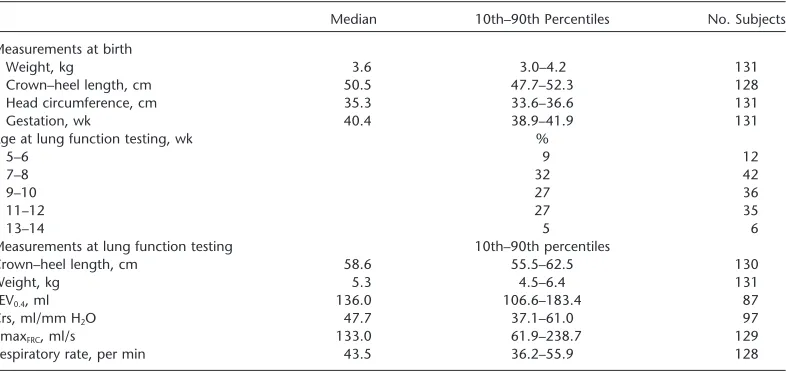
In a similar pattern to that found for FEV0.4, age- and sex-adjusted Crs fell by 7.0% for each SD decrease in birth weight (95% CI, 3.6 to 10.6, p⬍0.001), by 5.8% (95% CI, 2.2 to 9.5, p⫽0.002) for each SD decrease in neonatal head circumference, and by 7.1% (95% CI, 3.5 to 10.9, p ⬍ 0.001) for each SD decrease in neonatal crown–heel length. Age- and sex-adjusted Crs fell by 4.6% for each SD decrease in infant weight at lung function testing (95% CI, 0.8 to 8.6, p⫽ 0.02) and by 7.5%
TABLE 3. GEOMETRIC MEAN FEV0.4 (ml), ADJUSTED FOR AGE AT TESTING, ACCORDING
TO THIRDS OF BIRTH-WEIGHT SD SCORE AND WEIGHT-GAIN SD SCORE Weight-gain SD Score
Lowest Third Middle Third Highest Third
Birth-weight SD Score ⭐ ⫺0.54 SD ⫺0.54 to 0.21 SD ⬎0.21 SD Total
Lowest third,⭐ ⫺0.2 SD 124 (6) 129 (12) 116 (6) 125 (24)
Middle third,⫺0.2 to 0.58 SD 141 (11) 144 (10) 129 (12) 137 (33)
Highest third,⬎0.58 SD 148 (13) 128 (8) 143 (9) 141 (30)
Total 140 (30) 134 (30) 130 (27) 135 (87)
Each cell contains the back-transformed geometric mean, with the number of observations contributing to the mean in parentheses.
The geometric mean (SD of the logged values) for age-adjusted FEV0.4was 135 (0.20) ml.
(95% CI, 3.9 to 11.3, p⬍0.001) for each SD decrease in infant crown–heel length but was not related to infant weight gain (p⫽ 0.8). Table 4 presents mean age- and sex-adjusted Crs according to thirds of the infants’ birth weight and infant weight gain.
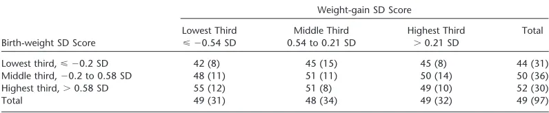
Age-adjusted V˙ maxFRCwas not related to birth weight (p⫽ 0.7) or to neonatal head circumference (p⫽0.8) and crown–heel length (p⫽0.4). Age-adjusted V˙ maxFRCwas also not related to crown–heel length SD score at the time of lung function testing (p⫽0.7) but did, however, decrease by 8.6% for each SD increase in weight at this time (95% CI, 0.3 to 16.2, p ⫽ 0.04) and by 11.0% for each SD increase in infant weight gain (95% CI, 3.2 to 18.2, p⫽0.007). Table 5 presents mean age-adjusted V˙ maxFRC according to thirds of the infants’ birth weight and infant weight gain. Although the effect of higher infant weight gain on lower V˙ maxFRCwas strongest in the lowest birth-weight grouping, there was no statistically significant interaction between the effects of birth weight and infant weight gain (data not shown).
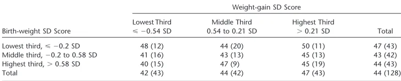
Respiratory rate rose by 2.9% (95% CI, ⫺0.1 to 5.8, p ⫽ 0.06) for each SD decrease in birth weight, by 2.7% (95% CI,
[image:3.603.92.491.624.707.2]Figure 1. Scatterplot of FVC-adjusted FEV0.4 against weight gain SD score, including regression line for the association.
infant weight gain (data not shown), in a simultaneous analysis, respiratory rate was inversely related to weight at birth (p⫽ 0.001) and positively related to weight at lung function testing (p⫽0.003).
Confirmatory analyses omitting the infant’s age from the anal-yses of FEV0.4and V˙ maxFRCand age and sex from the analyses of Crs showed little change in the associations between lower birth size and lower FEV0.4 and Crs; V˙ maxFRC remained not associated with size at birth. Omitting age as a co-variate in the analyses of weight gain SD score marginally strengthened the inverse associations with FEV0.4(p⫽0.03) and V˙ maxFRC(p⫽ 0.001); Crs remained not associated with weight gain (p⫽0.4). Back-transformed mean values of FEV0.4, Crs, V˙ maxFRC, and respiratory rate according to thirds of birth weight and infant weight gain are graphically portrayed in the online supplement (seeFigure E2 in the online supplement).
Maternal Smoking and Infant Feeding
Of the 131 mothers, 21% reported smoking at the time of lung function testing. Compared with the infants of mothers who did not smoke, the infants of smokers were lighter by 0.281 kg (p⫽ 0.005) and shorter by 1.2 cm (p ⫽ 0.003) at birth. Maternal smoking lowered FEV0.4by 7% (95% CI,⫺3% to 16%), FVC-adjusted FEV0.4by 1% (95% CI,⫺4% to 6%), Crsby 8% (95% CI,⫺1% to 16%), and V˙ maxFRCby 11% (95% CI, ⫺10% to 27%), but none of these associations was significant (p⫽0.2, 0.7, 0.07, and 0.3, respectively); respiratory rate was similar in infants born to smokers and nonsmokers (p⫽ 0.9). Findings were similar for smoking status before conception (data not
TABLE 4. GEOMETRIC MEAN CRS (ml/cm H2O) ADJUSTED FOR SEX AND AGE AT TESTING
ACCORDING TO THIRDS OF BIRTH-WEIGHT SD SCORE AND WEIGHT-GAIN SD SCORE Weight-gain SD Score
Lowest Third Middle Third Highest Third Total Birth-weight SD Score ⭐ ⫺0.54 SD 0.54 to 0.21 SD ⬎0.21 SD
Lowest third,⭐ ⫺0.2 SD 42 (8) 45 (15) 45 (8) 44 (31)
Middle third,⫺0.2 to 0.58 SD 48 (11) 51 (11) 50 (14) 50 (36)
Highest third,⬎0.58 SD 55 (12) 51 (8) 49 (10) 52 (30)
Total 49 (31) 48 (34) 49 (32) 49 (97)
Each cell contains the back-transformed geometric mean, with the number of observations contributing to the mean in parentheses.
Geometric mean (SD of the logged values) for age/sex-adjusted respiratory system compliance was 49 (0.18) ml/cm H2O.
shown). Of the 131 infants, 56% were breast-fed at the time of lung function testing; all lung function variables were similar in those breast-fed and bottle-fed (data not shown). Further analy-ses showed that the associations of infant anthropometry and weight gain with lung function were changed little by taking account of maternal smoking and infant feeding.
DISCUSSION
Age at Test
This is one of the largest studies measuring raised volume RTC in healthy infants of this age range and, therefore, contributes to the limited population-based data that have been published. Our study had a narrow age window for testing, but we found that age at test nonetheless had a major influence on FEV0.4, Crs, and V˙ maxFRC. This has important implications for accurate reporting of age at test in future studies. Other studies over much larger age ranges have also found a correlation between age and lung function (27, 28), but our data highlight that even an additional week can lead to a significant change in FEV0.4, Crs, and V˙ maxFRC. However, we found that among infants of below-average birth weight there was no increase in V˙ maxFRC with age. As our data are cross-sectional, further work is required to confirm and explain this observation. Nonetheless, if a high proportion of normal infants of below-average birth weight are essentially increasing their lung size over the first two months of life (greater FEV0.4and Crs) but failing to increase their maximal forced expiratory flows at FRC, this is a potentially important finding that could have major implications for respiratory health. Confirmation of how lung development evolves over the first months of life is needed, but using currently available techniques, the practical and ethical issues of conducting longitudinal studies to investigate lung function in infancy are considerable.
Maternal Smoking and Sex
[image:4.603.103.501.625.707.2]TABLE 5. GEOMETRIC MEAN V˙maxFRC (ml/s) ADJUSTED FOR AGE AT TESTING ACCORDING TO THIRDS OF BIRTH-WEIGHT SD SCORE AND WEIGHT-GAIN SD SCORE
Weight-gain SD Score
Lowest Third Middle Third Highest Third
Birth-weight SD Score ⭐ ⫺0.54 SD ⫺0.54 to 0.21 SD ⬎0.21 SD Total
Lowest third,⭐ ⫺0.2 SD 140 (12) 116 (18) 107 (11) 120 (41) Middle third,⫺0.2 to 0.58 SD 146 (16) 128 (14) 131 (14) 135 (44) Highest third,⬎0.58 SD 137 (15) 131 (10) 110 (19) 123 (44)
Total 141 (43) 123 (42) 115 (44) 126 (129)
Each cell contains the back-transformed geometric mean, with the number of observations contributing to the mean in parentheses.
The geometric mean (SD of the logged values) for age-adjusted V˙maxFRCwas 126 (0.47) ml/s.
cotinine, we cannot exclude misclassification of maternal smok-ing. However, we found a significant effect of maternal smoking on birth weight (mean reduction in birth weight 281 g) that is similar to the around 150 g reduction reported in a meta-analysis of maternal smoking (29). Moreover, we ascertained maternal smoking using an administered questionnaire both at the time of lung function testing and prospectively at home before con-ception; the associations that we found between infant birth weight and weight gain and the lung function parameters were changed little by taking account of maternal smoking at either time point.
Our data suggest that male infants have more compliant lungs, but we found no sex difference in V˙ maxFRC or FEV0.4. Some studies measuring V˙ maxFRCsuggest that girls demonstrate higher flows than boys (2, 8, 18, 19), although other studies, like ours, have found no difference (27, 30, 31). Studying forced expiratory flows from raised volumes, Jones and colleagues found that sex was only significant for FEF75, girls having higher flows than boys after accounting for body length and smoking during preg-nancy (28). The same study found no difference in FVC between girls and boys aged 3–149 weeks.
Size at Birth in Relationship to FEV0.4and Crs
Independent of the infant’s age and weight at the time of testing, we found that infants who had a lower birth weight and shorter neonatal crown–heel length had a lower FEV0.4. This effect was not apparent for FVC-adjusted FEV0.4, presumably because this correction will normalize for lung size. We have not studied severely compromised infants of low birth weight, but in a study of infants who were either small for gestational age or appropri-ately sized, Lum and colleagues reported diminished airway function in the low birth-weight infants (32). Our study excluded infants with major congenital abnormalities or neonatal prob-lems and was designed to recruit healthy, term infants
represen-TABLE 6. GEOMETRIC MEAN RESPIRATORY RATE (/min) ACCORDING TO THIRDS OF BIRTH-WEIGHT SD SCORE AND WEIGHT-GAIN SD SCORE
Weight-gain SD Score
Lowest Third Middle Third Highest Third
Birth-weight SD Score ⭐ ⫺0.54 SD 0.54 to 0.21 SD ⬎0.21 SD Total
Lowest third,⭐ ⫺0.2 SD 48 (12) 44 (20) 50 (11) 47 (43)
Middle third,⫺0.2 to 0.58 SD 41 (16) 43 (13) 45 (13) 43 (42)
Highest third,⬎0.58 SD 40 (15) 47 (9) 45 (19) 44 (43)
Total 42 (43) 44 (42) 47 (43) 44 (128)
Each cell contains the back-transformed geometric mean, with the number of observations contributing to the mean in parentheses.
The geometric mean (SD of the logged values) for respiratory rate was 44 (0.17).
tative of the local population; 7% of those we studied had a birth weight less than the 10th percentile for gestational age of unknown etiology, but the associations we found were graded across the range of size at birth. Our data suggest that restricted fetal growth is associated with particular impairment of lung and airway development, which are adversely affected more than one might expect for the infant’s size. Moreover, our data indi-cate that the reduced adult lung function of individuals of below-average birth weight (11, 12) at least in part originates from impaired lung and airway development in the prenatal and im-mediate postnatal period and not simply from increased suscepti-bility to respiratory illness after birth.
We found that Crs was highly correlated with birth weight and length, with larger babies having more compliant respiratory systems. Crs is influenced by chest wall compliance as well as lung compliance and interpretation of these data needs caution. Joyce and colleagues found that Crs was lower in term growth-restricted lambs than in control animals (33) and that the lung component was significantly lower in the growth-restricted group. If restricted fetal growth is associated with less compliant lungs, it has important implications for future respiratory health; if smaller infants have decreased elastic tissue laid downin utero, they are likely to have an increased rate of respiratory decline with aging.
[image:5.603.92.491.625.706.2](38, 39). Some of these abnormalities of lung development may be direct consequences of nutrient deficiencies. However, the nutritional stress may also induce fetal endocrine responses that themselves affect lung development (41).
Our data cannot definitively identify the timing of the intra-uterine effect on lung development and infant lung function. However, lower infant FEV0.4and Crs and faster respiratory rate had similar relationships with lower birth weight, neonatal head circumference, and crown–heel length, suggesting symmetrical growth restriction. Symmetrical restriction of fetal growth is thought to originate in early pregnancy, whereas asymmetrical growth restriction reflects impaired soft tissue accretion in late pregnancy. Our data provide weak evidence for an early preg-nancy effect on infant FEV0.4, Crs, and respiratory rate, perhaps mediated by impaired development of the bronchial tree, which is complete by 16-weeks gestation. Alveolar development does not begin until airway growth is complete but continues through both prenatal and postnatal life until 8–10 years of age; alveolar number at birth has been reported as being anything between 8% and 50% of the eventual adult number (42, 43).
Postnatal Weight Gain in Relationship to V˙maxFRC
and FVC-adjusted FEV0.4
In contrast to our observations for FEV0.4and Crs, we found that V˙ maxFRCwas not related to the infant’s weight and crown–heel length at birth but was lower in those who gained more weight between birth and testing. In consequence, V˙ maxFRCwas lower in those with a greater weight at testing; there was, however, no association between V˙ maxFRCand crown–heel length in infancy. Postnatal weight gain was not related to Crs and only weakly related to FEV0.4. Although both V˙ maxFRCand FEV in timet are influenced by lung size, FEV in timetis thought principally to reflect the size and function of larger airways, whereas V˙ maxFRCis influenced more by the function of the peripheral airways (24). Our observations raise the possibility that particular aspects of growth may have differing effects on these components of lung development. This is supported by our observation that when we normalized FEV0.4for FVC we found a strong inverse rela-tionship with postnatal weight gain. Previous studies of infants and young children of widely varying ages with respiratory disor-ders have reported that interpretation of FEV in timet/FVC is complicated by inverse associations with age and respiratory rate (24), but in the normal infants that we studied over a narrow age range, FEV in timet/FVC was not related to age or respira-tory rate.
We found that the lowest V˙ maxFRCwas in infants of below-average birth weight with above-below-average postnatal weight gain (Table 5). One explanation for these findings is that above-average postnatal weight gain may serve to identify fetuses that had followed a rapid trajectory of prenatal growth, which faltered in late pregnancy, impairing lung growth and development. Fe-tuses subject to such growth faltering in late gestation have increased postnatal weight gain unless they are exposed to severe or prolonged restriction in nutrient supplyin utero.
An alternative explanation for the association between in-creased postnatal weight gain and diminished lung function is that above-average weight gain is itself impairing lung develop-ment. There has been considerable interest in the relationship between asthma and obesity in recent years (44–47), but this is the first study to suggest that increased weight gain in early infancy is associated with worse lung function. It is unlikely that different feeding modes are responsible for this finding, as infants who were breast-fed and bottle-fed had similar lung function. As children who have high weight gain in early infancy tend to have a higher body mass index and a more central fat distribution
in childhood, our data offer a possible explanation for the associ-ation of asthma and obesity.
Respiratory Rate
We studied infants during quiet sleep augmented by chloral hydrate; respiratory rate measured in these circumstances pro-vides no more than a crude summary measure of lung function. Nonetheless, we found that infants that were of below-average birth weight and who had above-average postnatal weight gain had a faster respiratory rate. This observation again points to impaired lung development in these infants and complements our findings relating to the detailed measures of lung function. In conclusion, we describe associations of birth anthropome-try and early postnatal weight gain with lung function measured in the first few months of life. We have shown that even a 1-week increase in age is significant when reporting these infant lung function parameters. The absence of serial measurements in our study precludes definitive conclusions, but the observations sug-gest that lower rates of fetal growth and higher rates of early infancy weight gain are associated with impaired lung develop-ment. The association with higher infant weight gain appears paradoxical but could reflect catch-up in infants whose fetal growth faltered in late gestation. This may result in some infants having relatively small lungs, which have not grown at the same rate as the “infant.” These findings may have implications for respiratory health in childhood and later life.
Conflict of Interest Statement:J.S.L. does not have a financial relationship with a commercial entity that has an interest in the subject of this manuscript; H.M.I. does not have a financial relationship with a commercial entity that has an interest in the subject of this manuscript; K.M.G. does not have a financial relationship with a commercial entity that has an interest in the subject of this manuscript; C.T.F. does not have a financial relationship with a commercial entity that has an interest in the subject of this manuscript; J.O.W. is chairman of a scientific advisory board overseeing the conduct of two studies (ETAC and EPAAC) investigating the early life origins of allergy and the effect of cetirizine and levocetirizine for UCB Pharma and has given lectures for a number of pharmaceutical companies, includ-ing UCB Pharma, Novartis, SHS International, Merck Sharpe & Dohme, Glaxo-SmithKline, and AstraZeneca and over the last three years has received small grants from UCB Pharma for investigator-led research studies; R.K.G. does not have a financial relationship with a commercial entity that has an interest in the subject of this manuscript; J.B.C. does not have a financial relationship with a commercial entity that has an interest in the subject of this manuscript
Acknowledgment: The authors acknowledge the help of parents and infants who participated in this study. They are grateful to the staff of the Wellcome Trust Clinical Research Facility and Karen Collins for assisting with collection of infant lung function data. They are also grateful to the staff of the Southampton Women’s Survey for the collection of data on the mothers and for obtaining the birth anthropometric measurements. Dr. Jean Mulligan kindly provided assistance with derivation of the infant size SD scores. The authors would like to thank Professor Janet Stocks for her practical support and assistance in setting up and developing the Southampton Infant Lung Function Laboratory.
References
1. Clarke JR, Salmon B, Silverman M. Bronchial responsiveness in the
neonatal period as a risk factor for wheezing in infancy.Am J Respir
Crit Care Med1995;151:1434–1440.
2. Dezateux C, Stocks J, Dundas I, Fletcher ME. Impaired airway function and wheezing in infancy: the influence of maternal smoking and a
genetic predisposition to asthma.Am J Respir Crit Care Med1999;
159:403–410.
3. Martinez FD, Morgan WJ, Wright AL, Holberg CJ, Taussig LM. Dimin-ished lung function as a predisposing factor for wheezing respiratory
illness in infants.N Engl J Med1988;319:1112–1117.
4. Martinez FD, Morgan WJ, Wright AL, Holberg C, Taussig LM. Initial airway function is a risk factor for recurrent wheezing respiratory illnesses during the first three years of life: Group Health Medical
Associates.Am Rev Respir Dis1991;143:312–316.
5. Martinez FD, Wright AL, Taussig LM, Holberg CJ, Halonen M, Morgan WJ. Asthma and wheezing in the first six years of life: the Group
Health Medical Associates.N Engl J Med1995;332:133–138.
respiratory symptoms in a high risk population.Thorax2002;57:388– 392.
7. Stick SM, Burton PR, Gurrin L, Sly PD, LeSouef PN. Effects of maternal smoking during pregnancy and a family history of asthma on
respira-tory function in newborn infants.Lancet1996;348:1060–1064.
8. Young S, O’Keeffe PT, Arnott J, Landau LI. Lung function, airway responsiveness, and respiratory symptoms before and after
bronchio-litis.Arch Dis Child1995;72:16–24.
9. Nikolajev K, Heinonen K, Hakulinen A, Lansimies E. Effects of intra-uterine growth retardation and prematurity on spirometric flow values
and lung volumes at school age in twin pairs.Pediatr Pulmonol1998;
25:367–370.
10. Rona RJ, Gulliford MC, Chinn S. Effects of prematurity and intrauterine
growth on respiratory health and lung function in childhood.BMJ
1993;306:817–820.
11. Stein CE, Kumaran K, Fall CH, Shaheen SO, Osmond C, Barker DJ.
Relation of fetal growth to adult lung function in south India.Thorax
1997;52:895–899.
12. Barker DJ, Godfrey KM, Fall C, Osmond C, Winter PD, Shaheen SO. Relation of birth weight and childhood respiratory infection to adult
lung function and death from chronic obstructive airways disease.BMJ
1991;303:671–675.
13. Svanes C, Omenaas E, Heuch JM, Irgens LM, Gulsvik A. Birth character-istics and asthma symptoms in young adults: results from a
population-based cohort study in Norway.Eur Respir J1998;12:1366–1370.
14. Hanrahan JP, Tager IB, Segal MR, Tosteson TD, Castile RG, Van Vu-nakis H, Weiss ST, Speizer FE. The effect of maternal smoking during
pregnancy on early infant lung function.Am Rev Respir Dis1992;145:
1129–1135.
15. Tager IB, Ngo L, Hanrahan JP. Maternal smoking during pregnancy:
effects on lung function during the first 18 months of life.Am J Respir
Crit Care Med1995;152:977–983.
16. Young S, Le Souef PN, Geelhoed GC, Stick SM, Turner KJ, Landau LI. The influence of a family history of asthma and parental smoking on
airway responsiveness in early infancy.N Engl J Med1991;324:1168–
1173.
17. Stocks J, Dezateux C. The effect of parental smoking on lung function
and development during infancy.Respirology2003;8:266–285.
18. Gregory A, Doull I, Pearce N, Cheng S, Leadbitter P, Holgate S, Beasley R. The relationship between anthropometric measurements at birth:
asthma and atopy in childhood.Clin Exp Allergy1999;29:330–333.
19. Leadbitter P, Pearce N, Cheng S, Sears MR, Holdaway MD, Flannery EM, Herbison GP, Beasley R. Relationship between fetal growth and
the development of asthma and atopy in childhood.Thorax1999;54:
905–910.
20. Godfrey KM, Barker DJ, Osmond C. Disproportionate fetal growth and
raised IgE concentration in adult life.Clin Exp Allergy1994;24:641–
648.
21. Beasley R, Leadbitter P, Pearce N, Crane J. Is enhanced fetal growth a
risk factor for the development of atopy or asthma?Int Arch Allergy
Immunol1999;118:408–410.
22. Feher A, Castile R, Kisling J, Angelicchio C, Filbrun D, Flucke R, Tepper R. Flow limitation in normal infants: a new method for forced
expiratory maneuvers from raised lung volumes.J Appl Physiol1996;
80:2019–2025.
23. Turner DJ, Stick SM, Lesouef KL, Sly PD, LeSouef PN. A new technique to generate and assess forced expiration from raised lung volume in
infants.Am J Respir Crit Care Med1995;151:1441–1450.
24. Ranganathan SC, Hoo AF, Lum SY, Goetz I, Castle RA, Stocks J. Exploring the relationship between forced maximal flow at functional residual capacity and parameters of forced expiration from raised lung
volume in healthy infants.Pediatr Pulmonol2002;33:419–428.
25. Freeman JV, Cole TJ, Chinn S, Jones PR, White EM, Preece MA. Cross
sectional stature and weight reference curves for the UK, 1990.Arch
Dis Child1995;73:17–24.
26. Cole TJ. Conditional reference charts to assess weight gain in British
infants.Arch Dis Child1995;73:8–16.
27. Tepper RS, Reister T. Forced expiratory flows and lung volumes in
normal infants.Pediatr Pulmonol1993;15:357–361.
28. Jones M, Castile R, Davis S, Kisling J, Filbrun D, Flucke R, Goldstein A, Emsley C, Ambrosius W, Tepper RS. Forced expiratory flows and
volumes in infants: normative data and lung growth.Am J Respir Crit
Care Med2000;161:353–359.
29. Kramer MS. Determinants of low birth weight: methodological
assess-ment and meta-analysis.Bull World Health Organ1987;65:663–737.
30. Hanrahan JP, Tager IB, Castile RG, Segal MR, Weiss ST, Speizer FE. Pulmonary function measures in healthy infants: variability and size
correction.Am Rev Respir Dis1990;141:1127–1135.
31. Taussig LM, Landau LI, Godfrey S, Arad I. Determinants of forced
expiratory flows in newborn infants.J Appl Physiol1982;53:1220–1227.
32. Lum S, Hoo AF, Dezateux C, Goetz I, Wade A, DeRooy L, Costeloe K, Stocks J. The association between birthweight, sex, and airway
function in infants of nonsmoking mothers.Am J Respir Crit Care
Med2001;164:2078–2084.
33. Joyce BJ, Louey S, Davey MG, Cock ML, Hooper SB, Harding R. Compromised respiratory function in postnatal lambs after placental
insufficiency and intrauterine growth restriction.Pediatr Res2001;50:
641–649.
34. Wignarajah D, Cock ML, Pinkerton KE, Harding R. Influence of intra-uterine growth restriction on airway development in fetal and postnatal
sheep.Pediatr Res2002;51:681–688.
35. Maritz GS, Cock ML, Louey S, Joyce BJ, Albuquerque CA, Harding R. Effects of fetal growth restriction on lung development before and
after birth: a morphometric analysis.Pediatr Pulmonol2001;32:201–
210.
36. Maritz GS, Cock ML, Louey S, Suzuki K, Harding R. Fetal growth restriction has long-term effects on postnatal lung structure in sheep.
Pediatr Res2003;55:287–295.
37. Lin Y, Lechner AJ. Surfactant content and type II cell development in
fetal guinea pig lungs during prenatal starvation.Pediatr Res1991;
29:288–291.
38. Lechner AJ, Winston DC, Bauman JE. Lung mechanics, cellularity, and surfactant after prenatal starvation in guinea pigs.J Appl Physiol1986; 60:1610–1614.
39. Faridy EE. Effect of maternal malnutrition on surface activity of fetal
lungs in rats.J Appl Physiol1975;39:535–540.
40. Lechner AJ, Tull DS. Prenatal starvation retards development of the
ventilatory response to hypoxia in newborn guinea pigs.Pediatr Res
1986;20:920–924.
41. Harding R, Cock ML, Louey S, Joyce BJ, Davey MG, Albuquerque CA, Hooper SB, Maritz GS. The compromised intra-uterine environment:
implications for future lung health.Clin Exp Pharmacol Physiol2000;
27:965–974.
42. Angus GE, Thurlbeck WM. Number of alveoli in the human lung.J Appl
Physiol1972;32:483–485.
43. Davies G, Reid L. Growth of the alveoli and pulmonary arteries in
childhood.Thorax1970;25:669–681.
44. Chinn S, Rona RJ. Can the increase in body mass index explain the
rising trend in asthma in children?Thorax2001;56:845–850.
45. Figueroa-Munoz JI, Chinn S, Rona RJ. Association between obesity and
asthma in 4–11 year old children in the UK.Thorax2001;56:133–137.
46. Belamarich PF, Luder E, Kattan M, Mitchell H, Islam S, Lynn H, Crain EF. Do obese inner-city children with asthma have more symptoms
than nonobese children with asthma?Pediatrics2000;106:1436–1441.
47. Castro-Rodriguez JA, Holberg CJ, Morgan WJ, Wright AL, Martinez FD. Increased incidence of asthma like symptoms in girls who become
overweight or obese during the school years.Am J Respir Crit Care