S T U D Y P R O T O C O L
Open Access
Study Protocol - Accurate assessment of kidney
function in Indigenous Australians: aims and
methods of the eGFR Study
Louise J Maple-Brown
1,2*, Paul D Lawton
2, Jaquelyne T Hughes
1, Suresh K Sharma
1, Graham RD Jones
3,
Andrew G Ellis
4, Wendy Hoy
5, Alan Cass
6, Richard J MacIsaac
7, Ashim K Sinha
8, Mark AB Thomas
9,
Leonard S Piers
10, Leigh C Ward
11, Katrina Drabsch
1, Sianna Panagiotopoulos
7, Robyn McDermott
12, Kevin Warr
9,
Sajiv Cherian
13, Alex Brown
14, George Jerums
7, Kerin O
’
Dea
12Abstract
Background:There is an overwhelming burden of cardiovascular disease, type 2 diabetes and chronic kidney disease among Indigenous Australians. In this high risk population, it is vital that we are able to measure accurately kidney function. Glomerular filtration rate is the best overall marker of kidney function. However, differences in body build and body composition between Indigenous and non-Indigenous Australians suggest that creatinine-based estimates of glomerular filtration rate derived for European populations may not be appropriate for Indigenous Australians. The burden of kidney disease is borne disproportionately by Indigenous Australians in central and northern Australia, and there is significant heterogeneity in body build and composition within and amongst these groups. This heterogeneity might differentially affect the accuracy of estimation of glomerular filtration rate between different Indigenous groups. By assessing kidney function in Indigenous Australians from Northern Queensland, Northern Territory and Western Australia, we aim to determine a validated and practical measure of glomerular filtration rate suitable for use in all Indigenous Australians.
Methods/Design:A cross-sectional study of Indigenous Australian adults (target n = 600, 50% male) across 4 sites: Top End, Northern Territory; Central Australia; Far North Queensland and Western Australia. The reference measure of glomerular filtration rate was the plasma disappearance rate of iohexol over 4 hours. We will compare the accuracy of the following glomerular filtration rate measures with the reference measure: Modification of Diet in Renal Disease 4-variable formula, Chronic Kidney Disease Epidemiology Collaboration equation, Cockcroft-Gault formula and cystatin C- derived estimates. Detailed assessment of body build and composition was performed using anthropometric measurements, skinfold thicknesses, bioelectrical impedance and a sub-study used dual-energy X-ray absorptiometry. A questionnaire was performed for socio-economic status and medical history.
Discussion:We have successfully managed several operational challenges within this multi-centre complex clinical research project performed across remote North, Western and Central Australia. It seems unlikely that a single correction factor (similar to that for African-Americans) to the equation for estimated glomerular filtration rate will prove appropriate or practical for Indigenous Australians. However, it may be that a modification of the equation in Indigenous Australians would be to include a measure of fat-free mass.
* Correspondence: [email protected]
1Menzies School of Health Research, Institute of Advanced Studies, Charles
Darwin University, Darwin, Australia
Background
Indigenous Australians have rates of cardiovascular disease (CVD) mortality some 7-10 times higher than non-Indigenous Australians aged 25-64 years, a preva-lence of diabetes some 10 times higher (age 20-50 years), new cases of end stage kidney disease (ESKD) 10-15 times higher and life expectancy 15-20 years shorter [1-3]. Type 2 diabetes is common amongst Indigenous Australians with ESKD: 77% of Indigenous Australians with new ESKD in 2007 compared to 33% of non-Indigenous Australians have Type 2 diabetes as a comorbidity [4]. Rates of ESKD in Indigenous Aus-tralians are disproportionately highest in those in cen-tral and northern Auscen-tralia [5]. This group of people is widely dispersed and heterogenous: they live in urban, regional and remote settings and have wide variation in diet, body habitus, genetic admixture and socioeco-nomic background [6-8].
With the serious burden of ESKD in Indigenous Aus-tralians it is essential to be able to assess accurately renal function in these populations. Glomerular filtra-tion rate (GFR) is the best overall marker of renal func-tion in people with healthy or diseased kidneys [9] and is an independent predictor of renal and cardiovascular events [10,11]. At present there are no validated meth-ods for estimating GFR in Indigenous Australians [12,13]. Differences in body build and body composition between Indigenous and non-Indigenous Australians suggest that creatinine-based estimates of GFR (eGFR) derived for European populations may not be appropri-ate for Indigenous Australians, as muscle mass is a key determinant of serum creatinine levels. Compared to Australians of European background, Indigenous Aus-tralians from remote central and northern Australia have more fat for a given weight or BMI [7].
It is increasingly recognised that current creatinine-based formulae used to estimate GFR might not be gen-eralisable across all clinical presentations [14,15]. They also lack validation in ethnic populations apart from Caucasians and African Americans [12,14]. The Modifi-cation of Diet in Renal Disease 4-variable formula (MDRD) was derived from a study population with impaired renal function; and thus is less accurate within a“healthy range” GFR. To address this issue a revised equation (Chronic Kidney Disease Epidemiology Colla-boration, CKD-EPI) has been described [16].
An alternate approach is to use a different analyte from creatinine to estimate renal function and develop GFR prediction equations. Serum cystatin C has been proposed as a simple, reliable and accurate marker of GFR when compared to creatinine-based methods [17,18]. Although there is some evidence to suggest that cystatin C levels might be influenced by extra-renal
factors, numerous studies have suggested that cystatin C levels or GFR estimates based on cystatin C levels alone are more accurate indices of renal function than creati-nine-based methods [19]. However, at this time cystatin C assays are not routinely available, they are consider-ably more expensive than creatinine assays and are not yet fully standardised. Thus for the immediate future, optimisation of creatinine-based methods remains important.
The aim of the present study is to evaluate and improve if necessary the accuracy and precision of eGFR estimates in Indigenous Australians, taking into account the heterogeneity in body build and body composition across different populations. The study’s hypotheses are: (i) that differences in body build and body composition between Indigenous and non-Indigenous Australians will affect the validity of creatinine-based estimated measures of GFR (using existing predictive equations) and that accuracy of estimates of GFR will vary across different Indigenous population groups; and (ii) that a more accurate assessment of GFR will be obtained by a formula involving serum creatinine and percent fat free mass (obtained by a simple, validated bioimpedance measurement) or by using an alternative biochemical measure such as cystatin C. It is anticipated that this will be of use in both Indigenous and non-Indigenous Australians of variable body builds, particularly at GFR>60 ml/min/1.73 m2.
Methods/Design Setting & location
Centre, Meriba Dhoeynidhay Yabu Torres Strait & Northern Penninsula Area Community Council.
Participants and recruitment
Participants were Indigenous Australians (target n = 600 with equal numbers of males and females) aged 16 years and above, from urban, rural and remote regions of Northern and Central Australia across the following 5 strata:
(i) “healthy” group: nil diabetes, hypertension, chronic kidney disease (CKD), albuminuria; (ii) participants with diabetes or albuminuria &
eGFR (MDRD-4) >90 ml/min/1.73 m2; (iii) eGFR 60-90 ml/min/1.73 m2;
(iv) eGFR 30-59 ml/min/1.73 m2; (v) eGFR <15-29 ml/min/1.73 m2.
Indigenous Australians fulfilled the definition of
‘Aboriginal and/or Torres Strait Islander’ according to the standard method used in National Census data col-lection: “1) is of Aboriginal and/or Torres Strait Islander descent; 2) identifies as an Australian Aboriginal and/or Torres Strait Islander; and 3) is accepted as such by the community in which he or she lives or has lived”.
Exclusion criteria were: participants with rapidly chan-ging kidney function, participants receiving dialysis, women who were pregnant or breastfeeding and people with a history of allergy or adverse reaction to iodine-based contrast media.
Participants with CKD and/or diabetes were recruited from participating Aboriginal Medical Services and the nephrology, endocrinology and outreach kidney disease clinics associated with Royal Darwin Hospital, Alice Springs Hospital, Cairns Base Hospital and the Royal Perth Hospital. The identification and recruitment of the“healthy” group was through community networks, staff members of health services, word-of-mouth, self referral and family members of participants.
Funding
Major funding was provided by the National Health and Medical Research Council of Australia (NHMRC, pro-ject grant #545202). Prior to award of this grant, pilot funding was received from: Kidney Health Australia, the Colonial Foundation of Australia and Menzies School of Health Research. Additional funding for equipment was received from Rebecca L Cooper Medical Research Grant.
Ethics
The study was approved by the joint Menzies School of Health Research - Northern Territory Department of Health and Community Services Human Research Ethics
Committee. The project was considered and approved by both the Aboriginal sub-committee, which has absolute right of veto, and by the main committee. The study was also approved by the following Human Research Ethics Committees: Central Australian Human Research Ethics Committee, Western Australian Aboriginal Health Infor-mation and Ethics Committee, Royal Perth Hospital Ethics Committee and Cairns and Hinterland Health Ser-vices District Human Research Ethics Committee.
Staff
Six people were employed on contracts over the course of the study. Of these, four were Indigenous and two were non-Indigenous. Additional people were employed on a casual basis during visits to discrete communities; these community members acted as local facilitators. One post-graduate research student (who is Indigenous) also participated in the study and was substantially involved in data collection, whilst also making a signifi-cant contribution to study profile and media relations. The majority of staff members had prior health qualifi-cations and experience: three were Aboriginal Health Workers, two were registered nurses and the post-grad-uate research student was a specialist nephrologist. The staff member without a health qualification completed a venipuncture certification course.
Consent
Examination protocol
Following confirmation of contact details and eligibility and after obtaining informed consent, performance of the reference measure of GFR and a health examination was undertaken. This examination included: collection of blood and urine samples; clinical and anthropometric measurements; and the administration of questionnaires. The performance of the reference measurement of GFR required a minimum of 4 hours for participants, during which time participants were given a healthy meal and health promotion information. Details of the examina-tion protocol:
(i) Reference measure of GFR: determined by mea-suring the renal clearance of non-isotopic iohexol. Iohexol was injected (5.445 ml, 300 mg/ml “ Omni-paque”) into an antecubital vein (or forearm/hand vein if unable to use antecubital vein) and flushed with 10 ml of normal saline. The volume of iohexol includes the prime volume (0.445 ml) in the tubing of the butterfly cannula used to inject the iohexol. Extravasation was assessed by clinical inspection and palpation of the injection site. If there was any evi-dence of extravasation then the study was suspended and the participant invited to return in 3-7 days for assessment. Using the contralateral arm, venous blood samples (5 ml) were collected for measure-ment of iohexol at 120, 180 and 240 min post injec-tion. Participants were closely supervised for possible adverse reactions - whilst also receiving culturally appropriate education about chronic kidney disease, diabetes and/or healthy lifestyle.
Slope-intercept GFR was calculated from the formula:
Slope-intercept GFR (mL/min) = k × Iohexol dose (μg)/C0 (μg/mL) where k is the slope of the semilog plot of plasma Iohexol concentration versus time (plotted using 3 points, refer above), and C0 is the cal-culated Iohexol concentration at time zero (intercept). This value was multiplied by 1.73 and divided by the body surface area (BSA, calculated from the equation BSA = 0.20247 × height (metres)0.725 × weight (kg)0.425 [20]. The BSA-slope intercept GFR value (mL/min/1.73 m2) was corrected by the Brochner-Mortensen correc-tion factor = (0.990778 × GFR) - (0.001218 × GFR2) [21] providing a reference GFR value (mL/min/1.73 m2).
(ii) Blood and urine measures:
a. Serum creatinine measurements: performed both locally and at a central laboratory (The Department of Laboratory Medicine, Austin Health).
b. Estimates of GFR/creatinine clearance (where Scris serum creatinine concentration in μmol/L,
age in years, weight in kg, eGFR in mL/min/1.73 m2):
i. The adjusted MDRD-4 variable formula (for creatinine measurements traceable to the IDMS method and reported inμmol/L)
eGFR = 175 × [(Scr × 0.0113)-1.154] × (age)-0.203 ×
(0.742 if female) × (1.212 if African-American) [22]. The above African-American “correction factor” was not used for Indigenous Australians.
ii. The CKD-EPI formula [16]:
GFR = 141 × min(Scr × 0.0113/k, 1)a× max(Scr × 0.0113/k, 1)-1.209 × 0.993Age × 1.018 [if female] × 1.159 [if black], where Scr is serum creatinine, k is 0.7 for females and 0.9 for males, ais -0.329 for females and -0.411 for males, min indicates the minimum of Scr/k or 1, and max indicates the maximum of Scr/k or 1. As the correction factor“if black”applies to African-Ameri-cans this was not used for Indigenous Australians.
iii. The Cockcroft-Gault (C-G) formula: [23]
Creatinine Clearance (mL/min) = { [140 - age] × weight/[Scr × 0.814]} × (0.85 if female) *also calculated
using ideal body weight (IBW) according to Therapeutic Guidelines protocol when the patient is obese (BMI>30 kg/m2). IBW = 50 kg + 0.9 kg/each cm over 152 cm (-4.5 kg if female).
at 120 minutes for cystatin C, inflammatory mar-kers and a stored sample.
e. Urine: sample collected for microscopy and culture and determination of the albumin to creatinine ratio (ACR), performed as standard clinical care and analysed by local care providers. Urine cotinine was measured for quantitative assessment of cigarette smoking (recent nicotine consumption).
(iii) Clinical and anthropometric measures:
a. Blood pressure was measured three times after the participant had been seated quietly for at least 5 minutes. A Welch Allyn Spot Vital Signs monitor (Welch Allyn Medical Products, Skanea-teles Falls, USA) was used to measure systolic and diastolic pressure, with three minutes between readings. The pulse rate was recorded with each reading.
b. Height was measured to the nearest 0.1 cm using a wall-mounted stadiometer [24]. Partici-pants were shoeless and wore light clothing. They were instructed to stand facing forward with weight distributed evenly on both feet, with heels together and against the wall and arms hanging loosely by their sides. The participant’s head was positioned so that the Frankfort Plane was horizontal. The participant was then instructed to keep his or her eyes focused on a point straight ahead, breathe in deeply, and stretch to his or her fullest height, with heels still on the ground. The measuring plate was then lowered onto the scalp until it rested firmly on the top of the head and a reading to the nearest 0.1 cm was obtained and recorded. Following the measurement of weight (as described below), a second height measurement was obtained and recorded, again to the nearest 0.1 cm. If the two height measurements differed by more than 0.5 cm, a third measurement was taken and recorded.
c. Weight in kilograms was measured using a Seca digital portable scale (Model 767 and 841, Seca Deutschland, Hamburg, Germany). Partici-pants were asked to remove shoes, heavy gar-ments, heavy jewellery, belts, loose change, keys, mobile phones, and other items from pockets. With the scales at zero, participants were asked to stand on the centre of the base with feet together, arms hanging loosely at the sides and the head facing forward. Weight was recorded to the nearest 0.1 kilogram. Two separate measure-ments of weight were recorded and if the first two differed at all then a third measurement of weight was taken and recorded.
d. Waist and hip circumference were measured in centimetres using a 2-metre non-stretch flex-ible steel tape (Model W606PM Lufkin, Texas, USA). Following removal of outer clothing, tight-fitting garments, belts and heavy items from pockets, participants were asked to stand com-fortably erect in a relaxed manner, breathing normally, with weight balanced evenly on both feet, feet about 25-30 cm apart, and arms folded across the chest. Waist and hip measurements were taken alternately, with a minimum of two measurements for each. All measurements were taken and recorded to the nearest 0.1 centimetre. If the first two measurements of either waist or hip circumference differed by more than 1.0 cm, then a third measurement was taken for waist or hip, as relevant. Waist measurements were taken directly on the participant’s skin. Hip measure-ments were taken over the participant’s clothing, unless it was too tight or too baggy, in which case the participant was asked to remove it. The waist was defined as the midway point between the iliac crest and the costal margin. These two landmarks were identified and marked using a felt tip pen, and the distance between them mea-sured, with the midpoint marked. Once the tape was around the participant’s body at the appro-priate height and the tape was horizontal, the participant was asked to breathe out gently. The measurement was taken at the end of a normal expiration, with the tape pulled snug but not compressing the underlying soft tissue. If land-marks were unable to be identified (in the occa-sional very obese participant), waist measurements were omitted. The hip circumfer-ence was defined as the widest circumfercircumfer-ence over the buttocks and below the iliac crest. The hip circumference was measured at several posi-tions (starting with the widest part of the but-tocks when viewed from the side) and the widest circumference recorded. Fatty aprons were not included in the measurement of the hips. The measurement was taken with the tape in a hori-zontal position, and the tape was pulled to allow it to maintain its position without causing indentation.
insertion in CKD participants. The skinfold site was marked using a felt-tip pen and each site was identified as follows: biceps, anterior surface of the biceps midway between the acromial pro-cess and the elbow (antecubital fossa); triceps, posterior surface of triceps at midpoint from the acromial process (the most lateral and superior aspect of the acromial head) to the olecranon; subscapular, 2.5 cm infero-medially along a 45 degree line from inferior angle of the scapula; suprailiac, 2.5 cm supero-medially along a 45 degree line from where the medial border of the iliac crest tips in to the pelvis. The skin fold was then firmly grasped by the thumb and index fin-ger, using the pads at the tip of the thumb and finger, and the skin fold gently pulled away from the body. The caliper was placed perpendicular to the fold, on the site marked, dialled up, at approximately 1 cm below the finger and thumb. While maintaining the grasp of the skin fold, the caliper was released so that full tension was placed on the skin fold. The dial was read to the nearest 0.20 mm, 2 seconds after the grip was fully released. Three measurements were taken at each site were possible.
(iv) Whole body and segmental bioelectrical impe-dance measurements (BIA) were performed at a sin-gle frequency (50 kHz) using a four-terminal impedance plethysmograph (Model DF50, Impe-diMed, Brisbane). At study sites where a multi-fre-quency bioimpedance spectroscopy instrument (BIS, Model SFB7, ImpediMed, Brisbane) was available, this was used. BIS technology allowed prediction of body fluid volumes (intra- and extracellular water as well as total body water) at the theoretically optimal frequencies, providing precision and accuracy not achievable using the simpler BIA device that oper-ates at a single compromise frequency [26]. Predic-tion of body fluid volumes is particularly important in this study population at all stages of CKD - from hyperfiltration with“normal”renal function to ESKD (excluding dialysis). It is also worthy to note that BIS provides measures of both intra-and extracellu-lar water spaces and since muscle mass is strongly related to intracellular water space this will provide the opportunity to assess whether predictive power can be increased by inclusion of this variable in GFR formulae.
BIA/BIS was not performed if a participant had an indwelling electrical device (such as cardiac pace-maker). Participants were asked to void (empty blad-der) prior to the assessment. Assessment was performed after 5 minutes rest in the supine position with limbs abducted away from the trunk, ensuring
that thighs were not touching. Eight electrodes (elec-trocardiograph-style gel electrodes) were placed on bilateral dorsal surfaces of hands and feet (two elec-trodes on each hand and foot); after the surface was cleansed with an alcohol wipe. At the wrist, the proximal electrode was placed in the midline of the ulnar styloid process and the distal electrode was placed 5 cm distal to the proximal electrode. At the ankle, the proximal electrode was placed at the mid-point between the medial and lateral malleoli and the distal electrode was placed 5 cm distal to the proximal electrode. Whole body and segmental impedance measurements were according to pre-viously described methods [27].
(v) DXA Sub-study: Further assessment of body composition by dual-energy X-ray absorptiometry (DXA) assessment of total body fat mass, lean mass, bone mineral content and bone density was per-formed according to standard techniques in sites where this imaging technique was available: Darwin, Perth and Torres Strait. DXA is a reference method for assessing body composition but impedance methods, particularly inexpensive single frequency BIA, are much more practical in remote locations (where DXA is indeed not possible/available). Hence, the aim of the sub-study comparing DXA and BIA was to see if BIA is a reasonable substitute for DXA, thereby enabling body composition assess-ment in remote communities. In the present study all participants had body composition measured by BIA or BIS and anthropometry, and a subset (Dar-win, Perth and Torres Strait) also had DXA mea-surements. The DXA data will be used to validate the impedance data.
(vi) Questionnaires:
a. Medical history & medications - self report and from medical records (where available) including assessment of cigarette smoking: cur-rent status, past history and self-reported quantity.
b. Assessment of socio-economic status and related personal details by brief questionnaire: usual place of residence, first language, language spoken at home, ethnicity of grandparents, income source, ever worked, education level, number of bedrooms and occupants in house, housing tenure and landlord type.
provided in a plain language and culturally appropriate manner to individual participants and to their nomi-nated primary health care provider. Whenever possible, participants with abnormal results were first contacted by telephone or in person. The results packet was then mailed or hand-delivered, as appropriate. A $50 gift voucher to a local store was provided to participants with their results as a gift of appreciation for their parti-cipation in the study. Participants were not informed of this gift prior to their participation in the study and it was not used as an incentive to recruit participants to the study. The gift was agreed upon by study investiga-tors after it was suggested by several key organisations in our community consultation: Kimberley Aboriginal Medical Services Council and Wuchopperen Health Ser-vice. Permission to give the gift was approved by all relevant Human Research Ethics Committees.
Laboratory methods
Collected blood samples were centrifuged for 10 min at 3000 revolutions per min (RPM) within 4 hours of col-lection. If unable to be centrifuged immediately, blood was stored in a cool environment (fridge or esky) until centrifugation occurred. Following centrifugation, sam-ples were transported on ice to be stored at -80 degree freezer. For samples collected in remote locations, sto-rage for transportation was either on dry ice or in liquid nitrogen ("Biological Shipper”, CryoPak Series, Taylor-Wharton, AL, USA). Samples collected on Thursday Island, Queensland were stored at -30 degree for 2-14 days prior to transportation on dry ice to a -80 degree freezer in Cairns.
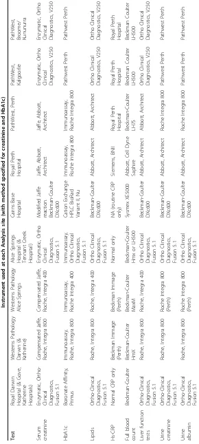
Serum creatinine and other metabolite assays were performed at each centre as part of standard clinical care. The creatinine assay at each centre was confirmed to have claimed traceability to the Isotope Dilution Mass Spectrometry (IDMS) reference method. The method and instrument used for measurement of each biochemical measure performed at each site are outlined in Table 1.
In addition, serum creatinine will be measured in all samples at Austin Health, using the kinetic Jaffe method and the Roche enzymatic method on a Coul-ter DxC 800 analyser (Fullerton, CA, USA). Beckman-Coulter claims traceability for its Jaffe methods to the IDMS reference method as it does for the Roche enzy-matic method. The Roche enzyenzy-matic method has been used as a reference method for the revision of the MDRD formula for IDMS aligned assays [28] and has undergone local independent validation [29]. Creatinine will be measured by both the Jaffe and enzymatic meth-ods as many laboratories in Australia measure creati-nine with a Jaffe method. This approach will allow for a calibration and standardization of the Jaffe methods
against the enzymatic method and assessment of varia-bility caused by the use of different routine methods. Cystatin C will be measured using an automated parti-cle-enhancing immunonephelometric assay on a BN II instrument (Dade Behring, Marburg, Germany). The intra- and interassay CVs for cystatin C were 2.58 and 3.95%, respectively, at a concentration of 1.54 mg/l. Cystatin C measures will be transformed to a GFR equivalent using the formua Cys-GFR = (86.7/cystatin C -4.2). This Cys-GFR method has been validated as an accurate marker of renal function across a wide-range of GFR values using isotopic GFR measurements (plasma dissapearance of 99 mTc-DTPA) as the refer-ence method [18,30].
Iohexol was measured by Austin Health, Melbourne using a validated HPLC assay modified from Niculescu-Duvaz et al [31]. Plasma samples were extracted using a solution of 5% perchloric acid containing internal stan-dard (sodium diatrizoate). Chromatographic separation was achieved using a Microsorb MV C18 5 μ column (Varian Australia) and detection at 244 nm. Analyte concentrations were calculated by comparison to multi-point linear standard curves derived from plasma sam-ples spiked to contain between 3.125 and 800 μg/mL Iohexol (reference material, US Pharmacopoeia). The lower limit of quantitation was 3.125 μg/mL. Intra- and inter-assay performance was assessed using donor plasma spiked with low (20 μg/mL), medium (100 μg/ mL) and high (400 μg/mL) concentrations of Iohexol. Precision studies showed a Coefficient of Variation of 1.0% or less, and inaccuracy ± 4.5% or less.
Samples were stored for analysis at a later date for: adiponectin and urine cotinine. For participants of the DXA sub-study, samples have been stored for analysis of 25-hydoxy Vitamin D and parathyroid hormone and bone turnover markers.
Data handling and statistical methods
strata. Sample size was determined using the methods of Bland-Altman [32,33] so that the limits of 95% confi-dence intervals for the mean were equal to pre-specified acceptability limits (a 10% difference in GFR was consid-ered to be acceptable). Sample size was calculated at 600 Indigenous participants across 4 sites (approximately 150 at each site), with considerable heterogeneity in body builds and composition expected both within and between sites. Regression equations will be derived using a similar approach to that used to develop the ori-ginal MDRD equations [34]. In addition, for the body composition substudy (DXAvs. BIA), we will be able to detect a difference ≥ 2% in fat free mass (FFM) esti-mated by the two methods, with a power >90% based on a sample size of 300, alpha = 0.05 and a CV of 10% in FFM.
Detailed analysis of demographic, clinical, biochemical and socioeconomic factors associated with CKD will involve cross-sectional analysis of the associations between these factors and current GFR. After determin-ing univariate associations between variables, established risk factors and variables with p < 0.2 on univariate ana-lysis will be selected for entry into logistic regression models using the backwards selection method, the out-come variable being current GFR/CKD stage. The degree of coupling of GFR and albuminuria will be determined in participants with type 2 diabetes (after accounting for the use of inhibitors of the renin-angio-tensin system). We have recently shown that there is a relatively high prevalence of non-albuminuric renal insufficiency in non-Indigenous participants with type 2 diabetes [35] and preliminary evidence suggests that the prevalence of non-albuminuric renal insufficiency is lower in Indigenous than non-Indigenous diabetic Aus-tralians [36].
Discussion
This study was designed with respect to what is practi-cal and achievable in very remote regions of Northern, Western and Central Australia. The choice of reference measure of GFR was limited by our remote setting and the available time of participants. Nuclear medicine facilities are not available in the vast majority of sites where this project was conducted. Study investigators agreed that 4-hour plasma disappearance of iohexol was the most appropriate reference measure of GFR for the remote setting. The benefits of this reference measure are that it is practical, safe, stable and reproducible [37,38]. In addition, several investigators had experience with use of this measure in the non-Indigenous urban setting (Melbourne) with accurate comparison to 99 mTc-DTPA in participants with diabetes [39]. Based on previous experience of investigators in Indigenous health research, we agreed that a reference measure that
required more than 4 hours of participants time would not be practical or achievable.
A potential limitation of our study is the use of Iohexol clearance from serum as the formal GFR mea-surement as opposed to the use of urinary clearance of iothalamate as the reference method used in the MDRD and CKD-EPI studies [16,34]. Iohexol serum clearance was however used in two studies used in the CKD-EPI validation group [40,41] however the limitation of 4 hours for the studies has the potential to increase inac-curacy in subjects with reduced GFR [40].
The study has encountered several operational chal-lenges to date that have been successfully managed. The first challenge is the significant labour intensity required to recruit each participant and the associated expenses. The proportion of eligible participants approached by the study team, who subsequently consented to and suc-cessfully completed the study, varied by site from approximately 30% in urban centres to up to 70% in some remote communities. We managed this challenge by adapting recruitment sites to those that had a higher success rate of obtaining complete data sets and expand-ing networks and recruitment sites beyond health ser-vice providers, e.g. Aboriginal Hostels Limited (provide temporary and transit accommodation in urban and regional centres).
The second challenge encountered by the eGFR study team was to recruit a diverse sample of participants including people living remotely and with poor access to regular medical services. We managed this challenge by: targeting a range of communities or regions; target-ing remote-livtarget-ing individuals whilst they were visittarget-ing regional centres (Darwin, Katherine and Nhulunbuy: health and non-health service sites); and selection of participants, stratified by study design, using electronic health record data held by urban and remote health services.
The third challenge was to conduct recruitment in such a way to facilitate complete data collection, in par-ticular complete 4 hour reference GFR measures. We commenced recruitment of participants in association with outpatient renal clinics (urban and regional). In the regional setting other commitments for remote partici-pants within the 4-hour duration of the study resulted in non-completion of the reference GFR measures. Rea-sons included the need to catch transport home (usually a plane) or logistic constraints such as inclement weather conditions. The study recruitment strategy was subsequently changed so that the eGFR study team tra-velled to remote communities to perform the study in
This study should provide evidence for clinical guide-lines in a field where evidence is currently lacking: a validated methodology to accurately assess renal func-tion in diverse populafunc-tions of Indigenous Australians. We expect to determine a validated and practical mea-sure of GFR suitable for use in all Indigenous Austra-lians. This may be cystatin C or eGFR (using MDRD or CKD-EPI formula with/without suitable revision). This measure would enable development of appropriate pro-tocols for investigation and management of CKD and subsequent improvements in clinical practice. We believe that the MDRD- or CKD-EPI derived method (modified as appropriate) is likely to emerge as the cheapest and most widely assessable means of estimat-ing GFR in the immediate future. It seems unlikely that a single correction factor (similar to that for Afro-Amer-icans) is appropriate or practical for Indigenous Austra-lians. However, it may be that a modification of the MDRD or CKD-EPI equation for Indigenous Australians (or for any population) would be to include a measure of fat-free mass. This measure may be obtained by mea-suring body composition using a simple, portable and inexpensive method such as BIA, once it has been suita-bly validated against DXA. Thereby this study may improve the utility of eGFR, using a modified MDRD or CKD-EPI formula, across different populations with dif-ferent body builds and compositions. Our revised for-mula for eGFR may indeed be of use for assessing kidney function in the broader Australian community, particularly those of variable body builds and/or eGFR>60 ml/min/1.73 m2.
Acknowledgements
The authors gratefully acknowledge the support of eGFR study participants, study staff, and partner organisations. The eGFR Study was funded by the National Health and Medical Research Council of Australia (NHMRC, Project Grant #545202), with additional support from Kidney Health Australia, Colonial Foundation, Rebecca L Cooper Foundation and SeaSwift, Thursday Island. LMB was supported by NHMRC Program Grant #320860 and the Centre of Clinical Research Excellence in Clinical Science in Diabetes, University of Melbourne. Alan Cass holds a Senior Research Fellowship from the NHMRC. Funding bodies had no role in the study design, in the collection, analysis or interpretation of data, in the writing of the manuscript or the decision to submit the manuscript for publication.
Author details
1Menzies School of Health Research, Institute of Advanced Studies, Charles
Darwin University, Darwin, Australia.2Division of Medicine, Royal Darwin Hospital, Darwin, Australia.3Chemical Pathology, St Vincent’s Hospital, Sydney, Australia.4University of Melbourne, Department of Medicine, Austin and Northern Health, Heidelberg, Victoria, Australia.5Centre for Chronic Disease, The University of Queensland, Australia.6The George Institute for International Health, University of Sydney, Sydney, Australia.7Department of Endocrinology, Endocrine Centre Austin Health & University of Melbourne, Heidelberg Repatriation Hospital, Heidelberg West, Victoria, Australia. 8Endocrine and Diabetes Unit, Cairns Base Hospital, Queensland, Australia. 9
Department of Nephrology, Royal Perth Hospital, Perth, Australia.10Centre for Health and Society, School of Population Health, University of Melbourne, Melbourne, Australia.11School of Chemistry and Molecular Biosciences, The University of Queensland, Australia.12Sansom Institute for Health Research,
UniSA, Adelaide, Australia.13Department of Renal Medicine, Alice Springs Hospital, Alice Springs, Australia.14Baker IDI Heart and Diabetes Institute, Alice Springs, Australia.
Authors’contributions
LMB drove the design of the study protocol for funding and Ethics applications, coordinated data collection and data management, analysed the data and drafted the manuscript. PL, RM, GJe, AS, KOD conceived of the study and participated in its design. SS was the study manager and participated in study design, ethics applications, data collection and data management. JH participated in study design, data collection and data management. GJo, AE, WH, AC participated in study design. MT, KW, CS participated in the design of the study and provided clinical expertise during the design and data collection phases. LP, LW, RMcD, AB participated in study design. All authors were involved in revising the manuscript for important intellectual content and read and approved the final manuscript.
Competing interests
LCW has consults to Impedimed Ltd. Impedimed Ltd. had no involvement, financial or otherwise, in the conception and execution of this study or in the preparation of the manuscript.
All remaining authors declare that they have no competing interests.
Received: 14 January 2010
Accepted: 19 February 2010 Published: 19 February 2010
References
1. Zhao Y, Dempsey K:Causes of inequality in life expectancy between Indigenous and non-Indigenous people in the Northern Territory, 1981-2000: a decomposition analysis.Med J Aust2006, 184:490-494.
2. Heart, stroke and vascular disease:Australian facts 2004. Cardiovascular Disease Series, No. 22.Canberra, Australian Institute of Health and Welfare and National Heart Foundation of Australia 2004.
3. O’Dea K, Patel M, Kubisch D, Hopper J, Traianedes K:Obesity, diabetes, and hyperlipidemia in a central Australian aboriginal community with a long history of acculturation.Diabetes Care1993,16:1004-1010.
4. ANZDATA Registry Report: Adelaide, Australia and New Zealand Dialysis and Transplant RegistryMcDonald S, Excell L, Livingston B 2009.
5. Cass A, Cunningham J, Wang Z, Hoy W:Regional variation in the incidence of end-stage renal disease in Indigenous Australians.Med J Aust2001,175:24-27.
6. Rutishauser IH:Body composition in Aboriginal Australians.Asia Pac J Clin Nutr1995,4:73-76.
7. Piers LS, Rowley KG, Soares MJ, O’Dea K:Relation of adiposity and body fat distribution to body mass index in Australians of Aboriginal and European ancestry.Eur J Clin Nutr2003,57:956-963.
8. Cass A, Cunningham J, Snelling P, Wang Z, Hoy W:End-stage renal disease in indigenous Australians: a disease of disadvantage.Ethn Dis2002, 12:373-378.
9. K/DOQI clinical practice guidelines for chronic kidney disease: evaluation, classification, and stratification. Am J Kidney Dis2005,39: S1-266.
10. Ninomiya T, Perkovic V, de Galan BE, Zoungas S, Pillai A, Jardine M, Patel A, Cass A, Neal B, Poulter N, Mogensen CE, Cooper M, Marre M, Williams B, Hamet P, Mancia G, Woodward M, Macmahon S, Chalmers J:Albuminuria and kidney function independently predict cardiovascular and renal outcomes in diabetes.J Am Soc Nephrol2009,20:1813-1821.
11. Astor BC, Hallan SI, Miller ER, Yeung E, Coresh J:Glomerular filtration rate, albuminuria, and risk of cardiovascular and all-cause mortality in the US population.Am J Epidemiol2008,167:1226-1234.
12. Mathew TH:Chronic kidney disease and automatic reporting of estimated glomerular filtration rate: a position statement.Med J Aust
2005,183:138-141.
13. Jose MD, Lawton PD:Chronic kidney disease and automatic reporting of estimated glomerular filtration rate.Med J Aust2006,184:42.
14. Zuo L, Ma YC, Zhou YH, Wang M, Xu GB, Wang HY:Application of GFR-estimating equations in Chinese patients with chronic kidney disease. Am J Kidney Dis2005,45:463-472.
Cockcroft-Gault Equations for Estimating Renal Function.J Am Soc Nephrol2005,16:763-773.
16. Levey AS, Stevens LA, Schmid CH, Zhang YL, Castro AF, Feldman HI, Kusek JW, Eggers P, Van Lente F, Greene T, Coresh J:A new equation to estimate glomerular filtration rate.Ann Intern Med2009,150:604-612. 17. Dharnidharka VR, Kwon C, Stevens G:Serum cystatin C is superior to
serum creatinine as a marker of kidney function: A meta-analysis.Am J Kidney Dis2002,40:221-226.
18. MacIsaac RJ, Tsalamandris C, Thomas MC, Premaratne E, Panagiotopoulos S, Smith TJ, Poon A, Jenkins MA, Ratnaike SI, Power DA, Jerums G:Estimating glomerular filtration rate in diabetes: a comparison of cystatin-C- and creatinine-based methods.Diabetologia2006,V49:1686-1689. 19. Madero M, Sarnak MJ, Stevens LA:Serum cystatin C as a marker of
glomerular filtration rate.Curr Opin Nephrol Hypertens2006,15:610-616. 20. DuBois D, DuBois EF:A formula to estimate the approximate surface area
if height and weight be known.Arch Int Med1916,17:863-871. 21. Brochner-Mortensen J:A simple method for the determination of
glomerular filtration rate.Scand J Clin Lab Invest1972,30:271-274. 22. Mathew TH, Johnson DW, Jones GR:Chronic kidney disease and
automatic reporting of estimated glomerular filtration rate: revised recommendations.Med J Aust2007,187:459-463.
23. Cockcroft DW, Gault MH:Prediction of creatinine clearance from serum creatinine.Nephron1976,16:31-41.
24. Norton K, Olds T:AnthopometricaUNSW Press 1996.
25. Lohman TG, Roche AF, Martorell R:Anthropometric standardization reference manualHuman Kinetics Books 1988.
26. Thomas BJ, Ward LC, Cornish BH:Bioimpedance spectrometry in the determination of body water compartments: accuracy and clinical significance.Appl Radiat Isot1998,49:447-455.
27. Cornish BH, Jacobs A, Thomas BJ, Ward LC:Optimizing electrode sites for segmental bioimpedance measurements.Physiol Meas1999,20:241-250. 28. Levey AS, Coresh J, Greene T, Marsh J, Stevens LA, Kusek JW, Van Lente F: Expressing the Modification of Diet in Renal Disease Study equation for estimating glomerular filtration rate with standardized serum creatinine values.Clin Chem2007,53:766-772.
29. Peake M, Whiting M:Measurement of serum creatinine - current status and future goals.Clin Biochem Rev2006,27:173-184.
30. Premaratne E, MacIsaac RJ, Finch S, Panagiotopoulos S, Ekinci E, Jerums G: Serial measurements of cystatin C are more accurate than creatinine-based methods in detecting declining renal function in type 1 diabetes. Diabetes Care2008,31:971-973.
31. Niculescu-Duvaz I, D’Mello L, Maan Z, Barron JL, Newman DJ, Dockrell MEC, Kwan JTC:Development of an outpatient finger-prick glomerular filtration rate procedure suitable for epidemiological studies.Kidney Int
2006,69:1272-1275.
32. Bland JM, Altman DG:Statistical methods for assessing agreement between two methods of clinical measurement.Lancet1986,1:307-310. 33. Altman DG, Bland JM:Measurement in Medicine: the Analysis of Method
Comparison Studies.The Statistician1983,32:307-317.
34. Levey AS, Bosch JP, Lewis JB, Greene T, Rogers N, Roth D:A more accurate method to estimate glomerular filtration rate from serum creatinine: a new prediction equation. Modification of Diet in Renal Disease Study Group.Ann Intern Med1999,130:461-470.
35. MacIsaac RJ, Tsalamandris C, Panagiotopoulos S, Smith TJ, McNeil KJ, Jerums G:Nonalbuminuric renal insufficiency in type 2 diabetes.Diabetes Care2004,27:195-200.
36. Thomas MC, Weekes AJ, Broadley OJ, Cooper ME:The assessment of kidney function by general practitioners in Australian patients with type 2 diabetes (NEFRON-2).Med J Aust2006,185:259-262.
37. Brown SC, O’Reilly PH:Iohexol clearance for the determination of glomerular filtration rate in clinical practice: evidence for a new gold standard.J Urol1991,146:675-679.
38. Gaspari F, Perico N, Ruggenenti P, Mosconi L, Amuchastegui CS, Guerini E, Daina E, Remuzzi G:Plasma clearance of nonradioactive iohexol as a measure of glomerular filtration rate.J Am Soc Nephrol1995,6:257-263. 39. Houlihan C, Jenkins M, Osicka T, Scott A, Parkin D, Jerums G:A comparison
of the plasma disappearance of iohexol and 99 mTc-DTPA for the measurement of glomerular filtration rate (GFR) in diabetes.Aust N Z J Med1999,29:693-700.
40. Grubb A, Nyman U, Bjork J, Lindstrom V, Rippe B, Sterner G, Christensson A: Simple cystatin C-based prediction equations for glomerular filtration
rate compared with the modification of diet in renal disease prediction equation for adults and the Schwartz and the Counahan-Barratt prediction equations for children.Clin Chem2005,51:1420-1431. 41. Mauer M, Zinman B, Gardiner R, Drummond KN, Suissa S, Donnelly SM,
Strand TD, Kramer MS, Klein R, Sinaiko AR:ACE-I and ARBs in early diabetic nephropathy.J Renin Angiotensin Aldosterone Syst2002,3:262-269.
Pre-publication history
The pre-publication history for this paper can be accessed here:http://www. biomedcentral.com/1471-2458/10/80/prepub
doi:10.1186/1471-2458-10-80
Cite this article as:Maple-Brownet al.:Study Protocol - Accurate
assessment of kidney function in Indigenous Australians: aims and methods of the eGFR Study.BMC Public Health201010:80.
Submit your next manuscript to BioMed Central and take full advantage of:
• Convenient online submission
• Thorough peer review
• No space constraints or color figure charges
• Immediate publication on acceptance
• Inclusion in PubMed, CAS, Scopus and Google Scholar
• Research which is freely available for redistribution