0022-538X/05/$08.00⫹0 doi:10.1128/JVI.79.2.1232–1243.2005
Copyright © 2005, American Society for Microbiology. All Rights Reserved.
Phosphorylation Site Mutations Affect Herpes Simplex Virus Type 1
ICP0 Function
David J. Davido,
1William F. von Zagorski,
1William S. Lane,
2and Priscilla A. Schaffer
1*
Departments of Medicine and Microbiology and Molecular Genetics, Harvard Medical School at Beth Israel DeaconessMedical Center, Boston,1and Microchemistry and Proteomics Analysis Facility, Department of Molecular and
Cellular Biology, Harvard University, Cambridge,2Massachusetts
Received 16 June 2004/Accepted 28 July 2004
The herpes simplex virus type 1 (HSV-1) immediate-early (IE) regulatory protein infected-cell protein 0 (ICP0) is a strong and global transactivator of both viral and cellular genes. In a previous study, we reported that ICP0 is highly phosphorylated and contains at least seven distinct phosphorylation signals as determined by phosphotryptic peptide mapping (D. J. Davido et al., J. Virol. 76:1077–1088, 2002). Since phosphorylation affects the activities of many viral regulatory proteins, we sought to determine whether the phosphorylation of ICP0 affects its functions. To address this question, it was first necessary to identify the regions of ICP0 that are phosphorylated. For this purpose, ICP0 was partially purified, and phosphorylation sites were mapped by microcapillary high-pressure liquid chromatography tandem mass spectrometry. Three phosphorylated re-gions containing 11 putative phosphorylation sites, all within or adjacent to domains important for the transactivating activity of ICP0, were identified. The 11 sites were mutated to alanine as clusters in each of the three regions by site-directed mutagenesis, generating plasmids expressing mutant forms of ICP0: Phos 1 (four mutated sites), Phos 2 (three mutated sites), and Phos 3 (four mutated sites). One-dimensional phosphotryptic peptide analysis confirmed that the phosphorylation state of each Phos mutant form of ICP0 is altered relative to that of wild-type ICP0. In functional assays, the ICP0 phosphorylation site mutations affected the subcel-lular and subnuclear localization of ICP0, its ability to alter the staining pattern of the nuclear domain 10 (ND10)-associated protein PML, and/or its transactivating activity in Vero cells. Only mutations in Phos 1, however, impaired the ability of ICP0 to complement the replication of an ICP0 null mutant in Vero cells. This study thus suggests that phosphorylation is an important regulator of ICP0 function.
Phosphorylation is a universal posttranslational modification that alters the activities of many viral regulatory proteins. Among the best examples of this effect is large T antigen of simian virus 40, a multifunctional 708-amino-acid nuclear phospho- and oncoprotein required for the replication of sim-ian virus 40 (reviewed in references 61 and 76). Many phos-phorylation sites have been identified on T antigen, and several lie adjacent to its nuclear localization signal and origin-binding domain, modulating simian virus 40 DNA replication (re-viewed in reference 61). Specifically, mutation of these sites alters T antigen’s origbinding activity, hexamer-hexamer in-teraction, nuclear localization, and/or DNA replication activ-ity, affecting both its biochemical and biological functions (11, 14, 41, 50–52, 54, 78).
In herpes simplex virus type 1 (HSV-1) infection, the first genes to be expressed are the immediate-early (IE) genes (39). These genes encode infected-cell protein (ICPs) 0, 4, 22, 27, and 47, which collectively exhibit diverse regulatory and im-munomodulatory functions. At least four of the five IE regu-latory proteins (ICPs 0, 4, 22, and 27) are known to be phos-phorylated, suggesting that phosphorylation is an important modulator of the functions of these proteins (1, 64, 82). In support of this hypothesis, studies by Xia et al. demonstrated that phosphorylation of the N-terminal region of ICP4, the
major transcriptional activator of HSV genes, is required for efficient viral replication in cells of neuronal lineage and in a mouse ocular model of HSV-1 latency (83). A second IE pro-tein, ICP22, essential for replication in selected cell types (73), is phosphorylated either directly or indirectly by the delayed-early viral kinase and structural protein UL13 (63, 64). Nota-bly, phosphorylation of ICP22 is required to activate the nor-mal program of viral gene expression in permissive cells (48, 66). A third IE protein, ICP27, an essential gene involved in transport and posttranslational processing of viral transcripts, is also phosphorylated (1, 85). In the study that mapped and mutated phosphorylation sites on ICP27, however, the mutants isolated and characterized were able to complement the growth of an ICP27 null mutant in cell culture (85).
ICP0, the focus of this study, is a 110-kDa nuclear phospho-protein that transactivates all classes of HSV-1 genes, IE, early (E), and late (L), as well as numerous cellular genes and genes of other viruses (9, 21, 27, 28, 34–36, 55, 65, 80). Mutant viruses affected in ICP0’s transactivating activity replicate, establish latency, and reactivate from latency inefficiently and are sen-sitive to the cellular antiviral factors interferons (6, 23, 37, 38, 47, 56, 68, 75). Although the precise mechanism by which ICP0 mediates its strong and broad transactivating activity is un-clear, this activity requires the cellular ubiquitin-proteasome pathway and correlates with the dispersal and/or degradation of cellular proteins linked to cellular transcription, prolifera-tion, differentiaprolifera-tion, and apoptosis (reviewed in reference 26). Many but not all of these proteins associate with nuclear struc-tures termed ND10s, and a subset of these proteins are
up-* Corresponding author. Mailing address: Beth Israel Deaconess Medical Center, 330 Brookline Ave., RN 123, Boston, MA 02215. Phone: (617) 667-2958. Fax: (617) 667-8540. E-mail: pschaffe@bidmc .harvard.edu.
1232
on November 8, 2019 by guest
http://jvi.asm.org/
regulated by interferons (reviewed in reference 57). Thus, ICP0’s ability to impair the interferon response during HSV infection appears to be associated with its transactivating ac-tivity.
Although early studies demonstrated that the phosphoryla-tion of ICP0 is a dynamic process (1, 80), the role of phorylation in ICP0 function, the location of specific phos-phorylation sites on ICP0, and the kinases that phosphorylate ICP0 are largely unknown. To date, two types of kinases have been shown to affect the phosphorylation state of ICP0: cellu-lar cyclin-dependent kinases (cdks) and the viral kinase, UL13. One report demonstrated that cdk-1 can phosphorylate the second exon (residues 20 to 241) of ICP0 in vitro (3). In the same study, the authors showed that a dominant-negative form of cdk-1 inhibited the expression of a late gene in HSV-in-fected cells; however, the phosphorylation state of ICP0 was not examined in these cells. We and others have shown that the posttranslational modification of ICP0, including its phosphor-ylation state, is altered in the presence of the cdk inhibitor roscovitine, which inhibits the activities of cdks-1, -2, likely -3, -5, -7, and -9 (2, 16, 53, 72, 79). This alteration correlated with a significant decrease in ICP0’s transactivating activity, sug-gesting a link between ICP0’s posttranslational modification state (including but not exclusively related to phosphorylation) and its transactivating activity (16). UL13, on the other hand, is required to achieve maximal levels of ICP0 phosphorylation during viral infection and can phosphorylate ICP0 in vitro (58). Notably, it has not yet been established that UL13-mediated phosphorylation of ICP0 is involved in ICP0 function. This study also established that ICP0 is phosphorylated to signifi-cant levels in the absence of UL13, indicating that other ki-nases, most likely cellular kiki-nases, also phosphorylate ICP0. The latter possibility was confirmed by Isler and Schaffer, who demonstrated that ICP0 expressed in cell culture in the ab-sence of other HSV proteins is extensively phosphorylated (40). Thus, although much is known about the phosphorylation of ICP0, the functional relevance of ICP0 phosphorylation to HSV replication has not been analyzed systematically.
The purpose of the present study was to determine which regions and sites on ICP0 are phosphorylated and assess what role(s) these regions and sites play in the many functions of the protein. To identify which of ICP0’s 133 potential phosphoac-ceptor sites are actually phosphorylated, microcapillary high-pressure liquid chromatography tandem mass spectrometry (LC-MS/MS) analysis was performed on ICP0 isolated from infected cell extracts. LC-MS/MS analysis identified three major regions of phosphorylation containing 11 potential phos-phorylation sites. Mutations which alter the phosphos-phorylation state of each of the three regions affect one or more of the activities of ICP0. This study therefore suggests a significant role for phosphorylation in ICP0 function.
MATERIALS AND METHODS
Cells and viruses.Vero cells, an African green monkey kidney cell line, were obtained from the American Type Culture Collection (Manassas, Va.) and prop-agated in Dulbecco’s modified Eagle’s medium (DMEM) supplemented with 10% fetal bovine serum as described previously (71). L7 cells, Vero cells stably transformed with the ICP0 gene of HSV-1, were passaged as described previ-ously (69). HSV-1, strain KOS (passage 11), was used as the wild-type virus and propagated as described (71). An ICP0 null mutant derived from KOS, 7134, was
propagated in Vero cells as previously described (7), and titers of 7134 were determined on L7 cells.
Partial purification of ICP0 forLC-MS/MS sequencing.Four 100-mm dishes
were seeded with 2⫻106
Vero cells, and 23 h after plating, cells were treated
with cycloheximide (50g/ml) for 1 h and infected with 5 PFU of KOS per cell
for 1 h at 37°C in the presence of cycloheximide. After 1 h of adsorption, the inoculum was removed, and the cells were washed three times with phosphate-buffered saline (PBS) containing cycloheximide; 5 ml of Vero cell medium containing cycloheximide was then added to each dish, and the cells subsequently
were incubated for 5 h (t⫽6 h postinfection) at 37°C. Att⫽6 h postinfection,
the medium was removed, and infected cells were washed three times with PBS
and incubated for an additional 6 h. Att⫽12 h postinfection, cells were washed
twice with ice-cold PBS, scraped into 2 ml of ice-cold PBS, and pelleted at 800
⫻gat 4°C. The supernatant fluid was removed, and the resulting cell pellets were
stored at⫺80°C. Subsequently, samples were thawed on ice, and resuspended in
2 ml of radioimmunoprecipitation assay (RIPA) lysis buffer [150 mM NaCl, 50 mM Tris (pH 7.5), 0.1% sodium dodecyl sulfate (SDS), 1% Nonidet P-40, and 0.5% deoxycholic acid] containing the protease inhibitors phenylmethylsulfonyl
fluoride (1 mM), 1g of leupeptin per ml, and 1g of aprotinin per ml; 8l of
J17, an ICP0 polyclonal rabbit antibody (86), was added per sample, and the suspensions were gently agitated overnight at 4°C.
The next day, 160l of protein A-agarose (Invitrogen Life Technologies,
Carlsbad, Calif.) was added to each sample and agitated for 2 h at 4°C. Immune
complexes were pelleted by centrifugation for 2 min at 3,300⫻gat 4°C. The
supernatant fluid was removed, and each pellet was washed and repelleted three times in RIPA buffer plus protease inhibitors. The resulting pellet was
resus-pended in 60l of 1x Laemmli buffer plus 1 mM phenylmethylsulfonyl fluoride
(46). Samples were heated at 100°C for 5 min, placed on ice for 2 min, and centrifuged to pellet the protein A-agarose. The supernatants were analyzed by sodium dodecyl sulfate-polyacrylamide gel electrophoresis (SDS-PAGE) on a 0.75-mm 6% acrylamide gel. Proteins were visualized by Coomassie blue
stain-ing, and an estimate of the total amount of ICP0 protein isolated (1.5g) was
determined relative to a standard curve of bovine serum albumin. Gel pieces containing ICP0 bands were marked, destained for Coomassie blue dye, excised,
and stored at⫺20°C until further analysis.
Identification of phosphorylation sites byLC-MS/MS.Phosphorylated
res-idues in ICP0 were detected withLC-MS/MS. With an in-house program,
Enzyme Optimizer, the ICP0 sequence was evaluated for a dual enzyme strategy which would optimize for coverage of S, T, and Y residues. The program con-siders peptide properties and experimental conditions that influence the recovery and detection of a predicted peptide rather than simple protein coverage. The band corresponding to ICP0 was then split in half for separate in-gel trypsin and chymotrypsin digestions after reduction and carboxyamidomethylation. The
re-sultant digests were pooled just prior toLC-MS/MS injection. Phosphorylated
peptide sequences were determined with a 75-m reverse-phase microcolumn
terminating in a custom nanoelectrospray source directly coupled to a Finnigan
LCQ DECA XP⫹quadrupole ion trap mass spectrometer (Thermo Electron).
The flow rate was nominally 250 nl/min. The ion trap repetitively surveyed the
rangem/z395 to 1,600, executing data-dependent MS/MS on the four most
abundant ions in each survey scan. MS/MS spectra were acquired with a relative collision energy of 30%, a 2.5-Da isolation width, and recurring ions dynamically excluded. Preliminary sequencing of peptides was facilitated by database corre-lation with the algorithm SEQUEST (19). The discovery of peptides carrying phosphorylation and subsequent manual validation of their MS/MS spectra were aided by the in-house programs Muquest and FuzzyIons, respectively (15).
Plasmids.Plasmid pIE3-CAT, which expresses the chloramphenicol acetyl-transferase (CAT) gene under the control of the HSV-1 IE ICP4 promoter, was
constructed as previously described (18). Plasmid pAlter-1⫹ICP0 was
con-structed by isolating a 4.6-kb EcoRI-HindIII fragment containing the ICP0 gene from the plasmid pSH (9) and cloned into the vector pAlter-1 (Promega Corp.
Madison, Wis.), with the same restriction enzyme sites. pAlter-1⫹ICP0 was
subsequently used as the parental vector to mutate the putative phosphorylation sites of ICP0 to alanine with mutagenic primers (IDT, Coralville, Iowa) accord-ing to the manufacturer’s protocol (Promega Corp., Madison, Wis.). The primers
used for the mutagenesis are Phos 1 (S224A, T226A, T231A, T232A), 5⬘-CTG
GGGGGGCACACGGTGAGGGCCCTagCGCCggCCCACCCTGAGCCggC
CgCGGACGAGGATGACGACGACCTGGAC-3⬘; Phos 2 (S365A, S367A,
S371A), 5⬘-GCAAACAACAGAGACCCCATAGTGATCgcCGAtgCCCCCCC
GGCCgCTCCCACAGGCCCCCCGCGGCGCCC-3⬘; and Phos 3 (S508A,
S514A, S517A, T518A), 5⬘-GCGGTGCGTCCGAGGAAGAGGCGCGGGgCc
GGCCAGGAAAACCCCgCCCCgCAGgCCgCGCGTCCCCCCCTCGCGCC
GGCAGGGG-3⬘. Lowercase letters indicate the nucleotides mutated relative to
on November 8, 2019 by guest
http://jvi.asm.org/
the wild-type (strain KOS) ICP0 sequences. Putative mutants were identified by restriction enzyme analysis (Fig. 1B) and confirmed by DNA sequencing.
Transfection/infection and32P and35S labeling of wild-type ICP0 and its
mutant forms.Vero cells were plated at 5⫻105cells per 60-mm dish, and at 2 h
prior to transfection (22 h postplating), the medium was changed. At 24 h after plating, transfections were performed with Lipofectamine 2000 (Invitrogen
Corp., Carlsbad, Calif.) according to the manufacturer’s protocol with 8g of
plasmid or salmon sperm DNA and 16l of Lipofectamine 2000 diluted in
Opti-MEM (Invitrogen Corp.). The DNA/Lipofectamine 2000 mixture was sub-sequently added to plates containing 5 ml of Opti-MEM in a dropwise manner and left on the cells for 4 h at 37°C. Four hours after transfection, cells were
treated for 1 h with 50g of cycloheximide per ml and mock infected or infected
with 5 PFU of KOS or 7134 per cell for 1 h at 37°C in the presence of cyclo-heximide. After 1 h of adsorption, the inoculum was removed, the cells were washed three times with PBS containing cycloheximide, 4 ml of Vero cell me-dium plus cycloheximide was added per dish, and the dishes were incubated for
4.5 h postinfection at 37°C. Att⫽5.5 h postinfection, the cells were incubated
for 0.5 h in phosphate- or methionine- and cysteine-free DMEM containing
cycloheximide. Att⫽6 h postinfection, the medium was removed, and the
infected cells were washed three times with phosphate- or methionine- and cysteine-free DMEM containing 1% fetal bovine serum. The cells were then
labeled with 500 Ci of 32P
i or 100 Ci of [35S]methionine/cysteine
(PerkinElmer Life Sciences, Inc., Boston, Mass.) in phosphate- or methionine-and cysteine-free DMEM containing 1% fetal bovine serum, respectively, for an
additional 6 h. Att⫽12 h postinfection, cells were washed twice with ice-cold
PBS, scraped into 1 ml of ice-cold RIPA lysis buffer containing protease inhib-itors as described above. Extract preparation, immunoprecipitation of ICP0, and SDS-PAGE analysis were performed as described previously (16). Proteins were visualized, and their signal intensities were quantified by PhosphorImager anal-ysis (Amersham Biosciences, Piscataway, N.J.).
ICP0 phosphotryptic peptide digestion and one-dimensional alkaline gel elec-trophoresis.32
P-labeled bands of wild-type ICP0 or its mutant forms were ex-cised from the SDS-PAGE gel described above and washed two times for 5 min in fresh ammonium bicarbonate (50 mM). Each gel piece was brought to a final
volume of 500l with sodium bicarbonate, homogenized with a pestle grinder,
and treated with 40g of TPCK [L-(tosyl/amido-z-phenyl)ethyl ketone)-treated
trypsin (Worthington Biochemicals, Lakewood, N.J.) while gently agitating at
34°C overnight. An additional 25g of TPCK-treated trypsin was added per
sample, incubating for a second time overnight at 34°C. Samples were spun at
20,800⫻gfor 10 min at room temperature, and the resulting supernatant was
removed. Remaining protein in the gel pieces was extracted twice by incubating
the pieces in 400l of acetonitrile-formic acid (1:1) for 20 min with gentle
rocking at room temperature. The acetonitrile-formic acid extracts and the
aqueous supernatant of the phosphotryptic digests were pooled (⬇1,200l) and
dried in a SpeedVac (Savant Instruments, Inc., Farmingdale, N.Y.).
Phosphola-beled peptides of ICP0 were suspended in 25l of sample buffer [0.125 M
Tris-HCl buffer (pH 6.8) and 6 M urea], loaded with equal Cherenkov counts
(⬇1,150 cpm), and separated on a 27-cm 30% (wt/vol) alkaline acrylamide gel as
described previously (81) at 10 mA for 44 h. Phosphotryptic peptides from the alkaline gel electrophoresis were visualized by PhosphorImager analysis.
Immunofluorescence.Vero cells were plated on coverslips in 12-well plates, and 22 h later, fresh Vero cell medium was added to each well 2 h prior to transfection. Transfections were performed with Lipofectamine 2000 according
to the manufacturer’s protocol with 3g of plasmid DNA and 6l of
Lipo-fectamine 2000 diluted in Opti-MEM (Invitrogen Corp.). The
DNA-Lipo-fectamine 2000 mixture was then added to each well containing 500l of
Opti-MEM in a dropwise manner and left on the cells for 5 h at 37°C. The medium was removed, and fresh medium was added, incubating the monolayers for an additional 19 h. Twenty-four hours posttransfection, the medium was removed, and the coverslips were washed twice with PBS. Transfected cells were fixed and permeabilized by formaldehyde and acetone treatments according to Zhu et al. (86).
Immunofluorescence staining for ICP0, its mutant forms, and PML was per-formed as previously described (17). The primary antibodies and the dilutions used for the dual staining of ICP0 and PML were ICP0 at 1:500 (H1112; mouse monoclonal antibody; Rumbaugh-Goodwin Institute for Cancer Research, Plan-tation, Fla.) and PML-14 at 1:500 (rabbit polyclonal antibody; Gerd Maul, Wistar Institute, Philadelphia, Pa.). The following secondary antibodies and dilutions were used for each primary antibody: ICP0 (H1112) at 1:100 (goat anti-mouse immunoglobulin G conjugated with fluorescein isothiocyanate) and PML-14 at 1:100 (goat anti-rabbit immunoglobulin G conjugated with rhodamine red X). All secondary antibodies were purchased from Jackson Immunoresearch (West Grove, Pa.). Following incubations with the primary and secondary antibodies,
the coverslips were washed, and 7l of Prolong antifade solution (Molecular
Probes, Eugene, Oreg.) was added per coverslip. Cells were viewed by
fluores-cence microscopy with a Nikon Eclipse TE300 fluoresfluores-cence microscope at⫻400
magnification and photographed with an RT Slider digital camera (Diagnostic Instruments, Sterling Heights, Mich.), and images were processed in Adobe
FIG. 1. Putative phosphorylated sequences on ICP0. (A) Location of phosphorylated sequences on ICP0 as determined byLS-MS/MS analysis. The 775 amino acids of ICP0, the locations of the major functional domains of ICP0, and the locations of the 11 putative phosphorylation sites in regions I, II, and III are shown. Beneath the diagram of ICP0, the amino acid sequences of regions I, II, and III are shown. The slash mark between D and A in region I represents the amino acid boundary between the second and third exons of ICP0. The codon numbers of the putatively phosphorylated serine (S) and thre-onine (T) residues are listed beneath the peptide sequence in each region. To the right of each serine or threonine are listed the cellular kinases which most likely target these residues, as determined by com-puter-based modeling with NetPhos 2.0 (Center for Biological Se-quence Analysis, Technical University of Denmark), ScanProsite (Swiss Institute for Bioinformatics), and MacVector 7.1.1 (GCG, Mad-ison, Wis.). Kinases include calmodulin kinase II (CaM II), protein kinase A (PKA), cyclin-dependent kinase 1 (cdk-1) and 2, casein ki-nase I (CKI), glycogen synthase kiki-nase 3 (GSK3), casein kiki-nase II (CKII), p70S6K kinase, protein kinase G (PKG), and protein kinase C (PKC). (B) Mutations in putative ICP0 phosphorylation sites. Serines and threonines in each phosphorylated region were mutated to alanine (A), resulting in the gain (⫹) or loss (⫺) of a specific restriction enzyme cleavage site, as shown beneath each substituted alanine res-idue. Forms of ICP0 in which phosphorylated serine (S) or threonine (T) residues in regions I, II and III have been changed to alanine (A) are designated Phos 1, 2, and 3, respectively, in the text.
on November 8, 2019 by guest
http://jvi.asm.org/
Photoshop (Adobe Systems Inc., Mountain View, Calif.). Images were assembled and labeled in Canvas 8 (Deneba Systems, Miami, Fla.).
At least 150 ICP0-stained cells from random fields were examined in each preparation and categorized as having nuclear only, nuclear and cytoplasmic, or cytoplasmic only staining. The percentage of cells in each category was deter-mined by dividing the number of cells in a given category by the total number of cells counted in all three categories.
Transient transfections and CAT assays.Vero cells (5⫻105
cells per 60-mm dish) were plated, and 2 h before transfection (22 h postplating), the medium was changed. Twenty-four hours after plating, the transfections were performed with
Lipofectamine 2000 according to the manufacturer’s protocol with a total of 8g
of DNA at 1g of CAT expression vector, salmon sperm testis DNA, and/or
increasing amounts of plasmid DNA (as indicated in Fig. 5) and 16l of
Lipofectamine 2000 diluted in Opti-MEM per dish. DNA-Lipofectamine 2000 was then added to dishes containing 5 ml of Opti-MEM in a dropwise manner and left on cells for 5 h at 37°C. The medium was removed, and fresh Vero cell medium was added, incubating the monolayers for an additional 43 h. At 48 h posttransfection, cells were washed three times with Tris-buffered saline (TBS),
harvested in 2 ml of TBS, and pelleted at 800⫻g; the supernatant was removed.
The resulting cell pellets were stored at⫺80°C. Samples were thawed on ice,
resuspended in 150l of TBS, sonicated for 20 s at 80% power in a Misonix
Sonicator 3000 (Misonix, Inc., Farmingdale, N.Y.), and cell debris was pelleted
for 10 min at 4°C at 20,800⫻g. The resulting supernatant was assayed for CAT
activity as performed by Seed and Sheen (74).
Complementation and plating efficiency of 7134.Vero cells were plated at 2⫻
105
cells per 35-mm dish. Twenty-two hours later, fresh medium was added to each well. Twenty four hours postplating, the transfections were performed with
Fugene 6 (Roche Diagnostic Corporation, Indianapolis, Ind.) with a total of 3g
of DNA (0.5g of infectious KOS or 7134 viral DNA) (9), salmon sperm DNA,
and/or 0.075g of plasmid DNA from pAlter-1 or wild-type ICP0- or Phos
mutant-expressing plasmids and 12l of Fugene 6 diluted in Opti-MEM. The
DNA/Fugene 6 mixture was divided in half, added dropwise to 35-mm dishes (for duplicate samples) containing 2 ml of Opti-MEM, and left on the cells for 5 h at 37°C. The medium was removed, fresh Vero cell medium was added, and the monolayers were incubated for an additional 43 h. Transfected cells were har-vested 48 h posttransfection and assayed for infectious virus by standard plaque assays on Vero cells for KOS or on L7 cells for 7134.
For the 7134 plating efficiency experiments, cells were transfected as described for the 7134 complementation experiments with the following modifications (8). Five hours posttransfection, the medium was removed from each culture and replaced with 2.5 ml of medium containing 0.5% methylcellulose and 10% fetal bovine serum in DMEM. Three days posttransfection, the methylcellulose-con-taining medium was removed from each plate, and the cells were washed twice
with PBS, fixed with 2% formaldehyde, and stained for-galactosidase activity
with 5-bromo-4-chloro-3-indolyl--D-galactopyranoside (X-Gal) as described by
Sanes et al. (70). X-Gal staining was performed to note the spread of 7134
infection (7134 contains thelacZgene in place of both copies of ICP0) by
counting cytopathic blue foci (CBF; clusters of eight or more cells exhibiting cytopathic effect) per quadrant (one-quarter of the area of each dish).
RESULTS
Identification of putative phosphorylation sites on ICP0 by
LC-MS/MS analysis. To identify the sites on ICP0 that are phosphorylated,LC-MS/MS analysis was performed on ICP0 partially purified from HSV-1-infected cells. ICP0 synthesized after removal of a cycloheximide block was immunoprecipi-tated from extracts of 8⫻106cells and separated by standard
SDS-PAGE. ICP0 was digested with trypsin and chymotrypsin in the gel and subjected toLC-MS/MS analysis. The results of
LC-MS/MS analysis are summarized in Fig. 1A. Three phos-phorylated regions on ICP0 were identified and designated regions I, II, and III. Three peptides were identified in region I, four in region II, and seven in region III. Each peptide was phosphorylated at one or two phosphoacceptor sites.
The largest peptide sequence is shown for each region (Fig. 1A). Each region lies within or adjacent to domains of ICP0 known to be important for its transactivating activity. Comput-er-based modeling indicated that 11 putative phosphorylation
sites (four within a 9-amino-acid stretch, three within a 7-ami-no-acid stretch, and four within an 11-ami7-ami-no-acid stretch for regions I, II, and III, respectively) are present on ICP0. Based on recognized kinase substrate/consensus motifs, these pep-tides and their putative phosphorylation sites were further analyzed by NetPhos 2.0, ScanProsite, and MacVector 7.1.1 to identify cellular kinases that might phosphorylate these sites. These analyses indicated that 9 of the 11 phosphoacceptor sites likely serve as targets of one or more cellular kinases (Fig. 1A). No cellular kinases were identified for Thr-232 and Ser-365 (Fig. 1A).
The 11 putative phosphorylation sites were mutated to ala-nine in clusters by site-directed mutagenesis, resulting in the loss or acquisition of a restriction enzyme cleavage site (Fig. 1B). Substitution of alanine for serine or threonine at similar sites in other proteins has been shown to inhibit the phosphor-ylation of these phosphoacceptor sites (31, 41). By clustering the mutations, we were able to determine which regions of phosphorylation are not required for ICP0 function, eliminat-ing the need to mutate and analyze individual sites. Multiply mutated ICP0 genes were screened for the presence of the mutations by restriction enzyme analysis and DNA sequencing. This mutagenesis strategy generated three mutant plasmids: Phos 1, containing four mutated sites; Phos 2, containing three mutated sites; and Phos 3, containing four mutated sites. These mutations were present in regions I, II, and III (Fig. 1B). The Phos 1, 2, and 3 plasmids were then tested in assays for ICP0 function.
Levels of ICP0 synthesis and phosphorylation state of ICP0 Phos mutants.We first examined the levels and phosphoryla-tion states of the wild-type and Phos mutant forms of ICP0 by SDS-PAGE with the protocol illustrated in Fig. 2A. The results of these tests indicate that the level of Phos 3 protein was modestly reduced as determined by35S labeling, whereas the
levels of the Phos 1 and 2 proteins were similar to that of wild-type ICP0 (Fig. 2B). The levels of phosphorylation of the Phos mutants were modestly reduced relative to that of wild-type ICP0, as determined by32P labeling (Fig. 2B). Compared
with wild-type ICP0, the relative level of phosphorylation of each Phos mutant as determined by32P labeling corresponded
closely with the level of each mutant protein as determined by
35S labeling (Fig. 2C). As expected, wild-type virus-infected
cells expressed high levels of phosphorylated ICP0, whereas mock-transfected and 7134-infected cells expressed no ICP0 (Fig. 2B).
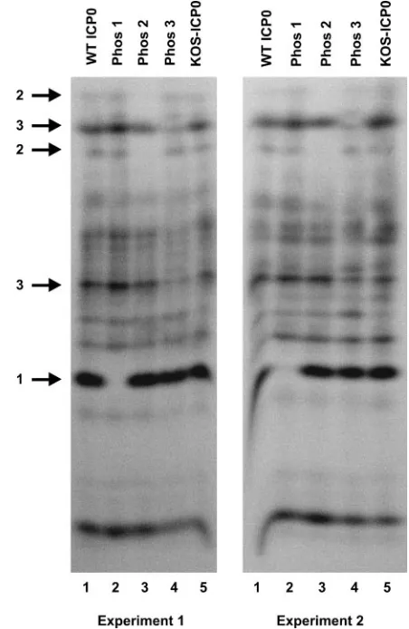
One-dimensional phosphotryptic peptide analysis. Among the 133 potential phosphorylation sites on ICP0,LC-MS/MS analysis identified subsets of sites in three regions that are actually phosphorylated. To confirm that mutagenesis resulted in the loss of phosphorylation of the Phos 1, 2, and 3 mutant forms of ICP0, we performed one-dimensional phosphotryptic peptide analysis with the phosphorylated samples shown in Fig. 2B. In two independent experiments, multiple phosphotryptic peptides were observed in gels of extracts of cells transfected with plasmids expressing wild-type ICP0 or the three Phos mutants and infected with 7134 (Fig. 3, lanes 1, 2, 3, and 4) or mock transfected and infected with wild-type virus (lane 5). As indicated by the arrows on the left of the figure, a loss or reduction in the signal of one or two specific phosphotryptic peptides was evident for each of the three mutant proteins. In
on November 8, 2019 by guest
http://jvi.asm.org/
contrast, wild-type ICP0 isolated from KOS-infected cells was essentially identical to the phosphotryptic peptide profile of wild-type ICP0 synthesized in transfected and infected cells (Fig. 3, lanes 1 and 5). Thus, the mutagenesis strategy was successful in altering the phosphorylation state of ICP0 ex-pressed from each of the mutant plasmids. As evident from the multiple phosphorylated bands not affected by any of the 11 alanine substitution mutations, LC-MS/MS analysis identi-fied only selected phosphorylation sites on ICP0.
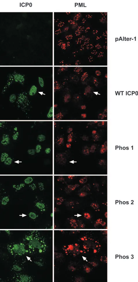
Immunofluorescence assays. (i) Subcellular localization of wild-type and Phos mutant forms of ICP0 in transfected cells.
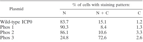
Having established that the phosphorylation states of the Phos 1, 2, and 3 mutant forms of ICP0 were altered with respect to wild-type ICP0, the effects of these clustered mutations on the subcellular localization of ICP0 and ICP0’s ability to alter the staining pattern of the ND10-associated protein promyelocytic leukemia (PML) were examined by immunofluorescence mi-croscopy. Focusing first on the subcellular localization of ICP0, transfected cells were classified in one of three categories based on their subcellular staining patterns: nuclear only, nu-clear and cytoplasmic, and cytoplasmic only. The staining pat-terns of wild-type ICP0, Phos 1, and Phos 2 were predomi-nantly nuclear and punctate (Fig. 4 and Table 1) (an example of each is indicated by the white arrow), as previously reported for wild-type ICP0 by Zhu and Schaffer (86). Specifically, 84 to 90% of transfected cells expressing these forms of ICP0 stained only in the nucleus, 8 to 15% stained both in the nucleus and in the cytoplasm, and less than 4% stained in the cytoplasm only. In contrast, the predominant subcellular location of the Phos 3 form of ICP0 was both nuclear and cytoplasmic (72.6% of cells; Fig. 4, white arrow), whereas⬇25% of cells exhibited staining exclusively in the nucleus (Table 1). Furthermore, Phos 3-expressing cells exhibited finer punctate, nuclear stain-ing and large cytoplasmic globular stainstain-ing relative to cells expressing wild-type ICP0. Thus, one or more of the four substitution mutations in Phos 3 are responsible for the altered subcellular and subnuclear staining pattern of ICP0.
(ii) Do the Phos mutations affect the staining pattern of PML? PML-specific immunofluorescence assays were per-formed with a primary rabbit polyclonal antibody which rec-ognizes PML. PML-specific staining in the absence of ICP0 (pAlter-1) was uniformly nuclear and punctate (Fig. 4). In cells that expressed wild-type ICP0 or an ICP0 Phos mutant, a reduction in PML-specific staining or an alteration in the PML staining pattern was observed. PML-specific staining was less intense in almost all wild-type ICP0- and Phos 1-expressing cells (Fig. 4, white arrows). PML staining was also less intense in many Phos 2-expressing cells; however, some large nuclear PML-specific bodies were present in nuclei expressing Phos 2 (white arrows), but they did not colocalize with Phos 2-specific staining, which was in the nuclear margin. In Phos 3-expressing cells, PML-specific nuclear staining was less intense overall, although PML-specific staining colocalized with large globular Phos 3 cytoplasmic bodies (white arrows) in a subset of cells.
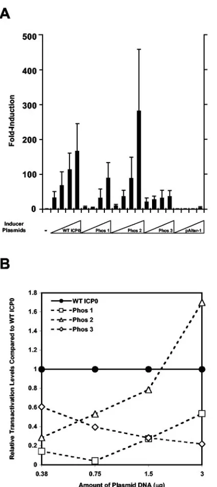
Transactivating activity of Phos mutants. To examine the transactivating activity of the Phos mutants, Vero cells were cotransfected with wild-type ICP0, Phos 1-, 2-, or 3-expressing plasmids, or pAlter-1 and the HSV-1 ICP4 promoter-CAT reporter construct (pIE3-CAT) or pIE3-CAT alone. As shown in Fig. 5A, CAT activity increased as a function of increasing
amounts of wild-type ICP0- and Phos mutant-expressing plas-mids transfected. Relative to wild-type ICP0, Phos 1 transac-tivating activity was impaired 7- to 20-fold at the two lowest amounts of plasmid transfected and only 2- to 3-fold at the two highest amounts transfected (Fig. 5A and B). The CAT activity induced by Phos 2 was reduced threefold at the lowest amount of plasmid transfected, but was similar (ⱕ2-fold differences) to wild-type ICP0 at the three highest amounts of the inducer plasmid tested (Fig. 5A and B). Phos 3 induced similar levels of CAT activity (ⱕ2-fold differences) to wild-type ICP0 at the two lowest amounts of plasmid transfected but was impaired 3- to 5-fold at the two highest amounts (Fig. 5A and B). Phos 3 was nonetheless able to induce limited CAT activity at least 20-fold above the basal level of the pIE3-CAT vector alone. Relative to wild-type ICP0, the altered levels of activity observed with the ranges of DNAs transfected may be a consequence of the residual transactivating activities of mutant forms of ICP0 and their interactions with other regulatory proteins.
7134-complementing activity of the Phos mutants.To deter-mine whether the Phos mutants are able to complement the replication of the ICP0 null mutant 7134 in Vero cells, tious wild-type or 7134 DNA was transfected alone or infec-tious 7134 DNA was cotransfected with pAlter-1 or a plasmid expressing a wild-type or Phos mutant form of ICP0 as de-scribed by Cai and Schaffer (9). Forty-eight hours after trans-fection, cells were harvested and lysates were assayed for the presence of infectious wild-type and 7134 virus by standard plaque assays in Vero and L7 cells, respectively. In these as-says, high titers of wild-type virus were detected (Table 2). In contrast, 7134 virus titers were low (⬇6 orders of magnitude lower than titers of wild-type virus) when 7134 DNA was trans-fected alone or in combination with pAlter-1. Complementa-tion of 7134 replicaComplementa-tion was achieved by transfecComplementa-tion of infec-tious 7134 viral DNA with the wild-type ICP0-expressing plasmid (⬇1,230-fold enhancement compared with 7134 DNA alone) (Table 2). 7134 titers were 20-fold lower when cotrans-fected with the Phos 1-expressing plasmid than with the plas-mid expressing wild-type ICP0, whereas viral titers following cotransfection of 7134 DNA with Phos 2 and Phos 3 were only 1.6- and 1.2-fold lower, respectively.
A second series of experiments tested the ability of Phos mutant and wild-type forms of ICP0 to enhance the replication of infectious 7134 DNA as measured by the production of cytopathic blue foci (CBF) in Vero cells. The ability of the plasmids to induce foci (clusters of eight or more cells exhib-iting cytopathic effect) when cotransfected with 7134 DNA was assessed 3 days posttransfection and visualized by X-Gal stain-ing of the cultures. X-Gal stainstain-ing was performed to visualize the spread of infection with 7134, which contains thelacZgene in place of both copies of ICP0. The production of CBF results primarily from complementation but may also result from re-combination (in this case rare [⬍5%] large plaques were ob-served). The ability of infectious 7134 viral DNA or 7134 DNA cotransfected with pAlter-1 to form CBF was limited, whereas cotransfection of 7134 DNA with the plasmid expressing wild-type ICP0 increased the number of CBF by at least 42-fold (Fig. 6). The ability of Phos 1 to induce CBF was reduced 4-fold relative to wild-type ICP0, and Phos 2 and 3 reduced the number of CBF by⬇1.7- and 1.4-fold, respectively. Further-more, the size of the Phos 1-induced CBF was smaller than
on November 8, 2019 by guest
http://jvi.asm.org/
that of those induced by wild-type ICP0, Phos 2, or Phos 3. Thus, the mutations in the Phos 1 form of ICP0 render this protein less capable of complementing the replication and plating efficiency of 7134 infectious DNA than the mutations in the Phos 2 or 3 forms of ICP0 in Vero cells.
FIG. 2. Protein levels and phosphorylation state of the Phos mu-tant forms of ICP0. (A) Experimental design. Vero cell monolayers plated 24 h previously were transfected with plasmids expressing wild-type ICP0, Phos 1, 2, or 3, or salmon sperm DNA (sasDNA) for 4 h. At 4 h posttransfection, transfected cells were treated for 1 h (t⫽ ⫺1) with the protein synthesis inhibitor cycloheximide (CHX), mock in-fected or inin-fected with 5 PFU/cell of wild-type (WT) or ICP0 null mutant (7134) (t⫽0), and incubated in the presence of cycloheximide for 6 h (t⫽6). IE transcripts were synthesized during the 6-h period. Half of the cultures were then released from the cycloheximide block into phosphate-free medium containing [32P]orthophosphate. The re-maining cultures were released into methionine- and cysteine-free medium containing [35S]methionine/cysteine. Radiolabeling was car-ried out for 6 h (t⫽12). (B) Immunoprecipitation and SDS-PAGE analysis of radiolabeled ICP0. Att⫽6 h postlabeling, extracts were prepared, ICP0 was immunoprecipitated with an ICP0 rabbit
poly-FIG. 3. One-dimensional phosphotryptic peptide analysis of wild-type and Phos mutant forms of ICP0.32P-labeled wild-type (WT) and Phos 1, 2, and 3 mutant forms of ICP0 expressed from Vero cells transfected with the corresponding plasmids and infected with 7134 or ICP0 expressed from KOS-infected cells was isolated following SDS-PAGE from the gels shown in Fig. 2B and digested with trypsin. The resulting phosphopeptides were separated on a 30% alkaline poly-acrylamide gel (81) and visualized by PhosphorImager analysis. The results of two independent experiments are shown. Numbered arrows on the left identify specific phosphotryptic peptides present in wild-type ICP0 that are absent or reduced in the Phos 1, 2, or 3 mutant forms of ICP0.
clonal antibody (J17), and resolved by SDS–6% PAGE.35S- and32 P-specific ICP0 bands were visualized by PhosphorImager analysis. Band intensities of wild-type ICP0 and Phos mutants were measured by PhosphorImager analysis, and the intensities of Phos mutant forms of ICP0 relative to wild-type ICP0 (expressed as 100%) for each radio-labeled band were determined. (C) Ratio of the relative32P to35S bands intensities of wild-type ICP0 and Phos 1, 2, and 3. The ratios of band intensities of wild-type and each Phos mutant form of ICP0 were determined by dividing the relative32P band intensity by the35S band intensity (shown in B) for wild-type ICP0 or Phos 1, 2, and 3.
on November 8, 2019 by guest
http://jvi.asm.org/
DISCUSSION
This study demonstrates that ICP0 is phosphorylated in at least three regions which collectively contain 11 putative
phos-phorylation sites as determined byLC-MS/MS analysis. With site-directed mutagenesis, potentially phosphorylated serine and threonine residues in these regions were changed to ala-nine. The resulting mutations in the three regions affect ICP0’s subcellular and subnuclear localization (Phos 3), ND10-dis-rupting activity in a subset of cells (Phos 2 and 3), and trans-activating activity (Phos 1 and 3) in Vero cells. Despite the altered phenotypes of all three Phos mutant forms of ICP0, only the Phos 1 form of ICP0 exhibited significantly reduced ability to complement the replication and plating efficiency of infectious 7134 DNA. This study represents the first attempt to fine map and mutate known posttranslational modification sites on ICP0 and determine their roles in ICP0 function, as the great majority of ICP0 structure-function analyses have employed nonsense, insertion, or deletion mutants. It is also acknowledged that introducing alanines in place of putative phosphoacceptor sites in clusters may alter the folding or con-firmation of ICP0 molecules and thus affect ICP0 function. Future studies designed to identify the specific phosphoryla-tion sites utilized in regions I, II, and III will allow us to prove or refute this possibility. WhileLC-MS/MS analysis of the peptides generated by trypsin and chymotrypsin digestion iden-tified three phosphorylated regions on ICP0 that are important for its diverse biological activities, this analysis clearly identi-fied only a subset of phosphorylated regions on ICP0, as illus-trated in Fig. 3. The location of additional regions and their role(s) in the functions of ICP0 will be determined by LC-MS/MS with alternative enzymes to generate phosphopep-tides.
Do the locations of the mutations in Phos 1, 2, and 3 cor-respond with domains or interaction sites known to be impor-tant for the activities of ICP0? (i)Phos 1. Based on LC-MS/MS analysis, each of the three phosphorylated regions of ICP0 identified lies within or overlaps domains reported to be important for ICP0’s transactivating activity. Thus, the four mutations in Phos 1 at positions 224, 226, 231, and 232 are adjacent to the RING finger motif of ICP0, which is required for its E3 ubiquitin ligase and transactivating activities (Fig. 1A) (5, 22, 24). This study has shown that the Phos 1 form of ICP0 is impaired in its transactivating activity and in its ability to complement the replication of an ICP0 null mutant but not its ability to degrade and/or disperse ND10. Insertion and deletion mutations in region I or between region I and ICP0’s RING finger have been shown to diminish both the transacti-vating activity of ICP0 and its ability to colocalize with conju-gated ubiquitin (20, 22, 24, 25); the latter activity is consistent with ICP0’s E3 ubiquitin ligase activity.
Mutations in the RING finger motif (residues 116 to 156) and an adjacent region (residues 162 to 188) show a range of ND10-disrupting phenotypes that are distinct from the pheno-type of wild-pheno-type ICP0 (20, 29, 49). To date, however, the ND10-disrupting activities of the majority of ICP0 mutants with mutations that lie in region I and between region I and the RING finger motif have not been reported. Additionally, mu-tation of Asp-199 (which lies between region I and the RING finger motif) to alanine negates the binding of ICP0 to cyclin D3, accelerating the destabilization of cyclin D3 (44). This mutation attenuates the pathogenesis of HSV-1 (77).
[image:7.585.44.282.65.550.2]Residues 20 to 241 of ICP0 have been shown to interact with the cellular transcription factor BMAL1 (43). The interaction
FIG. 4. ICP0- and PML-specific immunofluorescence in Vero cells transfected with plasmids expressing wild-type (WT) ICP0 or Phos 1, 2, or 3 mutant forms of ICP0. Vero cells were grown on coverslips in 12-well plates. Twenty-four hours after plating, cells were transfected with 3g of plasmid DNA expressing wild-type ICP0, Phos 1, 2, or 3 mutant forms of ICP0, or the cloning vector alone (pAlter-1). Twenty four hours posttrans-fection, cells were washed, fixed, permeabilized, and probed with primary and secondary antibodies to detect ICP0 and PML by immunofluores-cence. The white arrows show ICP0- and PML-specific staining in indi-vidual cells positive for ICP0 staining. Cells were viewed by fluorescence microscopy with a Nikon Eclipse TE300 Fluorescence microscope at ⫻400 magnification and photographed with a digital camera.
on November 8, 2019 by guest
http://jvi.asm.org/
of ICP0 with BMAL1 is thought to facilitate synergistic trans-activation of BMAL-1 responsive genes. Thus, it is possible that the effects that we observed with the Phos 1 mutations in Vero cells result from alterations in these adjacent regions. Of interest is the fact that the phenotypes of Phos 1 are strikingly similar to the phenotypes of ICP0 synthesized in the presence of the cdk inhibitor roscovitine (17). Specifically, ICP0 synthe-sized in the presence of roscovitine is impaired in its transac-tivating activity but not its ND10-disrupting activity. Thus, roscovitine-sensitive cdk-mediated phosphorylation of phos-phoacceptor sites in region I of ICP0, especially Ser-224 which is a potential cdk-1 or -2 phosphorylation site, may contribute to ICP0’s transactivating activity.
(ii) Phos 2.The mutations in Phos 2 at positions 365, 367, and 371 lie within a large proline-rich domain important for ICP0’s transactivating activity. (Fig. 1A) (80). Phos 2 was af-fected only in its capacity to disperse or degrade ND10-asso-ciated PML in a subset of Phos 2-expressing cells; the trans-activating- and 7134-complementing activities of Phos 2 were only minimally affected relative to wild-type ICP0. The study of Chen et al. noted a reduction in ICP0’s transactivating activity in a mutant form of ICP0 lacking residues 263 to 448, which includes all of region II (13). Consistent with the transactivat-ing potential of Phos 2, a report by Everett noted that an insertion in region II at residue 370 and deletion of residues 341 to 374 had marginal effects on ICP0-mediated transacti-vation in Vero cells (22). The ability of these deletion and insertion mutations to affect the dispersal or degradation of ND10-associated proteins (including PML) has not been re-ported. Notably, the magnitude of the impairment in the trans-activating activity of these mutants correlates with the severity of their mutations.
(iii) Phos 3.Mutations in Phos 3 at positions 508, 514, 517, and 518 overlap the putative nuclear localization signal of ICP0 and are adjacent to its multifunctional C-terminal do-main (9, 12, 13, 24, 27, 29, 49). Phos 3 was affected in its subcellular and nuclear localization and its ability to disperse or degrade PML in a subset of cells expressing Phos 3; how-ever, these mutations only minimally affected its ability to complement 7134 in Vero cells. The diminished transactivation potential of Phos 3 may also be due to its altered subcellular
and/or subnuclear localization. Similar observations have been reported for deletion mutations of ICP0 which eliminate its putative nuclear localization signal (24, 27). Notably, a propor-tion of cells expressing these delepropor-tion mutants colocalized with ND10-associated proteins in the cytoplasm, similar to our ob-servations with PML and Phos 3 (Fig. 4) (29, 49).
Because the mutations in Phos 3 are directly adjacent to the putative nuclear localization signal of ICP0 and because phos-phorylation of the nuclear localization signal may affect nu-clear import, Phos 3 may be inefficiently transported to or from the nucleus, as our immunofluorescence data suggest (Fig. 4). In support of this possibility, mutagenesis of potential phos-phorylation sites has been shown to regulate the subcellular localization of viral regulatory proteins from DNA-containing viruses, including the T antigen of simian virus 40 (41, 84), pp65 of human cytomegalovirus (32), latent nuclear antigen of Kaposi’s sarcoma-associated herpesvirus (60), IE63 protein of varicella zoster virus (4), and US11, an RNA-binding protein (67), and ICP27 of HSV-1 (10, 85).
Of the viral regulatory proteins just mentioned with the exception of US11, casein kinase II, cdk-1, and protein kinase A have been implicated in their subcellular localization, and putative phosphorylation sites for these cellular kinases have been identified in region III of ICP0. The subcellular localiza-tion of Phos 3 may also indirectly affect the phosphorylalocaliza-tion state of a portion of ICP0 molecules. For example, if ICP0 is efficiently phosphorylated by nuclear kinases which are re-quired for its biological activities, then impairment of ICP0’s nuclear translocation would prevent its phosphorylation by such kinases. Although the mutations in Phos 3 reduced its transactivating activity, they affected its 7134-complementating activity in Vero cells only minimally. These observations dem-onstrate that Phos 3 possesses sufficient transactivating activity to stimulate the replication of an ICP0 null mutant to levels similar to that of wild-type ICP0. This possibility is reinforced by two studies demonstrating that mutant forms of ICP0 ex-hibiting impaired transactivating activity are capable of sup-porting significant complementation of an ICP0 null mutant virus (9, 12). Finally, although we examined the phenotypic effects of the Phos 1, 2, and 3 mutations in Vero cells, it is conceivable that these mutations have cell type-specific effects in the functional assays used in this study, alter other known activities of ICP0 (e.g., E3 ubiquitin ligase activity, accumula-tion of conjugated ubiquitin, or cell cycle-blocking activity), and/or have distinct phenotypes in the context of viral infection in vivo. These possibilities warrant further investigation.
[image:8.585.43.284.89.161.2]Role of cellular and viral kinases in ICP0 function.While this study has identified 11 putative phosphorylation sites on ICP0, whether these sites are actually phosphorylated and, if they are, the identity of the cellular and viral kinases that phosphorylate these sites and the roles of specific phosphory-lation events in the reguphosphory-lation of ICP0 function remain to be determined. Of the three regions identified, our results indi-cate that the Phos 1 mutations had the greatest effect on ICP0-induced transactivation and viral replication, suggesting that the kinases that phosphorylate one or more of these sites are necessary for these activities of ICP0. From computer-based modeling, cellular kinases that potentially phosphorylate Ser-224, Thr-226, and Thr-231 are calmodulin kinase II, pro-tein kinase A, cdk-1 and -2, and casein kinase II (Fig. 1A). No
TABLE 1. Subcellular localization of wild-type and mutant forms of ICP0a
Plasmid
% of cells with staining pattern:
N N⫹C C
Wild-type ICP0 83.7 15.1 1.2
Phos 1 90.3 8.4 1.3
Phos 2 86.1 10.6 3.3
Phos 3 24.8 72.6 2.6
aThe subcellular localization of wild-type and Phos mutant forms of ICP0 in
individual Vero cells 24 h after transfection was determined. Cells that stained positive for ICP0 were classified into one of three groups based on the subcel-lular localization of ICP0: cells that displayed only nuclear staining (N), cells that
displayed both nuclear and cytoplasmic staining (N⫹C), and cells that displayed
only cytoplasmic staining (C). One hundred fifty or more ICP0-stained cells in each preparation were analyzed, and the percentage of cells exhibiting a specific pattern of subcellular localization was calculated by dividing the number of ICP0-expressing cells in a given category by the total number of cells counted in all three categories for each protein.
on November 8, 2019 by guest
http://jvi.asm.org/
cellular kinases were identified for T232 in our computer anal-ysis.
Do these cellular kinases play a role in the functions of HSV IE proteins, including ICP0? Indeed, several studies indicate that cellular kinases most likely modulate the functions of IE proteins through phosphorylation. As noted in the introduc-tion, phosphorylation of a serine-rich region of ICP4 from residues 140 to 210 by protein kinase A appears to be required for efficient HSV replication in PC12 cells (a rat cell line of neural lineage), efficient replication in the tear film and tri-geminal ganglia of mice during acute infection, and efficient reactivation from latency in the mouse ocular model (83). ICP27 has been shown to be phosphorylated on at least four major peptides in infected cell cultures (85). Two of the four peptides are phosphorylated by protein kinase A and casein kinase II in vitro. Mutation of these phosphorylation sites did not affect the ability of ICP27 to complement the growth of a virus lacking ICP27, indicating that these sites have no appar-ent role in viral replication in cell culture. The effects of these mutations in vivo will require further testing. A recent report noted that casein kinase II binds to ICP27, stimulating casein kinase II activity during viral infection (45). Casein kinase II subsequently phosphorylates ICP27, affecting its interaction with another ICP27-interacting cellular protein, heteroge-neous ribonucleoprotein, which has been implicated in tran-script transport. Cdk-1 has been shown to phosphorylate the second exon (residues 20 to 241) of ICP0 in vitro (3), and the portion of region I that lies within this exon contains an afore-mentioned putative cdk-1 or -2 phosphorylation site at Ser-224 (Fig. 1A; left of the hatchmark in region I). Consequently, cdk-1 or -2 may phosphorylate Ser-224, contributing to the biological activities of ICP0. The roles of the remaining cellular kinases identified in our computer analysis in ICP0 function (Fig. 1A; glycogen synthase kinase 3, casein kinase I, p70S6K kinase, protein kinase G, and protein kinase C) remain to determined.
[image:9.585.62.274.72.548.2]Based on sequence analysis and functional studies, HSV encodes three viral kinases: the products of the UL13 gene, a delayed-early tegument protein (64); the UL39 gene (ICP6), an E protein and the large subunit of ribonucleotide reductase
FIG. 5. Transactivating activity induced by wild-type (WT) and Phos mutant forms of ICP0. (A) Vero cells were plated and 24 h later cotransfected with 1g of pIE3-CAT reporter plasmid (ICP4 promot-er-CAT construct) (18) or with increasing amounts of inducer plasmids (0.375, 0.75, 1.5, and 3g) expressing wild-type ICP0 (ICP0), Phos 1, 2, and 3, or the cloning vector alone (pAlter-1). Forty-eight hours after transfection, cells were harvested, extracts were prepared, and CAT assays were performed. The induction (fold) of CAT activity relative to the basal CAT activity of the pIE3-CAT reporter plasmid transfected alone, given the arbitrary value of 1 (bar on the far left), is shown. (B) Transactivating activity of Phos mutants relative to wild-type ICP0 as a function of the amount of inducer DNA transfected. The relative level of transactivation of each Phos mutant was determined by divid-ing the CAT activity for a given amount of expression plasmid trans-fected over the CAT activity of an equivalent of amount of wild-type ICP0-expressing plasmid transfected, based on the data presented in panel A. In all cases, wild-type ICP0 was given the arbitrary value of 1 for each amount of wild-type ICP0-expressing plasmid transfected.
TABLE 2. Complementation of 7134 replication by ICP0 Phos mutant plasmidsa
Virus⫹plasmid
DNAs
Titerb
(PFU/ml)
Difference relative
to WT ICP0c
(fold)
KOS 1.33⫻107
7134 5
7134⫹pAlter-1 5
7134⫹WT ICP0 6,150 1
7134⫹Phos 1 315 220
7134⫹Phos 2 3,925 21.6
7134⫹Phos 3 5,000 21.2
aVero cells were transfected with 0.25g of KOS or 7134 infectious viral
DNA and 0.0375g of pAlter-1 or a plasmid expressing wild-type (WT) ICP0 or
a Phos mutant. Transfected cells were harvested 48 h posttransfection and assayed for infectious virus. The experiment was repeated twice, and results from one experiment are shown.
bAverage viral titers from duplicate samples were determined on Vero cells
(for KOS) or L7 cells (for 7134).
cDifference in viral titers for each Phos mutant-expressing plasmid relative to
the wild-type ICP0-expressing plasmid.
on November 8, 2019 by guest
http://jvi.asm.org/
[image:9.585.302.541.89.189.2](36, 59); and the US3 gene, an E protein implicated in cell survival (30, 33, 62). Of the three viral kinases, UL13 is the most extensively studied with regard to IE protein function. As previously discussed, ICP22 is phosphorylated, either directly or indirectly, by UL13, and this phosphorylation is required for ICP22 function in selected cell types (48, 63, 64, 66, 73). The phosphorylation of ICP0 is also mediated in part by UL13 in infected cells and in vitro with infected cell extracts, but the significance of this phosphorylation for the activities of ICP0 is unknown (58). Interestingly, one study reported that UL13 and cdk-1 can phosphorylate the same serine residue on the cellu-lar translation protein elongation factor 1␦(42). Consequently, UL13 may also phosphorylate the putative cdk-1 or -2 consen-sus motifs on ICP0 identified in our study.
Future studies of ICP0 phosphorylation will entail identify-ing the individual phosphorylation site or sites that affect the activities of each Phos mutant form of ICP0 and the roles of these sites in ICP0 functions as they relate to HSV-1 replica-tion. This fine-mapping approach will permit us to determine whether the effects of the clustered mutations in Phos 1, 2, and 3 are due to specific alterations in the phosphorylation state of ICP0 or to alterations in the structure of ICP0. Ultimately,
identifying the cellular and viral kinases that phosphorylate ICP0 and elucidating the roles of these kinases in the regula-tion of ICP0 funcregula-tion should provide insight into how virus-cell interactions influence the HSV life cycle.
ACKNOWLEDGMENTS
This work was supported by Public Health Service grant RO1CA20260 from the National Cancer Institute (to P.A.S.).
We thank Gerd Maul for providing the PML antibody, Joe Orlando for providing technical assistance with site-directed mutagenesis, and other members of the Schaffer lab and the Boston area herpesvirus group for helpful comments and suggestions.
REFERENCES
1.Ackermann, M., D. K. Braun, L. Pereira, and B. Roizman.1984. Character-ization of herpes simplex virus 1 alpha proteins 0, 4, and 27 with monoclonal
antibodies. J. Virol.52:108–118.
2.Advani, S. J., R. Hagglund, R. R. Weichselbaum, and B. Roizman.2001. Posttranslational processing of infected cell proteins 0 and 4 of herpes simplex virus 1 is sequential and reflects the subcellular compartment in
which the proteins localize. J. Virol.75:7904–7912.
3.Advani, S. J., R. R. Weichselbaum, and B. Roizman.2000. The role of cdc2 in the expression of herpes simplex virus genes. Proc. Natl. Acad. Sci. USA
97:10996–11001.
4.Bontems, S., E. Di Valentin, L. Baudoux, B. Rentier, C. Sadzot-Delvaux, and J. Piette.2002. Phosphorylation of varicella-zoster virus IE63 protein by
FIG. 6. Complementation of 7134 as determined by cytopathic blue focus (CBF) formation by wild-type ICP0 and Phos mutant forms of ICP0. Vero cells were transfected with 0.25g of 7134 infectious viral DNA alone or cotransfected with 0.0375g of pAlter-1 or plasmids expressing wild-type ICP0 or mutant forms of ICP0 (Phos 1, 2, and 3) per dish. 7134 contains thelacZgene in place of both copies of ICP0. Five hours after transfection, monolayers were overlaid with methylcellulose-containing medium. Three days posttransfection, monolayers were washed, fixed, and stained for-galactosidase activity with X-Gal and incubated for an additional 14 to 16 h at room temperature. The number and the differences (fold) in CBF (clusters of eight or more rounded, cytopathic cells) produced on monolayers transfected with 7134 infectious viral DNA alone, 7134 infectious viral DNA and pAlter-1, or 7134 and individual Phos mutant-expressing plasmids relative to 7134 cotransfected with a plasmid expressing wild-type ICP0 are shown beneath the label for each photo. Monolayers were viewed with a Nikon light microscope at⫻80 magnification and photographed with a digital camera.
on November 8, 2019 by guest
http://jvi.asm.org/
casein kinases influences its cellular localization and gene regulation activity.
J. Biol. Chem.277:21050–21060.
5.Boutell, C., S. Sadis, and R. D. Everett.2002. Herpes simplex virus type 1 immediate-early protein ICP0 and is isolated RING finger domain act as
ubiquitin E3 ligases in vitro. J. Virol.76:841–850.
6.Cai, W., T. L. Astor, L. M. Liptak, C. Cho, D. M. Coen, and P. A. Schaffer.
1993. The herpes simplex virus type 1 regulatory protein ICP0 enhances virus replication during acute infection and reactivation from latency. J. Virol.
67:7501–7512.
7.Cai, W., and P. A. Schaffer.1991. A cellular function can enhance gene expression and plating efficiency of a mutant defective in the gene for ICP0,
a transactivating protein of herpes simplex virus type 1. J. Virol.65:4078–
4090.
8.Cai, W., and P. A. Schaffer.1992. Herpes simplex virus type 1 ICP0 regulates expression of immediate-early, early, and late genes in productively infected
cells. J. Virol.66:2904–2915.
9.Cai, W. Z., and P. A. Schaffer.1989. Herpes simplex virus type 1 ICP0 plays a critical role in the de novo synthesis of infectious virus following
transfec-tion of viral DNA. J. Virol.63:4579–4589.
10.Catez, F., M. Erard, N. Schaerer-Uthurralt, K. Kindbeiter, J. J. Madjar, and J. J. Diaz.2002. Unique motif for nucleolar retention and nuclear export
regulated by phosphorylation. Mol. Cell. Biol.22:1126–1139.
11.Cegielska, A., I. Moarefi, E. Fanning, and D. M. Virshup.1994. T-antigen kinase inhibits simian virus 40 DNA replication by phosphorylation of intact
T antigen on serines 120 and 123. J. Virol.68:269–275.
12.Chen, J., C. Panagiotidis, and S. Silverstein.1992. Multimerization of ICP0,
a herpes simplex virus immediate-early protein. J. Virol.66:5598–5602.
13.Chen, J. X., X. X. Zhu, and S. Silverstein.1991. Mutational analysis of the
sequence encoding ICP0 from herpes simplex virus type 1. Virology180:
207–220.
14.Chen, Y. R., S. P. Lees-Miller, P. Tegtmeyer, and C. W. Anderson.1991. The human DNA-activated protein kinase phosphorylates simian virus 40 T
an-tigen at amino- and carboxy-terminal sites. J. Virol.65:5131–5140.
15.Chittum, H. S., W. S. Lane, B. A. Carlson, P. P. Roller, F. D. Lung, B. J. Lee, and D. L. Hatfield.1998. Rabbit beta-globin is extended beyond its UGA stop codon by multiple suppressions and translational reading gaps.
Bio-chemistry37:10866–10870.
16.Davido, D. J., D. A. Leib, and P. A. Schaffer.2002. The cyclin-dependent kinase inhibitor roscovitine inhibits the transactivating activity and alters the posttranslational modification of herpes simplex virus type 1 ICP0. J. Virol.
76:1077–1088.
17.Davido, D. J., W. F. Von Zagorski, G. G. Maul, and P. A. Schaffer.2003. The differential requirement for cyclin-dependent kinase activities distinguishes
two functions of herpes simplex virus type 1 ICP0. J. Virol.77:12603–12616.
18.DeLuca, N. A., and P. A. Schaffer.1985. Activation of immediate-early, early, and late promoters by temperature-sensitive and wild-type forms of herpes
simplex virus type 1 protein ICP4. Mol. Cell. Biol..5:1197–1208.
19.Eng, J. K., A. L. McCormack, and J. R. Yates, III.1994. An approach to correlate tandem mass spectral data of peptides with amino acid sequences
in a protein database. J. Am. Soc. Mass Spectrom.5:976–989.
20.Everett, R., P. O’Hare, D. O’Rourke, P. Barlow, and A. Orr.1995. Point mutations in the herpes simplex virus type 1 Vmw110 RING finger helix affect activation of gene expression, viral growth, and interaction with
PML-containing nuclear structures. J. Virol.69:7339–7344.
21.Everett, R. D.1985. Activation of cellular promoters during herpes virus
infection of biochemically transformed cells. EMBO J.4:1973–1980.
22.Everett, R. D.1988. Analysis of the functional domains of herpes simplex
virus type 1 immediate-early polypeptide Vmw110. J. Mol. Biol.202:87–96.
23.Everett, R. D.1989. Construction and characterization of herpes simplex virus type 1 mutants with defined lesions in immediate early gene 1. J. Gen.
Virol.70:1185–1202.
24.Everett, R. D.1987. A detailed mutational analysis of Vmw110, a trans-acting transcriptional activator encoded by herpes simplex virus type 1. EMBO J.
6:2069–2076.
25.Everett, R. D.2000. ICP0 induces the accumulation of colocalizing
conju-gated ubiquitin. J. Virol.74:9994–10005.
26.Everett, R. D.2000. ICP0, a regulator of herpes simplex virus during lytic and
latent infection. Bioessays22:761–770.
27.Everett, R. D.1988. Promoter sequence and cell type can dramatically affect the efficiency of transcriptional activation induced by herpes simplex virus type 1 and its immediate-early gene products Vmw175 and Vmw110. J. Mol.
Biol.203:739–751.
28.Everett, R. D.1984. Trans activation of transcription by herpes virus prod-ucts: requirement for two HSV-1 immediate-early polypeptides for
maxi-mum activity. EMBO J.3:3135–3141.
29.Everett, R. D., and G. G. Maul.1994. HSV-1 IE protein Vmw110 causes
redistribution of PML. EMBO J.13:5062–5069.
30.Frame, M. C., F. C. Purves, D. J. McGeoch, H. S. Marsden, and D. P. Leader.1987. Identification of the herpes simplex virus protein kinase as the
product of viral gene US3. J. Gen. Virol.68:2699–2704.
31.Fu, X. D., P. T. Tuazon, J. A. Traugh, and J. Leis.1988. Site-directed
mutagenesis of the avian retrovirus nucleocapsid protein, pp 12, at Serine 40,
the primary site of phosphorylation in vivo. J. Biol. Chem.263:2134–2139.
32.Gallina, A., E. Percivalle, L. Simoncini, M. G. Revello, G. Gerna, and G. Milanesi.1996. Human cytomegalovirus pp65 lower matrix phosphoprotein
harbours two transplantable nuclear localization signals. J. Gen. Virol.77:
1151–1157.
33.Galvan, V., and B. Roizman.1998. Herpes simplex virus 1 induces and blocks apoptosis at multiple steps during infection and protects cells from exoge-nous inducers in a cell-type-dependent manner. Proc. Natl. Acad. Sci. USA
95:3931–3936.
34.Gelman, I. H., and S. Silverstein.1985. Identification of immediate early genes from herpes simplex virus that transactivate the virus thymidine kinase
gene. Proc. Natl. Acad. Sci. USA82:5265–5269.
35.Gius, D., and L. A. Laimins.1989. Activation of human papillomavirus type 18 gene expression by herpes simplex virus type 1 viral transactivators and a
phorbol ester. J. Virol.63:555–563.
36.Goldstein, D. J., and S. K. Weller.1988. Herpes simplex virus type 1-induced ribonucleotide reductase activity is dispensable for virus growth and DNA
synthesis: isolation and characterization of an ICP6lacZinsertion mutant.
J. Virol.62:196–205.
37.Halford, W. P., and P. A. Schaffer. 2001. ICP0 is required for efficient reactivation of herpes simplex virus type 1 from neuronal latency. J. Virol.
75:3240–3249.
38.Halford, W. P., and P. A. Schaffer.2000. Optimized viral dose and transient immunosuppression enable herpes simplex virus ICP0-null mutants to
es-tablish wild-type levels of latency in vivo. J. Virol.74:5957–5967.
39.Honess, R. W., and B. Roizman.1974. Regulation of herpesvirus macromo-lecular synthesis. I. Cascade regulation of the synthesis of three groups of
viral proteins. J. Virol.14:8–19.
40.Isler, J. A., and P. A. Schaffer.2001. Phosphorylation of the herpes simplex
virus type 1 origin binding protein. J. Virol.75:628–637.
41.Jans, D. A., M. J. Ackermann, J. R. Bischoff, D. H. Beach, and R. Peters.
1991. p34cdc2-mediated phosphorylation at T124 inhibits nuclear import of
SV-40 T antigen proteins. J. Cell Biol.115:1203–1212.
42.Kawaguchi, Y., K. Kato, M. Tanaka, M. Kanamori, Y. Nishiyama, and Y. Yamanashi.2003. Conserved protein kinases encoded by herpesviruses and cellular protein kinase cdc2 target the same phosphorylation site in
eukary-otic elongation factor 1delta. J. Virol.77:2359–2368.
43.Kawaguchi, Y., M. Tanaka, A. Yokoymama, G. Matsuda, K. Kato, H. Ka-gawa, K. Hirai, and B. Roizman.2001. Herpes simplex virus 1 alpha regu-latory protein ICP0 functionally interacts with cellular transcription factor
BMAL1. Proc. Natl. Acad. Sci. USA98:1877–1882.
44.Kawaguchi, Y., C. Van Sant, and B. Roizman.1997. Herpes simplex virus 1 alpha regulatory protein ICP0 interacts with and stabilizes the cell cycle
regulator cyclin D3. J. Virol.71:7328–7336.
45.Koffa, M. D., J. Kean, G. Zachos, S. A. Rice, and J. B. Clements.2003. CK2 protein kinase is stimulated and redistributed by functional herpes simplex
virus ICP27 protein. J. Virol.77:4315–4325.
46.Laemmli, U. K.1970. Cleavage of structural proteins during the assembly of
the head of bacteriophage T4. Nature227:680–685.
47.Leib, D. A., D. M. Coen, C. L. Bogard, K. A. Hicks, D. R. Yager, D. M. Knipe, K. L. Tyler, and P. A. Schaffer.1989. Immediate-early regulatory gene mutants define different stages in the establishment and reactivation of
herpes simplex virus latency. J. Virol.63:759–768.
48.Long, M. C., V. Leong, P. A. Schaffer, C. A. Spencer, and S. A. Rice.1999. ICP22 and the UL13 protein kinase are both required for herpes simplex virus-induced modification of the large subunit of RNA polymerase II. J.
Vi-rol.73:5593–5604.
49.Maul, G. G., and R. D. Everett.1994. The nuclear location of PML, a cellular member of the C3HC4 zinc-binding domain protein family, is rearranged during herpes simplex virus infection by the C3HC4 viral protein ICP0.
J. Gen. Virol.75:1223–1233.
50.McVey, D., L. Brizuela, I. Mohr, D. R. Marshak, Y. Gluzman, and D. Beach.
1989. Phosphorylation of large tumour antigen by cdc2 stimulates SV40
DNA replication. Nature341:503–507.
51.McVey, D., S. Ray, Y. Gluzman, L. Berger, A. G. Wildeman, D. R. Marshak, and P. Tegtmeyer.1993. cdc2 phosphorylation of threonine 124 activates the
origin-unwinding functions of simian virus 40 T antigen. J. Virol.67:5206–
5215.
52.McVey, D., B. Woelker, and P. Tegtmeyer.1996. Mechanisms of simian virus
40 T-antigen activation by phosphorylation of threonine 124. J. Virol.70:
3887–3893.
53.Meijer, L., A. Borgne, O. Mulner, J. P. Chong, J. J. Blow, N. Inagaki, M. Inagaki, J. G. Delcros, and J. P. Moulinoux.1997. Biochemical and cellular effects of roscovitine, a potent and selective inhibitor of the cyclin-dependent
kinases cdc2, cdk2 and cdk5. Eur. J. Biochem.243:527–536.
54.Montenarh, M., and D. Muller.1987. The phosphorylation at Thr 124 of simian virus 40 large T antigen is crucial for its oligomerization. FEBS Lett.
221:199–204.
55.Mosca, J. D., D. P. Bednarik, N. B. Raj, C. A. Rosen, J. G. Sodroski, W. A. Haseltine, G. S. Hayward, and P. M. Pitha.1987. Activation of human immunodeficiency virus by herpesvirus infection: identification of a region