JOURNAL OF VIROLOGY, May 1421-1426 0022-538X/87/051421-06$02.00/0
Copyright© 1987, AmericanSociety for Microbiology
Simian
Immunodeficiency
Virus
Induces
Expression
of Class
IIMajor
Histocompatibility
Complex Structures
on
Infected
Target
Cells In Vitro
MARIKANNAGI, MASAYA KIYOTAKI, NORVAL W. KING, CAROL I. LORD, AND NORMAN L. LETVIN*
HarvardMedical School, New England Regional Primate ResearchCenter, Southborough, Massachusetts 01772 Received 14October1986/Accepted7January 1987
The human immunodeficiency virus (HIV) and the closely related simian immunodeficiency virus (SIV)
induce profound immune dysfunction in primate species. The present studies show that cell populations infected in vitro with SIV exhibit increases in major histocompatibility complex (MHC) class II antigen expression. Cell lineschronically infectedwith both the monkey and human virusesexpresssubstantiallymore
MHCclassHbut notmorelineage-restrictedoractivation antigensontheir membranes than do uninfected cell
lines. Furthermore, 2'-deoxy-5-iodouridine increased MHC class II antigen expression on SIV-infectedcell
linesin parallel with increased expression of viral antigens. MHC class II induction does not appear tobe mediated through the production of a soluble factor, such as gamma interferon, by SIV-infected cells.
Interestingly, studies of the kinetics ofantigen expression by cell lines after SIV infection indicate that the inductionof MHC class IIstructuresisalateevent.Immunoelectron microscopy revealedthatMHC classII antigenisexpressednotonlyonthesurfaces oftheSIV-infected cells but alsoonthe envelope of virus particles
derived from those cells. MHC antigen expression on virus-infected cells and the expression of those
determinantsby the virusmayplayarole inthe pathogenesis of acquiredimmunodeficiencysyndrome andthe autoimmune abnormalities observed inHIV-infected individuals.
The humanimmunodeficiency virus (HIV) is the etiologic agentinacquired immunodeficiency syndrome (AIDS), and the closely related simian immunodeficiency virus (SIV), previously called simian T-lymphotropic virus type III, induces anAIDS-like syndrome inmacaque monkeys (1, 4,
16). Theprecise mechanisms by which these viruses induce immune abnormalities remain unclear. They infect T4+
(helper/inducer) lymphocytes (11, 12) and cells of the monocyte/macrophage lineage (9). After infection with these agents,adramatic decrease in the number ofcirculating T4+
lymphocytes occurs (15, 16). Other consequences of
infec-tions with this family of viruses which may account for immunedysfunction remain tobe delineated.
Major histocompatibility complex (MHC)-encoded struc-tures onthe surfaces of immune cells playacrucial role in
the interactions of these cells. MHC class I (MHC-I) cell surface molecules are recognized by T8+ (suppressor/ cytotoxic) lymphocytes, and MHCclass II (MHC-II) mole-culesarerecognized byT4+ lymphocytes (17). These struc-turesarecritically importantintherecognition ofselfbythe immune cell. In the present studieswe have demonstrated thatcellpopulationsinfected with SIV exhibitanincrease in MHC-II-encodedmembrane antigenexpression.
MATERIALS ANDMETHODS
Cells and viruses. The cell linesusedwereH9,H9 infected with SIV (H9-SIV), HUT78, HUT78 infected with SIV (HUT78-SIV), andH9 infected withtwodifferent isolates of HIV(H9-HIVAandH9-HIVB).Thechronicallyinfected cell lines had been maintained for at least 18 months after infection. The SIV isolate used for acute infection in this study had been isolated from a rhesus monkey with a
lymphoma (16). Culture supernatants of phytohemag-glutinin-stimulatedhumanperipheralbloodlymphocytes
in-*Correspondingauthor.
fected with this isolate or those of H9 cells chronically
infected with this isolatewereused asavirus source.
MAbs. Monoclonal antibodies (MAbs) reactive with Ti (24T6G12), T3 (2Ad2A2), T4 (19Thy5D7), MHC-I (W6/32), MHC-II (I-2,949), the interleukin-2 (IL-2) receptor(1HT4), and the T-cell activation antigen Ta.1 (4EL) were kindly
provided by S. Schlossman (Dana-Farber Cancer Institute, Boston, Mass.). The anti-T3 MAbSP34wasprovided by C.
Terhorst (Dana-Farber Cancer Institute). The anti-MHC-II MAb LB3.1 was provided by J. Strominger (Dana-Farber
Cancer Institute). All the anti-MHC-II MAbs used
recog-nized HLA-DR.
Cellularradioimmunoassay. Between 2 x 10⁵and 5 < 105
infected cells and equivalent numbers of uninfected cells
were platedper well in triplicate intoflat-bottom wells ofa
flexible96-well microtiterplate (BectonDickinsonLabware, Oxnard, Calif.) coated with 100 ,ug ofpoly-L-lysine (Sigma ChemicalCo.,St. Louis, Mo.)perml and incubated for2h atroomtemperature.Thecellsweredried,2%bovineserum
albumin inphosphate-buffered salinewasincubated in each
wellfor 1 h andthen50,ul ofMAbwasaddedtothewells for 45 min. The wells were washed two timeswith phosphate-buffered saline and then incubated for 45 minwith 50 ,ul of '25I-labeled goat anti-mouse immunoglobulin (DAKO, Santa Barbara, Calif.). After four washes withphosphate-buffered saline, the wells were dried and cut, and the radioactivity bound toeach wellwas measured.Todetect SIVantigen,a
one-stepradioimmunoassaywasdoneonthesecellsby using
125I-labeled immunoglobulin purified from the plasma ofa
rhesusmonkeywhich had been inoculatedwith SIV. Immunoprecipitation and gel electrophoresis. H9 and H9-SIV were surface labeled with 1 mCi of 1251 (Amersham Corp., Arlington Heights, Ill.) by the lactoperoxidase method. The labeledcellsweresolubilizedwithlysatebuffer
(1%TritonX-100,0.05 M Trishydrochloridebuffer(pH 7.2), 0.15 M NaCl, 1 mM EDTA, 1% aprotinin, 1 mM
1421
on November 10, 2019 by guest
http://jvi.asm.org/
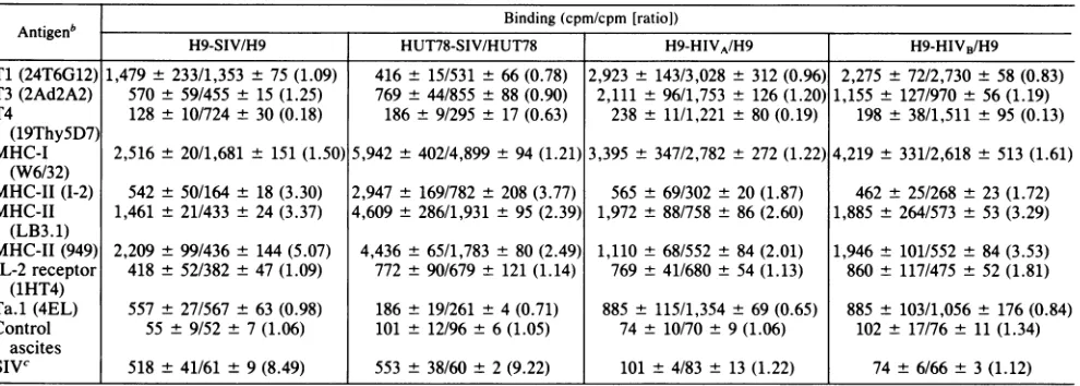
TABLE 1. Antigenexpressionofhuman T-cell lineschronically infectedwith SIVor HIV'
Binding (cpm/cpm [ratio])
H9-SIV/H9 HUT78-SIV/HUT78 H9-HIVA/H9 H9-HIVB/H9
Ti (24T6G12) 1,479 ± 233/1,353 ±75 (1.09) 416 ± 15/531 ± 66 (0.78) 2,923 ± 143/3,028 ± 312 (0.96) 2,275 ± 72/2,730± 58 (0.83) T3 (2Ad2A2) 570 ± 59/455 ± 15 (1.25) 769 ± 44/855 ± 88 (0.90) 2,111 ± 96/1,753 ± 126 (1.20) 1,155 ± 127/970 ± 56 (1.19) T4 128 ± 10/724 ± 30 (0.18) 186± 9/295 ± 17 (0.63) 238 ± 11/1,221 ± 80 (0.19) 198 ± 38/1,511 ± 95 (0.13)
(19ThySD7)
MHC-I 2,516 ± 20/1,681 ± 151 (1.50) 5,942 ± 402/4,899± 94 (1.21)3,395 ± 347/2,782 ± 272 (1.22) 4,219 ± 331/2,618 ± 513 (1.61) (W6/32)
MHC-II (1-2) 542 ± 50/164 ± 18 (3.30) 2,947 ± 169/782 ± 208 (3.77) 565 ± 69/302 ± 20(1.87) 462 ± 25/268 ± 23 (1.72) MHC-II 1,461 ± 21/433 ± 24 (3.37) 4,609± 286/1,931 ± 95(2.39) 1,972 ± 88/758 ± 86(2.60) 1,885 ± 264/573 ± 53 (3.29)
(LB3.1)
MHC-II(949) 2,209 ± 99/436 ± 144 (5.07) 4,436± 65/1,783 ± 80 (2.49) 1,110± 68/552 ± 84(2.01) 1,946± 101/552± 84 (3.53) IL-2 receptor 418 ± 52/382 ± 47 (1.09) 772 ± 90/679 ± 121 (1.14) 769 ± 41/680 ± 54(1.13) 860 ± 117/475± 52 (1.81)
(1HT4)
Ta.1 (4EL) 557 ± 27/567 ± 63 (0.98) 186± 19/261 ± 4 (0.71) 885± 115/1,354 ± 69 (0.65) 885 ± 103/1,056± 176 (0.84) Control 55 ± 9/52± 7(1.06) 101 ± 12/96 ± 6(1.05) 74 ± 10/70± 9(1.06) 102± 17/76± 11(1.34)
ascites
SIVC 518 ±41/61 ± 9(8.49) 553 ± 38/60 ± 2(9.22) 101 ± 4/83± 13(1.22) 74± 6/66± 3(1.12)
aAcellular radioimmunoassay was performed on the cells chronically infected with SIV or HIV and equivalent numbers of uninfected cells.
bTheparticularMAbs used in these studies are noted in parentheses after the antigens.
cTodetect SIV antigens, immunoglobulin purified from the plasma of a rhesus monkey which was inoculated with SIV was directly labeled with
125I-Na
and bound to the cells in the wells.phenylmethylsulfonyl fluoride), clearedby
microcentrifuga-tion,andprecleared with rabbitanti-mouseimmunoglobulin (DAKO)-coatedprotein A-SepharoseCL-4B beads(Sigma).
Precleared cell lysates were incubated with MAb-coated
protein A beads overnight at 4°C. After
being washed,
immunoprecipitates were eluted from the beads withLaemmli sample buffer and analyzed in a 10% sodium
dodecyl sulfate-polyacrylamidegelwitha3.5%
stacking
gel underreducing conditions. The gels were dried andvisual-ized by autoradiography.
IFN--y assays. Two different assay systems were usedto
detect gamma interferon
(IFN-y)
in culture supernatants.The induction of MHC-II expression on peripheral blood
mononuclear cellsfromapatient with chronicmyelogenous leukemia (CML) was determinedafter incubation with cul-turesupernatants as afunctionalassayfor
IFN-y
(6). Atotal of106CMLcellswereincubatedin 1 ml of RPMI 1640plus 10% fetal calf serum (FCS) containing variousconcentra-tions of recombinant IFN--y(Biogen, Cambridge, Mass.) or culture supernatants of H9-SIV cellsat 37°C for24h. Each
cellsamplewasthen stained with anti-MHC-IIMAb(949)or
control mouse ascites followed by fluorescein-labeled goat
anti-mouse immunoglobulin (DAKO). The mean channel fluorescence of these CML cellswasmeasuredbya fluores-cence-activated cell sorter (Epics-C; Coulter Electronics, Inc., Hialeah, Fla.). A solid-phase radioimmunoassay for IFN--y was also done with a commercially
available
kit (Centocor, Pa.). Briefly, culture supernatant samples wereincubated with polystyrene beads coated with a MAb
spe-cific for human IFN--y. These beads were then incubated with an
125I-labeled
MAb against human IFN-y, and theradioactivitybound to thebeads was measured.
Kinetic studiesof antigenexpression. A total of
107
H9 cells wereincubatedat 37°C with supernatants containing SIV or control medium overnight and washed. The infected anduninfected cells were then maintained for 40 to 70 days in RPMI 1640plus 10% FCS under the same conditions. The cellconcentrationsof thepaired flasks were adjusted to 5 x
105/ml
every 3 to 4 days, and antigen expression on these cells wasdeterminedat the same time by a cellular radioim-munoassay with MAbs whichrecognizeTi,
T3,T4,MHC-I,MHC-II,
the IL-2 receptor,Ta.1,
and with an SIV-specificheterologous antiserum. A binding ratio was calculated as the radioactivity bound to infected cells divided by that
boundtotheequivalentnumber of uninfected cells. Reverse
transcriptase activitywasmeasuredaspreviouslydescribed (11).
Immunoelectron microscopy. Chronic H9-SIV cells were
incubated with an anti-MHC-II MAb (LB3.1) for 45 min, washed, andthenincubated with
gold-conjugated
goatanti-mouse
immunoglobulin
G(IgG)
(Boehringer
Mannheim Biochemicals, Indianapolis, Ind.) for 45 min. Cells werewashed, fixed with 1% glutaraldehyde in 0.1 M cacodylate buffer overnight, and then processed for electron
micros-copy. H9-SIV cells incubated with negative-controlmouse
ascites and gold-conjugated anti-mouse IgG showed no staining with gold particles.
RESULTS
MHC antigen expressiononhumanT-cell lineschronically infected with HIV and SIV. Preliminary studies were per-formed to determine if human T-cell lines chronically
in-fected withHIV or SIVhavealterationsintheirexpression
of MHCantigens. Antigen expressiononcells was assessed
by a cellularradioimmunoassay by using a panelof MAbs
(Table 1).Theexpressionof MHC-IIantigensrecognizedby three different MHC-II-specific MAbs was 3 to 5 times greater on the chronically SIV-infected cells than on the
uninfected H9 cells. This increase of MHC-II antigen
ex-pression was also observed on HUT78-SIV and H9-HIVA and H9-HIVB. MHC-I antigen expression by H9-SIV and oneof thetwostudied H9-HIVlineswasapproximately50% greater than that of uninfected H9 cells; the increase of MHC-I was approximately 20% on HUT78-SIV and the other H9-HIV line whencompared with theuninfected cell lines. T4 expression was substantially less on the infected cell lines than on theuninfectedcelllines. Theexpression of
otherT-cell antigens, including
Ti,
T3, the IL-2 receptor, and theactivation antigen Ta.1, varied byless than 35%in the infected and uninfected cell lines. One of the H9-HIV lines showed an81%increase in the expression of the IL-2 receptor.Student's t tests were performed on log10-transformed counts-per-minute data to compare the binding of each
on November 10, 2019 by guest
http://jvi.asm.org/
antibodytoinfected anduninfected cells. Thefourseparate
experiments shown in Table 1 were combined to facilitate this statistical analysis. T4 expression was significantly
lower (P < 0.005) and MHC-II, as detected by any of the
three MAbs, was significantly higher (P < 0.05) on the
infected than on the uninfected cells. No significant
differ-ence could be demonstrated between infected and uninfected cells in the expression of the other antigens studied. These data suggest that cellular MHC antigen expression, especially class II antigen expression, is specif-ically increased when the cell is chronspecif-ically infected with HIVor SIV.
The monkey immunoglobulin used for the detection of SIVantigens did not reactin the cellular radioimmunoassay with H9-HIV cells,apopulationwhichwasstronglypositive
forMHCantigens. Furthermore,noneof the anti-MHC-Ior
-II MAbs tested blocked the binding of the SIV-specific antibodytoH9-SIV cells, nordid theSIV-specific antibody
block anti-MHC-MAbs binding to those cells. These data
suggestthattheapparentincrease of MHC expression in the human T-cell lines infected with SIVorHIVwasunlikelyto
representaserologic cross-reaction between MHC and viral
antigens.
Surface-labeled cellular protein from uninfected and H9-SIVcellswasanalyzed byimmunoprecipitation with MAbs
followedby sodium dodecylsulfate-polyacrylamide gel elec-trophoresis (Fig. 1). This study confirmed that MHC-II (29-and 34-kilodalton) and MHC-I (44-kilodalton) molecules
weresignificantly increased onH9-SIVcells compared with
uninfected H9 cells, whereas the expression of T3 remained unchanged. The radioactivity (infected/uninfected) of each band, measured by5-mincounts on agammacounter, was
I-J
1n m
I -j
Z*
-a b
200
-93 -66
-45-
a
N\ ^ :> O
c-4
I'4I 0
a b a b a b a b a b
*
A.
31-
e
22
-FIG. 1. Celllysatesof125I-surface-labeledH9(a)and H9-SIV(b)
wereincubated with antibody-coated protein A-Sepharose beads,
andresulting immunoprecipitateswereanalyzed bysodiumdodecyl sulfate-polyacrylamide gel electrophoresis. Anti-SIV serum was
obtained from a rhesus monkey inoculated with SIV. Negative
mouseasciteswasusedasthenegativecontrol. Numberstothe left
of thegelindicatethe molecular massinkilodaltons.
a-z 0
so
1000
00
T T3 IL2R T4 MHCI MHC-:E MHC-3I SIV (1-2) (LB3.1)
FIG. 2. Induction ofantigens on H9-SIV cells with IUDR. H9 and H9 chronicallyinfected with SIV (H9-SIV)wereincubated in RPMI1640containing10% FCS withorwithout 50,uwgof IUDRper ml.After 72 h of incubationat37°C, H9(0),H9treated with IUDR (0; ), H9-SIV (Ki), and H9-SIV treated with IUDR (E) were
harvested, and the antigens expressed on equivalent numbers of cellswerequantitated byacellularradioimmunoassay.
asfollows:
3,614/540
for MHC-II(LB3.1);
873/428for MHC-II(I-2);
1,156/568
for MHC-I(W6/32);
476/538for T3(SP34);
and
1,866/595,
and1,566/427
foranti-SIV serum,withback-ground
countsof200. SIV immunemonkey
serum precipi-tated both the 160-kilodaltonenvelope
glycoprotein
and the32-kilodalton transmembrane
portion
of the viralenvelope
structure in thecontrollane.
Induction of MHC and viral
antigen expression.
2'-Deoxy-5-iodouridine(IUDR)
induces theexpression
of viralanti-gens on virus-infected cells.
We,
therefore,
assessed therelationship
between MHC and viralantigen
expression
on H9-SIV cellsby
inducing
viralantigens
onchronically
infected cells with IUDR. MHC-II and SIV
antigens
were induced 21 to26%on H9-SIVcells aftera72-h incubationwith IUDR
(P
<0.05)
compared
with cellstreatedsimilarly,
but without IUDR
(Fig. 2).
Nosignificant
increase wasobserved in the
expression
ofTl, T4, MHC-I,
and IL-2receptors onthese cells. Uninfected H9cells did not show any induction of
MHC-antigen
expression
after treatment with IUDR. These data suggest that increased MHC-IIexpression
isassociatedwith increased SIVantigen
expres-sion.Assessment of
IFN-y
in cultures. MHCantigens
are in-duced in some systemsthrough
theelaboration of humoralfactors,
such asIFN-y,
by
virus-infected cells(7).
The [image:3.612.323.562.72.219.2]production
ofsuchhumoralfactorsinducing
MHC-II in theTABLE 2. MHC-IIinductiononCML cells with culture supernatantsof H9-SIV
Samplesa Meanchannel fluorescenceb
Control medium ... 79.91 IFN--y(3 U/ml) ... ... 95.70
IFN--y (10 U/ml)... 100.47
IFN--y(100 U/ml)... 101.24
Culturesupernatantof H9-SIV
(25%)
... ... 70.47Culturesupernatantof H9-SIV
(50%)
... ... 64.43Culture
supernatant
of H9(50%)... 74.21aSamplesweredilutedinthecontrol medium(RPMI1640plus
10o
FCS). bMeanchannel fluorescencerepresentsexperimental(cellsstaining posi-tivelywithananti-MHC-IIMAb)minuscontrol(cellsstainingpositivelywithacontrolmouseascites). 61,
410
40
Mb
- Imon November 10, 2019 by guest
http://jvi.asm.org/
[image:3.612.71.298.407.657.2]A
10 1 104
I ~~~~~~~~~~~~~~~E
63 10
(0 10 2 0 4 0 2 0 4
[image:4.612.59.299.63.252.2]DAYS OF CULTURE
FIG. 3. Kinetics of antigen expression onH9 cells acutely in-fected with SIV. The antigens shown are MHC-II (1-2)(0),MHC-II (LB3.1) (A), MHC-I (O), T4 (A), IL-2 receptor (x), and SIV-encoded determinants (0). A binding ratio was calculated as the radioactivity bound to H9-SIV divided by theradioactivity boundto theequivalentnumber of H9 uninfected cells, as determined by a cellular radioimmunoassay. Reverse transcriptase activity in the infected culture(0--0) was also monitored. SIV isolates used for infection in this studywere culture supernatantsof human periph-eralbloodlymphocytes infected with SIV (A) and culture superna-tantsof H9chronically infected with SIV and maintained inRPMI 1640 with10% FCS (B).
supernatants of H9-SIV was therefore assessed. This was
donebyusingas afunctionalassay theinduction ofMHC-II
expression on CML cells after incubating culture superna-tants ofH9-SIV cells (Table 2). The mean channel
fluores-cenceof CML cells stained withanti-MHC-II MAb signifi-cantly increasedwhen the cells wereincubated with
.3
U ofrecombinant
IFN-y
per ml. This induction ofMHC-II onCML cells was blocked by an anti-IFN--y MAb (data not
shown). MHC-II expression by CML cells incubated with culture supernatants from H9-SIVwas notgreaterthan that
A
ofCML cellsincubatedwithRPMI 1640
plus
10% FCS. TheinductionofMHC-IIonuninfectedH9cells afterincubation
with
IFN-y
wasalso assessed.MHC-II inductiononH9 cells withIFN-y
was not seen after a24-hperiod
ofincubation.After 72 h of incubation with 300 U of
IFN-y
perml, the percentage ofMHC-II-positive
cells increased from 5.6 to13.9%. The culture supernatantsofH9-SIV
cells,
however,
didnot affectMHC-II
expression
onH9 cells after 24or72 h of incubation. Aradioimmunoassay
was also done to detectIFN-y
in the culture supernatant ofH9-SIVby
using
aMAbspecific
forhumanIFN--y.
NoIFN-y
wasmeasurable in H9-SIV culture supernatantsby
this approach, anassay system sensitive enough to detect 0.5 U ofIFN--y per ml. These data suggest thatMHC-II inductiononH9-SIVcells is notlikely
toresultfromautoregulation mediatedbyIFN-y.
Kinetics ofMHC-II induction in vitro. We then assessed thekinetics of theexpression ofMHC-IIandviral
antigens
on H9 cells
acutely
infected with SIV. H9 cells wereincubatedwithSIVovernight, washed,and then cultured for 40 to 70
days (Fig. 3).
Reversetranscriptase activityin the supernatants was detectable by day 10 after infection and reachedapeak
betweendays
10 and 20. ThekineticsofSIVantigen
expression correlated with that of thegenerationof reverse transcriptase. T4 antigen expression decreased asviral antigen expression increased and thereafter remained low. An increase in MHC-II antigen expression was not
observed until between
days
20 and 30 after infection. Similarresultswere seenwhen thevirussource wasfromanIL-2-dependent
humanperipheral
bloodlymphocyteculture or H9 cells chronically infected with SIV maintained in RPMI 1640 plus 10% FCS. In two additional experiments, MHC-II antigen inductionfirst occurred ondays46 and53,
whereas SIV antigen was detected on days 14 and 17, respectively, after infection (data not shown). MHC-I anti-genwas notsignificantly inducedin any ofthese
experiments
duringtheperiod ofculture.Acquisition of MHC-II by virus during budding. To deter-mine whether theincreasein MHC-II on themembranes of
infectedcellsaffectsthe SIV particles, we examinedbudding
viral particles for evidence of MHC-II on their surfaces. Immunoelectron microscopy was used to visually localize MHC-II antigen on budding and mature SIV particles.
B
ti;^*M
t..
..
It-14 $--, I-e-I-r
%t 'It. -1
0,,-'jai=
A(ft
v? -4. 4r.
FIG. 4. Electronmicrographs ofH9-SIVcellsincubated with (A) mouse anti-MHC-II MAb or (B) negative-control mouse ascites fluid and thenincubated with agold-conjugated goat anti-mouse IgG serum. Gold particles indicating the presence of MHC-II antigen are aligned along thecellmembrane and around the envelope of mature viral particles in panel A but not in panelB.
e I..1
e,
4- 41
..'A'
on November 10, 2019 by guest
http://jvi.asm.org/
[image:4.612.61.553.504.692.2]CLASS II MHC ANTIGEN INDUCTION BY SIV 1425 Virus-infected H9 cells were incubated first with a mouse
anti-MHC-II MAb and then with a gold-conjugated goat
anti-mouse IgG serum. Gold particles were seen on the surfaces of a significant percentage of infected cells and also onthe surfaces ofsomebudding and mature virus particles
(Fig. 4). Thus, by this technique it appears that MHC-II
antigens may be acquired by some viral particles while
buddingfrom cell membranes.
DISCUSSION
The present studies indicate that MHC antigen
expres-sion, most strikingly class II antigen expression, is
specifi-cally increasedwhen cellsareinfected in vitro withSIV.SIV
andthe human AIDS virus HIV are morphologically
indis-tinguishableandantigenically related; SIV and HIV share a
tropism for T4+ lymphocytes and both induce analogous
immunodeficiency syndromes (3, 10, 11, 16). Therefore, it is
likely that the present observations with SIV will also hold true for HIV. In fact, we have shown that MHC-II is
increased in H9 cells chronically infected with HIV (Table 1).
Although MHC-I as well as MHC-II antigens are ex-pressed in greater quantity on the surfaces of chronically
SIV-infected cells than onuninfectedcells, we were unable to demonstrate the induction of MHC-I expression with IUDR or its increased expression after the acute infection of cell lines in vitro. It is therefore not clear whether the
infectionof cell populations with SIV reproducibly induces
MHC-I expression or if such an infection induces MHC-I expression byadifferent mechanism.
Inthepresentstudy,we haveshown that H9-SIV cells did not produce detectable IFN--y by using afunctional assay systemsensitiveenough todetect3 Uof
IFN-y
per ml and aradioimmunoassay sensitiveenough to detect 0.5 UofIFN--y per ml. MHC-II expression was shown to increase only
slightlyonuninfectedH9cellswhen thecellswereincubated with highconcentrations of exogenous IFN--y.Furthermore, theincrease of MHC-IIon H9cells after infection with SIV occurred long after virus production could be detected by
thesecells. Itis thereforeunlikelythattheMHC-IIinduction on H9-SIVcells is mediatedby IFN--y.
The mechanism by which this MHC-II induction occurs
remains undefined. It is, however, interesting to speculate thatageneproductof SIV mightbe responsible for regulat-ing this induction. A human T-lymphotropic virus type
II-encodedtrans-activating proteinhasbeenrecently shown
toinduce cellular IL-2receptorexpression (5), andHIV has
been clearly shown to encode for proteins which similarly
canactivate gene expressionintrans(18).
The observation that MHC-II can be acquired
by
SIV during buddingisprovocative. Thisacquisition,while it maybemerely passive, might alsooccur asaresult ofpreferential interaction between viral structural proteins and MHC-II.
Furthermore, becauseMHC-IIpreferentially recognizes T4, theexpression of MHC-II onthesurface ofthe virus
might
increase the tropism of the virus for T4+ cells. Furtherstudies will be needed to determine whether MHC-II is
preferentially acquired by
budding
virus or ismerely
pas-sively acquired.
Changes
in MHC-IIexpression
by
antigen-presenting
cell(APC)populationsinpatientswith AIDS has been
reported.
Infact, MHC-II expressiononthe
circulating
monocytes of these individuals has been shown to besubstantially
less than thatofhealthycontrols(8),andMHC-IIexpression
onLangerhan's cells, the APCs of the
skin,
is also less inindividuals with AIDS than in healthy controls (2). Interest-ingly, however, and consistent with the findings in the present studies, Heagy et al. (8) showed that MHC-II
expression onthe monocytes of patients with AIDS-related complex is actually greater than that of normal individuals. Thus, anincreased MHC-II expression by APCs may occur in vivo in the early stages of an HIV infection.
Ithas been suggested that autoimmune mechanisms may play an important part in the pathogenesis of the basic immune defects in AIDS (13). Such potentially autoimmune phenomena as thrombocytopenia, hypergammaglob-ulinemia, glomerulonephritis, circulating immune com-plexes, and the generation of circulating anti-lymphocyte antibodies clearly contribute to the morbidity associated with this disease (14). Increased MHC-II expression on
virus-infected APC populations might initiate a cycle of increased immune reactivity to self antigens which could leadto these
sequelae.
ACKNOWLEDGMENTS
WethankStephen Cannistra for assistanceinperforming IFN--y assays, Keith Reimann for assistance in thestatistical analysis of data, and Christopher Rudd for valuable conversations. We also thank John MacKey for skillful assistance in the electron micro-scopic studies and Bettye-Jean Roy and Debbie Brosseau for the preparation of this manuscript.
This work was supported by Public Health Service grants Al 20729 andCA38205fromtheNational Institutes ofHealth andRR 00168from the Division ofResearch Resourcesandbya contract fromtheMassachusetts Departmentof Public Health.N. L.Letvin is the recipient of an American Cancer Society Junior Faculty Research Award.
LITERATURECITED
1. Barre-Sinoussi, F., J. C. Cherman, F. Rey, M. T. Nugeyre, S. Chamaret, J. Gruest, C. Dauguet, C. Axler-Blin, F. Vezinet-Brun, C. Rouzioux, W. Rozenbaum, and L. Montagnier. 1983. Isolation ofaT-lymphotropic retrovirus from apatientatrisk for acquired immune deficiency syndrome (AIDS). Science 220:868-871.
2. Belsito, D. V., M. R. Sanchez, R. L. Baer, F. Valentine, and G.J.Thorbecke. 1984. ReducedLangerhanscell Iaantigenand ATPaseactivityinpatientswiththeacquired immunodeficiency syndrome. N.Engl. J. Med310:1279-1282.
3. Daniel, M. D., N. L. Letvin,N. W. King, M. Kannagi, P. K. Sehgal,R. D.Hunt,P.J.Kanki,M.Essex,and R.C. Desrosiers. 1985. Isolation of T-celltropic HTLV-III-like retrovirusfrom macaques. Science 228:1201-1204.
4. Gallo,R.C., S. Z.Salahuddin,M.Popovic, G. M.Shearer,M. Kaplan, B. F. Haynes, T. J. Palker, R.Redfield, J. Oleske,B. Safai,G.White, P. Foster, and P. D. Markham. 1984. Frequent detection and isolation ofcytopathicretroviruses (HTLV-III) from patients with AIDS and at risk for AIDS. Science 224:500-503.
5. Greene, W. C., W.J. Leonard, Y. Wano,P. B. Svetlik,N.J. Peffer, J. G. Sodroski, C. A. Rosen, W. C. Goh, and W. A. Haseltine. 1986. trans-activatorgene of HTLV-IIinduces IL-2 receptor and IL-2 cellular gene expression. Science 232: 877-880.
6. Griffin, J. D., K. D. Sabbath, F. Herrmann, P. Larcom, K. Nichols, M. Kornacki, H. Levine, and S. A. Cannistra. 1985. Differentialexpression of HLA-DRantigens in subsets of
hu-manCFU-GM. Blood 66:788-797.
7. Halloran, P. F., A. Wadgymar, and P. Autenried. 1986. The regulation ofexpression of major histocompatibility complex products.Transplantation41:413-420.
8. Heagy, W.,V.E.Kelly,T. B.Strom,K.Mayer,H. M.Shapiro, R. Mandel, and R. Finberg. 1984. Decreased expression of
on November 10, 2019 by guest
http://jvi.asm.org/
human class II antigens on monocytes from patients with acquired immune deficiency syndrome. J. Clin. Invest. 74: 2089-2096.
9. Ho, D. D., T. R. Rota, and M. S. Hirsch. 1986. Infection of monocyte/macrophages by human T lymphotropic virus type
III. J. Clin.Invest.77:1712-1715.
10. Kanki, P. J., M. F. McLane, N. W. King, Jr., N. L. Letvin, R. D. Hunt, P. Sehgal, M. D. Daniel, R. C. Desrosiers, and M. Essex. 1985. Serologic identification andcharacterization ofa macaqueT-lymphotropic retrovirus closely related to HTLV-III.Science228:1199-1201.
11. Kannagi, M., J. M. Yetz, and N. L. Letvin.1985.In vitrogrowth characteristics of simian T-lymphotropic virus type III. Proc. Natl.Acad. Sci. USA 82:7053-7057.
12. Klatzmann, D., F. Barre-Sinoussi, M. T. Nugeyre, C. Dauguet, E. Vilmer, C. Griscelli, F. Brun-Vezinet, C. Rouzioux, J. C. Gluckman, J.-C. Chermann, and L. Montagnier.1984. Selective tropism of lymphadenopathy associatedvirus (LAV)for helper-inducerT lymphocytes. Science 225:59-63.
13. Klatzmann, D.,and L.Montagnier. 1986. ApproachestoAIDS therapy.Nature(London)319:10-11.
14. Lane, H. C., and A. S. Fauci. 1985.Immunologic abnormalities
in theacquired immunodeficiency syndrome. Annu. Rev. Im-munol. 3:477-500.
15. Lane, H.C., H. Masur, E. P. Gelmann, D. L. Longo, R. G.Steis, T. Chused, G. Whalen, L. C. Edgar, and A. S. Fauci. 1985. Correlation betweenimmunologicfunction and clinical subpop-ulations of patients with theacquired immune deficiency
syn-drome. Am. J. Med. 78:417-422.
16. Letvin, N. L., M. D. Daniel, P. K.Sehgal,R.C. Desrosiers,R. D. Hunt, L. M. Waldron, J. J. MacKey, D. K. Schmidt, L. V. Chalifoux, and N. W. King. 1985. Induction of AIDS-like disease in macaque monkeys with T-cell tropic retrovirus
STLV-III. Science 230:71-73.
17. Meuer,S.C.,S. F.Schlossman,and E. L. Reinherz.1982.Clonal analysis of human cytotoxic T lymphocytes: T4+ and T8+ effector T cellsrecognize productsof differentmajor histocom-patibility complex regions. Proc. Natl. Acad. Sci. USA 79:4395-4399.
18. Rosen,C. A., J.G.Sodroski,K.Campbell, and W. A. Haseltine. 1986. Construction of recombinant murine retroviruses that
express the human T-cell leukemia virus type II and human T-cell lymphotropic virus type III trans activator genes. J.
Virol. 57:379-384.