0022-538X/89/083213-07$02.00/0
Copyright© 1989, American Society for Microbiology
Functional Characterization of
a
Complex Protein-DNA-Binding
Domain Located within the
Human
Immunodeficiency
Virus
Type 1
Long
Terminal Repeat Leader Region
MICHAELH. MALIM, RANDY FENRICK, DEANW. BALLARD, JOACHIM HAUBER, ERNSTBOHNLEIN,
ANDBRYAN R. CULLEN*
Howard Hughes MedicalInstitute andDepartmentof Medicine, Duke UniversityMedical Center,
Durham,
North Carolina 27710 Received 19October 1988/Accepted 8 April 1989Transcriptionaltransactivation of the human immunodeficiency virus type 1 (HIV-1)long terminalrepeat (LTR) by the viraltattransactivator ismediated by an LTR-specificsequencelocatedimmediately 3' tothe start of transcription initiation. We have used a range of molecular techniques to examine DNA-protein interactions that occurin the vicinity of this cis-acting sequence. Our results demonstrate the existence ofa
sequence-specific DNA-protein interaction involving the HIV-1 leader DNA and map this binding event to between -2 and +21 base pairs relativetotheHIV-1 LTR transcriptionstartsite.Evidence suggesting that this interaction involves three distinct protein-DNA contact sites extending along one side of the DNA helix is
presented. Mutation ofthese siteswasfoundtoablate protein-DNA bindingyetwasobservedtohavenoeffect
oneither thebasalortattrans-activated level of HIV-1 LTR-specificgeneexpression. We therefore conclude
that this DNA-protein interaction has a function distinct from the regulation of HIV-1 LTR-specific gene
expression.
The pathogenic human retrovirus human immunodefi-ciencyvirustype 1(HIV-1) encodesanonstructural protein, termedtat, whose functional expression is required for viral replication in vitro (8, 12). Thetatprotein is localizedtothe nucleusofexpressing cells (15) and actstogreatly enhance the expression ofviral or heterologous genes linkedto the HIV-1 long terminal repeat (LTR) promoter element (1, 6, 25, 28, 29, 34, 39). Althoughevidence exists for a posttran-scriptional component of this tat-mediated trans activation (6, 10, 29, 39), the primary effect oftatistoenhancetherate oftranscription from the HIV-1 LTR (15, 17, 20, 31). This enhancement is in turn mediated by an HIV-1 LTR
se-quence, termed the trans-activation response (TAR)
ele-ment, whichhasbeenmapped toasite locatedimmediately
3' to the start of viral mRNAtranscription (11, 14, 17, 30) (Fig. 1).
Transcriptional trans activation is normally mediated by thesequence-specific bindingoftranscriptionfactorstosites within the affected promoter DNA sequence (22). A good exampleof thisgeneralizationisprovidedbythe HIV-1 LTR U3regionitself, which contains functional bindingdomains for several transcription factors, including Spl and NF-KB (4, 18, 26). A number of promoters have been shown to contain DNAregulatoryelements that extend 3'tothestart oftranscription (16, 23, 36, 38). It was therefore of great
interest whenDNase Ifootprinting and DNase I hypersen-sitivity analyses (13, 14) revealed the presence ofa strong DNA-protein interactionthat extended 3'tothe HIV-1 LTR transcription startsiteto theregionof the TAR element.
Inthis study, we attempted to characterize andprecisely identify the target sequences involved in this particular HIV-1 LTRDNA-binding event. Our data demonstrate the existence of three adjacent, highly cooperative
protein-* Correspondingauthor.
tPresent address: Sandoz Research Institute, A1235 Vienna,
Austria.
binding sites, located between nucleotides -2 and +21 relative to the HIV-1 LTR transcription start site, which interact with constitutively expressed cellularDNA-binding proteins. These binding sites are shown to partly overlap with the HIV-1LTR TARelement butarealso showntobe functionally fullydistinct from TAR. Wetherefore conclude that this HIV-1 LTR DNA-binding event, although highly specific, is not directly involved in the tat-mediated trans
activation ofHIV-1-specific gene expression.
MATERIALS AND METHODS
Construction of molecular clones. The HIV-1 LTR-based chloramphenicol acetyltransferase (CAT) gene expression vectorpBC12/HIV/CAT hasbeen described previously (3). Cleavage of pBC12/HIV/CAT at the unique HIV-1 LTR PvuII andBglII sitespermitted the introduction of oligonu-cleotides containing site-specific mutations (Fig. 1). The primarystructureof these mutationswasconfirmedbyDNA
sequenceanalysis. The pcTAT/dhfr plasmidexpressesboth the HIV-1 tatgene under the control ofa cytomegalovirus immediate-earlypromoteranda mousedihydrofolate reduc-tase (dhfr) gene under simian virus 40 early-promoter
con-trol. pcTAT/dhfr was constructed by insertion of a 1.8-kilobase-pairPvuII-BamHI dhfrgenefragment from pSV2-dhfr(37)into theuniqueStuIsiteof thetat gene expression vector pcTAT (21). Plasmid pTAR contains the PvuII-to-HindIIl (-18 to +81) region of the HIV-1 LTR (Fig. 1) inserted into the polylinker region ofpSP68(Promega Bio-tech). A probe (-18/+81 probe) derived from this plasmid
wasused for themethylationinterferenceanalysisdescribed below.
Cell culture and transfection. HeLa and Jurkat cells were
maintainedaspreviouslydescribed(4, 6).Forpreparationof theHeLa/cTATcellline,HeLa cellsweretransfected witha
mixture of 250ngofpSV2neo (35) and 5 ,ugofpcTAT/dhfr (both linearized by cleavage withPvuI), as well as 5 jigof high-molecular-weighthuman carrierDNA, usingacalcium
3213
on November 10, 2019 by guest
http://jvi.asm.org/
3214 MALIM ET AL.
G6ATI
-27 TA TA AGC A G C TGCT T TTTGC C TGT A CT GGG +3
TATA PvulI
v
LCAP
SITE+4 T C T C T C T G G T T A G ACC AGAT CTGAGCC TG G +33
A A A AA BglII
A 1 2 3 4
___
B
\
o o i~i
-Z lbZ
+34 GAGCTCTCTGGCTAACTAGGGAACCCACTG 63
Sac
+83
+64 CTTAAGCCTCAATAAAGCTT Hindlll
FIG. 1. Sequence of the HIV-1 LTRaroundthesite of transcrip-tion initiatranscrip-tion. The core TAR element, as defined by mutational analysis(14, 17), extendsfrom -+20 to -+44nucleotides relative to the cap site. Also shown are locations of the TATA box and relevant restriction sites. For simplicity, this entire element is referred to in the text as the HIV-1 leaderregion. Boxedsequences indicate the mutations introduced into the HIV-1leaderregion by oligonucleotide substitution between the Pvull and BgIII sites, termed Al, A2, and A3, respectively, in order from the 5' to 3' direction. These mutationsweredesignedtoeliminate bases shown by methylation interference analysis to be important for factor bindingtothe HIV-1 leaderDNA(A).
phosphate transfection technique (7). After 48h, theculture was subjected to selection in 0.8 mg of G418 per ml. This eventually resulted in the appearance of -100 independent Neor colonies. These were pooled and subjectedto a second selection with 5 x 10⁻⁸ Mamethopterin. We hadpreviously observed that our HeLa cell culturesonly very rarely (<1in
107)are able to spontaneously give rise to colonies resistant to this level ofamethopterin. However, pooled populations ofpreselected Neor cells cotransfected with a dhfr
expres-sion vectorreadily yield viable colonies under these
condi-tions. Experiments using a number of model genes have demonstrated that thesedoubly selected cells expressahigh
level not onlyof dhfr but also of anylinkedgeneof interest. This level is -100-fold higher than that obtained by simple coselection with the neo gene alone (data notshown). The resultant dhfr+ Neor cells were pooled and termed HeLa/ cTAT. High-level, functional expression of the HIV-1 tat genewasconfirmedbyimmunoprecipitation analysiswithan
anti-tat antipeptide antiserum (15, 21) (Fig. 2B) and by
transfection with the indicator construction pBC12/HIV/
CAT (3)(data not shown).
Theexpression phenotypes of site-specific mutants of the HIV-1 LTRin thepBC12/HIV/CATbackground were tested
bycalcium phosphate-mediated cotransfection (7) of HeLa cellswith the tatexpression vectorpgTAT or the negative
control vectorpBC12/CMV (6, 21). CAT expressionlevels were determined at 60 hposttransfection by the method of Neumannetal. (27).
Radiolabeling of restriction fragments and oligonucleotides. The DNA probes used for gel retardation studies were normally generated by cleavage of pBC12/HIV/CAT at the unique HindlIl site (Fig. 1) and filling in with Klenow DNA
polymerase I in the presence of
o-32P-labeled
deoxynucle-otide triphosphates (6). In some cases, these probes were prepared by cleavage of pBC12/HIV/CAT withPvuII (Fig. 1), followed by sequential treatment with alkaline phos-phatase and T4 polynucleotide kinase in the presence of[_y-32P]ATP
(6). Probes were isolated by preparativepoly-acrylamide gel electrophoresis after specific secondary
cleavage asindicated. Similarly, the probes used for
meth-ylation interference were prepared by digestion of pTAR with HindIII, followed by dephosphorylation with alkaline
phosphatase. The positive strand was labeled with Klenow
_prR'l
43.0) 25.7-
18.4-14.3- AM
6.2-C' "I
.Ap$Sfp>
7r.
FIG. 2. Gel retardation analysis using an HIV-1 LTR leader region probe. The assay shown in panelA used 10 ,ug ofbovine serum albumin (negative control) or 10 1Lg of nuclear proteins derivedfrom HeLa, HeLa/cTAT, orJurkat cells. These proteins were preincubated with 2 p.g of poly(dI-dC) competitor before addition of-0.5 ngoftheradiolabeled-18/+81 probe. All nuclear protein samples tested yielded a single retarded band of similar mobility (<-). The HeLa/cTAT line, which was shownto express high levels of the 15.5-kilodalton HIV-1 tat trans activator by immunoprecipitation analysis (21), yieldedthe samebinding pheno-type asdidtheparental HeLacell line(panel A, lanes 2 and 3).
DNA polymerase I in the presence of [cx-32P]dATP; the negative strandwaslabeled with T4polynucleotide kinasein the presence of
[y-32P]ATP.
The labeled DNA was then cleaved at a pSP68-derived HaeIII site, and the resultant 135-base-pair probe fragment was isolated as described above. Synthetic oligonucleotides were isolated and an-nealed aspreviously described(2, 4).1 1234562 3 4 5 6
- -~
t
k
':"(I
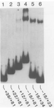
PFIG. 3. Mapping of theHIV-1 leaderregion protein-bindingsite. End-labeledprobes extendingacrosstheindicated nucleotide posi-tions were prepared by restriction endonuclease cleavage of the full-length -18/+81 probe at sites indicated in Fig. 1. Equivalent levels ofall probes were then tested for protein binding by gel retardationanalysis. This experimentdemonstrated that the HIV-1 leader region protein-binding site was contained between nucleo-tides -18 to +23.
J. VIROL.
on November 10, 2019 by guest
http://jvi.asm.org/
[image:2.612.362.514.79.280.2] [image:2.612.61.301.81.176.2] [image:2.612.396.483.486.661.2]Preparation of nuclear protein extracts. The isolation of nuclear proteins from HeLa, HeLa/cTAT, and Jurkat cells was based on the method of Dignam et al. (9). The cells were washed with cold phosphate-buffered saline (Ca2"-Mg2+
free; GIBCO Laboratories), harvested, and suspended in ice-cold 10 mM N-2-hydroxyethylpiperazine-N'-2-ethane-sulfonic acid (HEPES)-NaOH (pH7.9)-10mMKCl-1.5 mM
MgCl2-200
mM sucrose-0.5 mMdithiothreitol-1x PAL (0.5 mM phenylmethylsulfonyl fluoride [Sigma Chemical Co.], 3,ug
of aprotinin per ml [Boehringer Mannheim Biochemi-cals], 2 pg of leupeptin per ml [Boehringer Mannheim]). The cells were lysed with 10 strokes of a tissue homogenizer, and the nuclei were collected by a low-speed spin (500 x g). The nuclei were suspended in (per108
starting cells) 300pl
of ice-cold 10 mM HEPES-NaOH (pH 7.9)-400 mM KCl-1.5mM
MgCl2-0.1
mM EDTA-300 mM sucrose-5% (vol/vol)glycerol-0.5 mM dithiothreitol-1x PAL and agitated for 30
min
at4°C.
The suspension was transferred to Eppendorf tubes and centrifuged for 30minat4°C,
and the supernatant was dialyzed against 20 mM HEPES-NaOH (pH7.9)-50 mMKCl-0.5
mM
MgCl2-0.2mM EDTA-20%(vol/vol) glycerol-0.5 mM dithiothreitol-Ix PAL at 40C. The extract was transferred to Eppendorf tubes and centrifuged for 15 min at4°C.
The supernatant was then divided into equal portions, frozen in liquid nitrogen, and stored at-70°C.
Protein concentrations were assayed by the method of Bradford (5). Gel retardation assays. Protein-DNA-binding reactions were performed basically as described by Singh et al. (33). For a standard reaction (15 to 25,ul),
approximately 10 p.g of extracted nuclear proteins was mixed with 2 p.g ofpoly(dI-dC)
(Boehringer Mannheim) in extract buffer and incubated at 250C for 10 to 15 min. Radiolabeled DNA (-0.5 ng at 5 x10³
to 20 x103
cpm per reaction) was then added, and the reaction was maintained at 250C for a further 15 to 20 min. Specific oligonucleotide competitors were added 15 min before the addition of32P-labeled fragment at an -100-fold molar excess. Retarded probe samples were resolved by electrophoresis in 5% nondenaturing polyacrylamide gels and were visualized by autoradiography.Methylation interference assays. A method similar to that of Sen and Baltimore (32) was used to assay methylation interference. A scaled-up binding reaction was performed with 80
,ug
of nuclear protein extract, 16[Lg
of poly(dI-dC), and105
cpm of partially methylated (24) radiolabeled probe. The retarded and free DNA fragments were excised from the wet gel and electroeluted onto NA-45 paper (Schleicher & Schuell, Inc.) for 2 to 4 h in 0.25x TBE at 50 V. Fragments were then eluted by heating in 1 ml of 1 MNaCl-10 mM Tris hydrochloride (pH7.5)-l
mM EDTA at 68°C for2 h, with 2,ug
of yeast tRNA added as a carrier. The recovered nucleic acids were cleaved at methylated bases by using piperidine and resolved together with adjacent A+G and G ladders, on an 8% sequencing gel (24). The resultant autoradiographs were quantitatively scanned with an Ultrascan XL densitom-eter (LKB Instruments, Inc., Rockville, Md.).RESULTS
Detection of a DNA-protein complex involving the HIV-1 LTR leader region. The impetus for this work was provided by the demonstration by Garcia et al. (13) that an HIV-1 LTR domain extending from -13 to +52 was protected from in vitro DNase I digestion by protein(s) present in a HeLa cell nuclear extract. These investigators proposed that this factor, which they termed the TAR factor, might be impor-tantly involved in mediating the trans activation of HIV-1
LTR-specific transcription by the viral tat protein. The biological relevance of this DNA-binding interaction was further suggested by the demonstration of an in vivo DNase
I-hypersensitive site at this location within the HIV-1 LTR (14). The extraordinary size of the DNase Ifootprint defined by Garcia et al. (13) suggested the potential for multiple protein-binding events within this sequence.
Our initial approach to the dissection of this extensive DNA-protein interaction was to examine whether an end-labeled DNA probe, extending from -18 to +81 nucleotides relative to the site of transcription initiationwithin the HIV-1 LTR (Fig. 1),would demonstrate specific protein binding as determined by gel retardation analysis (33). Thisexperiment demonstrated that proteins present in a HeLa cell nuclear extract yielded only a single retarded complex in the pres-ence of excess nonspecific competitor DNA (Fig. 2A, lane 2). Similarly, a nuclear extract derived from the human T-cell line Jurkat yielded a single retarded band of similar
mobility but lower intensity (Fig. 2A, lane 4). To test whether expression of the HIV-1 tat gene would affect this DNA-protein interaction, we used gene-linked coamplifica-tion (7) to prepare a HeLa cell line (HeLa/cTAT) that expressed highly elevated levels of the tat protein as deter-mined either by immunoprecipitation analysis(Fig. 2B) orby phenotype (data not shown). A nuclearextractderived from
this cell line yielded a single retarded complex similar in mobility and intensity to the complex observed by using the parental HeLa cells (Fig. 2A, lanes 2 and 3). These results therefore suggested that the tatprotein is not a componentof this DNA-protein-binding event. The single retarded band observed in these initial binding experiments also suggested that this HIV-1 DNA sequence maysupport theformationof only one major DNA-protein complex.
The HIV-1 LTR leader region contains multiple protein-binding sites. To more closely define the sequence
require-ments for this HIV-1 LTRDNA-protein interaction,we next truncated the initial -18/+81 probe fromeither the 5' orthe 3' end (Fig. 3). These results demonstrated that afragment extending from -18 to +23 retained full binding activity,
whereas fragments extending from +12 to +81 or +22 to +81 retained little or no binding activity, respectively.
To identify specific G residues within the -18/+81 region
thatparticipate in binding-site recognition, we performedgel retardation studies that used DNA probes modified by
partial G methylation (24, 32). DNAs corresponding to the free and protein-bound forms were eluted from gel slices,
cleaved at methylated G residues with piperidine (24), and analyzed on a denaturing polyacrylamide gel. A repre-sentative methylation interference gel analysis is shown in Fig. 4A; quantitation of the level of interference due to methylation ofindividual G residues located on each DNA strand is presented in Fig. 4B. Interestingly, interference with DNA binding at any particular G residue never reached 100%. However, a major area of interference (80 to 90%)
was noted at positions +9, +11, and +12. In addition, two regions exhibitingpartial interference (50 to70%)were noted
at positions -2 and +18/+19. It is of interest to note that these three sites are separated by -10 base pairs, i.e.,
by
approximately one turn of the DNA helix, which raises the possibility that this DNA-protein interaction extends alongone side of the DNA helix. Overall, these results demon-strated that thisHIV-1 leader regionprotein-binding domain extended from at least positions -2 to +21 and suggested
that protein-DNA contact occurred at at least three distinct sites within this larger sequence. DNA
footprinting
ofthis protein-DNA complex, using 1,10-phenanthraline copperason November 10, 2019 by guest
http://jvi.asm.org/
3216 MALIM ET AL.
A
1_2-
B
__ ~~10
p*:#4: T
r
-10
-_ - a 4
,,- 4 C
+10-- T
C
T
+20-PG|
T
T
A
+30- G
A c *c
A
G
+40- A
T C
T G A
+50--- C
T
4% G
O 5
@0<
G 0% INTERFERENCE
80 60 40 20 0
I -10
t- '
.--1 r <-5i
1~~~~~-
+ 1L iL
L _.__
_ - j
i.
~~~~~~~~~~-1
i___<+15
_ q+20
+25
+30
1~~~J+35
FIG. 4. Methylation interference analysis ofthe HIV-1 leader DNAprotein-binding site. The partially methylated(24) probes used extendedfrom-18 to +81andcontaineda36-base-pair 5'extension consisting of procaryoticsequencesderived fromthepTARvector (see Materials and Methods). Gel retardation and oligonucleotide competition analyses demonstratedthatthesemodifiedprobes dis-playedtheexpected protein-binding pattern(datanot shown). (A) Representative methylation interference (32) sequencing gel, using theantisense strand.(B)Quantitativerepresentation ofthe average levelof interference with protein binding observed for individual G residuespresentinthe sense(=)andantisense(E1) strands. *, Residuesyieldingmorethan 50% interference.
thecleavagereagent,also revealedprotectionoverthissame sequence element (datanot shown).
Toconfirm the importance of each of these three
protein-DNAinteraction sites in theformation ofthe overall
DNA-protein complex, we prepared a series of double-stranded
oligonucleotides extending from the PvuII site at position -18 to theBglII site at position +23 in the HIV-1 LTR. A
wild-type oligonucleotide served as the control; other
syn-thetic oligonucleotides contained a triple point mutation at
positions -2, -1, and +1 (Al), +9, +10and +11 (A2), or
+17, +18, and +19 (A3) (Fig. 1). Oligonucleotides contain-ing two (A1+3) or all three (A1+2+3) of these mutations were also prepared. All of the oligonucleotides were an-nealed under identical conditions. The 4-nucleotide
BgIII
overhang present in each double-stranded oligonucleotide was then filled in with Klenow DNA polymerase in the presence of either cold deoxynucleotide triphosphates or a trace amount of
32P-labeled
deoxynucleotides. Equal amountsof thelabeled, annealed oligonucleotides were thenanalyzed by acrylamide gel electrophoresis. This analysis
(Fig. 5B) confirmed that all of the oligonucleotides were of the same, expected mobility and were equivalently labeled
byKlenow DNA polymerase, thus demonstrating equivalent
double strandedness. The unlabeled, annealed
oligonucleo-tides were then tested forthe ability to compete for factor
A
(
'1V'iV
I 0% '0%v v
/ /
..~~ ~ ~ ~ ~ U.
[image:4.612.365.513.77.371.2]4W ~ ~ 4
FIG. 5. Oligonucleotide competitionanalysis using HIV-1 lead-er-derived oligonucleotides. Gel retardation assays (A) were per-formedasdescribed forFig.2exceptthat thenuclearproteinswere
preincubated with synthetic double-stranded oligonucleotides
ex-tending from -18 to +24 relative to the HIV-1 LTR. These
oligonucleotideseither had thewild-type (W.T.)HIV-1 sequenceor contained thesingleormultipleclusteredpointmutationsdescribed in the legendtoFig. 1, asindicated. Theequivalentdouble strand-edness of allcompetitor oligonucleotideswasconfirmedbytreating
equal portionsof the annealedsynthetic DNAs with Klenow DNA polymerase in the presence of a trace amount of a-12P-labeled
deoxynucleotide triphosphate,followedby analytical acrylamidegel
electrophoresis (B). Unlabeled competitor oligonucleotides were usedatan-100-fold molarexcess overthe labeled -181+81leader DNAprobe.
binding
to thefull-length
-18/+81 TAR DNAprobe.
Asexpected,
a100-fold excess of thewild-type
oligonucleotide
essentially completely
ablated detectablebinding
(Fig.
5A,lane2). In contrast, thesame excessof the AlorA2 mutant
oligonucleotide
reducedbinding by only
-3-fold(lanes
3 and4). The A3 mutation did notappearto affect
binding
signifi-cantly
whenpresentalone, asthisoligonucleotide competed
effectively
forbinding
to thewild-type
sequence at this concentration (lane 5). However, theoligonucleotide
con-taining the A1+3 double mutation was
reproducibly
a less effectivecompetitor
than the Aloligonucleotide,
and both thiscompetitor
and thetriple
mutation (A1+2+3) had nodetectable
specific
effect onbinding
to thewild-type probe
(lanes6and7).These resultsthereforeconfirmed the
impor-tanceofthe threesites identifiedbymethylation
interference inmediating
formation of this leaderDNA-protein complex.
The HIV-1 leaderDNA-binding protein
is nota transcrip-tion factor. To test whether these same leader mutations would have any effect on the basal or tat trans-activated level of HIV-1LTR-specific
geneexpression,
each of theoligonucleotideswasintroducedinto the HIV-1 LTRpresent
J. VIROL.
on November 10, 2019 by guest
http://jvi.asm.org/
[image:4.612.63.303.77.362.2]TABLE 1. Levelsof expression fromthe HIV-1 leader DNA mutants inthe presence and absenceoftat"
Relative CAT aictivity(cpm)"
Clonetransfected
_ tilt +t(It activation
pBC12/HIV/CAT 100 40.790 408
p l 40 12'560 314
pA2 40 13.710 343
pA3 60 15,740 262
pA1+3 60 20,810 347
pA1+2+3 90 22,970 255
pD+35/+38 70 110 <2
' HeLa cellcultures weretrainsfected (7) withequimolair-amountsof the
HIV-1 LTR constructions together with either the tot expression vector
pgTAT or the control vectorpBC12/CMV (21).CAT expression levels were
determinedat 60 hposttransfectionby thediffusion assaiyof Neumann et al.
(27).
"Corrected for the level of activityobser-ved in aculture trainstectedwith pBC12/CMV alone (i.e.. 40 cpm). The smalldiffer-encesobserved in thisassaiV
were neither reproducible norsignificant.
in the previously described CAT gene expression vector pBC12/HIV/CAT, using the Pill,1 and Bglll sites (Fig. 1). Each of these vectors was then transfected into HeLa cells in the presence or absence of the tait expression vector pgTATand analyzed for the level of CAT expression at 60 h posttransfection. These leader DNA mutations had no
sig-nificant effect on either the basal or traitis-activated level of HIV-1 LTR-specific gene expression when compared with the wild-type HIV-1 LTR present in pBC12/HIV/CAT (Table 1). MutantpAl+2+3, whichwas mutated at all three protein-DNA interaction sites,isparticularly notableinthat itdisplayed levels of CAT activity, bothinthepresence and intheabsence of tat, that differed by less than twofold from levelsof the wild-type HIV-1 LTR construction. Incontrast.
apreviously described (14) deletion mutant lacking nucleo-tides +35 to +38 (pD+35/+38) was found to be entirely
refractory to tat trans activation. DISCUSSION
In this study, we have attempted to delineate the HIV-1 LTR DNA sequencesrequired forbinding of theTARfactor
initially defined by Garcia et al. (13) and to understand the
importance of this interaction in the tranis activation of HIV-1 LTR gene expression bytat.Our results confirmthat thereisareadily detectable, sequence-specific DNA-protein interaction involving the HIV-1 LTR leader and map this
binding event between nucleotide positions -2 and +21 relative to the HIV-1 transcription start site. Despite the
large size of this binding site, only a single specific DNA-protein complex was detected by gel retardation analysis. This complex, however, appeared to be divisible into three protein-DNA contact sites by methylationinterference
anal-ysis (Fig. 4B). Inparticular, thistechniquedetectedastrong core interaction centered on nucleotide +10 as well as two
flanking interactions, one located immediately 5' tothe cap site and asecond centered on nucleotide +19.
Oligonucleo-tide competition analyses (Fig. 5) support the hypothesis that this interaction represents a single, highly cooperative
binding event rather than three distinct protein-DNA inter-actions. In particular, mutation of only one of these three sites was, in the case ofA1 and
zA2,
sufficient to markedlyreduce theability of oligonucleotidestocompete for protein
binding to the wild-type leader DNA probe (Fig. 5). In addition, none of the oligonucleotide competition
experi-ments led to the detection of a protein-DNA complex of
increased mobility, as might be predicted if one of several binding proteins present in the complex were specifically
competed. This finding is particularly noteworthy since recent evidence suggests that the TAR factor, renamed UBP-1 by Wu et al., consists of at least three distinct polypeptide species (40;ourunpublished results).
The second aim of this study was to understand the importance ofthis leader DNA-binding event for tat-medi-ated traniiis activation of the HIV-1 LTR. The relevant observations are as follows: (i) coexpression of tat has no effectontheelectrophoretic mobilityortheextentof forma-tionof the leaderDNA-protein complex (Fig. 2); (ii) deletion of sequences 3' toposition +23, which have been shown to be critical fortait-mediated t(Itilis activation (11, 14, 17), has noeffectonthisprotein-DNA-bindingevent(Fig. 3);and(iii) mutation of sequences shown by methylation interference
analysis (Fig. 4) to have an important role in this DNA-protein interaction results in the predicted loss of this
DNA-protein-binding event (Fig. 5). However, insertion of thesesame mutationsinto the HIV-1 LTRhad nosignificant
effectoneither the basalortait tr-atis-activated levelofHIV-1
LTR-specific gene expression (Table 1). In conclusion, we believe that the evidence presented here strongly suggests that this HIV-1 LTR DNA-protein interaction, although readily detectable and highly sequence specific, does not have a major role in the tait-mediated tr-anis activation of HIV-1 gene expression.
The major impetusfor this studywas the hypothesis that a sequence-specific transcriptional tr-a01s activation must involve a sequence-specific DNA-protein interaction, yet ourdatasuggest that the only readilydetectable interaction in the area of the TAR element is not relevant to this trans-activationevent.Oneexplanationfor this result is that a second, as yet undetected, HIV-1 leader DNA-protein
interaction exists. Alternatively, tat tranitis activation might
instead be mediated by recognition of TAR as an RNA,
rather than aDNA,sequence. Indeed, recentresultsfroma numberof laboratories suggest that trOatIs activationbytat is
dependent on the integrity of a predicted RNA stem-loop
structure that coincides with the TAR element (11, 17,
25).
Before completion of this work, Jones et al. (19) used a somewhat different approach,
i.e.,
a combination ofscan-ning mutagenesis and DNase I
footprinting analysis,
to examine the sequencerequirements
forprotein binding
to the HIV-1 LTR leader DNAregion.
Ingeneral
agreement with our results, they conclude that this leader bindingdomain extendsfrom -17 to +27 and thatit can befurther subdivided into three binding sites approximately centered onnucleotides -1, +11,and+18,
respectively. They
further propose that this interaction results from the presence of three equivalentbinding
sites for asingle
protein(leader
binding protein, or LBP-1), each
having
the consensus 5'-XCTGG-3'(or5'-CCAGT-3'). Although
ourresults donotdirectly address the
validity
of this latterhypothesis,
it is of interest to notethat themethylation
interference patternsatthese three sites are not identical; for
example,
methylation
oftheGat
position
+2appears tohavelittleeffect,
whereasmethylation ofthe
equivalent
G atposition
+12 appearstogreatly inhibit protein
binding.
However, the major distinc-tion between ourresults and the data ofJones et al. is that we have been unable to confirm any role for this DNA-protein interaction inenhancing
either the basal or tattranli.s-activated
level ofexpression
from the HIV-1 LTR promoter. Infact,mutagenesis
ofthe HIV-1 LTRsequencesresponsible for the
binding
ofLBP-1 resulted in no detect-ablephenotypic change,
as determinedby
on November 10, 2019 by guest
http://jvi.asm.org/
3218 MALIM ET AL.
sion assays (Table 1). Nevertheless, the demonstration by
Jones et al. (19) that this binding event is fully conserved between HIV-1 and the related butdistinct retrovirus HIV-2 does strongly suggest that this interaction has some
physio-logical
role. Potential functions include a role in DNAreplication,
in theorganization
ofproviral
chromatin struc-ture (14), orperhaps
in some aspect of HIV-1 proviralintegration.
It will beof interest toexamine whether muta-tions that interfere with thisbinding
event induce any detectable phenotypic change in the in vitroreplication
of HIV-1.ACKNOWLEDGMENTS
We thank Sabine Bohnleinand Hal Bogerd for technical
assis-tanceandSharon Goodwin for secretarial assistance. We also thank George Pavlakis for the HeLa cells, Kathy Theisen and Richard Randall foroligonucleotide synthesis,and Wendy Mauryfor useful discussions.
LITERATURECITED
1. Arya, S. K., C. Guo,S. F. Josephs, and F. Wong-Staal. 1985. Trans-activator gene of human T-lymphotropic virus type III (HTLV-I11). Science 229:69-73.
2. Ballard,D. W., E. Bohnlein, J. W. Lowenthal, Y.Wano, B. R. Franza, and W. C. Greene. 1988. HTLV-I tax inducescellular proteinsthat activatekB element in the IL-2 receptor co gene. Science241:1652-1655.
3. Berger, J., J. Hauber,R. Hauber,R.Geiger,and B. R.Cullen. 1988. Secreted placentalalkaline phosphatase: a powerful new
quantitative indicator ofgene expression in eukaryotic cells. Gene66:1-10.
4. Bohnlein, E., J. W. Lowenthal, M. Siekevitz, D. W. Ballard,
B. R. Franza, and W. C. Greene. 1988. The same inducible
nuclearproteins regulate mitogen activation ofboth the inter-leukin-2receptor-alpha geneandtype 1 HIV. Cell53:827-836. 5. Bradford, M. M. 1976. A rapid and sensitive method for the quantitation of microgram quantities of protein utilizing the principleofprotein-dye binding.Anal. Biochem.72:248-254. 6. Cullen, B. R. 1986. Trans-activation of human
immunodefi-ciencyvirus occurs viaabimodal mechanism. Cell46:973-982. 7. Cullen,B. R. 1987. Useofeukaryoticexpression technologyin the functional analysis of cloned genes. Methods Enzymol. 152:684-703.
8. Dayton, A. I., J. G. Sodroski, C. A. Rosen, W. C. Goh, and
W. A.Haseltine. 1986.The trans-activatorgeneof the human T celllymphotropic virus type III isrequiredforreplication.Cell 44:941-947.
9. Dignam, J. D., R. M. Lebovitz, and R. G. Roeder. 1983. Accurate transcription initiation by RNA polymerase ll in a
solubleextractfrom isolated mammalian nuclei. Nucleic Acids Res. 11:1475-1489.
10. Feinberg,M.B., R. F.Jarrett,A.Aldovini,R.C.Gallo,andF. Wong-Staal.1986.HTLV-IIIexpressionandproductioninvolve complex regulation at the levels ofsplicingand translation of viralRNA. Cell46:807-817.
11. Feng, S.,and E. C. Holland. 1988. HIV-1 tat trans-activation requires the loop sequence within tar. Nature (London) 334: 165-167.
12. Fisher, A. G., M. B. Feinberg, S. F. Josephs, M. E. Harper,
L. M.Marselle,G.Reyes,M. A.Gonda,A.Aldovini,C.Debouk,
R.C. Gallo,and F.Wong-Staal. 1986. Thetrans-activator gene ofHTLV-I11 is essential forvirus replication. Nature(London) 320:367-371.
13. Garcia, J. A.,F. K.Wu,R.Mitsuyasu,and R. B.Gaynor. 1987. Interactions of cellularproteins involved in thetranscriptional regulation of the human immunodeficiency virus. EMBO J. 6:3761-3770.
14. Hauber, J., and B. R. Cullen. 1988. Mutationalanalysis of the
Ir(Ins-activation-responsive
region of the human immunodefi-ciencyvirus type 1longterminalrepeat. J. Virol. 62:673-679. 15. Hauber, J., A. Perkins,E. P. Heimer,and B. R. Cullen. 1987.Trans-activation of humanimmunodeficiency virus gene expres-sion is mediatedby nuclear events. Proc. Natl. Acad. Sci. USA 84:6364-6368.
16. Hultmark, D., R. Klemenz, and W. J. Gehring. 1986. Transla-tional and transcripTransla-tional control elements in the untranslated leader of the heat-shock genehsp22. Cell 44:429-438.
17. Jakobovits, A., D. H.Smith, E. B. Jakobovits, and D. J. Capon. 1988. A discrete element 3' of humanimmunodeficiency virus 1 (HIV-1) and HIV-2 mRNA initiation sites mediates transcrip-tional activation by an HIV trans activator. Mol. Cell. Biol. 8:2555-2561.
18. Jones, K. A., J. T.Kadonaga,P. A.Luciw, and R. Tjian. 1986. Activation of the AIDS retrovirus promoter by the cellular transcription factor, Spl. Science 232:755-759.
19. Jones, K. A., P. A. Luciw, and N. Duchange. 1988. Structural arrangements oftranscription control domains within the 5'-untranslated leaderregions of the HIV-1 and HIV-2 promoters. Genes Dev. 2:1101-1114.
20. Kao, S. Y.,A. F.Calman,P. A.Luciw, and B. M. Peterlin. 1987. Anti-termination oftranscriptionwithinthelongterminal repeat of HIV-1by tatgeneproduct. Nature(London) 330:489-493. 21. Malim, M. H., J. Hauber, R. Fenrick, and B. R. Cullen. 1988.
Immunodeficiency virus rev transactivator modulates the expression of the viral regulatory genes. Nature (London) 335:181-183.
22. Maniatis, T., S.Goodbourn,andJ. A. Fischer. 1987.Regulation of inducible and tissue-specific gene expression. Science 236: 1237-1245.
23. Mansour,S.L.,T.Grodzicker,and R.Tjian. 1985. Downstream sequences affect transcription initiation from the adenovirus majorlate promoter. Mol. Cell. Biol. 5:2633-2641.
24. Maxam, A. M., and W. Gilbert. 1977. A new method for sequencingDNA. Proc. Natl. Acad. Sci. USA 74:560-564. 25. Muesing,M.A.,D. H.Smith,and D.J.Capon. 1987.Regulation
of mRNA accumulation by a human immunodeficiency virus trans-activatorprotein. Cell 48:691-701.
26. Nabel, G., andD. Baltimore. 1987. An inducibletranscription factor activatesexpression ofhumanimmunodeficiencyvirus in Tcells. Nature(London)326:711-713.
27. Neumann, J. R., C. A. Morency, and K. 0. Russian. 1987. A novel rapid assay forchloramphenicol acetyl transferasegene expression. Biotechniques5:444-447.
28. Peterlin, B. M., P. A. Luciw, P. J. Barr, and M. D. Walker. 1986. Elevated levels of mRNAcan account forthe transacti-vation of human immunodeficiency virus (HIV). Proc. Natl. Acad. Sci. USA 83:9734-9738.
29. Rosen, C. A., J. G. Sodroski, W. C. Goh, A. I. Dayton, J. Lippke,and W. A.Haseltine. 1986. Post-transcriptional
regula-tion accountsfor the trans-activation ofthe human
T-lympho-tropicvirus type 111. Nature(London)319:555-559.
30. Rosen,C. A., J. G. Sodroski, andW. A. Haseltine. 1985. The location ofCis-acting regulatorysequencesin the human T cell lymphotropic virus type III (HTLV-III/LAV) long terminal repeat. Cell 41:813-823.
31. Sadaie,M.R., T. Benter,andF.Wong-Staal. 1988. Site-directed mutagenesisoftwotrcans-regulatorygenes(tat-III, trs)of HIV-1. Science239:910-914.
32. Sen, R., and D. Baltimore. 1986. Multiple nuclear factors
interact with the immunoglobulin enhancer sequences. Cell 46:705-716.
33. Singh, H., R. Ser, D. Baltimore, and P. A. Sharp. 1986. A
nuclear factor that binds to a conserved sequence motif in transcriptionalcontrolelements ofimmunoglobulingenes.
Na-ture(London)319:154-158.
34. Sodroski, J., R. Patarca, and C. Rosen. 1985. Location of the transactivation regiononthe genomeof human T-cell lympho-tropicvirus type lll.Science 229:74-77.
35. Southern,P.J., and P. Berg. 1982. Transformationof mamma-Han cells to antibiotic resistance with a bacterial gene under controlof theSV40early regionpromoter. J. Mol.Appl. Genet. 1:327-341.
36. Stenlund, A., G. L. Bream, and M. R. Botchan. 1987. A promoter withaninternalregulatorydomain is part of theorigin J. VIROL.
on November 10, 2019 by guest
http://jvi.asm.org/
ofreplication in BPV-1. Science 236:1666-1671.
37. Subramani, S., R. Mulligan, and P. Berg. 1981. Expression of the mouse dihydrofolate reductase complementary
deoxyribo-nucleic acid in simian virus 40 vectors. Mol. Cell. Biol. 1:
854-864.
38. Theill, L. E., 0. Wiborg, and J. Vuust. 1987. Cell-specific
expression of the human gastrin gene: evidence for a control
elementlocated downstream of the TATA box. Mol. Cell. Biol.
7:4329-4336.
39. Wright, C. M., B. K. Felber, H.Paskalis, and G. N. Pavlakis. 1986. Expression and characterization of the trans-activator of HTLV-11I/LAV virus. Science 234:988-992.
40. Wu, F. K., J. A. Garcia, D.Harrich, and R. B. Gaynor. 1988.
Purification of the human immunodeficiency virus type 1
en-hancer and TARbinding proteins EBP-1 and UBP-1. EMBOJ. 7:2117-2129.