Open Access
BMC Infectious Diseases2002,
2 x
Research article
Anti-HIV-1 activity of anionic polymers: a comparative study of
candidate microbicides
A Robert Neurath*, Nathan Strick and Yun-Yao Li
Address: Biochemical Virology Laboratory, The Lindsley F. Kimball Research Institute of the New York Blood Center, New York, NY 10021, USA
E-mail: A Neurath* - [email protected]; Nathan Strick - [email protected]; Yun-Yao Li - [email protected] *Corresponding author
Abstract
Background: Cellulose acetate phthalate (CAP) in soluble form blocks coreceptor binding sites
on the virus envelope glycoprotein gp120 and elicits gp41 six-helix bundle formation, processes involved in virus inactivation. CAP is not soluble at pH < 5.5, normal for microbicide target sites. Therefore, the interaction between insoluble micronized CAP and HIV-1 was studied. Carbomer 974P/BufferGel; carrageenan; cellulose sulfate; dextran/dextrin sulfate, poly(napthalene sulfonate) and poly(styrene-4-sulfonate) are also being considered as anti-HIV-1 microbicides, and their antiviral properties were compared with those of CAP.
Methods: Enzyme linked immunosorbent assays (ELISA) were used to (1) study HIV-1 IIIB and BaL
binding to micronized CAP; (2) detect virus disintegration; and (3) measure gp41 six-helix bundle formation. Cells containing integrated HIV-1 LTR linked to the β-gal gene and expressing CD4 and coreceptors CXCR4 or CCR5 were used to measure virus infectivity.
Results: 1) HIV-1 IIIB and BaL, respectively, effectively bound to micronized CAP. 2) The
interaction between HIV-1 and micronized CAP led to: (a) gp41 six-helix bundle formation; (b) virus disintegration and shedding of envelope glycoproteins; and (c) rapid loss of infectivity. Polymers other than CAP, except Carbomer 974P, elicited gp41 six-helix bundle formation in HIV-1 IIIB but only poly(napthalene sulfonate), in addition to CAP, had this effect on HIV-HIV-1 BaL. These polymers differed with respect to their virucidal activities, the differences being more pronounced for HIV-1 BaL.
Conclusions: Micronized CAP is the only candidate topical microbicide with the capacity to
remove rapidly by adsorption from physiological fluids HIV-1 of both the X4 and R5 biotypes and is likely to prevent virus contact with target cells. The interaction between micronized CAP and HIV-1 leads to rapid virus inactivation. Among other anionic polymers, cellulose sulfate, BufferGel and aryl sulfonates appear most effective in this respect.
Background
Several anionic polymers were shown to have HIV-1 in-hibitory activity and are being considered and evaluated as vaginal topical microbicides for prevention of
transmis-sion of sexually transmitted disease (STD) pathogens, in-cluding HIV-1. These polymers belong to several classes: (1) sulfate esters of polysaccharides (dextrin and dextran sulfates [1–11]; cellulose sulfate [12–15]; sulfate esters of
Published: 21 November 2002
BMC Infectious Diseases 2002, 2:27
Received: 14 August 2002 Accepted: 21 November 2002
This article is available from: http://www.biomedcentral.com/1471-2334/2/27
galactose + 3,6-anhydrogalactose copolymers {carrageen-an; [1,16–19]}); (2) polymers containing sulfonated ben-zene or naphthalene rings {poly(styrene-4-sulfonate) [20,21]} and naphthalene sulfonate polymers {PRO2000 [22–24]}; and (3) polycarboxylates (acrylic acid polymers {Carbomer 974P; BufferGel [25–29]}); and acetyl phthaloyl cellulose {cellulose acetate phthalate (CAP) [30–37]}.
The mechanisms involved in sexual transmission of HIV-1 appear to be complex and are not yet fully defined [38,39]. Thus, a microbicide with HIV-1 inhibitory activi-ty could provide protection in vivo only if both the com-pound and the virus reach susceptible cells before productive infection could occur. This might be difficult to accomplish. For this reason, it is preferable that micro-bicides irreversibly inactivate the virus upon the first com-pound/HIV-1 encounter in the vaginal environment, i.e. microbicides should have direct virucidal activity. Such activity was reported for dextran sulfate after 2 h incuba-tion with HIV-1 [2] but results of other studies indicated that dextran sulfate failed to neutralize virus infectivity [4,10]. Inactivation of HIV-1 by carrageenan was reported in a review article [18]. BufferGel is virucidal against HIV-1 by providing a buffered low pH environment (pH 3.9 [25]), but virus inactivation might be incomplete and pos-sibly compromised by minor pH increases due to the pres-ence of seminal fluid [40]. To be effective in preventing infection by direct virus inactivation, anti-HIV-1 microbi-cides must be virucidal against both X4 and R5 viruses, utilizing CXCR4 and CCR5 as coreceptors [39,41], respec-tively, R5 HIV-1 being most frequently sexually transmit-ted. Recent results [36,37], (R. Shattock, personal communication) indicate that CAP in soluble form meets these requirements. Since CAP, unlike the other polymers listed above, is not soluble at low pH, characteristic for a normal vaginal environment [42], micronized forms of CAP had to be used in microbicidal formulations of this compound [30–34]. Therefore, it was of interest to deter-mine how CAP in micronized form and other candidate polymeric microbicides affect X4 and R5 viruses repre-sented by HIV-1 IIIB and BaL, respectively.
Methods
ReagentsAquateric (the micronized form of CAP containing ⬇
66% CAP and ⬇ 34% of Poloxamer and distilled acetylat-ed monoglycerides) was obtainacetylat-ed from the FMC Corpora-tion, Philadelphia, PA. Cellulose acetate phthalate (CAP) was a gift from Eastman Chemical Company, Kingsport, TN, and micronized using a SPEX 6800 freezer mill (SPEX CertiPrep, Metuchen, NJ) with the following settings: pre-cooling time 10 min, pre-cooling time 1 min, input frequency 15; 3 cycles of 3 min each. The following additional poly-mers were obtained from commercial sources likely to be
different from those for proprietary products being devel-oped as microbicides: carrageenans κ and λ (Sigma, St. Louis, MO; mixed at a 1:1 (w/w) ratio in all experiments); cellulose sulfate (Across Organics, Piscataway, NJ); dex-tran sulfate (M.W. 500,000; Pharmacia, Kalamazoo, MI); poly(naphthalene sulfonate) (BASF, Parsippany, NJ); po-ly(styrene-4-sulfonate) (Polysciences, Inc., Warrington, PA); and Carbomer 974P (B. F. Goodrich, Cleveland, OH). Poloxamer {an α-Hydro-γ -hydroxypoly(oxyethyl-ene)poly(oxypropylene)poly(oxyethylene) block copoly-mer} was from Spectrum, New Brunswick, NJ. Goat serum was from Atlanta Biologicals, Norcross, CA. Buffer-Gel was a gift from Dr. Kevin J. Whaley, John Hopkins University. The mouse monoclonal antibody (mAb), NC-1, raised against the gp41 six/helix bundle from HIV-1 IIIB was prepared as described [43] and shown to react also with six-helix bundles from HIV-1 BaL [37]. Rabbit anti-bodies against the gp41 six-helix bundle and against HIV-1 IIIB gpHIV-120 were prepared as described [44,45]. The latter antibodies were shown to crossreact with HIV-1 BaL gp120 [37]. Rabbit antibodies against HIV-1 tat as well as recombinant HIV-1 IIIB gp120 and biotinylated soluble CD4 (sCD4) were from ImmunoDiagnostics, Inc., Woburn, MA. Horseradish peroxidase (HRP) labeled streptavidin was from Zymed, South San Francisco, CA. HRP labeled goat anti-rabbit IgG was from Southern Bio-technology Associates, Inc., Birmingham, AL. HRP was quantitated using a kit from Kirkegaard & Perry Laborato-ries, Inc., Gaithersburg, MD. Biotin labeled mouse IgG was obtained from the Roche Diagnostics Corporation, Indianapolis, IN. Recombinant protein A/G was from Pierce, Rockford, IL. Enzyme linked immunosorbent as-say (ELISA) kits for the HIV-1 p24 antigen were from Beckman Coulter, Inc., Miami, FL. Pelletted, 1000-fold concentrates of HIV-1 IIIB (6.8 × 10¹⁰ virus particles/ml)
and BaL (2.47 × 10¹⁰ virus particles/ml) [46] were from
Advanced Biotechnologies, Inc., Colombia, MD. Human seminal fluid was purchased from New England Immu-nology Associates, Cambridge, MA. HeLa-CD4-LTR-β-gal and MAGI-CCR5 cells were obtained from the AIDS Re-search and Reference Reagent Program contributed by Drs. M. Emerman and J. Overbaugh. Dulbecco's modified Eagle medium (DMEM) was from GIBCO™ Invitrogen Corporation, Carlsbad, CA. The Galacto-Light Plus chemi-luminescence reporter assay for β-galactosidase was from Applied Biosystems, Foster City, CA.
Citrate/phosphate buffers in the pH range of 2.9 – 5.0 were prepared as described [47].
Enzyme-linked immunosorbent assays (ELISA)
6000 (PEG) in order to separate virus particles from excess compounds. The pellets were dissolved in lysis buffer [1% Nonidet P40 {NP40}, 100 µg/ml bovine serum albumin (BSA) in phosphate buffered saline (PBS)], incubated for 30 min at 20°C and then added to wells coated with rabbit polyclonal antibodies to the gp41 core. Control virus was treated similarly. The lysis buffer was used as a safety pre-caution to minimize work with potentially infectious vi-rus. Similar results were obtained when PBS was used for resuspension of virus particles instead of lysis buffer. After incubation at 4°C overnight, the bound six-helix bundles were detected by adding mAb NC-1 (1 µg/ml in PBS/1% BSA/1% gelatin {100 µl/well} for 1 h at 37°C. Subse-quently the wells were washed three times with PBS/ 0.05% Tween 20, and biotin labeled anti-mouse IgG (100
µl/well; 125 ng/ml diluted in PBS containing 1% dry fat-free milk) was added. After incubation for 1 h at 37°C the wells were washed and HRP-streptavidin (125 ng/ml in PBS containing 10% goat serum; 100 µl/well) was added. After incubation for 1 h at 37°C, the wells were washed six times with PBS/0.05% Tween 20. Bound HRP was detect-ed using the test kit from Kirkegaard & Perry Laboratories following the manufacturer's protocol, and the absorb-ance was read at 450 nm.
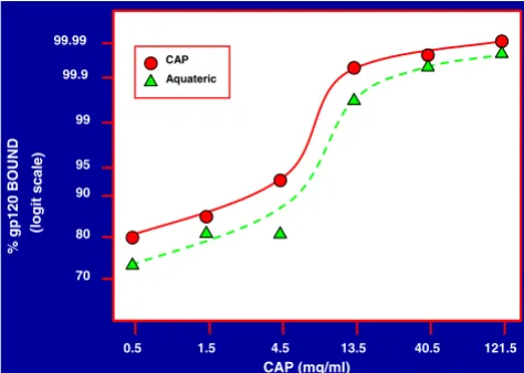
Recombinant gp120 HIV-1 IIIB in the presence of a 10-fold excess of BSA and gelatin, respectively, was quantitat-ed by adding the gp120/BSA/gelatin mixtures (before and after binding to micronized CAP or Aquateric; Fig. 1) at se-rial 5-fold dilutions (2-fold to 3.9 × 10⁶-fold in 0.14 M
NaCl-0.01 M Tris {TS}) to 96-well polystyrene plates (Im-mulon II, Dynatech Laboratories, Inc. Chantilly, VA). Af-ter overnight incubation at 4°C, the wells were washed and rabbit anti-gp120 (diluted 1:1,000 in 20% goat serum – 0.05% Tween 20 in PBS pH 7.4) was added. After 2 h at 37°C the wells were washed and HRP labeled goat anti-rabbit IgG (diluted in 1% BSA – 0.25% gelatin – 0.05% Tween 20 in PBS pH 7.4; {250 ng/well}) was added. After 2 h at 37°C bound HRP was detected using the kit from Kirkegaard & Perry Laboratories. The quantity of gp120 not adsorbed to CAP/Aquateric was determined from cal-ibration curves relating absorbance readings to the con-centration of gp120 not exposed to CAP/Aquateric.
The envelope glycoprotein gp120 released from HIV-1 BaL was quantitated as follows: Wells of polystyrene plates were first coated with protein A/G (1 µg/well) in 0.1 M Tris buffer, pH 8.8 for 2 h at 20°C followed by polyclo-nal rabbit anti-gp120 (diluted 500-fold) for 1 h at 20°C. Serial 2-fold dilutions in PBS containing 1% BSA and 1% gelatin (PBS-BG) of preparations containing gp120 were added to the wells. After 4 h at 20°C the wells were washed and bound gp120 was detected by addition of biotinyl sCD4 (1 µg) followed by HRP streptavidin. Bound HRP was quantitated as described above. The experiments were done in triplicate.
Measurements of HIV-1 infectivity
Two-fold serial dilutions of HIV-1 IIIB treated with dis-tinct anionic polymers, and separated from these poly-mers by precipitation with 3% PEG or by centrifugation at 14,000 rpm for 1 h, and control virus (100 µl), respective-ly, were added to HeLa-CD4-LTR-β-gal cells which had been plated a day before infection in 96-well plates at 1 × 10⁴ cells/well in 100 µl of DMEM medium containing
[image:3.612.56.293.87.256.2]10% fetal bovine serum (FBS). In some experiments, anti-HIV-1 tat IgG (25 µg) was added to treated and control vi-rus to suppress the effect of extracellular tat, which may have been present in the virus preparations, on virus infec-tion [48,49]. After incubainfec-tion at 37°C for 48 h, the culture supernatant fluids were removed and the cells washed once with PBS. Subsequently, 50 µl of lysis buffer from the Galacto-Light Plus kit were added to the wells for 1 h at 20°C. Aliquots (20 µl) of the cell lysates were transferred into wells of 96-well microplates and β-galactosidase was quantitated using the Galacto-Light Plus System chemilu-minescence reporter assay in a Microlight ML 2250 lumi-nometer (Dynatech Laboratories, Inc. Chantilly, VA). The infectivity of treated and control HIV-1 BaL was measured
Figure 1
Binding of HIV-1 IIIB gp120 to micronized CAP and Aqua-teric, respectively. Graded quantities of micronized CAP and Aquateric, respectively, in 0.5 ml of 0.14 M NaCl were mixed with 100 µl of a solution containing 1 µg of HIV-1 IIIB gp120, and 10 µg each of bovine serum albumin (BSA) and gelatin. After incubation for 5 min at 37°C the mixtures were cooled on ice, centrifuged at 10,000 rpm and the supernatant fluids were filtered through 0.45 µ filters which had been pre-washed with a solution containing 100 µg/ml of each BSA and gelatin in 0.14 M NaCl. gp120 in the filtrates was determined as described in the Methods section. The percentage of gp120 recovered in the filtrates was determined from cali-bration curves relating absorbance readings to the concen-tration of gp120.
CAP (mg/ml)
0.5 1.5 4.5 13.5 40.5 121.5
%
gp120
B
OUND
(l
o
git
sc
al
e)
70 80 90 95 99 99.9 99.99
by the same method except that MAGI-CCR5 cells were used.
Measurement of HIV-1 infectivity in seminal fluid is im-peded by the cytotoxicity of seminal plasma [50,51]. con-tributed to by spermine [52]. Such effects were minimized by using for titrations of virus infectivity pellets after pre-cipitation with 3% PEG or after centrifugation (14,000 rpm for 1 h).
Results
Micronized CAP induces gp41 six-helix bundle formation and is virucidal against HIV-1
Earlier studies [36,37]. indicated that soluble CAP at neu-tral pH binds to the CXCR4 and CCR5 coreceptor binding sites on HIV-1 IIIB and BaL, respectively, and induced the formation of "dead-end" gp41 six-helix bundles. These two properties of soluble CAP appear to be involved in the virus inhibitory and virucidal activities of this compound. However, CAP is not soluble at < pH 5.5 and would
pre-cipitate in a healthy human vaginal environment which is mildly acidic with a pH range of 3.5 – 4.5 [53]. The pre-cipitate would have drastically reduced antiviral activity. To overcome this problem, CAP in a micronized form was used and proven to have virucidal activity against HIV-1 IIIB [31] and shown to be effective in reducing vaginal in-fection by simian immunodeficiency virus (SIV) in a macaque model [34]. Micronized CAP provides a low pH buffering system and this may have contributed to its viru-cidal properties. In order to correlate the properties of CAP in soluble and micronized forms, it was necessary to establish whether or not micronized CAP would bind HIV-1 virus particles and HIV-1 envelope glycoproteins, and induce the formation of gp41 six-helix bundles, a marker of irreversible virus inactivation, as did soluble CAP [37]. First it was established that HIV-1 gp120 bound to micronized CAP (provided in the form of micronized CAP particles or of a commercial product, "Aquateric", containing about 66% of CAP and used for enteric coating of tablets and capsules in the pharmaceutical industry [54]); (Fig. 1). Similar results were obtained when gp120 was diluted in seminal fluid (diluted 1:1 with 0.14 M Na-Cl). Under these conditions, ≥ 99.9997% of gp120 bound to micronized CAP and Aquateric (final concentration of CAP: 121.5 mg/ml), respectively. HIV-1 IIIB and BaL virus particles also bound effectively in the presence of seminal fluid to Aquateric formulated into a topical gel (Fig. 2).
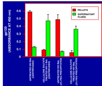
As a result of interaction with micronized CAP (in the form of Aquateric), the integrity of both HIV-1 IIIB and HIV-1 BaL virus particles was strikingly affected as indicat-ed by the observation that most of the p24 antigen after virus contact with micronized CAP was no longer precipi-table by 3% polyethylene glycol 6000 (PEG) unlike p24 antigen within untreated virus particles which precipitates under these conditions (Fig. 3). Furthermore, interaction with micronized CAP elicited the formation of gp41 six-helix bundles which were undetectable or present at low levels in untreated virus particles [37]. The six-helix bun-dles detectable by ELISA were mostly not precipitable by 3% PEG indicating that they were no longer associated with virus particles (Fig. 3). Furthermore, they were de-tected partially (HIV-1 IIIB) or entirely (HIV-1 BaL) in a form not bound anymore to the micronized CAP parti-cles, unlike most of the virus-derived p24 antigen. This suggests that interaction of virus particles with micronized CAP resulted in stripping off of the viral envelopes which was more noticeable with HIV-1 BaL than with HIV-1 IIIB.
[image:4.612.62.287.86.257.2]To confirm that gp41 six helix bundle formation was caused by contact of virus particles with the micronized form of CAP (= Aquateric) and not by low pH contributed by CAP, purified HIV-1 III B and BaL, respectively, was ex-posed for 5 min at 37°C to acidic buffers (final pH 2.9, 3.5, 4.0, 4.5 and 5.0). The suspensions were neutralized
Figure 2
Binding of HIV-1 IIIB and BaL, respectively, to Aquateric in the presence of human seminal fluid. Five hundred µl aliquots of a formulation containing Aquateric (18% Aquateric, 6% hydroxypropyl methylcellulose, 76% glycerol) were mixed with graded volumes of seminal fluid (range: 100 µl – 1,000
µl) each containing equal amounts of purified HIV-1 IIIB (6.8
× 109 particles) and BaL (2.5 × 10⁹ particles), respectively. After incubation for 5 min at 37°C the mixtures were cooled in ice, centrifuged at 10,000 rpm for 10 min in order to pellet Aquateric. The supernatant fluids were all adjusted to identi-cal volumes (1.5 ml) and NP40 was added to a final concen-tration of 1%. After incubation for 30 min at 37°C, serial 5-fold dilutions (1/1,000 to 1/3.9 × 108) were prepared and p24 antigen in each dilution was measured by ELISA. The per-centage of p24 antigen (representing a relative measure of the number of HIV-1 virus particles) was determined from calibration curves relating absorbance readings to the con-centration of p24 antigen.
% SEMINAL FLUID
10 20 30 40 50 60 70
99.9999
99.999
99.99
99.9
99
95
85
%
VIRUS
BOUND
(LOGIT
SCALE)
and assayed by ELISA for six-helix bundles. The results were negative (absorbance in the 0 to 0.068 range, the same as for viruses exposed to pH 7.0 in the absence of CAP). Poloxamer, a constituent of Aquateric (≤ 33%) at a final concentration of 30 mg/ml also did not induce the formation of six-helix bundles.
Results shown above indicated that HIV-1 adsorbed onto micronized CAP and that the CAP – virus interaction lead to the formation of gp41 six-helix bundles, a hallmark for HIV-1 inactivation. [37] To confirm that contact of virus with micronized CAP leads to loss of HIV-1 infectivity, HIV-1 IIIB and BaL, respectively, were diluted in seminal fluid and the diluted virus preparations were mixed with an Aquateric formulation (for composition, see legend to Fig. 2) at volume ratios of 3:1, 1:1 and 1:3 for 5 min at 37°C. Subsequently, the micronized Aquateric particles were sedimented by centrifugation. The supernatant flu-ids were brought to pH 7.0 and the pellets were dissolved
by addition of Na3PO4 (final pH = 7.0). All fractions were
then precipitated by 3% PEG and the redissolved pellets tested at 2- to 256-fold dilutions for infectivity (see Meth-ods section). Infectious virus was undetectable (≥ 99.6% and ≥ 99.2% inactivation of HIV-1 IIIB and BaL, respec-tively). Similar results were obtained with other CAP for-mulations (data not shown).
Induction of six-helix bundle formation by polymeric ani-onic candidate microbicides
Since elicitation of gp41 six-helix bundles might be in-volved in, or contribute to the virucidal activity of candi-date microbicides, it was of interest to determine whether anionic polymers other than CAP also induced six-helix bundle formation in HIV-1 IIIB and BaL virus particles.
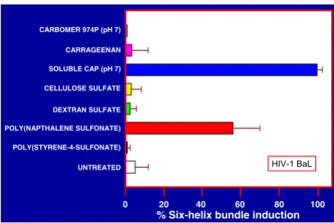
Results in Fig. 4 show that carrageenan, cellulose sulfate, dextran sulfate, poly(napthalene sulfonate) and poly(sty-rene-4-sulfonate) all elicit gp41 six-helix bundle forma-tion in HIV-1 IIIB but somewhat less effectively in comparison with CAP. On the other hand, Carbomer 974P, the active ingredient in BufferGel, was inactive in this respect. Strikingly different results were obtained for HIV-1 BaL (Fig. 5), and show that only CAP and to a lesser extent poly(napthalene sulfonate) elicited gp41 six-helix bundles detectable by ELISA.
Effect of anionic polymeric microbicide candidates on the integrity of HIV-1 particles
Since micronized CAP caused the dissociation of HIV-1 particles [31]; (Fig. 3), it was of interest to determine whether anionic polymeric candidate microbicides in sol-uble form at neutral pH would have similar effects. While no such effects were seen with HIV-1 IIIB, the integrity of HIV-1 BaL appeared to be partially diminished by treat-ment with CAP and drastically affected by cellulose sulfate (Fig. 6). The latter finding was confirmed by the observa-tion that HIV-1 BaL gp120 was no longer precipitable by 3% PEG or pellettable by centrifugation after treatment of the virus with cellulose sulfate, unlike gp120 associated with untreated virus particles (Fig. 7). This was not seen with cellulose sulfate treated HIV-1 IIIB (data not shown).
Comparative virucidal activity of polymeric anionic micro-bicide candidates
[image:5.612.76.269.85.255.2]HIV-1 IIIB and BaL, respectively, were treated with the dis-tinct polymers (10 mg/ml) for 5 min at 37°C and the in-fectivity of the treated viruses was compared with that of control untreated HIV-1. In repeated experiments with HIV-1 IIIB, soluble and micronized CAP, cellulose sulfate, poly(naphthalene sulfonate), poly(styrene-4-sulfonate) and BufferGel, respectively, caused a ≥ 140-fold reduction in infectivity, while dextran sulfate, carrageenan and Car-bomer 974P (pH 7), respectively, were less effective (4- to
Figure 3
Aquateric causes the disintegration of HIV-1 IIIB and BaL virus particles and induces the formation of gp41 six-helix bundles. Equal volumes of HIV-1 IIIB and BaL virus prepara-tions (6.8 × 10¹⁰ and 2.47 × 10¹⁰ virus particles, respectively, per ml) and the Aquateric formulation (see legend to Fig. 2) were mixed and incubated for 5 min at 37°C. Aquateric was pelletted by centrifugation at 10,000 rpm for 10 min and the supernatant fluids were neutralized by addition of 1 M Na2HPO4and filtered through 0.45 µ filters. The pellets con-taining Aquateric and adsorbed virus antigens were resus-pended and brought to pH 7.0 by addition of 1 M Na3HPO4. The supernatant fluids and the resuspended pellets, respec-tively, were mixed with a solution of PEG 6000 (final concen-tration 3%). The supernatant fluids and the resuspended pellets after PEG precipitation were mixed with NP40 (final concentration 1%). After incubation for 30 min at 37°C and 20°C, respectively, p24 antigen and six-helix bundles in the supernatant fluids and resuspended pellets were measured by ELISA.
0 20 40 60 80 100
P24 ANTIGEN SIX HELIX BUNDLES
A B
NOT ADSORBED TO CAP; PRECIPITATED BY PEG
C
D ADSORBED TO CAP; NOT PRECIPITATED BY PEG NOT ADSORBED TO CAP; NOT PRECIPITATED BY PEG ADSORBED TO CAP; PRECIPITATED BY PEG
%
ANT
IG
EN
A B C D A B C D
25-fold reduction in infectivity). A typical example for HIV-1 IIIB is shown in Fig. 8.
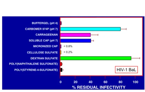
Representative results shown in Fig. 9 indicate that the polymers differed from each other more substantially with respect to their capacity to inactivate HIV-1 BaL, mi-cronized CAP, cellulose sulfate, BufferGel and the aryl sul-fonates being most effective.
Inhibitory effect of polymeric anionic microbicide candi-dates on blood clotting
For the selection of the most promising candidate micro-bicides, it is important to compare not only their virus in-hibitory and virucidal activities, but to also study their potential untoward effects. Since heparin and other ani-onic polymers are known to inhibit blood clotting [55], the effect of anionic polymeric microbicide candidates on blood clotting was measured. While CAP, carrageenan and BufferGel (Carbomer 974P) had moderate or no
ef-fects, cellulose sulfate, dextran sulfate, poly(napthalene sulfonate) and poly(styrene-4-sulfonate) greatly inhibited clotting of ACD plasma (Fig. 10). The increase in clotting time might be potentially undesirable for topical applica-tions under condiapplica-tions of local preexisting injury or bleed-ing.
Discussion
Several anionic polymers are being considered as topical microbicides to prevent sexual transmission of HIV-1. They include: (1) sulfate esters of polysaccharides {carra-geenan [1,16–19], cellulose sulfate [12–15], and dextran or dextrin sulfate [1–11]}; (2) aryl sulfonates {poly(naph-thalene sulfonate) [22–24] and poly(styrene-4-sulfonate) [20,21]}; and (3) aliphatic {Carbomer 974P/BufferGel [25–29]} and aromatic carboxylates {CAP [30–37]}. The anti-HIV-1 properties of these polymers are expected to depend on their complexation with virus proteins which is likely to occur mainly through the formation of salt linkages or ion pairs between oppositely charged groups on the polymers and virus proteins, respectively [56]. However, the anti-HIV-1 properties of these polymers may depend not only on their charge density, but also on the characteristics of their uncharged portions, which may be involved in hydrophobic and hydrogen bonding inter-actions which can elicit profound conformational chang-es in hydrophobic regions of proteins and cause disassembly of complex proteins consisting of subunits [57]. For these reasons it is not be expected that all anionic
Figure 4
gp41 six-helix bundle induction in HIV-1 IIIB by distinct ani-onic polymers representing candidate microbicides. Purified HIV-1 IIIB (7 × 10⁹ particles in TS) was precipitated with 3% PEG. The pellets were resuspended in 100 µl of phosphate buffer saline (PBS) to separate the virus from soluble HIV-1 antigens in the preparation. The different anionic polymers (listed on the ordinate) were added to a final concentration of 5 mg/ml except for Carbomer 974P (2.5 mg/ml). After incubation for 5 min at 37°C, the mixtures were cooled in ice and PEG was added (final concentration 3%). The pellets after centrifugation were washed once with 3% PEG in PBS, repelletted and finally resuspended in PBS containing 1% NP40 and 100 µg/ml BSA and incubated at 20°C for 30 min. The formation of six-helix bundles was determined by ELISA (see Methods section). All experiments were done in tripli-cate, the absorbance readings are shown as percentages of the absorbance corresponding to six-helix bundles elicited by CAP (= 100%).
CARBOMER 974P (pH 7)
CARRAGEENAN
DEXTRAN SULFATE
POLY(NAPTHALENE SULFONATE)
POLY(STYRENE-4-SULFONATE) SOLUBLE CAP (pH 7)
CELLULOSE SULFATE
UNTREATED
% Six-helix bundle induction
0 20 40 60 80 100
[image:6.612.313.554.99.260.2] [image:6.612.55.295.102.259.2]HIV-1 IIIB
Figure 5
gp41 six-helix bundle induction in HIV-1 BaL by distinct ani-onic polymers respresenting candidate microbicides. Purified HIV-1 BaL (2.5 × 10⁹ particles in Dulbecco's modified Eagle medium {DMEM} + 20% fetal bovine serum {FBS}) were pre-cipitated with 3% PEG and subsequently treated as described in the legend for Fig. 4.
% Six-helix bundle induction
CARBOMER 974P (pH 7)
CARRAGEENAN
DEXTRAN SULFATE
POLY(NAPTHALENE SULFONATE)
POLY(STYRENE-4-SULFONATE) SOLUBLE CAP (pH 7)
CELLULOSE SULFATE
UNTREATED
0 20 40 60 80 100
0 20 40 60 80 100
polymers, considered as anti-HIV-1 microbicides, would have similar biological activities.
In vivo protection by anti-HIV-1 compounds against
sexu-al transmission of the virus could only be accomplished if the compounds at sufficient concentrations, and the virus both reach susceptible cells within genital mucosa before productive infection could occur [38,39]. Considering the complex events involved in sexual transmission of HIV-1, this might not be easily accomplished. Therefore, inactiva-tion of HIV-1 infectivity preceding virus contact with sus-ceptible target cells should be considered a preferred mechanism of protection against infection. For this rea-son it was of interest to study the virucidal activity of ani-onic polymeric microbicide candidates against both X4 (using CXCR4 as coreceptor) and R5 (using CCR5 as core-ceptor) viruses, the latter being most frequently transmit-ted sexually [39].
Soluble CAP at neutral pH was shown to bind strongly to the coreceptor binding sites on gp120 of both X4 and R5 viruses [36] and to elicit the formation of gp41 six-helix bundles, the hallmark of irreversible virus inactivation [37]. Thus, HIV-1 exposed to CAP at sufficient
concentra-tions is rendered noninfectious, and continued presence of CAP is not required to maintain lack of infectivity. CAP differs from all other anionic polymers listed above, in that it is not soluble at pH < 5.5. Therefore, CAP would be expected to precipitate in a normal acidic vaginal environ-ment. To avoid this, CAP in micronized form (which does not aggregate at low pH), instead of CAP in soluble form is being considered as a topical microbicide [30–35]. Mi-cronized CAP (Aquateric) was shown to be virucidal against HIV-1, herpesviruses and several nonviral sexually transmitted disease (STD) pathogens [30–34]. The viru-cidal activity of micronized CAP could at least partly be explained by its buffering capacity at low pH [40] since it is a free acid while other anionic polymeric microbicide candidates (except BufferGel, the active ingredient of which is Carbomer 974P [27]) are sodium salts. Results presented here explain how micronized CAP inactivates HIV-1. Both the envelope glycoprotein gp120 (Fig. 1) and HIV-1 IIIB and BaL virus particles (Fig. 2) in the presence of an excess of nonviral proteins, including seminal fluid, bind effectively to micronized CAP. Under in vivo condi-tions (when the virus concentracondi-tions are by one to four or-ders of magnitude lower than those used here under experimental conditions [58,59], (Fig. 2)), this would be expected to physically remove virus particles by adsorp-tion onto micronized CAP (Aquateric) so that they would cease to be available to initiate the complex sequence of events leading to mucosal infection. Furthermore,
interac-Figure 6
Effect of anionic polymeric microbicide candidates on the integrity of HIV-1 IIIB and BaL virus particles. Purified HIV-1 IIIB (7 × 10⁹ particles/100 µl) and BaL (2.5 × 10⁹ particles/100
µl) untreated or treated with the polymeric substances shown on the abscissa (final concentrations 5 mg/ml except for Carbomer 974P {2.5 mg/ml}) for 5 min at 37°C. The mix-tures were cooled on ice and the virus particles were pellet-ted by centrifugation (1 h at 14,000 rpm in a Beckman 5415 C microfuge). The pellets were resuspended in lysis buffer (1% NP40, 100 µg/ml BSA in PBS) and incubated for 30 min at 37°C. p24 antigen was determined in serial dilutions of the lysates by ELISA. The levels of p24 antigen in the treated virus preparations were compared with those corresponding to lysed untreated virus (= 100%).
CARBOMER 974P (pH 7)
CARRAGEENAN
DEXTRAN SULFATE
POLY(NAPTHALENE SULFONATE)
POLY(STYRENE-4-SULFONATE) SOLUBLE CAP (pH 7)
CELLULOSE SULFATE
UNTREATED
0.001 0.01 0.1 1 10 100 1000
% HIV-1 (p24 ANTIGEN)INPELLET
[image:7.612.328.533.85.258.2]Top bars HIV-1 IIIB Bottom bars HIV-1 BaL
Figure 7
Release of gp120 from HIV-1 BaL by treatment with cellulose sulfate. Purified HIV-1 BaL (2.5 × 10⁹ particles/100 µl) was treated with cellulose sulfate (final concentration 5 mg/ml) for 5 min at 37°C. The mixture was cooled in ice and divided into 2 aliquots. One aliquot was centrifuged for 1 h at 14,000 rpm and the 2nd aliquot was precipitated with PEG 6000 (final concentration 3%). gp120 in the respective supernatant fluids and pellets (resuspended in PBS) was determined by ELISA as described [37]; (see Methods section).
[image:7.612.53.297.109.237.2]tion with micronized CAP elicits the formation of gp41 six-helix bundles and causes the stripping off of HIV-1 en-velope glycoproteins (Fig. 3). The formation of gp41 six-helix bundles was not due to exposure to a low pH envi-ronment provided by the micronized CAP, since exposure to low pH buffers did not elicit gp41 six-helix bundle for-mation. In summary, the contact of HIV-1 IIIB and HIV-1 BaL, representatives of X4 and R5 viruses respectively, with micronized CAP, resulted in a complex sequence of events involving virus adsorption, exposure to low pH, in-duction of gp41 six-helix bundles, and stripping off of vi-ral envelopes and virus disintegration, ultimately resulting in loss of infectivity.
[image:8.612.310.555.103.239.2]In order to contribute to the understanding of the poten-tial virucidal activity of anionic polymers, their capacity to induce gp41 six-helix bundle formation was measured and compared with that of CAP in soluble form (pH 7.0). All polymers except Carbomer 974P elicited gp41 six-he-lix bundle formation in HIV-1 IIIB, albeit in some cases less efficiently in comparison with CAP (Fig. 4). On the other hand, gp41 six-helix bundle formation in HIV-1 BaL was detectable only for CAP and poly(naphthalene sul-fonate) treated virus (Fig. 5) suggesting differences among the anionic polymers with respect to their activity against the R5 virus HIV-1 BaL. The induction of six-helix bundles in HIV-1 does not seem to be the only mechanism in-volved in virucidal activity of anionic polymers (compare
Figure 8
[image:8.612.53.299.109.250.2]Inactivation of HIV-1 IIIB by polymeric anionic candidate microbicides. HIV-1 IIIB (7 × 10⁹ virus particles/100 µl) was treated with the compounds indicated on the abscissa (final concentrations 10 mg/ml except for Carbomer 974P [2.5 mg/ ml; neutralized to pH 7.0; at higher concentrations the solu-tions became too viscous to handle]) for 5 min at 37°C. The final dilution of BufferGel was 4-fold (final pH = 4.0). The preparations were cooled on ice and virus particles were separated from the polymers and their infectivity determined as described in the Methods section. For comparison, results obtained with a formulation of micronized CAP (= Aquateric; see legend for Fig. 2) are shown.
Figure 9
Inactivation of HIV-1 BaL by polymeric anionic candidate microbicides. HIV-1 BaL (2.5 × 10⁹ virus particles/100 µl) was treated as described in the legend for Fig. 8.
POLY(STYRENE-4-SULFONATE) POLY(NAPHTHALENE SULFONATE) DEXTRAN SULFATE CELLULOSE SULFATE SOLUBLE CAP (pH 7) CARRAGEENAN BUFFERGEL (pH 4)
CARBOMER 974P (pH 7)
MICRONIZED CAP
% RESIDUAL INFECTIVITY
0 20 40 60 80 100
< 0.2% < 0.2%
< 0.2%
HIV-1 IIIB
POLY(STYRENE-4-SULFONATE) POLY(NAPHTHALENE SULFONATE) DEXTRAN SULFATE CELLULOSE SULFATE SOLUBLE CAP (pH 7) CARRAGEENAN BUFFERGEL (pH 4)
CARBOMER 974P (pH 7)
MICRONIZED CAP
% RESIDUAL INFECTIVITY
0 20 40 60 80 100
< 0.8%
< 0.2%
[image:8.612.51.297.456.613.2]HIV-1 BaL
Figure 10
Effect of polymeric anionic candidate microbicides on clotting of ACD plasma. The compounds indicated on the abscissa (final concentrations of 100 and 1000 µg/ml, respectively) were added to fresh citrated plasma prewarmed to 37°C. CaCl2 was added to a final concentration of 10 mM, and the time when clot formation had started was measured. The increase in clotting time as compared with the time required for clotting of plasma in the absence of any added com-pounds is shown.
CARBOMER 974P (pH 7)
CARRAGEENAN
DEXTRAN SULFATE
POLY(NAPTHALENE SULFONATE)
POLY(STYRENE-4-SULFONATE) SOLUBLE CAP (pH 7)
CELLULOSE SULFATE
INCREASE IN CLOTTING TIME
1x 2x 3x 4x 5x 6x >500x
Fig. 5 with Figs. 8 and 9) and there might be other contrib-uting processes.
Since CAP in micronized form was shown to cause distegration of HIV-1 virus particles [31], (Fig. 3), it was of in-terest to determine whether any of the other anionic polymeric microbicide candidates affect the integrity of vi-rus particles. While HIV-1 IIIB was not affected significant-ly by any of the posignificant-lymers tested, CAP to some extent, but surprisingly cellulose sulfate affected the integrity of HIV-1 BaL particles (Fig. 6). These results were supported by the observation that HIV-1 BaL gp120 after treatment with cellulose sulfate, unlike virus associated gp120, did not become precipitated with 3% PEG and failed to pellet by centrifugation under conditions leading to sedimentation of intact virus particles (Fig. 7)]. The differential sensitivity of an R5 virus, HIV-1 BaL, as compared to an R4 virus, HIV-1 IIIB, to the same negatively charged polyelectrolyte appears unprecedented to the best of our knowledge.
Among the three polysaccharide sulfate esters (carrageen-an, cellulose sulfate and dextran sulfate), cellulose sulfate appeared to be more effective against HIV-1 BaL than the other polymers. This might be due to the polysaccharide backbone of cellulose [60] with a propensity to form strong intermolecular hydrogen bonds and to be involved in hydrophobic interactions more than soluble dextrans [61] and galactose + 3,6-anhydrogalactose copolymers (= the saccharide moiety of carrageenan) [62,63]. Among the two polymers containing aryl sulfonates, poly(napthalene sulfonate) was more effective than poly(styrene-4-sul-fonate) with respect to induction of gp41 six-helix bun-dles in HIV-1 BaL. This difference may possibly be due to the high propensity of the uncharged part of the molecule {poly(naphtalene)} to be involved in hydrophobic inter-actions. Among the two carboxylate polymers (Carbomer 974P and CAP), only soluble CAP (at neutral pH) was able to elicit the formation of gp41 six-helix bundles in both HIV-1 IIIB and BaL and was virucidal against both of these viruses. The insoluble, micronized form of CAP had similar properties. The differences in biological activities of these polymers is probably due to their noncharged portions, i.e. mainly the occurrence of benzene rings in CAP.
Polycarboxylates (soluble CAP and Carbomer 974P; Fig. 10) had a minimal effect on plasma coagulation. Among the sulfonates and polysaccharide sulfates, carrageenan and poly(napthalene sulfonate) had lower anti-coagulant activity in comparison with cellulose sulfate, dextran sul-fate and poly(styrene-4-sulfonate) (Fig. 10). Considera-tion of these side effects might contribute to the process of selection of preferred microbicides for future large-scale use.
Conclusions
Among anionic polymers considered as topical microbi-cides to prevent sexual transmission of HIV-1 {Carbomer 974P/BufferGel, carrageenan, CAP, cellulose sulfate, dex-tran/dextrin sulfate, poly(napthalene sulfonate) and po-ly(styrene-4-sulfonate)}, CAP formulated in a micronized form, is the only compound which has a capacity to re-move from physiological fluids infectious virus by adsorp-tion onto the micronized particles. The interacadsorp-tion between HIV-1 IIIB, a representative of X4 viruses, and HIV-1 BaL, a representative of R5 viruses, with the micro-nized form of CAP has the following consequences: 1) elicitation of "dead-end" gp41 six-helix bundles expected to render the virus incompetent to fuse with target cells; 2) loss of virus integrity and stripping off of the envelope glycoproteins; and 3) inactivation of virus infectivity. Elic-itation of gp41 six-helix bundles and virucidal activity rep-resent shared properties between micronized and soluble (at sufficient concentrations) forms of CAP [37].
As determined by ELISA, the aforementioned polymers other than CAP (except Carbomer 974P) also elicited gp41 six-helix bundle formation in HIV-1 IIIB, albeit less effectively than CAP. On the other hand, only poly(nap-thalene sulfonate), in addition to CAP, elicited detectable six-helix bundle formation in HIV-1 BaL. This suggests that X4 and R5 viruses may be differently affected by some of the polymeric compounds being considered as topical microbicides. This was confirmed by the results of assays for virucidal activity.
Abbreviations used
ACD, acid citrate dextrose; CAP, cellulose acetate phtha-late; BSA, bovine serum albumin; DMEM, Dulbecco's modified Eagle medium; ELISA, enzyme-linked immuno-sorbent assay; FBS, fetal bovine serum; HIV-1, human im-munodeficiency virus type 1; HRP, horseradish peroxidase; mAb, monoclonal antibodies; PEG 6000, pol-yethylene glycol 6000; PBS, phosphate buffered saline; PBS-BG, 1% BSA/1% gelatin in PBS; sCD4, soluble CD4; STD, sexually transmitted disease; TS, 0.14 M NaCl, 0.01 M Tris, pH 7.0;
Competing interests
None declaredAuthors' contributions
Acknowledgements
We thank Ms. V. Kuhlemann for preparation of the manuscript and figures, Dr. S. Jiang and Dr. A. K. Debnath for comments and Dr. K. J. Whaley and Dr. S. Jiang for providing BufferGel and mAb NC-1, respectively. This study was supported by NIH grant (P01 HD41761) and the Marilyn M. Simpson Charitable Trust.
References
1. Baba M, Snoeck R, Pauwels R, De Clercq E: Sulfated polysaccha-rides are potent and selective inhibitors of various enveloped viruses, including herpes simplex virus, cytomegalovirus, ve-sicular stomatitis virus, and human immunodeficiency virus.
Antimicrob Agents Chemother 1988, 32:1742-1745
2. Bagasra O, Lischner HW: Activity of dextran sulfate and other polyanionic polysaccharides against human immunodeficien-cy virus. J Infect Dis 1988, 158:1084-1087
3. Baba M, Schols D, Pauwels R, Nakashima H, De Clercq E: Sulfated polysaccharides as potent inhibitors of HIV-induced syncy-tium formation: A new strategy towards AIDS chemothera-py. J Acquir Immune Defic Syndr 1990, 3:493-499
4. McClure MO, Moore JP, Blanc DF, Scotting P, Cook GM, Keynes RJ, Weber JN, Davies D, Weiss RA: Investigations into the mecha-nism by which sulfated polysaccharides inhibit HIV infection in vitro . AIDS Res Hum Retroviruses 1992, 8:19-26
5. Lynch G, Low L, Li S, Sloane A, Adams S, Parish C, Kemp B, Cunning-ham AL: Sulfated polyanions prevent HIV infection of lym-phocytes by disruption of the CD4-gp120 interaction, but do not inhibit monocyte infection. J Leukoc Biol 1994, 56:266-272 6. Shaunak S, Gooderham NJ, Edwards RJ, Payvandi N, Javan CM,
Bag-gett N, MacDermot J, Weber JN, Davies DS: Infection by HIV-1 blocked by binding of dextrin 2-sulphate to the cell surface of activated human peripheral blood mononuclear cells and cultured T-cells. Br J Pharmacol 1994, 113:151-158
7. Davies DS: Dextrin sulfates as anti HIV-1 agents and compo-sition thereof. US Patent 5,439,892
8. Javan CM, Gooderham NJ, Edwards RJ, Davies DS, Shaunak S: Anti-HIV type 1 activity of sulfated derivatives of dextrin against primary viral isolates of HIV type 1 in lymphocytes and monocyte-derived macrophages. AIDS Res Hum Retroviruses
1997, 13:875-880
9. Stafford MK, Cain D, Rosenstein I, Fontaine EA, McClure M, Flanagan AM, Smith JR, Taylor-Robinson D, Weber J, Kitchen VS: A placebo-controlled, double-blind prospective study in healthy female volunteers of dextrin sulphate gel: A novel potential intrav-aginal virucide. J Acquir Immune Defic Syndr Hum Retrovirol 1997, 14:213-218
10. Baba M, Pauwels R, Balzarini J, Arnout J, Desmyter J, De Clercq E: Mechanism of inhibitory effect of dextran sulfate and heparin on replication of human immunodeficiency virus in vitro . Proc Natl Acad Sci U S A 1988, 85:6132-6136
11. Rosenstein IJ, Stafford MK, Kitchen VS, Ward H, Weber JN, Taylor-Robinson D: Effect on normal vaginal flora of three intravagi-nal microbicidal agents potentially active against human im-munodeficiency virus type 1. J Infect Dis 1998, 177:1386-1390 12. Yamamoto I, Takayama K, Honma K, Gonda T, Matsuzaki K, Hatanaka
K, Uryu T, Yoshida O, Nakashima H, Yamamoto N, et al: Synthesis, structure and antiviral activity of sulfates of cellulose and its branched derivatives. Carbohydrate Polymers 1991, 14:53-63 13. Anderson RA, Zaneveld LJD, Usher TC: Cellulose sulfate for use
as antimicrobial and contraceptive agent. US Patent 6,063,773
14. Mauck C, Frezieres R, Walsh T, Robergeau K, Callahan M: Cellulose sulfate: tolerance and acceptability of penile application. Con-traception 2001, 64:377-381
15. Mauck C, Weiner DH, Ballagh S, Creinin M, Archer DF, Schwartz J, Pymar H, Lai JJ, Callahan M: Single and multiple exposure toler-ance study of cellulose sulfate gel: a Phase I safety and col-poscopy study. Contraception 2001, 64:383-391
16. Carlucci MJ, Pujol CA, Ciancia M, Noseda MD, Matulewicz MC, Da-monte EB, Cerezo AS: Antiherpetic and anticoagulant proper-ties of carrageenans from the red seaweed Gigartina skottsbergii and their cyclized derivatives: correlation be-tween structure and biological activity. Int J Biol Macromol 1997, 20:97-105
17. Coggins C, Blanchard K, Alvarez F, Brache V, Weisberg E, Kilmarx PH, Lacarra M, Massai R, Mishell D Jr, Salvatierra A, et al:
Prelimi-nary safety and acceptability of a carrageenan gel for possi-ble use as a vaginal microbicide. Sex Transm Infect 2000, 76:480-483
18. Schaeffer DJ, Krylov VS: Anti-HIV activity of extracts and com-pounds from algae and cyanobacteria. Ecotoxicol Environ Saf
2000, 45:208-227
19. Maguire RA, Bergman N, Phillips DM: Comparison of microbi-cides for efficacy in protecting mice against vaginal challenge with herpes simplex virus type 2, cytotoxicity, antibacterial properties, and sperm immobilization. Sex Transm Dis 2001, 28:259-265
20. Anderson RA, Feathergill K, Diao X, Cooper M, Kirkpatrick R, Spear P, Waller DP, Chany C, Doncel GF, Herold B, et al: Evaluation of poly(styrene-4-sulfonate) as a preventive agent for concep-tion and sexually transmitted diseases. J Androl 2000, 21:862-875
21. Christensen ND, Reed CA, Culp TD, Hermonat PL, Howett MK, An-derson RA, Zaneveld LJ: Papillomavirus microbicidal activities of high-molecular-weight cellulose sulfate, dextran sulfate, and polystyrene sulfonate. Antimicrob Agents Chemother 2001, 45:3427-3432
22. Rusconi S, Moonis M, Merrill DP, Pallai PV, Neidhardt EA, Singh SK, Willis KJ, Osburne MS, Profy AT, Jenson JC, et al: Naphthalene sul-fonate polymers with CD4-blocking and anti-human immun-odeficiency virus type 1 activities. Antimicrob Agents Chemother
1996, 40:234-236
23. Van Damme L, Wright A, Depraetere K, Rosenstein I, Vandersmissen V, Poulter L, McKinlay M, Van Dyck E, Weber J, Profy A, et al: A phase I study of a novel potential intravaginal microbicide, PRO2000 in healthy sexually inactive women. Sex Transm Dis
2000, 76:126-130
24. Weber J, Nunn A, O'Connor T, Jeffries D, Kitchen V, McCormack S, Stott J, Almond N, Stone A, Darbyshire J: 'Chemical condoms' for the prevention of HIV infection: evaluation of novel agents against SHIV89.6PD in vitro and in vivo. AIDS 2001, 15:1563-1568 25. Olmsted SS, Dubin NH, Cone RA, Moench TR: The rate at which human sperm are immobilized and killed by mild acidity. Fer-til Steril 2000, 73:687-693
26. Clarke JG, Peipert JF, Hillier SL, Heber W, Boardman L, Moench TR, Mayer K: Microflora changes with the use of a vaginal micro-bicide. Sex Transm Dis 2002, 29:288-293
27. Zeitlin L, Hoen TE, Achilles SL, Hegarty TA, Jerse AE, Kreider JW, Ol-msted SS, Whaley KJ, Cone RA, Moench TR: Tests of BufferGel for contraception and prevention of sexually transmitted diseas-es in animal models. Sex Transm Dis 2001, 28:417-423
28. Mayer KH, Peipert J, Fleming T, Fullem A, Moench T, Cu-Uvin S, Bent-ley M, Chesney M, Rosenberg Z: Safety and tolerability of Buff-erGel, a novel vaginal microbicide, in women in the United States. Clin Infect Dis 2001, 32:476-482
29. van de Wijgert J, Fullem A, Kelly C, Mehendale S, Rugpao S, Kumwen-da N, Chirenje Z, Joshi S, Taha T, Padian N, et al: Phase 1 trial of the topical microbicide BuffferGel: Safety results from four international sites. J Acquir Immune Defic Syndr 2001, 26:21-27 30. Gyotoku T, Aurelian L, Neurath AR: Cellulose acetate phthalate
(CAP): an 'inactive' pharmaceutical excipient with antiviral activity in the mouse model of genital herpesvirus infection.
Antivir Chem Chemother 1999, 10:327-332
31. Neurath AR, Strick N, Li Y-Y, Lin K, Jiang S: Design of a "microbi-cide" for prevention of sexually transmitted diseases using "inactive" pharmaceutical excipients. Biologicals 1999, 27:11-21 32. Neurath AR, Li YY, Mandeville R, Richard L: In vitro activity of a cellulose acetate phthalate topical cream against organisms associated with bacterial vaginosis. J Antimicrob Chemother 2000, 45:713-714
33. Neurath AR: A microbicide for prevention of sexually trans-mitted diseases using a pharmaceutical excipient. AIDS Patient
Care STDS 2000, 14:215-219
34. Manson KH, Wyand MS, Miller C, Neurath AR: The effect of a cel-lulose acetate phthalate topical cream on vaginal transmis-sion of simian immunodeficiency virus in rhesus monkeys.
Antimicrob Agents Chemother 2000, 44:3199-3202
35. Kawamura T, Cohen SS, Borris DL, Aquilino EA, Glushakova S, Mar-golis LB, Orenstein JM, Offord RE, Neurath AR, Blauvelt A: Candi-date microbicides block HIV-1 infection of human immature Langerhans cells within epithelial tissue explants. J Exp Med
36. Neurath AR, Strick N, Li YY, Debnath AK: Cellulose acetate phthalate, a common pharmaceutical excipient, inactivates HIV-1 and blocks the coreceptor binding site on the virus en-velope glycoprotein gp120. BMC Infect Dis 2001, 1:17
37. Neurath AR, Strick N, Jiang S, Li YY, Debnath AK: Anti-HIV-1 ac-tivity of cellulose acetate phthalate: Synergy with soluble CD4 and induction of "dead-end" gp41 six-helix bundles. BMC Infect Dis 2002, 2:6
38. Greenhead P, Hayes P, Watts PS, Laing KG, Griffin GE, Shattock RJ: Parameters of human immunodeficiency virus infection of human cervical tissue and inhibition by vaginal virucides. J Vi-rol 2000, 74:5577-5586
39. Shattock RJ, Doms RW: AIDS models: Microbicides could learn from vaccines. Nat Med 2002, 8:425
40. O'Connor TJ, Kinchington D, Kangro HO, Jeffries DJ: The activity of candidate virucidal agents, low pH and genital secretions against HIV-1 in vitro. Int J STD AIDS 1995, 6:267-272
41. Berger EA, Doms RW, Fenyo EM, Korber BT, Littman DR, Moore JP, Sattentau QJ, Schuitemaker H, Sodroski J, Weiss RA: A new classi-fication for HIV-1. Nature 1998, 391:240
42. Larsen B: Vaginal flora in health and disease. Clin Obstet Gynecol
1993, 36:107-121
43. Jiang S, Lin K, Lu M: A conformation-specific monoclonal anti-body reacting with fusion-active gp41 from the human im-munodeficiency virus type 1 envelope glycoprotein. J Virol
1998, 72:10213-10217
44. Neurath AR, Strick N, Li Y-Y, Jiang S: Improbability of harmful au-toimmune responses resulting from immunization with HIV-1 envelope glycoproteins. AIDS Res Hum Retroviruses 1993, 9:1195-1208
45. Jiang S, Lin K, Zhang L, Debnath AK: A screening assay for antivi-ral compounds targeted to the HIV-1 gp41 core structure us-ing a conformation-specific monoclonal antibod J Virol Methods
1999, 80:85-96
46. Gartner S, Markovits P, Markovitz DM, Kaplan MH, Gallo RC, Popo-vic M: The role of mononuclear phagocytes in HTLV-III/LAV infection. Science 1986, 233:215-219
47. Stoll VS, Blanchard JS: Buffers: Principles and Practice In Methods in Enzymology, Guide to Protein Purification (Edited by: Deutscher MP) San Diego, CA., Academic Press, Inc 1990, 182:24-38
48. Huang L, Bosch I, Hofmann W, Sodroski J, Pardee AB: Tat protein induces human immunodeficiency virus type 1 (HIV-1) core-ceptors and promotes infection with both macrophage-trop-ic and T-lymphotropmacrophage-trop-ic HIV-1 strains. J. Virol 1998, 72:8952-8960 49. Secchiero P, Zella D, Capitani S, Gallo RC, Zauli G: Extracellular HIV-1 tat protein up-regulates the expression of surface CXC-chemokine receptor 4 in resting CD4+ T cells. J Immunol
1999, 162:2427-2431
50. Rasheed S, Li Z, Xu D: Human immunodeficiency virus load. Quantitative assessment in semen from seropositive individ-uals and in spiked seminal plasma. J Reprod Med 1995, 40:747-757
51. Krieger JN, Coombs RW, Collier AC, Ho DD, Ross SO, Zeh JE, Co-rey L: Intermittent shedding of human immunodeficiency vi-rus in semen: implications for sexual transmission. J Urol 1995, 154:1035-1040
52. Allen RD, Roberts TK: Role of spermine in the cytotoxic effects of seminal plasma. Am J Reprod Immunol Microbiol 1987, 13:4-8 53. Masters W, Johnson V: Sexual physiology and AIDS protection.
In AIDS and Sex (Edited by: Voeller B, Reinisch J, Gottlieb M) New York, Oxford University Press 1990, 371-374
54. Goskonda SR, Lee JC: Cellulose Acetate Phthalate. In Handbook
of Pharmaceutical Excipients (Edited by: Kibbe AH, Washington, DC)
American Pharmaceutical Association 2000, 99-101
55. de Raucourt E, Mauray S, Chaubet F, Maiga-Revel O, Jozefowicz M, Fischer AM: Anticoagulant activity of dextran derivatives. J
Bi-omed Mater Res 1998, 41:49-57
56. Izumi T, Hirata M: Complexation of papain with strong poly-anions and enzymatic activities of the resulting complexes. J
Macromol Sci, Pure Appl Chem 1994, A31(1):39-51
57. Sedlak E, Antalik M: Coulombic and noncoulombic effect of polyanions on cytochrome c structure. Biopolymers 1998, 46:145-154
58. Dyer JR, Kazembe P, Vernazza PL, Gilliam BL, Maida M, Zimba D, Hoffman IF, Royce RA, Schock JL, Fiscus SA, et al: High levels of hu-man immunodeficiency virus type 1 in blood and semen of
seropositive men in sub-Saharan Africa. J Infect Dis 1998, 177:1742-1746
59. Chakraborty H, Sen PK, Helms RW, Vernazza PL, Fiscus SA, Eron JJ, Patterson BK, Coombs RW, Krieger JN, Cohen MS: Viral burden in genital secretions determines male-to-female sexual trans-mission of HIV-1: a probabilistic empiric model. AIDS 2001, 15:621-627
60. Kadla JF, Gilbert RD: Cellulose structure: A review. Cellulose
Chemistry and Technology 2000, 34:197-216
61. Cheetham NWH, Fiala-Beer E: Dextran structural details from high-field proton NMR spectroscopy. Carbohydrate Polymers
1991, 14:149-158
62. Reilly WJ Jr: Carrageenan. In Handbook of Pharmaceutical Excipients (Edited by: Kibbe AH, Washington, DC) American Pharmaceutical Associa-tion 2000, 91-93
63. Caram-Lelham N, Sundelof LO: The effect of hydrophobic char-acter of drugs and helix-coil transition of κ-carrageenan on the polyelectrolyte-drug interaction. Pharm Res 1996, 13:920-925
Pre-publication history
The pre-publication history for this paper can be accessed here:
http://www.biomedcentral.com/1471-2334/2/27/prepub
Publish with BioMed Central and every scientist can read your work free of charge
"BioMed Central will be the most significant development for disseminating the results of biomedical researc h in our lifetime."
Sir Paul Nurse, Cancer Research UK
Your research papers will be:
available free of charge to the entire biomedical community
peer reviewed and published immediately upon acceptance
cited in PubMed and archived on PubMed Central
yours — you keep the copyright
Submit your manuscript here:
http://www.biomedcentral.com/info/publishing_adv.asp