Evolution-Guided Structural and Functional Analyses of the
HERC Family Reveal an Ancient Marine Origin and
Determinants of Antiviral Activity
Ermela Paparisto,
aMatthew W. Woods,
aMacon D. Coleman,
aSeyed A. Moghadasi,
aDivjyot S. Kochar,
aSean K. Tom,
aHinissan P. Kohio,
aRichard M. Gibson,
aTaryn J. Rohringer,
aNina R. Hunt,
aEric J. Di Gravio,
aJonathan Y. Zhang,
aMeijuan Tian,
aYong Gao,
aEric J. Arts,
aStephen D. Barr
aaWestern University, Schulich School of Medicine and Dentistry, Department of Microbiology and
Immunology, London, Ontario, Canada
ABSTRACT
In humans, homologous to the E6-AP carboxyl terminus (HECT) and
regulator of chromosome condensation 1 (RCC1)-like domain-containing protein 5
(HERC5) is an interferon-induced protein that inhibits replication of evolutionarily
di-verse viruses, including human immunodeficiency virus type 1 (HIV-1). To better
un-derstand the origin, evolution, and function of HERC5, we performed phylogenetic,
structural, and functional analyses of the entire human small-HERC family, which
in-cludes HERC3, HERC4, HERC5, and HERC6. We demonstrated that the
HERC
family
emerged
⬎
595 million years ago and has undergone gene duplication and gene
loss events throughout its evolution. The structural topology of the RCC1-like
do-main and HECT dodo-mains from all HERC paralogs is highly conserved among
evolu-tionarily diverse vertebrates despite low sequence homology. Functional analyses
showed that the human small HERCs exhibit different degrees of antiviral activity
to-ward HIV-1 and that HERC5 provides the strongest inhibition. Notably, coelacanth
HERC5 inhibited simian immunodeficiency virus (SIV), but not HIV-1, particle
produc-tion, suggesting that the antiviral activity of HERC5 emerged over 413 million years
ago and exhibits species- and virus-specific restriction. In addition, we showed that
both HERC5 and HERC6 are evolving under strong positive selection, particularly
blade 1 of the RCC1-like domain, which we showed is a key determinant of antiviral
activity. These studies provide insight into the origin, evolution, and biological
im-portance of the human restriction factor HERC5 and the other HERC family
mem-bers.
IMPORTANCE
Intrinsic immunity plays an important role as the first line of defense
against viruses. Studying the origins, evolution, and functions of proteins responsible
for effecting this defense will provide key information about virus-host relationships
that can be exploited for future drug development. We showed that HERC5 is one
such antiviral protein that belongs to an evolutionarily conserved family of HERCs
with an ancient marine origin. Not all vertebrates possess all HERC members,
sug-gesting that different HERCs emerged at different times during evolution to provide
the host with a survival advantage. Consistent with this, two of the more recently
emerged HERC members, HERC5 and HERC6, displayed strong signatures of having
been involved in an ancient evolutionary battle with viruses. Our findings provide
new insights into the evolutionary origin and function of the HERC family in
verte-brate evolution, identifying HERC5 and possibly HERC6 as important effectors of
in-trinsic immunity in vertebrates.
KEYWORDS
HERC3, HERC4, HERC5, HERC6, restriction factor, HIV-1, positive
selection, antiviral, retroviruses, interferon, HERC, evolution, human
Received28 March 2018Accepted10 April
2018
Accepted manuscript posted online18
April 2018
CitationPaparisto E, Woods MW, Coleman MD,
Moghadasi SA, Kochar DS, Tom SK, Kohio HP, Gibson RM, Rohringer TJ, Hunt NR, Di Gravio EJ, Zhang JY, Tian M, Gao Y, Arts EJ, Barr SD. 2018. Evolution-guided structural and functional analyses of the HERC family reveal an ancient marine origin and determinants of antiviral activity. J Virol 92:e00528-18.https://doi.org/10 .1128/JVI.00528-18.
EditorFrank Kirchhoff, Ulm University Medical
Center
Copyright© 2018 Paparisto et al. This is an
open-access article distributed under the terms of theCreative Commons Attribution 4.0 International license.
Address correspondence to Stephen D. Barr, [email protected].
crossm
on November 6, 2019 by guest
http://jvi.asm.org/
immunodeficiency virus, innate immunity, interferons, intrinsic immunity, simian
immunodeficiency virus
V
ertebrates possess multiple defense mechanisms to inhibit the replication of
viruses. This defense system is largely composed of specialized hematopoietic cells
that react nonspecifically to pathogens (innate immunity), an antibody-dependent and
cell-mediated response (adaptive immunity), and core cellular effector proteins called
restriction factors (intrinsic immunity). Restriction factors are considered to be the front
line of defense against viral infection, since their activity typically does not require
virus-triggered signaling or intercellular communication (1). The importance of intrinsic
immunity in vertebrates is highlighted by the evolutionarily ancient origin and broad
antiviral activity of restriction factors, such as bone marrow stromal antigen 2 (BST-2)/
tetherin (2). Other restriction factors, such as apolipoprotein B mRNA-editing enzyme
catalytic polypeptide-like 3G (APOBEC3G) and tripartite motif protein 5 alpha (TRIM5
␣
),
are unique to placental mammals and appear to play more specialized antiviral roles by
targeting a more limited range of viruses, largely retroviruses (3–8).
Interferon-stimulated gene 15 (ISG15) and/or its conjugation to newly translated
proteins (referred to as ISGylation) exhibits broad antiviral activity toward evolutionarily
diverse viruses, including those belonging to the families
Retroviridae,
Orthomyxoviri-dae,
Flaviviridae,
Togaviridae,
Herpesviridae,
Poxviridae,
Arteriviridae, and
Pneumoviridae
(9–31). The main cellular E3 ligase responsible for ISGylation activity is “homologous to
the E6-AP carboxyl terminus (HECT) and regulator of chromosome condensation 1
(RCC1)-like domain-containing protein 5” (HERC5), an interferon (IFN)-induced
restric-tion factor that has evolved under strong positive selecrestric-tion in vertebrates (11, 28–30,
32–36). HERC5 belongs to a subfamily of four small HERC proteins, HERC3 to -6.
Although referred to as “small,” the small HERC proteins are
⬃
116 kDa in size, each
containing a single amino-terminal RCC1-like domain and a carboxyl-terminal HECT
domain. The small HERCs are classified as E3 ligases due to the presence of their HECT
domains and their ability to conjugate ubiquitin or ubiquitin-like molecules to proteins
(32–34). Although the biological functions of the small-HERC family have not been fully
defined, their E3 ligase activities have been implicated in a variety of biological
processes, such as protein degradation, cell signaling, spermatogenesis, tumor
sup-pression, and antiviral defense (reviewed in reference 37).
By virtue of their RCC1-like domains, HERCs also belong to the phylogenetically
widespread RCC1 superfamily of proteins (38, 39). The prototypical member of this
superfamily is RCC1, characterized by the presence of seven repeats of 51 to 68 amino
acids that assume a 7-bladed

-propeller structure. RCC1 is localized in the nuclei of
eukaryotic cells and activates the GTPase Ras-related nuclear (Ran) protein (40). RCC1
maintains a
⬎
1,000-fold higher level of RanGTP in the nucleus than in the cytoplasm,
which is critical for Crm1-dependent nuclear export of macromolecules (41, 42). We
previously showed that human HERC5 inhibits the Crm1-dependent nuclear export of
incompletely spliced human immunodeficiency virus type 1 (HIV-1) RNA, resulting in a
severe reduction in the level of intracellular HIV-1 Gag protein and production of virus
(29). This mechanism is independent of its E3 ligase activity. Blade 1 of the 7-bladed

-propeller RCC1-like domain structure of HERC5 was critical for this inhibition and
contained numerous residues predicted to be evolving under positive selection,
iden-tifying the region as a potential key antiviral interface between HERC5 and viruses (29).
Thus far, antiviral activity has been demonstrated only for human HERC5 and its
functional homolog in mice, HERC6 (11, 28, 30, 32–36). Here, we investigate the
evolutionary origins and antiviral activities of the HERC family members, providing a
better understanding of the role these proteins play in intrinsic immunity.
RESULTS
The small-
HERC
gene family has an ancient marine origin more than 595
million years ago.
With the sequencing of many evolutionarily diverse vertebrate and
mammalian genomes, we can approximate the emergence and divergence of gene
July 2018 Volume 92 Issue 13 e00528-18 jvi.asm.org 2
on November 6, 2019 by guest
http://jvi.asm.org/
families throughout evolution. We analyzed the most recent genome assemblies (UCSC
Genome Browser [https://genome.ucsc.edu]) and NCBI gene and protein sequence
databases for the presence of small
HERC
gene members. The oldest small-HERC
member is
HERC4, which is present in one of the only surviving lineages of jawless fish,
sea lampreys (originating
⬃
595 million years ago [mya]) (43). To better understand the
evolution of the small-HERC
family, we investigated the emergence and divergence of
HERC
genes in evolutionarily diverse vertebrates. The elephant shark is among the
oldest and most slowly evolving jawed vertebrates and has accumulated a small
number of chromosomal rearrangements (44). This allowed us to look for evidence of
gene expansion at an early point in vertebrate evolution (
⬃
476 mya) (43). A single copy
of
HERC4
and multiple copies of
HERC3
are present in elephant sharks. Two copies of
HERC3
are located immediately adjacent to
HERC4, likely representing an early point in
vertebrate evolution (
⬃
476 to 595 mya), just after the small-HERC
family expanded with
the duplication and divergence of
HERC4
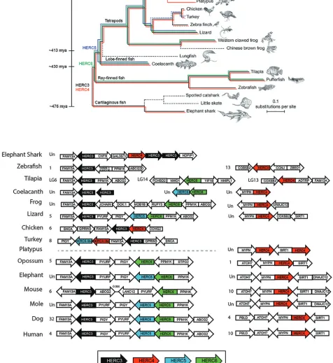
(Fig. 1).
HERC3
and
HERC4
are present in all the
jawed vertebrates examined except the platypus, which appears to be the only
vertebrate that contains two copies of
HERC4. Since the only available assembly for
platypus is considered low coverage, future improvements in the assembly are needed
to help explain this apparently unique composition of the
HERC
family in these
mammals. Chromosomal rearrangement likely occurred sometime after the divergence
of ray-finned fish from cartilaginous fish (
⬃
430 mya), giving rise to two different
chromosomal
HERC
loci in most vertebrates, where the
HERC3-HERC5-HERC6
locus is
flanked by
FAM13A
and
PIGY-PYURF
and
HERC4
by
MYPN
and
SIRT1
(Fig. 1B).
The
HERC
family expanded further after the divergence of cartilaginous fish (
⬃
430
mya), with the emergence of
HERC6, which is present in most jawed vertebrates with
an apparent absence in platypus and some fish (e.g., zebrafish) and bird (e.g., chicken,
turkey, and zebra finch) species (Fig. 1). The last expansion of the
HERC
family occurred
after the divergence of ray-finned fish (
⬃
413 mya), with the likely duplication of
HERC6,
giving rise to
HERC5
(Fig. 1B).
HERC5
is present in the coelacanth, one of the earliest
predecessors of tetrapods, and appears to have been lost in some species of frogs, birds
(e.g., chicken), metatherian (marsupial) mammals (e.g., opossum), and rodents (e.g.,
mouse) (Fig. 1) (43, 45). A partial
HERC5-like gene was identified in turkey, and a partial
HERC4-like gene was identified in turkey and finch, possibly indicating erosion of the
gene family in birds. In species that appear to have lost
HERC
orthologs, we cannot
rule out the possibility that the orthologs are present but were missed due to low
sequence homology and/or incomplete genome annotation. Together, these
find-ings indicate that the small-HERC
family has an ancient marine origin at least 595
mya, before the emergence of jawed vertebrates, and has undergone chromosomal
rearrangement, gene duplication, and potential gene loss events during vertebrate
evolution.
Evolutionarily distant RCC1-like domains and HECT domains are well
con-served.
Phylogenetic analysis of
HERC
sequences showed segregation of the small
HERC
genes into four major clusters consisting of
HERC3,
HERC4,
HERC5, and
HERC6
(Fig.
2A). Most
HERC
orthologs have high sequence homology, ranging from
⬃
70 to 100%
amino acid identity, whereas most
HERC
paralogs have low homology, ranging from
⬃
34 to 57% (see Fig. S1 in the supplemental material). Notably, coelacanth and lizard
HERC5
sequences clustered on their own, showing more sequence similarity to
HERC6
genes than to other
HERC5
genes, possibly indicating that these genes are actually
HERC6
or a hybrid of
HERC5
and
HERC6. Similar tree topologies regarding the main
branching were predicted using several tree-generating algorithms (maximum
likeli-hood, minimal evolution, unweighted pair group method using average linkages
[UPMGA], and neighbor-joining methods). Consistent with the approximate emergence
times of the small-HERC
family members shown in Fig. 1,
HERC4
is the oldest of the
HERC
paralogs, followed by
HERC3,
HERC6, and then
HERC5.
The small HERCs are believed to have arisen from a gene fusion event between an
RCC1-like domain and a HECT domain (46). Although the approximate date of this
event is unknown, the presence of HERC4 in jawless fishes (e.g., lampreys) suggests that
on November 6, 2019 by guest
http://jvi.asm.org/
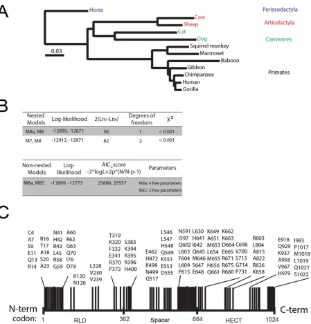
FIG 1Emergence of the small-HERCfamily. (A)HERC3(black),HERC4(red),HERC5(blue), andHERC6(green) sequences were searched in genome assemblies using the UCSC Genome Browser (https://genome.ucsc.edu) and NCBI gene and protein sequence databases. The presence ofHERCorthologs in species is
(Continued on next page)
July 2018 Volume 92 Issue 13 e00528-18 jvi.asm.org 4
on November 6, 2019 by guest
http://jvi.asm.org/
[image:4.585.58.534.170.689.2]the fusion event occurred more than 595 mya. Typically, the primary amino acid
sequences of RCC1-like domains have low sequence homology in the RCC1
superfam-ily; however, their tertiary structures are highly conserved (38, 39). To assess how
conserved the predicted tertiary structures are among the different small-HERC
mem-bers, we generated a phylogenetic tree based on the Q
Hstructural measure derived
from alignment of the predicted tertiary structures of RCC1-like domains (Fig. 2B to F;
see Table S1 in the supplemental material). The models were predicted using 3D-Jigsaw
v2.0 (https://bmm.crick.ac.uk/~populus/) and showed that each of the RCC1-like
do-mains of HERC3 to -6 adopted the characteristic

-propeller structure of the
superfam-ily, despite their low sequence homology (47–51). Alignment of the structures was
carried out using the program Structural Alignment of Multiple Proteins (STAMP), which
is a tool for aligning protein sequences based on their three-dimensional structures
(52). The STAMP algorithm minimizes the C
␣
distance between aligned residues of each
molecule by applying globally optimal rigid-body rotations and translations. STAMP
analysis revealed that the RCC1-like domains of the HERC orthologs are well conserved
overall, with HERC3 and HERC4 being the most conserved (Fig. 2B to F). Several
paralogs showed greater similarity to each other than to their orthologous counterparts
(e.g., lizard HERC4 and human HERC6). Chimpanzee HERC3 differed substantially from
the other HERCs in that it lacks blade 3 of the

-propeller. Notably, the amino-terminal
blade 1 of each RCC1-like domain is the least conserved region and adopts a more
extended conformation than the other blades.
Analysis of the predicted HECT domain structures showed that they all adopted the
typical bilobal structure of HECT domains (Fig. 2G to K). STAMP analysis showed that
the different orthologs are well conserved overall, with HERC3 and HERC4 being the
most conserved. Some HERC5 and HERC6 paralogs shared more similarity to each other
than to their respective orthologs. Notably, coelacanth and mouse HERC6 proteins were
more similar to HERC5 paralogs than to other HERC6 orthologs, potentially indicating
conservation in structure and function of these HECT domains (Fig. 2K). This is
consis-tent with the finding that mouse HERC6 is the functional homolog of human HERC5;
these are the main cellular E3 ligases for ISG15 in mice and humans, respectively.
Together, these data show that despite low sequence homology, evolutionarily
diver-gent
HERC
genes share remarkable similarity in the predicted structures of their
RCC1-like domains and HECT domains.
Human HERC3 to -6 differentially inhibit HIV-1 particle production.
Given the
remarkable similarity in the predicted structures of the HECT and RCC1-like domains,
we asked whether human HERC3, HERC4, and HERC6 inhibited HIV-1 particle
produc-tion like HERC5. To test the effect on HIV-1 replicaproduc-tion of knocking down endogenous
HERC protein levels, we first screened different HERC short hairpin RNA (shRNA)
constructs for the ability to knock down endogenous HERC mRNA and protein levels.
As shown in Fig. 3A, several of the shRNA constructs knocked down HERC mRNA levels
by 2- to 5-fold. For each shRNA construct used, no significant differences in mRNA
levels were detected for any of the other related small HERCs, demonstrating specificity
(Fig. 3B). Unfortunately, we were unable to readily measure endogenous levels of HERC
proteins using several different commercially available antibodies. This could be due to
poor recognition of endogenous HERC proteins by the antibodies and/or tightly
controlled cytosolic levels of HERC proteins, as previously observed (34). As such, HERC
protein knockdown efficiencies of the shRNA constructs were instead determined using
exogenously expressed Flag-tagged HERC constructs (Fig. 3C). ShRNAs that knocked
FIG 1Legend (Continued)
indicated by colored lines. The approximate dates of divergence among the organisms are indicated by the time line on the left from the perspective of humans, as previously described by Hedges et al. (43). The Bayesian tree was obtained from a multiple-sequence alignment of 251 genes with a 1:1 ratio of orthologs in 22 vertebrates, rooted on cartilaginous fish (support was 100% for all clades but armadillo and elephant, with 45%), as described previously (45). The dashed black lines indicate that noHERCorthologs were identified in the species. The dashed red and blue lines indicate the presence of partial HERC4-like andHERC5-like sequences, respectively. (Adapted from reference 45 with permission of the publisher [Springer Nature].) (B) Syntenic relationships of the genomic contexts of the small-HERCloci in evolutionarily diverse vertebrates. Chromosome numbers are indicated on the left of each locus. Un, unplaced scaffold.
on November 6, 2019 by guest
http://jvi.asm.org/
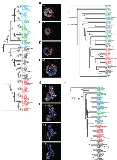
FIG 2(A) Molecular phylogenetic analysis using the maximum likelihood method. The evolutionary history was inferred by using the maximum likelihood method based on the JTT matrix-based model (89). The bootstrap consensus tree, inferred from 100 replicates, was taken to represent the evolutionary
(Continued on next page)
July 2018 Volume 92 Issue 13 e00528-18 jvi.asm.org 6
on November 6, 2019 by guest
http://jvi.asm.org/
[image:6.585.63.527.68.700.2]down Flag-tagged HERC3, HERC4, HERC5, and HERC6 protein levels by 10.1-, 7.2-, 5.0-,
and 3.6-fold, respectively, were used for subsequent experiments.
HOS-CD4-CXCR4 cells were cotransfected with plasmids carrying HERC shRNA and
HIV-1 R9 (a full-length replication-competent NL4-3 derivative). After 72 h of replication
(
⬃
2 or 3 rounds), quantitative Western blot analysis of cell lysates or virions produced
from cells showed that cells knocked down for HERC5 expression exhibited a significant
increase in Gag particle production (
⬃
8-fold), whereas HERC3, -4, and -6 released
modestly more virions into the supernatant than the control cells (
⬃
2-fold) (Fig. 3D).
The amount of infectious HIV-1 in the supernatant was also measured by infecting the
TZM-bl indicator cell line, which enabled quantitative analysis of HIV-1 using luciferase
as a reporter (53–57). Cells knocked down for endogenous HERC5 expression failed to
inhibit production of infectious HIV-1, whereas cells knocked down for HERC3, HERC4,
or HERC6 expression released levels of infectious virions similar to those of the control
cells (Fig. 3E).
To test the effect of increased HERC expression on single-round HIV-1 particle
production, human 293T cells, which do not support multiround replication, were
cotransfected with plasmids carrying HIV-1 (R9) and either empty vector, HERC3, HERC4,
HERC5 or HERC6. As expected, HERC5 potently inhibited HIV-1 particle production (Fig.
3F). HERC3 or HERC4 also significantly inhibited particle production, but not as potently
as HERC5. In contrast, HERC6 modestly inhibited HIV-1 particle production but did not
achieve statistical significance. As expected, HERC5 also potently inhibited the
produc-tion of infectious HIV-1, whereas HERC3, HERC4, and HERC6 modestly inhibited
pro-duction of infectious HIV-1 (Fig. 3G). Each transfected HERC construct exhibited robust
mRNA expression, although HERC3 and HERC4 levels were less than those of HERC5
and HERC6 (Fig. 3H). Taken together, these data show that upregulated expression of
HERC3 to -6 inhibited HIV-1 particle production and replication to varying degrees, with
HERC5 exhibiting the most potent activity and HERC6 the weakest activity. Notably,
only endogenous levels of HERC5 significantly inhibited production of infectious HIV-1.
Human HERC3 to -6 differentially inhibit nuclear export of incompletely spliced
RNA.
We previously showed that human HERC5 blocked nuclear export of
Rev-dependent HIV-1 RNA (29). To determine if HERC3, HERC4, and HERC6 also blocked
nuclear export of Rev-dependent HIV-1 RNAs, we cotransfected 293T cells with
plas-mids carrying full-length HIV-1 R9 and either empty vector, HERC3, HERC4, HERC5, or
HERC6. A plasmid encoding enhanced green fluorescent protein (eGFP) was also
cotransfected to serve as a transfection control. Total RNA was harvested from the total
cell extract or the cytoplasmic extract only and subjected to quantitative PCR (qPCR)
with primers specific for either Gag (unspliced HIV-1 genomic RNA), Rev (fully spliced
RNA),

-actin (loading control), or eGFP. Each of the small HERC proteins exhibited
significant reductions in the amount of HIV-1 genomic RNA present in the cytoplasm,
with HERC5 exhibiting the strongest activity (Fig. 4A). In contrast, no significant
reductions in the export of fully spliced HIV-1 transcripts were observed (Fig. 4B).
To further support this finding, we tested the abilities of the small HERCs to inhibit
Gag expression from Rev-dependent (e.g., GagPol-RRE) and Rev-independent (e.g.,
FIG 2Legend (Continued)
histories of the taxa analyzed. Branches corresponding to partitions reproduced in less than 50% of the bootstrap replicates were collapsed. The percentages of replicate trees in which the associated taxa clustered together in the bootstrap test (100 replicates) are shown next to the branches. Initial trees for the heuristic search were obtained automatically by applying neighbor-joining and BioNJ algorithms to a matrix of pairwise distances estimated using a JTT model and then selecting the topology with a superior log likelihood value. The analysis involved 91 amino acid sequences. All positions containing gaps and missing data were removed. There were a total of 433 positions in the final data set. Evolutionary analyses were conducted in MEGA7 (83). H3, HERC3; H4, HERC4; H5, HERC5; H6 HERC6. (B to F) Evolutionary conservation of HERC RCC1-like domains. (B to E) Predicted structures of the RCC1-like domains were generated using 3D-Jigsaw (v2.0). Multiple structural alignments were generated based on the QHstructural measure using the program STAMP, a plug-in in the MultiSeq interface of the Visual Molecular Dynamics (VMD) software (v1.9.2). (F) Three-dimensional representation of the structural data colored by structural conservation. Each amino acid is colored according to the degree of conservation within the alignment: blue, highly conserved; white, somewhat conserved; and red, very low or no conservation. The structure-based cladogram was derived from sequence and structural alignments of the predicted tertiary structures of multiple mammalian HERC RCC1-like domains. (G to K) Evolutionary conservation of HERC HECT domains. (G to J) Three-dimensional representations of the predicted HECT domain structural data colored by structural conservation. (K) Structure-based cladogram derived from sequence and structural alignments of the predicted tertiary structures of multiple mammalian HERC HECT domains. See Table S1 in the supplemental material for the parent structure scaffolds used to generate all the models.
on November 6, 2019 by guest
http://jvi.asm.org/
GagPol-4
⫻
CTE) constructs, as previously described (29, 58). HIV-1 Rev promotes nuclear
export of incompletely spliced HIV-1 mRNAs by binding to a specific
cis-acting element
called the Rev-response element (RRE) located within an HIV-1 intron (Fig. 4C). HIV-1
mRNA containing four copies of the Mason-Pfizer monkey virus constitutive export
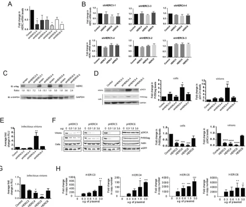
FIG 3HERC3 to -6 differentially restrict HIV-1 particle production. (A and B) HOS-CD4-CXCR4 cells were transfected with a plasmid carrying either scrambled shRNA (control) or shRNAs to each of the different HERCs independently. HERC mRNA levels were measured by qPCR using HERC-specific primers. The average fold changes in HERC mRNA levels (with standard errors of the mean [SEM]) from the results of 3 independent experiments are shown. One-way analysis of variance (ANOVA) and Dunnett’s multiple-comparison test with the control were performed. (C) 293T cells were first transfected with plasmids carrying HERC shRNA and 24 h later with the respective Flag-tagged HERC constructs. HERC protein levels were measured by quantitative Western blotting (immunoblotting [IB]) using anti (␣)-Flag or anti-GAPDH (loading control). The numbers below the blots represent the average fold changes in HERC protein levels compared to the control cells after densitometric quantification of the Flag-tagged HERC bands. scram, scrambled shRNA. (D) HOS-CD4-CXCR4 cells were transfected with pR9 and plasmids carrying either scrambled shRNA (control) or shRNAs to each of the different HERCs independently. Seventy-two hours after transfection, cell lysates and HIV-1 virions in the supernatant were harvested. HIV-1 particle production was measured by quantitative Western blotting using anti-p24CA or anti-GAPDH. (E) Average (plus SEM) densitometric quantifications of Pr55Gag (cells) and p24CA (virions) bands. Virions produced from the cells were used to infect the luciferase reporter cell line TZM-bl to measure the number of infectious virions. The average fold changes in relative light units (RLU) from the results of at least 3 independent experiments are shown. (F and G) 293T cells were cotransfected with pR9 and peGFP (transfection control) and increasing amounts of either empty vector, pHERC3, pHERC4, pHERC5, or pHERC6. Virions in the supernatant and total cell lysates were subjected to quantitative Western blot analysis using anti-p24CA, anti-eGFP, and anti--actin as a loading control. The average (plus SEM) densitometric quantification of p24CA and Pr55Gag levels from Western blot images of virions or cell lysate (3.6g HERC lane) are shown on the right. The values were normalized to-actin and eGFP levels. (G) Virions produced from cells were used to infect the luciferase reporter cell line TZM-bl to measure the number of infectious virions. (H) 293T cells were transfected with either empty vector or increasing amounts of plasmids encoding the different HERCs. Twenty hours posttransfection, total RNA was harvested from the cells and subjected to qPCR to measure HERC mRNA levels. Relative fold changes in HERC mRNA compared to the control cells are shown. Statistical significance was determined using one-way ANOVA and Dunnett’s multiple-comparison test with the control cells.*,P⬍0.05;**,P⬍0.01;***,P⬍0.001;****,P⬍0.0001.
July 2018 Volume 92 Issue 13 e00528-18 jvi.asm.org 8
on November 6, 2019 by guest
http://jvi.asm.org/
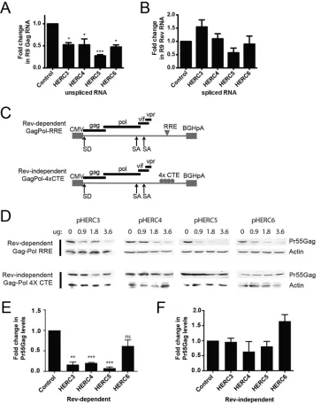
[image:8.585.47.540.72.486.2]FIG 4HERC3 to -5 inhibit cytoplasmic accumulation of unspliced HIV-1 RNA. (A and B) 293T cells were cotransfected with pR9 and peGFP (transfection control) and either empty vector, pHERC3, pHERC4, or pHERC5. Forty-eight hours after transfection, total RNA was extracted and reverse transcribed into cDNA from whole-cell lysates or from the cytoplasmic fraction only. Quantitative PCR was performed on each fraction using primers specific for unspliced HIV-1 genomic RNA (e.g., Gag), fully spliced RNA (e.g., Rev), -actin (loading control), or eGFP (transfection control). The proportion of unspliced or fully spliced HIV-1 RNA in the cytoplasmic fraction compared to the total amount of HIV-1 RNA (nuclear plus cytoplasmic) was determined for control cells and cells expressing HERC after normalization to-actin and eGFP levels. The fold changes in copy numbers relative to control cells are shown. The data shown represent the averages (plus SEM) from the results of four independent experiments. (C) Schematic depicting the different Gag-Pol constructs used in the experiment shown in panel D. CMV, cytomegalovirus; SA, splice acceptor; SD, splice donor. (D) 293T cells were cotransfected with increasing amounts of plasmids encoding HERC3, HERC4, or HERC5 and either Rev-dependent GagPol-RRE (plus pRev) or Rev-independent GagPol-4⫻CTE. The total DNA transfected was kept equal with the empty-vector plasmid. Gag levels within the cell lysates were analyzed by quantitative Western blotting using anti-p24CA and anti--actin as a loading control. (E and F) Densitometric quantification of Pr55Gag bands from the lanes containing the largest amount of each HERC in the Western blots from panel D was performed. Shown are the average fold changes (plus SEM) in Pr55Gag levels relative to the empty-vector control after normalization to -actin levels. Statistical significance was determined using ANOVA with Dunnett’s multiple-comparison test.*,P⬍0.05;**,P⬍0.01,***,P⬍0.001; ns, not significant.
on November 6, 2019 by guest
http://jvi.asm.org/
[image:9.585.43.406.73.529.2]element (4
⫻
CTE) in place of the RRE is not dependent on Rev for nuclear export and
thus serves as a Rev-independent control (59). Successful export of incompletely spliced
RNA can be assessed by Western blotting for Gag protein expression. 293T cells were
cotransfected with a plasmid encoding Rev and increasing concentrations of plasmids
encoding HERC, with or without pGagPol-RRE or pGagPol-4
⫻
CTE. As shown in Fig. 4D
and E, each of the small HERCs differentially inhibited nuclear export of Rev-dependent
RNA, where HERC5 exhibited the highest level of inhibition and HERC6 the weakest
inhibition. In contrast, none of the HERCs significantly inhibited nuclear export of
Rev-independent RNA (Fig. 4D and F). Together, these findings indicate that the
small-HERC members differentially inhibit nuclear export of Rev-dependent RNAs.
Antiviral activity of HERC5 evolved more than 413 million years ago.
We next
asked whether the antiretroviral activity of human HERC5 has an evolutionarily ancient
origin. Since the coelacanth was the oldest vertebrate in which we identified
HERC5, we
tested the ability of coelacanth HERC5 to inhibit HIV-1 virus production. To assess
potential virus-specific effects, we also tested the antiviral activity toward another,
related nonhuman retrovirus, simian immunodeficiency virus (SIV) (SIVmac239, a
full-length rhesus macaque derivative lacking the 5
=
long terminal repeat [LTR]), which is
thought to be at least 32,000 years older than HIV-1 (60). For comparison, we included
human HERC5 and human HERC6, which exhibited the strongest and weakest
anti-HIV-1 activities, respectively. Human 293T cells were cotransfected with plasmids
carrying SIVmac239 or HIV-1 R9 and increasing concentrations of either empty vector,
coelacanth HERC5, human HERC5, or human HERC6. Forty-eight hours after
transfec-tion, virus released into the supernatant was measured by Western blotting. As
expected, human HERC5 exhibited strong inhibition, whereas both coelacanth HERC5
and human HERC6 exhibited little inhibition of HIV-1 (Fig. 5A to C). In contrast, each of
the HERCs inhibited SIVmac239 virus production, with human HERC5 being the most
potent (Fig. 5D to F). Together, these results demonstrate that the antiretroviral activity
of HERC5 has an ancient marine origin at least 413 mya and that HERC5 and HERC6
exhibit species- and virus-specific antiviral activity.
HERC6 is evolving under positive selection.
We previously showed that HERC5,
especially blade 1 of its RCC1-like domain, has been evolving under positive selection
for
⬎
100 million years (29). We performed a similar analysis for each of the small-HERC
members to determine if the other members of the small-HERC family have been
evolving under positive selection. HERC evolution in mammals was evaluated under
several standard models of sequence evolution using the Server for the Identification
of Site-Specific Positive Selection and Purifying Selection (Selecton) program (61–64).
This comprised two nested pairs of models (M8a and M8; M7 and M8), in which the first
model of each pair is nested in the second model. The M8 model, but not the M8a or
M7 model, allows sites to evolve under positive selection. A nonnested-pair (M8a and
MEC) model comparison was also performed. The MEC model differs from the other
models in that it takes into account the differences between amino acid replacement
rates (61). The nested models were compared using the likelihood ratio test.
Analysis of 12 evolutionarily diverse HERC sequences using Selecton revealed that
HERC6, but not HERC3 and HERC4, is evolving under positive selection (Fig. 6; see Table
S2 in the supplemental material). Allowing sites to evolve under positive selection (M8)
gave a significantly better fit to the HERC6 sequence data than the corresponding
model without positive selection (M8a and M7) (Fig. 6B). The MEC model, which allows
positive selection, was compared with the M8a null model, which does not allow
positive selection. Comparison of the Akaike Information Criterion (AICc) scores (M8a,
25,806; MEC, 25,557) revealed that the MEC model fits the HERC6 data better than the
M8a model. The results of the MEC analysis were projected onto the primary sequence
of human HERC6 (Fig. 6C). Notably,
⬃
23% (23 of 102) of the codons cluster within the
first 80 amino acids of the amino terminus of the RCC1-like domain, encompassing
blade 1 of its predicted

-propeller structure. Another
⬃
32% (33 of 102) cluster at the
carboxyl terminus of the spacer region (amino acids
⬃
630 to 680). These results show
July 2018 Volume 92 Issue 13 e00528-18 jvi.asm.org 10
on November 6, 2019 by guest
http://jvi.asm.org/
that strong positive selection is operating on HERC6, with a large number of codons in
blade 1 of the RCC1-like domain and the carboxyl terminus of the spacer region
evolving under positive selection.
Blade 1 of the RCC1-like domain of human HERC6 is an important determinant
of anti-HIV-1 activity.
Given the evolutionary similarities between human HERC5 and
human HERC6, we asked why they differed in their antiviral activities. Since we
previously showed that blade 1 of HERC5 is required for its anti-HIV-1 activity and that
blades 1 of both HERC5 and HERC6 contain numerous residues evolving under positive
selection, we asked if blade 1 of HERC5 can confer antiviral activity on HERC6. We
replaced either the entire RCC1-like domain (H6:H5RLD) or blade 1 (H6:H5blade1) from
human HERC5 with the corresponding region in human HERC6. We then measured the
abilities of these HERC6 mutants to inhibit HIV-1 particle production. As shown in Fig.
7A, the H6:H5RLD and H6:H5blade1 mutants potently inhibited HIV-1 particle
produc-tion similarly to wild-type HERC5. This inhibiproduc-tion occurred despite levels of H6:H5RLD
and H6:H5blade1 protein expression slightly lower than that of wild-type HERC5 (Fig.
7B). This result indicates that blade 1 is an important determinant of antiviral activity.
DISCUSSION
We showed here that the small-HERC
family has an ancient marine origin, where
HERC4
emerged at least 595 mya and expansion of the family occurred sometime after
FIG 5Coelacanth HERC5 restricts SIV, but not HIV-1, particle production. 293T cells were cotransfected with a plasmid carrying HIV-1 (pR9) (A to C) or SIV (pSIVmac239) (D to F) and increasing amounts of plasmids encoding either coelacanth HERC5, human HERC5, or human HERC6. Forty-eight hours posttransfection, virus released into the supernatant was measured by quantitative Western blotting of Gag proteins using monoclonal anti-p24CA (183-H12-5C) or anti-SIVp17 (KK59). The average relative fold changes (plus SEM) in HIV p24CA or SIV p24CA protein levels compared to the control cells after densitometric quantification of 3 independent Western blot images are shown. Statistical significance was determined using one-way ANOVA with Dunnett’s multiple-comparison test with the control cells.**,P⬍0.01;***,P⬍0.001;****,P⬍0.0001.
on November 6, 2019 by guest
http://jvi.asm.org/
[image:11.585.46.536.70.432.2]the divergence of jawed vertebrates from jawless vertebrates (
⬃
476 to 595 mya).
Elephant sharks are among the oldest and most slowly evolving jawed vertebrates and
have accumulated a small number of chromosomal rearrangements (44). Thus, analysis
of their genome allows us to gain insight into the early evolution and expansion of
gene families. The presence of a single copy of
HERC4
and multiple copies of
HERC3
in
FIG 6HERC6is evolving under strong positive evolutionary selection. (A) Neighbor-joining phylogenetic tree for progressive alignment of 12 differentHERC6species using COBALT for multiple protein sequences. The branch lengths are proportional to the amount of inferred evolutionary change. (B) Analysis for positive selection was performed using HERC6 amino acid sequences from human (Homo sapiens), chimpanzee (Pan troglodytes), gorilla (Gorilla gorilla gorilla), marmoset (Callithrix jacchus), baboon (Papio anubis), squirrel monkey (Saimiri boliviensis), gibbon (Nomascus leucogenys), horse (Equus caballus), sheep (Ovis aries), cow (Bos taurus), dog (Canis lupus familiaris), and cat (Felis catus). Evolutionary analysis for positive selection inHERC6used various models of evolution, where M8 and MEC allowed sites to evolve under positive selection and M7 and M8a did not. L represents the likelihood of the model given the data, p represents the number of free parameters, and N represents the sequence length. The lower the AICCscore, the better the fit of the model to the data, and hence, the model is considered more justified. (C) Schematic showing the results of a Bayesian analysis approach identifying positively selected sites with a ratio of the number of nonsynonymous substitutions per nonsynonymous site (Ka) to the number of synonymous substitutions per synonymous site (Ks) (Ka/Ks) with a value of⬎1.5 and a 95% confidence interval larger than 1 and therefore considered statistically significant. TheHERC6reference sequence accession number is
NM_017912.3.
July 2018 Volume 92 Issue 13 e00528-18 jvi.asm.org 12
on November 6, 2019 by guest
http://jvi.asm.org/
[image:12.585.48.497.68.537.2]the elephant shark likely represents an early time in vertebrate evolution when the
HERC4
ancestral gene duplicated and evolved into
HERC3. Although the evolutionary
pressures in vertebrates triggering expansion of the small-HERC
family are unknown, it
is plausible, given their antiretroviral activity, that this expansion involved retroviruses.
Endogenous retroviruses (ERVs) comprise a substantial portion of vertebrate genomes
and appear to have an ancient marine origin, with evidence of ERV sequences found in
the genomes of elephant shark, coelacanth, and possibly lamprey (65–68). A recent
panvertebrate comparative genomic analysis showed that retroviruses have an
unprec-edented capacity for rampant host switching among distantly related vertebrates,
undoubtedly exerting substantial evolutionary pressure on their hosts (68). Pressures
like these can trigger antiviral gene duplication and neofunctionalization events in the
hosts, allowing them to evolve more rapidly in order to maintain evolutionary
domi-nance over viruses. Several examples where gene duplication/neofunctionalization has
given rise to restriction factor families in primates are
MX1,
IFITM,
TRIM5, and
APOBEC3
(5, 6, 69–74). These genes, including
HERC5
and
HERC6, exhibit strong signatures of
positive selection, which is consistent with repeated exposure to such evolutionary
pressures.
An interesting feature of the small-HERC family is the highly conserved topology of
the RCC1-like domain, despite limited sequence homology. By allowing numerous
amino acid substitutions while maintaining the overall protein configuration and
antiviral activity, these HERC proteins may be able to interfere with the binding of
diverse viral antagonists. Evidence of such an evolutionary battle lies in the strong
signatures of positive selection in both human HERC5 and HERC6, especially blades 1
of their RCC1-like domains, which we have shown to be important determinants of
antiviral activity. HERCs are not the only restriction factors likely to have played an
important antiviral role early in vertebrate evolution. BST2/tetherin has also been
shown to have an ancient marine origin, emerging
⬎
450 mya, before the separation of
cartilaginous fish from bony vertebrates (2). Like the HERC family, the general topology
of BST2/tetherin orthologs are also highly conserved despite low sequence homology,
and it is the overall protein configuration that is important for its antiviral activity (2, 75,
76). This evolutionary strategy may help BST2/tetherin and HERC proteins maintain
evolutionary dominance over viruses.
We observed that some small
HERC
genes have been lost in some species, most
notably birds and rodents. For birds, this is not too surprising, given that their genomes
have been subjected to lineage-specific erosion of repetitive elements, large segmental
deletions, and gene loss (
⬎
1,000 genes), resulting in a smaller repertoire of immune
genes than in humans (77, 78). Although
HERC5
and
HERC6
are missing in most bird
FIG 7Blade 1 of the human HERC5 RCC1-like domain confers potent antiviral activity on human HERC6. (A) 293T cells were cotransfected with pR9, peGFP, and either empty vector, pHERC5, pHERC6, pH 6:H5RLD, or pH6:H5blade1. Forty-eight hours posttransfection, virions in the supernatant and total cell lysates were subjected to quantitative Western blot analysis using anti-p24CA or anti--actin. Densitometric quantification of p24CA (virions) and Pr55Gag (cells) from the results of three independent experiments is shown beside the blots after normalization to the-actin loading controls. The error bars indicate SEM.***, P⬍ 0.001;****, P⬍ 0.0001. (B) Cell lysates from panel A were subjected to Western blot analysis using anti-Flag, anti-eGFP, or anti-GAPDH. Average densitometric quantification of the HERC bands is shown below the blot after normalization to anti-eGFP or anti-GAPDH from the results of three independent experiments.
on November 6, 2019 by guest
http://jvi.asm.org/
[image:13.585.51.540.73.197.2]species,
HERC3
and/or
HERC4
is present. Given their modest antiretroviral activity in
humans, it will be interesting to learn if HERC3 and HERC4 play an antiviral role in birds,
perhaps compensating for the loss of
HERC5
and
HERC6. Other potent antiviral
restric-tion factor genes, such as
BST2/tetherin,
TRIM22,
TRIM5, and
APOBEC3G, are also notably
absent from birds; however, they do possess other members of the
BST,
TRIM, and
APOBEC
families, whose antiviral activities remain largely uncharacterized in birds.
Rodents also have
HERC3
and
HERC4
but also possess at least one of the
HERC5
and
HERC6
genes. For example, mice, rats, and hamsters possess
HERC6
but lack
HERC5,
ground squirrels possess
HERC5
but lack
HERC6, and guinea pigs possess both
HERC5
and
HERC6. Since no rodent lacks both
HERC5
and
HERC6, it is likely that one of these
genes has assumed the role of the main cellular E3 ligase for ISG15 in the absence of
the other, potentially adding a new antiviral defense for rodents. For instance, this is the
case in mice, which possess only
HERC6, which encodes the main cellular E3 ligase for
ISG15 and serves as a critical antiviral defense mechanism in mice (79, 80). Our
phylogenetic analysis, where we showed that the predicted structures of the HECT
domains of mouse HERC6 and human HERC5 (the main cellular E3 ligase for ISG15 in
humans) share a high degree of similarity, also supports this finding. One possibility for
the loss of
HERC5
or
HERC6
in rodents is that retroviruses or other viral pathogens have
not provided constant selective pressure to maintain both genes in these species. A
similar dynamic history of gene expansion and loss is evident for other restriction
factors, such as
TRIM22,
TRIM5, and
BST2/tetherin
(2, 7, 81).
As genes duplicate, neofunctionalize, and diverge in response to evolutionary
pressures, reliance on the activities of the ancestral genes may diminish, or they may be
replaced altogether by their more advantageous descendants (reviewed in reference
82). Therefore, it is not surprising that the human small-HERC family exhibits differential
antiviral activity. Despite still possessing antiviral activity when overexpressed
in vitro,
it is unlikely that HERC3 and HERC4 play significant biological roles as antiviral proteins
in humans, since they are not IFN induced, nor have they been evolving under positive
selection. This role was likely assumed by HERC5 and HERC6 after the divergence of
ray-finned fish from cartilaginous fish (
⬃
430 mya). However,
HERC3
and
HERC4
do
exhibit differential tissue-specific expression (reviewed in reference 37). Therefore, it is
possible that HERC3 and HERC4 play more dominant antiviral roles in tissues where
their basal expression is already much more elevated, such as in the brain, heart, and
stomach for HERC3 and the brain, lung, and testis for HERC4. Moreover, species such as
elephant shark, coelacanth, and platypus that contain duplicated copies of
HERC3
or
HERC4
genes may also express higher levels of these HERC proteins due to increased
gene copy numbers. Although we did not test the antiviral activities of other
evolu-tionarily diverse HERC3 or HERC4 proteins, our phylogenetic analyses demonstrated
that the HECT and RCC1-like domains of these proteins show remarkable structural
similarity to their human counterparts, which do exhibit antiviral activity at elevated
levels. It will be interesting to determine if the antiviral function of HERC3 and HERC4
is conserved in these ancient vertebrates, before the emergence of HERC5 and HERC6.
Interactions between host antiviral proteins and their viral-protein targets can be a
critical requirement for their antiviral activity. When viral proteins mutate to evade such
interactions, the antiviral protein frequently develops rapid amino acid replacements at
the protein-protein interface in an attempt to restore those interactions and maintain
evolutionary dominance over the virus. HERC5 is known to interact with evolutionarily
diverse viral proteins and, like HERC6, is evolving under strong positive selection (11,
28–30, 32, 35, 36). Therefore, it is highly likely that these proteins contain one or more
protein-protein interaction interfaces between viral and host proteins. Our findings that
blades 1 of the RCC1-like domains of HERC5 and HERC6 contain numerous positively
selected residues and that these residues are important determinants of antiretroviral
activity indicate that blade 1 is likely one such interface. Although there is currently no
evidence that retroviruses have driven positive selection of blade 1, it is interesting that
blade 1 of HERC5 is sufficient to confer antiretroviral activity on HERC6. It is possible
that the topology of blade 1 from HERC5 is such that it promotes interaction with a
July 2018 Volume 92 Issue 13 e00528-18 jvi.asm.org 14
on November 6, 2019 by guest
http://jvi.asm.org/
cellular protein required for activity that blade 1 of HERC6 prevents. However, our
finding that wild-type HERC6 potently inhibited SIVmac239, but not HIV-1, in the same
cell type suggests that virus-specific differences are more likely to account for the
observed differential antiviral activity between HERC5 and HERC6. Additional
structure-function studies are needed to differentiate between these possibilities and others.
In conclusion, the small-HERC family has an evolutionarily ancient origin more than
595 mya, with the latest expansion of the family occurring more than 413 mya. We
showed that the structural topologies of the HECT and RCC1-like domains are highly
conserved despite low sequence homology and that the antiretroviral activity of HERC5
has an ancient marine origin. HERC5 and HERC6 are evolving under strong positive
selection, and a patch of positively selected residues in blade 1 of the RCC1-like domain
is a strong determinant of antiviral activity. Altogether, our study highlights the
potential importance of the HERC family in intrinsic immunity.
MATERIALS AND METHODS
Cell lines.293T cells were obtained from the American Type Culture Collection. HOS-CD4-CXCR4 and TZM-bl cells were from the NIH AIDS Reagent Program. The cells were maintained in standard growth medium (Dulbecco’s modified Eagle’s medium [DMEM]) supplemented with 10% heat-inactivated fetal bovine serum (FBS), 100 U/ml penicillin, and 100g/ml streptomycin at 37°C with 5% CO2.
Analyses of sequences and synteny.The MEGA 7.0 package was used for phylogenetic analysis (83). The amino-terminal end of the small HERCs varies in length among the different members and was not included for phylogenetic analysis. The first 30 amino acids were omitted from HERC3 and HERC4 and the first 23 amino acids from HERC5 and HERC6. The accession numbers used were as follows: for HERC3, Homo sapiens(human) (NP_055421.1),Gorilla gorilla gorilla(gorilla) (XP_004039158.1),Pan troglodytes (chimpanzee) (XP_517337.3),Nomascus leucogenys(gibbon) (XP_003265945.1),Papio anubis(baboon) (XP_003898995.1),Saimiri boliviensis boliviensis(squirrel monkey) (XP_003924058.1),Callithrix jacchus (marmoset) (XP_002745644.1),Bos taurus(cow) (NP_001077132.1),Ovis aries(sheep) (XP_004009758.1), Felis catus(cat) (XP_003985228.1),Canis lupus familiaris(dog) (XP_535653.3),Ailuropoda melanoleuca (giant panda) (XP_002913643.1),Equus caballus(horse) (XP_001496703.3),Callorhinchus_milii(elephant shark) (XM_007902532.1), Danio rerio (zebrafish) (NM_001145624.1), Takifugu rubripes (pufferfish) (XM_011612933.1),Oreochromis niloticus(tilapia) (XM_005456948.2),Latimeria chalumnae(coelacanth) (XM_006005071.2), Xenopus tropicalis (frog) (XM_002938630.3), Anolis carolinensis (lizard) (XM_008111037.2), Geospiza fortis (finch) (XM_005418124.1), Meleagris gallopavo (turkey) (XM_003205471.2), Gallus gallus (chicken) (XM_015276202.1), Monodelphis domestica (opossum) (XM_007495699.2), Mus musculus (mouse) (NM_028705.3), and Dasypus novemcinctus (armadillo) (XM_004480548.2); for HERC4, human (NP_071362.1), gorilla (XP_004049535.1), chimpanzee (XP_001167753.1), gibbon (XP_003258260.1), baboon (XP_003903924.1), squirrel (XP_003928728.1), marmoset (XP_002756356.1), cow (NP_001070362.2), sheep (XP_004021455.1), dog (XP_849808.1), panda (XP_002913788.1), horse (XP_001503636.1), elephant shark (XM_007897346.1), zebrafish (XM_005173035.3), pufferfish (XM_011602912.1), tilapia (XM_005473848.2), coelacanth (XM_005992449.2), frog (NM_001128650.1), lizard (XM_008115175.2), finch (XM_005428501.2), turkey (XM_010714265.1), chicken (XM_015278711.1),Ornithorhynchus anatinus(platypus) (XM_007667149.1), opossum (XM_016421882.1), mouse (NM_030114.2), armadillo (XM_012529817.1), and Petromyzon marinus (lamprey) ENSPMAG00000001626; for HERC5, human (NP_057407.2), chimpanzee (XP_003310459.1), gorilla (XP_004039179.1), marmoset (XP_002745648.1), baboon (XP_003898997.1), squirrel monkey (XP_003924055.1), gibbon (XP_003265940.1), horse (XP_001915115.2), giant panda (XP_002913645.1), sheep (XP_004009762.1), cow (NP_001095465.1), dog (XP_535652.3), cat (XP_003985249.1), coelacanth (XM_014498805.1), lizard (XM_008111035.2), and armadillo (XM_012520464.1); for HERC6, human (NP_060382.3), gorilla (XP_004039178.1), chimpanzee (XP_001160851.1), gibbon (XP_003265938.1), baboon (XP_003899001.1), squirrel (XP_003924053.1), mar-moset (XP_002745681.1), cow (NP_001179573.1), sheep (XP_004010096.1), cat (XP_003985250.1), dog (XP_851549.1), horse (XP_001494887.1), pufferfish (XM_011618146.1), tilapia (XM_005474674.1), coela-canth (XM_014498807.1), frog (XM_002938624.2), lizard (XM_008111034.2), opossum (XM_007495704.1), mouse (NM_025992.2), and armadillo (XM_012520459.1).
Synteny.Synteny maps were derived using the following reference assemblies:Callorhinchus milii 6.1.3 GCF_000165045.1 (elephant shark), Danio rerio GRCz10 GCF_000002035.5 (zebrafish), Takifugu rubripes FUGU5 GCF_000180615.1 (pufferfish), Oreochromis niloticus Orenil1.1 GCF_000188235.2 (tilapia), Latimeria chalumnaeLatCha1 GCF_000225785.1 (coelacanth), Xenopus tropicalis Xtropicalis_v7 GCF_000004195.2 (frog),Anolis carolinensisAnoCar2.0 GCF_0000090745.1 (lizard), Geospiza fortis GeoFor_1.0 GCF_000277835.1 (finch), Meleagris gallopavo Turkey_5.0 GCF_000146605.2 (Turkey),Gallus_gallus-5.0 GCF_000002315.4 (chicken),Ornithorhynchus_anatinus -5.0.1 GCF_000002275.2 (platypus),Monodelphis domesticaMonDom5 GCF_000002295.2 (opossum), Dasypus novemcinctus Dasnovv3.0 GCF_000208655.1 (armadillo), Loxodonta africana Loxafr3.0 GCF_000001905.1 (elephant),Mus musculusGRCm38.p3 GCF_000001635.23 (mouse),Rattus norveg-ivcusRnor_6.0 GCF_000001895.5 (rat),Elephantulus edwardiiEleEdw1.0 GCF_000299155.1 (shrew), Chrysochloris asiaticaChrAsi1.0 GCF_000296735.1 (cape golden mole),Canis lupus familiaris Can-Fam3.1 GCF_000002285.3 (dog), andHomo sapiensGRCh38.p2 GCF_000001405.28 (human).
on November 6, 2019 by guest
http://jvi.asm.org/
Positive selection.Positive-selection analysis was performed as previously described (29). HERC sequences were aligned, and a phylogenetic tree was generated using COBALT (constraint-based alignment tool) (http://www.ncbi.nlm.nih.gov/tools/cobalt/) (84). The following HERC sequences were obtained from GenBank: for HERC3,Homo sapiens(human) (NP_055421.1),Gorilla gorilla gorilla(gorilla) (XP_004039158.1), Pan troglodytes (chimpanzee) (XP_517337.3), Nomascus leucogenys (gibbon) (XP_003265945.1),Papio anubis(baboon) (XP_003898995.1),Saimiri boliviensis boliviensis(squirrel monkey) (XP_003924058.1),Callithrix jacchus(marmoset) (XP_002745644.1),Bos taurus(cow) (NP_001077132.1), Ovis aries (sheep) (XP_004009758.1), Felis catus(cat) (XP_003985228.1),Canis lupus familiaris (dog) (XP_535653.3), Ailuropoda melanoleuca(giant panda) (XP_002913643.1), and Equus caballus(horse) (XP_001496703.3); for HERC4, human (NP_071362.1), gorilla (XP_004049535.1), chimpanzee (XP_001167753.1), gibbon (XP_003258260.1), baboon (XP_003903924.1), squirrel (XP_003928728.1), mar-moset (XP_002756356.1), cow (NP_001070362.2), sheep (XP_004021455.1), dog (XP_849808.1), panda (XP_002913788.1), and horse (XP_001503636.1); for HERC5, human (NP_057407.2), chimpanzee (XP_003310459.1), gorilla (XP_004039179.1), marmoset (XP_002745648.1), baboon (XP_003898997.1), squirrel monkey (XP_003924055.1), gibbon (XP_003265940.1), horse (XP_001915115.2), giant panda (XP_002913645.1), sheep (XP_004009762.1), cow (NP_001095465.1), dog (XP_535652.3), and cat (XP_003985249.1); for HERC6, human (NP_060382.3), gorilla (XP_004039178.1), chimpanzee (XP_001160851.1), gibbon (XP_003265938.1), baboon (XP_003899001.1), squirrel (XP_003924053.1), mar-moset (XP_002745681.1), cow (NP_001179573.1), sheep (XP_004010096.1), cat (XP_003985250.1), dog (XP_851549.1), and horse (XP_001494887.1). At least 2 independent sequences were available for human, sheep, baboon, marmoset, gibbon, and squirrel monkey. The following sequences were not indepen-dently validated: cat, dog, cow, horse, sheep, and giant panda. The identification of site-specific positive selection and purifying selection was generated using the Selecton server (http://selecton.tau.ac.il/index .html). The HERC5 phylogenetic tree was used in the Selecton analysis. Nested pairs of models (M8a and M8; M7 and M8) and a nonnested pair (M8a and MEC) were compared using the likelihood ratio test implemented in the Selecton program.
Plasmids, transfections, antibodies, and Western blotting. (i) Plasmids. Plasmids encoding Flag-tagged HERC3, HERC4, HERC5, and HERC6 were created by first PCR amplifying the various HERC coding regions from their respective templates (as described above) using the following primers: for HERC3, forward, 5=ACG TGA ATT CCA TGT TAT GTT GGG GAT ATT GG 3=, and reverse, 5=ACG TGG TAC CTC AGG CCA AAC TAA ACC CTT CAT AGT TGT C 3=; for HERC4, forward, 5=ACG TGA ATT CTA TGT TGT GCT GGG GAA ATG C 3=, and reverse, 5=ACG TTC TAG ATT ATA TTA AAC TGA AGC CTT CAT TGT G 3=; for HERC5, forward, 5=AAT CGA GAT CTT ATG GAG CGC CGC AGC 3=, and reverse, 5=TAT GCG GAT CCT CAG CCA AAT CCT CTG 3=; and for HERC6, forward, 5=AGA TAA GAT CTT ATG TAC TTC TGT TGG GGC 3=, and reverse, 5=TAG GAG ATA TCT TAT GAC TGT GTG AGC ATG 3=. The amplified products were then cloned into p3xFLAG-CMV10 using the following restriction enzymes: for HERC3, EcoRI and KpnI; for HERC4, EcoRI and XbaI; for HERC5, BglII and BamHI; and for HERC6, BglIII and EcoRV. The resulting plasmids were named pHERC3, pHERC4, pHERC5, and pHERC6.
To generate pH 6:H5RLD, the HERC5 RCC1-like domain was PCR amplified from pHERC5 using the following primers: forward, 5=GGA TGA CGA TGA CAA GAT GGA GCG CCG CAG CC 3=, and reverse, 5=TAT GTT CCA GCA AAA ATT ATT AAC TCC TTT TCT GAG GTA TGG CTT TCA AG 3=. The backbone of pHERC6 was PCR amplified using the following primers: forward, 5=TTT TTG CTG GAA CAT ATG CCA ACT TTG 3=, and reverse, 5=CTT GTC ATC GTC ATC CTT GTA ATC GAT G 3=. The two amplified fragments were cloned using the fast cloning technique (85).
To generate pH6:H5blade1, blade 1 of HERC5 (amino acids 1 to 100) was PCR amplified from pHERC5 using the following primers: forward, 5=GGA TGA CGA TGA CAA GAT GGA GCG CCG CAG CCG CCC CAA CAG AAG TAC ATC TTG TCA TCG TCA TCC TTG TAA TCG ATG 3=, and reverse, 5=GCT CCT TCC CGC AGC TCA CGT GGA TCT TCA TGT TCT TGC CCA GC 3=. The backbone of pHERC6 was PCR amplified using the following primers: forward, 5=GCT GGG CAA GAA CAT GAA GAT CCA CAG CTG CGG GAA GGA GCA C 3=, and reverse, 5=GGC TGC GGC GCT CCA TCT TGT CAT CGT CAT CCT TGT AAT CGA TG 3=. The two amplified fragments were cloned using the Gibson cloning technique per the manufacturer’s instructions (New England Biolabs).
To generate pH6:R10A, site-directed mutagenesis was performed on pHERC6 using the following primers: forward, 5=TTC TGT TGG GGC GCC GAC TCC GCG GAG CTG CAG CGC CGG AGG 3=, and reverse, 5=CCT CCG GCG CTG CAG CTC CGC GGA GTC GGC GCC CCA ACA GAA 3=. pH6:E67A was generated similarly using the following primers: forward, 5=GCA GCG CGG GGA GCT GCC AGC ACC AAT TCA GGC ATT GGA AAC C 3=, and reverse, 5=GGT TTC CAA TGC CTG AAT TGG TGC TGG CAG CTC CCC GCG CTG C 3=. pH6:R10A/E67A was generated similarly, except pH6:R10A was used as the template with the following primers: forward, 5=GCA GCG CGG GGA GCT GCC AGC ACC AAT TCA GGC ATT GGA AAC C 3=, and reverse, 5=GGT TTC CAA TGC CTG AAT TGG TGC TGG CAG CTC CCC GCG CTG C 3=. pR10G was generated similarly, using the following primers: forward, 5=CGC CGA CTC CGG GGA GCT GCA 3=, and reverse, 5=TGC AGC TCC CCG GAG TCG GCG 3=. pHERC3-ΔRLD was generated similarly, using the following primers: forward, 5=GAC GAT GAC AAG ATG AGC TCA CCA CCA GAT GTT GAA G 3=, and reverse, 5=CAT CTG GTG GTG AGC TCA TCT TGT CAT CGT CAT CCT TGT AAT CG 3=. pHERC4-ΔRLD was generated similarly, using the following primers: forward, 5=ACG ATG ACA AGA TGA ATT GGT ACC CCT ATA ATG GGC AGT G 3=, and reverse, 5=TAG GGG TAC CAA TTC ATC TTG TCA TCG TCA TCC TTG TAA TCG 3=. pHERC5-ΔRLD was generated previously (86). pHERC6-ΔRLD was generated similarly, using the following primers: forward, 5=CGA TGA CAA GAT GAT TTT TGC TGG AAC ATA TGC CAA C 3=, and reverse, 5=GTT CCA GCA AAA ATC ATC TTG TCA TCG TCA TCC TTG T 3=. pHERC3-C1018A was generated similarly, using the following primers: forward, 5=CGG TGG CCC ACA CTG CTT ACA ACC TTC TTG 3=, and reverse, 5=GAG
July 2018 Volume 92 Issue 13 e00528-18 jvi.asm.org 16
on November 6, 2019 by guest
http://jvi.asm.org/
GTC AAG AAG GTT GTA CGC AGT GTG GGC C 3=. pHERC4-C1025A was generated similarly, using the following primers: forward, 5=CCC AGT TTC CCA TAC TGC TTT TAA TCT TCT G 3=, and reverse, 5=GAA GAT CCA GAA GAT TAA AAG CAG TAT GGG AAA C 3=. pHERC5-C994A was generated previously (86). pHERC6-C985A was generated similarly, using the following primers: forward, 5=CCA ACA TCA ATA ACT GCT CAT AAT ATT CTC TCC C 3=, and reverse, 5=GGG AGG GAG AGA ATA TTA TGA GCA GTT ATT GAT G 3=.
The promoterless empty-vector plasmid pGL3, p3xFLAG-CMV10, and peGFP were obtained from Promega, Sigma, and Clontech, respectively. pLKO.1/scrambled shRNA and pLKO.1/HERC5 shRNA were previously described (29, 86). The following pLKO.1 shRNA constructs were obtained from Dharmacon: HERC3-#1 (TRCN0000000291), HERC3-#3 (TRCN0000000293), HERC4-#4 (TRCN0000034302), HERC6-#2 (TRCN0000160017), and HERC6-#3 (TRCN0000160044). The coding regions ofHERC3,HERC4,HERC5, and HERC6were obtained from the following sources:HERC3(NM_014606.2),HERC6(NM_017912.3) (87), and HERC5(NP_057407.2) (33).HERC4(NM_015601.3) was isolated from HeLa cells by first reverse transcribing total RNA and then PCR amplification of cDNA using the following primers: forward, 5=ACG TGA ATT CTA TGT TGT GCT GGG GAA ATG C 3=, and reverse, 5=ACG TTC TAG ATT ATA TTA AAC TGA AGC CTT CAT TGT G 3=. All the constructs were verified by sequencing. Transfections were performed using Lipofectamine 2000 (Invitrogen) according to the manufacturer’s instructions unless otherwise indicated. Cotransfec-tions of HERC plasmids with pR9 were performed at a ratio of 10:1 unless otherwise noted. Standard Western blot analyses were performed as previously described (29). Densitometric analysis was per-formed using ImageJ 1.43u software 64-bit version (NIH).
For the construction of pSIVmac239 (pREC_nfl_SIV239), the SIVmac239 Spx vector was obtained from the NIH AIDS Reagent Program. We previously constructed pREC_nfl_HIV and pCMV_cplt vectors for Saccharomyces cerevisiae-based cloning of diverse HIV-1 strains (88). We developed a similar method for SIV cloning. To generate pREC_nfl_SIV239, the 5=half of the HIV genome in the pREC_nfl_HIV vector was first replaced with URA3 and then with the 5=half of the SIV239 genome through the yeast recombi-nation technique described below. Yeast colonies were selected on C-leu plates supplemented with uracil but lacking leucine for selection of pREC_nfl_HIVΔ5=HIV/URA3 and on C-leu supplemented with 5-fluoro-1,2,3,6-tetra-hydro-2,6-dioxo-4-pyrimidine carboxylic acid (5-FOA) for selection of pREC_nfl_ 5=SIV239/3=HIV. C-leu plates allow growth only when a plasmid containing the leucine gene is trans-formed into the yeast. The 3=half of SIV239 was introduced using the same procedure to form the vector pREC_nfl_SIV239; this vector contains nearly the full-length SIV239 genome and lacks the 5=repeat (R) and unique (U5) regions. Approximately 95% of the FOA-resistant yeast colonies harbored pREC_nfl_ SIV239. A crude yeast lysate was then used to transform bacteria and to amplify these ampicillin-resistant DNA plasmids for purification, as described previously (88). For yeast recombination,S. cerevisiaeHanson (MYA-906) (MAT␣ ade6 can1 his3 leu2 trp1 URA3) was obtained from the American Type Culture Collection (ATCC). The yeast was grown at 30°C in appropriate medium (yeast extract peptone dextrose [YEPD] or complete [C] minimal medium C-LEU-URA3, C-LEU, or C-LEU/5-FOA), depending on the cloning step. Transformations/recombinations were performed using the lithium acetate (LiAc) method. Briefly, the linearized vector DNA (⬃1g) and PCR product (⬃3g) were added to competent cells at a 1:3 ratio, along with 50g of single-stranded salmon sperm carrier DNA (BD Biosciences/Clontech, Palo Alto, CA) and sterile polyethylene glycol (50%)-TE (10 mM Tris-Cl, 1 mM EDTA)-LiAc (100 mM). Following agitation for 30 min at 30°C, the yeast was heat shocked at 42°C for 15 min and plated on C-leu agar plates containing the appropriate selection.
(ii) Antibodies.The following reagents were obtained through the NIH AIDS Research and Reference Reagent Program, Division of AIDS, NIAID, NIH: HIV-1 p24 monoclonal antibody (183-H12-5C) from Bruce Chesebro and Kathy Wehrly and anti-SIVmac p17 monoclonal antibody (KK59) from Karen Kent and Caroline Powell. Anti-FLAG and anti-hemagglutinin (HA) (clone 3F10) were purchased from Sigma, anti-myc and anti--actin were purchased from Rockland, anti-eGFP was purchased from Clontech, and anti-GAPDH (glyceraldehyde-3-phosphate dehydrogenase) (clone 6C5) was purchased from EMD/Milli-pore. Anti-HERC3 (H00008916-B01P), anti-HERC4 (H00026091-A01), anti-HERC5 (H00051191-A01), and anti-HERC6 (H00055008-A01) were purchased from Abnova.
Quantitative PCR.Total RNA was extracted using the PureLink RNA minikit (Ambion, Life Technol-ogies). Three micrograms of RNA was reverse transcribed to cDNA using Moloney murine leukemia virus (MMLV) reverse transcriptase and oligo(dT) primers (Life Technologies). Prior to qPCR, the cDNA samples were diluted 1:5 with water. Each PCR mixture consisted of 10l of SYBR green master mix, 2l of gene-specific primers (1l of 10M forward primer and 1l of 10M reverse primer), 5l of diluted cDNA, and water to a total volume of 20l. For quantification of incompletely and fully spliced HIV RNAs, qPCR was run on the Rotor-Gene 6000 qPCR machine (Corbett Life Science) under the following cycling conditions: 10 min at 95°C and 40 cycles of 10 s at 95°C, 15 s at 60°C, and 20 s at 72°C. The Rotor-Gene 6000 series software (version 1.7) was used to determine the cycle threshold (CT) for each PCR. The
gene-specific forward and reverse primer sets used were as follows: Gag- (forward, 5=CAT ATA GTA TGG GCA AGC AGG G 3=; reverse, 5=CTG TCT GAA GGG ATG GTT GTA G); Rev (forward, 5=GAG CTC ATC AGA ACA GTC AGA C 3=; reverse, 5=CGA ATG GAT CTG TCT CTG TCT C 3=). Quantification of endogenous HERC mRNA was run on the QuantStudio5 qPCR machine (Applied Biosystems) under the following cycling conditions: 2 min at 95°C and 40 cycles of 5 s at 95°C, 10 s at 60°C, and 20 s at 72°C. QuantStudio design and analysis desktop software (version 1.4) was used to determine theCTfor each PCR. The primer pairs
were as follows: HERC3 (forward, 5=CAG TGC CCA GGT TAA TAC AAA AG 3=; reverse, 5=GAA CTC CTT CCC TAA GCC AAG 3=), HERC4 (forward, 5=TTC ATG TGG AGA AGC TCA TAC G 3=; reverse, 5=CAT CAG AAT CGA GAC CCC AAG 3=), HERC5 (forward, 5=ATG AGC TAA GAC CCT GTT TGG 3=; reverse, 5=CCC AAA TCA GAA ACA TAG GCA AG 3=), HERC6 (forward, 5=GCG TCA ATT AAG TCA AGC TGA AGC 3=; reverse, 5=GAA
on November 6, 2019 by guest
http://jvi.asm.org/
ACC ACA TGC AGG AAC CC 3=), GAPDH (forward, 5=CAT GTT CGT CAT GGG TGT GAA CCA 3=; reverse, 5= AGT GAT GGC ATG GAC TGT GGT CAT 3=), and eGFP (forward, 5=GACAACCACTACCTGAGCAC 3=; reverse, 5=CAGGACCATGTGATCGCG3=). To ensure no carryover of DNA into each total purified RNA sample, 3g of the purified RNA was used directly as the template without reverse transcription for qPCR, using the primer sets described above.
Statistical analyses.GraphPad Prism v6 was used for all statistical analyses mentioned in the text. ThePvalues and statistical tests used are mentioned where appropriate.Pvalues of less than 0.05 were deemed significant.
SUPPLEMENTAL MATERIAL
Supplemental material for this article may be found at
https://doi.org/10.1128/JVI
.00528-18.
SUPPLEMENTAL FILE 1,
PDF file, 0.2 MB.
SUPPLEMENTAL FILE 2,
XLSX file, 0.1 MB.
SUPPLEMENTAL FILE 3,
XLSX file, 0.3 MB.
ACKNOWLEDGMENTS
We thank J. L. Rosa (Universitat de Barcelona) for providing the HERC3 construct, L.S.
Xu for cloning the HERC4 construct, and A. Meadows for cloning the HERC-ΔHECT
mutants.
The research was funded in part by an Ontario HIV Treatment Network Salary Award
to S.D.B., a Natural Sciences and Engineering Research Council of Canada (NSERC) grant
(386404-2012) to S.D.B., Canadian Institutes of Health Research (CIHR) grants
(HBF134179 and HBF137693) to S.D.B., and an Ontario Graduate Scholarship and
CIHR-Canadian Association of HIV Research (CAHR) Doctoral Fellowship to M.W.W. E.J.A.
was funded by NIAID/NIHAI49170 and the CWRU/UH Center for AIDS Research (P30
AI036219). E.J.A. currently holds the Canadian Research Chair in HIV-1 Pathogenesis and
Viral Control. The funders had no role in study design, data collection and analysis,
decision to publish, or preparation of the manuscript.
REFERENCES
1. Bieniasz PD. 2004. Intrinsic immunity: a front-line defense against viral attack. Nat Immunol 5:1109 –1115.https://doi.org/10.1038/ni1125. 2. Heusinger E, Kluge SF, Kirchhoff F, Sauter D. 2015. Early vertebrate
evolution of the host restriction factor Tetherin. J Virol 89:12154 –12165.
https://doi.org/10.1128/JVI.02149-15.
3. Münk C, Willemsen A, Bravo IG. 2012. An ancient history of gene duplications, fusions and losses in the evolution of APOBEC3 muta-tors in mammals. BMC Evol Biol 12:71.https://doi.org/10.1186/1471 -2148-12-71.
4. Johnson WE, Sawyer SL. 2009. Molecular evolution of the antiretroviral TRIM5 gene. Immunogenetics 61:163–176. https://doi.org/10.1007/ s00251-009-0358-y.
5. Conticello SG, Thomas CJ, Petersen-Mahrt SK, Neuberger MS. 2005. Evolution of the AID/APOBEC family of polynucleotide (deoxy)cytidine deaminases. Mol Biol Evol 22:367–377.https://