Replication, Genomic Integration, and Lymphomagenesis
Annachiara Greco, Nadine Fester, Annemarie T. Engel, Benedikt B. Kaufer
Institut für Virologie, Freie Universität Berlin, Berlin, Germany
ABSTRACT
Marek’s disease virus (MDV) is a cell-associated alphaherpesvirus that causes generalized polyneuritis and T-cell lymphomas in chickens. MDV is able to integrate its genome into host telomeres, but the mechanism of integration is poorly understood. The MDV genome harbors two arrays of telomeric repeats (TMR) at the ends of its linear genome: multiple telomeric repeats (mTMR), with a variable number of up to 100 repeats, and short telomeric repeats (sTMR), with a fixed number of 6 repeats. The mTMR have recently been shown to play an important role in MDV integration and tumor formation; however, the functions of the sTMR have remained unknown. In this study, we demonstrate that deletion of the sTMR in the MDV genome abrogates virus replication, while extensive mutation of the sTMR does not, indicating that the presence of the sTMR but not the sTMR sequence itself is important. Furthermore, we generated a panel of truncation mutants to determine the minimal length of the sTMR and observed a direct correlation between sTMR length and MDV replication. To address the role of sTMR in MDV replication, inte-gration, and tumorigenesis, sTMR sequences were replaced by a scrambled repeated sequence (vsTMR_mut). vsTMR_mut repli-cated comparably to parental and revertant virusesin vitro. In vivo, however, a significant reduction in disease and tumor inci-dence was observed in chickens infected with vsTMR_mut that also correlated with a reduced number of viral integration sites in tumor cells. Taken together, our data demonstrate that the sTMR play a central role in MDV genome replication, pathogenesis, and MDV-induced tumor formation.
IMPORTANCE
Marek’s disease virus (MDV) is a highly oncogenic alphaherpesvirus that infects chickens and causes high economic losses in the poultry industry. MDV integrates its genetic material into host telomeres, a process that is crucial for efficient tumor formation. The MDV genome harbors two arrays of telomeric repeats (TMR) at the ends of its linear genome that are identical to host telo-meres and that are termed mTMR and sTMR. mTMR have been recently shown to be involved in MDV integration, while the functions of sTMR remain unknown. Here, we demonstrate that the presence and length of sTMR sequence, but not the exact nucleotide sequence, are crucial for MDV replication. Furthermore, the sTMR contribute to the high integration frequency of MDV and are important for MDV pathogenesis and tumor formation. As a number of herpesviruses harbor arrays of telomeric repeats (TMR), MDV serves as a model to determine the role of the herpesvirus TMR in replication, integration, and
pathogenesis.
M
arek’s disease virus (MDV) is a highly oncogenic alphaher-pesvirus, also known as gallid herpesvirus 2 (GaHV-2). MDV infects chickens and causes neurological disorders, immune suppression, and malignant T-cell lymphomas (1). Upon entry at the respiratory tract, MDV is taken up by macrophages and trans-ported to lymphoid organs (2). MDV initially replicates in B cells that subsequently transfer the virus to T cells. MDV establishes latency in predominantly CD4⫹T cells that transport the virus to the feather follicle epithelial cells, where cell-free virus is produced and shed into the environment (2). In addition, MDV is able to transform CD4⫹T cells, resulting in solid lymphomas in visceral organs and mortalities of 90 to 100% in susceptible animals (3).Over the years, a number of MDV genes have been identified that contribute to lymphomagenesis. The major oncogene is
meq, which encodes the Marek’s EcoRI-Q-encoded protein, a basic leucine zipper protein that functions as a transcriptional regulator for viral and cellular genes (1,4). In addition, Meq contributes to T-cell transformation via interaction with cellu-lar factors, including the tumor suppressor proteins p53 and retinoblastoma (Rb) as well as the transcriptional corepressor C-terminal binding protein (CtBP) (1,3,5–7). Another factor involved in MDV-induced tumorigenesis is the viral
telome-rase RNA (vTR), a homologue of cellular telometelome-rase RNA (8– 10). vTR is incorporated into the telomerase complex, resulting in increased telomerase activity (8,11). In addition, vTR can promote tumor formation independently of its presence in the telomerase complex (10). Several other factors are also in-volved in lymphomagenesis, including the MDV chemokine viral interleukin-8 (vIL-8) and MDV-encoded microRNAs (miRNAs) (4,12–14).
In MDV-induced tumors, the viral genome has been shown to integrate into the telomeres of host chromosomes (15,16). Inte-gration of MDV is not a dead end as the virus can readily mobilize its genome from the integrated state and initiate lytic replication. MDV and human herpesvirus 6 (HHV-6), a betaherpesvirus that
Received22 August 2014 Accepted22 September 2014
Published ahead of print1 October 2014
Editor:L. Hutt-Fletcher
Address correspondence to Benedikt B. Kaufer, [email protected].
Copyright © 2014, American Society for Microbiology. All Rights Reserved.
doi:10.1128/JVI.02437-14
on November 7, 2019 by guest
http://jvi.asm.org/
causes roseola infantum and has been associated with a number of other diseases (17), are also found integrated in telomeres of la-tently infected cells (18). Integration is one mechanism that facil-itates maintenance of the herpesviral genome during latency (16, 18), but most herpesviruses maintain their genomes in the host cell nuclei as circular episomes during latent infection (17). MDV, HHV-6, and several other herpesviruses harbor telomeric repeats (TMR) at either end of their genomes (16,18,19), suggesting that viral TMR may be involved in the integration of all herpesviral genomes into host telomeres (20).
The MDV genome is comprised of a unique long (UL) and a
unique short (US) sequence flanked by terminal (TRLand TRS,
respectively) and internal (IRLand IRS, respectively) repeat
re-gions (Fig. 1A).a-likesequences are located at the junction be-tween the repeat long (RL) and repeat short (RS) regions. During
herpesvirus replication, these sequences are often duplicated at the genome termini (Fig. 1A) (21). Thea-likesequences harbor two telomeric repeat regions: multiple telomeric repeats (mTMR), with a variable number of repeats, and short telomeric repeats (sTMR), with a fixed number of 6 repeats (Fig. 1A). The
mTMR and sTMR regions are located between the conserved packaging signals (pac-1 and pac-2) and the genome cleavage site (direct repeat 1 [DR-1]) that act incisand are essential for virus replication (22). Cleavage of the concatemeric genome was re-cently shown to occur adjacent to the sTMR (21).
Previous studies addressed the role of mTMR in MDV replica-tion and tumorigenesis (16). Kaufer and colleagues demonstrated that deletion of the mTMR or substitution in both the mTMR and sTMR did not affect MDV replicationin vitro. However, these recombinant viruses were severely impaired in terms of MDV pathogenesis and tumor formationin vivo. Integration of MDV with mutations of both mTMR and sTMR was limited to a single integration site in tumor cells and did not occur in host telomeres but occurred elsewhere in the chromosomes as concatemers (16). In addition, reactivation of the mutant viruses from the integrated state was severely impaired (16). While the role of the mTMR in viral replication, integration, and pathogenesis has been ad-dressed, the role of the highly conserved sTMR has remained un-known.
In this study, we investigated the role of the sTMR in MDV
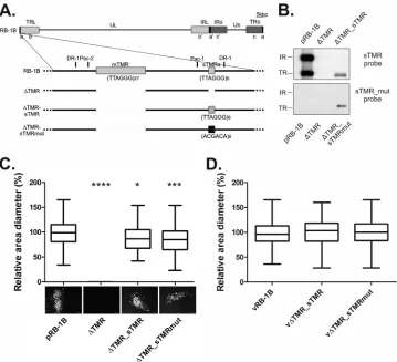
FIG 1Characterization of telomere deletion mutants. (A) Schematic representation of the MDV genome (RB-1B strain) with a focus on one of thea-likeregions.
⌬TMR contains a deletion of mTMR and sTMR in botha-likesequences present at either the terminal repeat (TR) or internal repeat (IR) region. Either the sTMR in⌬TMR were restored (⌬TMR_sTMR) or a scrambled repeated sequence was introduced (⌬TMR_sTMRmut). (B) Southern blotting using a TMR-specific probe (upper panel) or a mutant repeat probe (lower panel) after BamHI digestion of the indicated BAC clones. (C) Plaque size assay of RB-1B,⌬TMR,
⌬TMR_sTMR, and⌬TMR_sTMRmut viruses at 6 days posttransfection Plaque sizes are shown as box plots with minimums and maximums. Representative plaque images are shown below. Scale bar, 100m. Results are shown as the means of three independent experiments. *,P⬍0.05 compared to RB-1B; ***,P⬍ 0.001 compared to RB-1B; ****,P⬍0.0001 compared to all other samples (n⫽126; one-way ANOVA). (D) Plaque size assay after infection with 100 PFU of vRB-1B, v⌬TMR_sTMR, and v⌬TMR_sTMRmut. Plaque sizes are shown as box plots with minimums and maximums. Results are the means of three independent experiments (P⬎0.05 by one-way ANOVA;n⫽200).
on November 7, 2019 by guest
http://jvi.asm.org/
[image:2.585.113.472.65.392.2]replication, tumor formation, and genome integration. We dem-onstrate that deletion of sTMR sequences abrogates MDV repli-cation but that the exact sTMR sequence is not important for production of progeny virusin vitro. A panel of sTMR truncation mutants confirmed that the exact length of the sTMR is crucial for efficient MDV replication. In addition, mutation of the sTMR reduced integration frequency and impaired MDV pathogenesis and tumor formation.
MATERIALS AND METHODS
Cells and viruses.Chicken embryo cells (CEC) were prepared from Valo specific-pathogen-free (SPF) embryos as described previously (23). Re-combinant viruses were reconstituted by transfection of CEC with puri-fied bacterial artificial chromosome (BAC) DNA using CaPO4 transfec-tion (13,16,24,25). Virus was propagated on CEC for 2 to 4 passages, and infected cells were stored in liquid nitrogen. To confirm the presence of the introduced mutations, viral DNA was extracted from infected cellsin vitroandex vivousing an RTP DNA/RNA Virus minikit (Invitek), and the target region was analyzed by DNA sequencing.
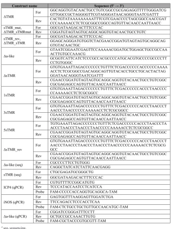
Generation of sTMR mutant viruses.sTMR mutant viruses were gen-erated using two-step Red-mediated mutagenesis from pRB-1B, an infec-tious BAC clone of the highly oncogenic RB-1B MDV strain (26,27). Briefly, the sequence of interest, including an I-SceI-aphAIcassette, was amplified from either the pEPKan-S or pTE2 transfer plasmid as previ-ously described (16,26). The⌬sTMR PCR product was used as the tem-plate for the generation of 1sTMR, 3sTMR, and 5sTMR truncation mu-tants and a revertant (⌬sTMR_rev). Primers used for the mutagenesis are listed inTable 1. In the first recombination step, the mutation was intro-duced into pRB-1B by electroporation of the PCR product into recombi-nation- and electrocompetent GS1783 cells. The I-SceI-aphAIselection marker cassette was subsequently removed by a second Red recombina-tion step (26). All resulting clones were screened by multiple restriction fragment length polymorphism (RFLP) analyses, Southern blotting, and sequencing of the target region as described previously (26,28).
Southern blotting.To confirm the recombinant BAC clones, South-ern blot analyses were performed as previously described (16). Briefly, BAC DNA was digested with various restriction enzymes, resolved on an agarose gel, and transferred onto a positively charged nylon membrane (NytranSPC membrane; Whatman). TMR or mutant repeats were de-tected using digoxigenin (DIG)-labeled oligonucleotide probes specific for the telomeric repeats (TTAGGG)6or the scrambled repeat sequence (ACGACA)6(Eurofins MWG) (16).
Growth kinetics and plaque size assays.Replication properties of re-combinant viruses were determined by multistep growth kinetics as de-scribed previously (28). Cell-to-cell spread of the recombinant viruses was determined by plaque size assays (28). Images of 20 to 50 randomly se-lected plaques per well were taken, and plaque areas were determined using ImageJ software (NIH).
Analysis of⌬a-like restoration.DNA was isolated from CEC infected with a virus with a deletion of thea-likesequence in the IR (v⌬a-like) 6 days after transfection (passage 0, P0) and at passages 1 (P1) and 2 (P2) after transfection using an RTP DNA/RNA Virus minikit (Invitek). Quan-titative PCR (qPCR) was performed using primers and probe specific for thea-likedeletion site (Table 1). Data were normalized against the num-ber of viral genomes detected using primers and a probe specific for in-fected cell protein 4 (ICP4) as described previously (10).
In vivo experiments.One-day-old Valo SPF chickens (Lohmann Tierzucht, Germany) were infected intra-abdominally with 1,000 (exper-iment 1) or 200 (exper(exper-iment 2) PFU of either vRB-1B, vsTMR_mut, or vsTMR_rev. Naive chickens were housed with infected animals to deter-mine transmission by the natural route of infection(experiment 1). Blood samples were taken from infected animals at 4, 7, 10, 14, and 28 days postinfection (p.i.) and from contact animals at 21, 28, and 35 days p.i. to determine MDV genome copy numbers in the blood, as described previ-ously (24,29,30). Chickens were monitored for clinical symptoms of MD
on a daily basis. Animals were examined for tumor lesions postmortem once clinical symptoms were evident or after termination of the experi-ments. Experiment 1 was terminated at 91 days and experiment 2 was terminated at 84 days p.i. Animal experiments were approved by the Landesamt für Gesundheit und Soziales in Berlin, Germany (approval number G0026/08).
Isolation of tumor cells from solid organ tumors.Lymphocytes were isolated from solid tumors as previously described (16,31). Briefly, tu-mors were extracted postmortem, and tissues were disrupted by mincing using a cell strainer (Falcon). Lymphocytes were isolated using a Ficoll gradient (density, 1.077 g/ml; Biochrom). Cells were subsequently washed with phosphate-buffered saline (PBS) and cultured in LM Hahn medium (32).
Quantification of MDV genome copy numbers in chicken whole blood.DNA was isolated from the blood of infected animals using an E-Z96 96-well blood DNA isolation kit (Omega Biotek, USA) according to the manufacturer’s instructions. qPCR using specific primers and a probe for ICP4 was used to determined MDV genome copy numbers. ICP4 copy numbers were normalized against cellular inducible nitric ox-ide synthase (iNOS) as described previously (24,29,30).
Metaphase preparation and FISH.Metaphase chromosomes were prepared from primary tumor cells and analyzed for the presence of the MDV genome by fluorescencein situhybridization (FISH) (16,33,34). Briefly, MDV integration sites were detected by a DIG-labeled MDV whole-genome probe (34) and visualized using a fluorescein isothiocya-nate (FITC)-conjugated anti-DIG antibody (Sigma-Aldrich). Metaphase FISH preparations were analyzed using an Axio Imager M1 system and AxioVision software (Carl Zeiss, Inc.).
Statistical analyses.Statistical analyses were performed using Graph-Pad Prism, version 7, and SPSS software (SPSS, Inc.). Data sets were first tested for normal distribution. Plaque size data of MDV recombinant viruses were analyzed as plaque diameters using one-way analysis of vari-ance (ANOVA), with a Bonferroni correction for multiple comparisons in the case of aPvalue of⬍0.05. qPCR data of MDV genome copy numbers in whole-blood samples and growth kinetics were analyzed using Kruskal-Wallis and Mann-Whitney U tests. For tumor incidence, groups were compared by a Kruskal-Wallis test.
RESULTS
Generation of TMR deletion mutants.To characterize the role of TMR, we generated a mutant in which the mTMR and sTMR were deleted in both copies of thea-likesequences (Fig. 1A,⌬TMR) present in pRB-1B byen passantmutagenesis (26). Mutants were analyzed by multiple RFLP analyses, sequencing, and Southern blotting using a probe specific for the telomeric repeats (Fig. 1B, upper panel). Telomeric repeats were detectable in the parental pRB-1B in both copies of thea-likesequences but not in the dou-ble deletion mutant (⌬TMR) (Fig. 1B, upper panel). The⌬TMR mutant virus (v⌬TMR) did not replicate upon virus reconstitu-tion in CEC, and only single infected cells were detected after transfection (Fig. 1C), while the parental virus was efficiently re-constituted. The mTMR were previously shown to be dispensable for MDV replication (16), which led us to conclude that the in-ability of the v⌬TMR mutant virus to replicate was caused by the absence of the sTMR. To determine if the absence of the sTMR was responsible for the lethal phenotype of v⌬TMR, we restored the sTMR in the terminal copy of thea-likesequences of v⌬TMR (Fig. 1A,⌬TMR_sTMR). Further, to examine if the exact sTMR se-quence is important for MDV replication, the scrambled repeat sequence (ACGACA)6 was inserted in place of the authentic
sTMR repeats (Fig. 1A,⌬TMR_sTMRmut). Southern blot analy-sis using a specific probe against either the parental (Fig. 1B, upper panel) or the mutated (Fig. 1B, lower panel) sequence indicated
on November 7, 2019 by guest
http://jvi.asm.org/
that in both cases the target sequence was introduced only into the terminal copy of thea-likesequences. Restoration of a sin-gle sTMR locus was sufficient to allow reconstitution of v⌬TMR_sTMR (Fig. 1C), while the second locus was restored during virus replication (data not shown). Insertion of a scram-bled sTMR sequence in the v⌬TMR_sTMRmut virus also restored virus replication (Fig. 1C), indicating that not the exact sTMR sequence but its function as a spacer sequence is essential for virus growth. Plaque sizes upon transfection of CEC with
⌬TMR_sTMR and ⌬TMR_sTMRmut viruses were mildly re-duced compared to those of the parental vRB-1B (Fig. 1C), which was likely due to slightly lower transfection efficiencies. Stan-dard plaque size assays confirmed that v⌬TMR_sTMR and v⌬TMR_sTMRmut viruses replicated with kinetics that were comparable to those of the parental virus (Fig. 1D).
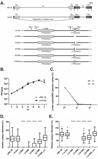
Generation and characterization of v⌬a-like virus.To sim-plify further analysis of the sTMR sequences, the internala-like
region of vRB-1B was deleted byen passantmutagenesis (Fig. 2A,
⌬a-like). It was shown recently that deletion of most of the IRLwas
[image:4.585.122.466.85.540.2]completely restored by homologous recombination events during viral DNA replication after only two passages in cell culture (13). We therefore hypothesized that the deletion of thea-likesequence would also be repaired within a few passages in CEC. Thereby, any mutation introduced into the remaininga-likesequence would be copied into the deleted locus. v⌬a-like was successfully reconsti-tuted in CEC, revealing that onea-like region is sufficient for MDV replication. Plaque size assays and multistep growth kinetics confirmed that v⌬a-like replicated with an efficiency that is com-parable to that of the parental vRB-1B (Fig. 2B). To determine if the deleteda-likeregion was indeed restored, we performed qPCR
TABLE 1Primer and probes for qPCR, DNA sequencing, and construction of recombinant virusesa
aseq, sequencing.
on November 7, 2019 by guest
http://jvi.asm.org/
analysis using specific primers and probes for the deletion. After just one passage in cell culture, the deletion of thea-likesequences was barely detectable, indicating that the locus was rapidly re-paired during virus replication (Fig. 2C). DNA sequencing
con-firmed that the deleted region was completely restored (data not shown).
Generation and characterization of⌬sTMR and sTMR trun-cation mutants.Next, we determined the minimal number of
FIG 2In vitrocharacterization of⌬a-like and sTMR truncation mutants. (A) Schematic representation of the MDV genome strain RB-1B,⌬a-like, and sTMR truncation mutants. Thea-likesequence at the IR was deleted (⌬a-like) byen passantmutagenesis. Then, the sTMR locus in the TR region was deleted (⌬sTMR). Subsequently 1 (1sTMR), 3 (3sTMR), 5 (5sTMR), or 6 (⌬sTMR_rev) repeats were introduced into⌬sTMR. (B) Multistep growth kinetics of vRB-1B and the v⌬a-like mutant. Data are shown as mean titers of three independent experiments with standard errors (P⬎0.05 for all time points by a Mann-Whitney U test;n⫽9). dpi, days postinfection. (C) qPCR amplification of the⌬a-like region in CEC transfected with⌬a-like virus. Results from two independent transfections (T1 and T2) are shown. Amplification of the deleted region was normalized against ICP4. The amplification level posttransfection (P0) was set to 100%. (D) Plaque size assay after transfection of the indicated BAC clones (****,P⬍0.0001 compared to pRB-1B by one-way ANOVA;n⫽148). Plaque sizes are shown as box plots with minimums and maximums. Results are the means of three independent experiments. (E) Plaque size assay after infection with the indicated viruses. Mean plaque sizes are shown as box plots. Scale bar, 100m. Results are the means of three experiments (****,P⬍0.0001 compared to vRB-1B, v⌬a-like, and v⌬sTMR_rev viruses by one-way ANOVA;n⫽84).
on November 7, 2019 by guest
http://jvi.asm.org/
[image:5.585.135.449.64.575.2]sTMR sequences necessary for MDV replication. Therefore, we generated a panel of mutants harboring 0, 1, 3, or 5 telomeric repeats in the sTMR locus (Fig. 2A). To verify that the sTMR are indeed essential for replication, we completely deleted the sTMR in the v⌬a-like mutant virus (v⌬sTMR) (Fig. 2A). Telomeric re-peats were subsequently reinserted into v⌬sTMR. In addition, a revertant clone was generated in which the entire sTMR sequence was restored (v⌬sTMR_rev). Replication properties were ana-lyzed by plaque size assays upon transfection of CEC. As expected, the v⌬sTMR mutant was not able to replicate in CEC (Fig. 2D) as only single infected cells were detected (data not shown), confirm-ing that the sTMR are indeed essential for MDV replication. In-triguingly, a gradual restoration of MDV replication dependent on the number of repeats in the sTMR region was observed. How-ever, only v⌬sTMR_rev replicated with efficiencies comparable to those of the parental vRB-1B (Fig. 2D). The growth properties of all the mutants were confirmed by standard plaque size assays (Fig. 2E). To ensure that the numbers of repeats were maintained during passaging of the virus, we sequenced all viruses after vari-ous passages to ensure that the mutation was stable (data not shown). Taken together, our data demonstrated that the sTMR are essential and that more than one of the six telomeric repeats present in the sTMR is required for minimal MDV replication.
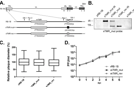
Generation andin vitrocharacterization of vsTMR_mut.To characterize the role of sTMR in viral pathogenesis and integra-tion, we generated a mutant in which only the sTMR were
re-placed by scrambled repeats (sTMR_mut) (Fig. 3A). In addition, a revertant virus in which the original sequence was restored (sTMR_rev) was generated to exclude spurious effects of second-ary mutations that might have occurred during mutagenesis. Pos-itive clones were confirmed by multiple RFLP analyses, Southern blotting using a specific probe for the sTMR_mut sequence (Fig. 3B), and DNA sequencing. Both the vsTMR_mut and vsTMR_rev viruses replicated efficiently upon reconstitution in CEC. vsTMR_ mut replication was comparable to that of both the parental and revertant viruses with respect to plaque sizes induced (Fig. 3C) and growth properties as assessed by multistep growth kinetics (Fig. 3D). We concluded from the results that the exact sTMR sequence is not important for replicationin vitro.
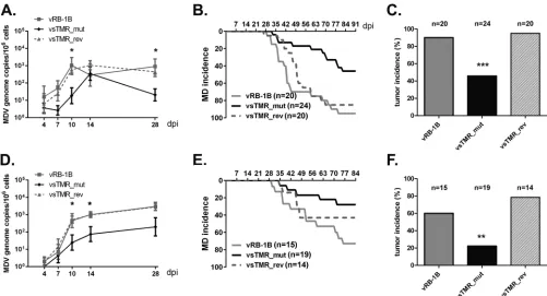
In vivocharacterization of vsTMR_mut.To determine the role of the sTMR in MDV pathogenesis and integration into host chromosomes, 1-day-old chickens were infected intra-abdomi-nally with either 1,000 (experiment 1) or 200 (experiment 2) PFU of vRB-1B, vsTMR_mut, or vsTMR_rev virus. To determine if replication of vsTMR_mut was affected in infected chickens, we analyzed MDV genome copy numbers in peripheral blood. In both experiments, viral loads of vsTMR_mut-infected birds were significantly reduced at the later stages of infection (Fig. 4Aand D). In addition, MD incidence with vsTMR_mut infection was severely decreased compared to rates with the parental and rever-tant viruses in both experiments (Fig. 4BandE), indicating that the sTMR sequences play an important role in MDV pathogenesis.
FIG 3The exact sTMR sequence is not important for MDV replication. (A) Schematic representation of MDV genome strain RB-1B with a focus on one of the a-likeregions. In the sTMR_mut construct, both sTMR loci were replaced by scrambled repeats, (ACGACA)6. In the sTMR_rev construct, the original sequence
was restored. (B) Southern blot analysis of a BamHI digestion of the indicated BAC clones using a probe specific for the scrambled repeats. (C) Plaque size assay after infection with vRB-1B, vsTMR_mut, and vsTMR_rev viruses. Plaque sizes are shown as box plots with minimums and maximums. Results are shown as the means of three independent experiments (P⬎0.05 by one-way ANOVA;n⫽150). (D) Multistep growth kinetics of vRB-1B, vsTMR_mut, and vsTMR_rev shown as means with standard errors of the means (df⫽2,P⬎0.05; Kruskal-Wallis). One representative of three independent experiments is shown.
on November 7, 2019 by guest
http://jvi.asm.org/
[image:6.585.77.504.65.345.2]At final necropsy, a drastic reduction in tumor incidence in vsTMR_mut-infected animals was detected, while tumors were found in most animals infected with the parental and revertant viruses (Fig. 4CandF).
To assess if transmission by the natural route was affected for vsTMR_mut, naive animals were housed together with inoculated chickens. vsTMR_mut was able to spread to contact animals as the MDV genome was readily detected in the blood of contact animals by qPCR (Fig. 5A); however, none of the vsTMR_mut contact animals showed clinical symptoms (Fig. 5B). At the final necropsy, only very few vsTMR_mut contact animals had MDV-induced tumors, while parental vRB-1B and vsTMR_rev efficiently in-duced tumors in contact animals (Fig. 5C). Taken together, our data showed that the sTMR sequence plays an important role in disease incidence and tumor formationin vivo.
Role of sTMR in MDV genome integration.As discussed ear-lier, MDV commonly integrates into the telomeres of multiple chromosomes in MDV-induced tumor cells, while integration of recombinant viruses that lack the mTMR was restricted to a single intrachromosomal integration site (16). To determine if mutation of the sTMR affects MDV genome integration, lymphocytes were isolated from tumors of animals infected with either vRB-1B, vsTMR_mut, or vsTMR_rev. Fluorescencein situhybridization (FISH) was performed to determine integration frequency. As ex-pected, multiple integration sites were detected in samples derived from vRB-1B- or vsTMR_rev-infected cells (Fig. 6AtoD). The
average number of integration sites of vsTMR_mut-induced tu-mor cells (2.5) was reduced compared to the number of integra-tion sites of tumor cells derived from parental (4.0) and revertant (4.4) viruses (Fig. 6D). The vsTMR_mut tumor cells still harbored up to four integration sites, suggesting that the sTMR play only a minor role in MDV integration and that other sequences may complement their function.
DISCUSSION
MDV, HHV-6, and a number of other herpesviruses harbor TMR arrays at the ends of their linear genomes. The MDV genome harbors two such TMR arrays located within thea-likesequence: one of multiple telomeric repeats (mTMR) and one of short telo-meric repeats (sTMR). Previously, we characterized the role of the mTMR (16) and demonstrated that these sequences are dispens-able for replicationin vitrobut play a crucial role in lymphom-agenesis, pathogenesis, and integration into telomeres of host chromosomes; however, the role of the sTMR, which are com-pletely conserved among all MDV strains, had remained un-known.
In this study, we investigated the role of sTMR in virus repli-cation, pathogenesis, and integration. Since mutagenesis of the herpesvirus repeat regions that harbor the sTMR is challenging and time-consuming, we initially established a system that facili-tates rapid and efficient mutagenesis of the sTMR. We recently demonstrated that deletion of most of the IRLwas completely FIG 4Mutation of the sTMR impairs disease development and tumor formationin vivo. Characterization of sTMR_mut and sTMR_rev viruses after intra-abdominal infection with 1,000 PFU (experiment 1) and 200 PFU (experiment 2). (A and D) qPCR analysis of the viral ICP4 gene and host iNOS gene. Blood samples of animals infected with the indicated viruses were taken at 4, 7, 10, 14, and 28 days p.i. Viral titers in the blood are shown as mean MDV genome copy numbers per 1⫻106cells of eight infected chickens per group in experiment 1 (df⫽2; *,P⬍0.05 compared to vRB-1B; Kruskal Wallis test) (A) and experiment
2 (df⫽2; *,P⬍0.05 for vsTMR_mut compared to vRB-1B; Kruskal Wallis test) (D) (B and E) MD incidence in chickens infected in experiment 1 (B) and experiment 2 (E) with vRB-1B, vsTMR_mut, or vsTMR_rev. (C and F) Tumor incidence in chickens infected with the indicated viruses in experiment 1 (df⫽ 2; ***,P⬍0.001 for vsTMR_mut compared to both vRB-1B and vsTMR_rev; Kruskal-Wallis) (C) and experiment 2 (df⫽2; **,P⬍0.01 for vsTMR_mut compared to vsTMR_rev; Kruskal-Wallis). Tumor incidence is shown as the percentage of animals per group.
on November 7, 2019 by guest
http://jvi.asm.org/
[image:7.585.41.542.65.336.2]restored after two passages in cell culture (13) due to homologous recombination events that occur during herpesvirus replication (35). Therefore, we deleted one copy of thea-likesequences (v⌬ a-like) and hypothesized that its deletion would be restored as de-scribed for the IRL. We observed that deletion of the internala-like
region did not affect MDV replicationin vitroand that the deleted sequence was indeed rapidly restored. Therefore, v⌬a-like virus could be used as platform for efficient modifications of the MDV
a-likesequences beyond the sTMR mutants described in this pub-lication.
In our initial experiment, we observed that deletion of both mTMR and sTMR (v⌬TMR virus) abrogated MDV replication. Restoration of the sTMR (v⌬TMR_sTMR) resulted in a virus that
replicated comparably to wild-type virus, suggesting that the sTMR play an important role in MDV replication. To confirm the essential role of the sTMR in MDV replication, we generated a virus that lacks only the sTMR sequence and not the mTMR (v⌬sTMR). As expected, deletion of the sTMR sequences abro-gated MDV replication.
To determine if the exact sTMR sequence itself was essential for MDV replication, we inserted scrambled repeats instead of the original sTMR into v⌬TMR (v⌬TMR_sTMRmut). Insertion of these completely unrelated repeat sequences efficiently restored MDV replication, indicating that the exact sTMR sequence is not important for MDV replication. MDV sTMR are flanked by the packaging signal pac-1 and the DR-1 cleavage site. These regula-tory sequence elements act incisto facilitate correct assembly of the machinery that is required to ensure correct cleavage and packaging of the concatemeric viral genome during replication (22). Volkening and Spatz recently demonstrated that cleavage of the MDV genome occurs adjacent to the sTMR repeats (21), ap-proximately 8 bp upstream of the DR-1 cleavage site. We specu-lated that sTMR repeats ensure the proper spacing between regu-latory sequences and the site of cleavage, which has been shown to be crucial for efficient cleavage of the herpesvirus genome (22). To determine if the spacing between DR-1 and the pac-1 could play a role in MDV replication, we generated a panel of truncation mu-tants containing 1, 3, or 5 repeats in the sTMR region. Efficiency of MDV replication was indeed a direct function of the length of the sTMR sequences, indicating that the distance between the pac-1 and DR-1 cleavage site is important for MDV replication.
To investigate the role of the sTMR in MDV pathogenesis and tumor formation, we mutated the sTMR in the highly oncogenic RB-1B strain. In two independent animal experiments with either a high- or low-dose inoculum, we observed that the development
FIG 5Disease and tumor incidence upon infection via the natural route. (A) qPCR detecting MDV genome copies in the blood of chicken infected with the indicated virus via the natural route of infection. Mean MDV genome copy numbers per 1⫻106cells are shown for the indicated time
points (df⫽2; **,P⬍0.01 for vsTMR_mut compared to vRB-1B; Kruskal Wallis). (B) MD incidence in contact animals infected with vRB-1B, vsTMR_mut, and vsTMR_rev via the natural route of infection during experiment 1. (C) Tumor incidence in contact birds infected with the indicated viruses in experiment 1 (df⫽2; *,P⬍0.05 for vsTMR_mut com-pared to both vRB-1B and vsTMR_rev; Kruskal-Wallis). Tumor incidence is shown as the percentage of animals per group.
FIG 6Mutation of the sTMR impairs MDV integration. Representative meta-phase images are shown of tumors derived from chickens infected with vRB-1B, vsTMR_mut, and sTMR_rev. Arrows indicate integration sites of the MDV genome (anti-DIG-FITC; green) in host chromosomes stained with 4=,6=-diamidino-2-phenylindole (blue). Scale bar, 5m. Images were taken with an Axio Observer M1 system (Zeiss). (D) Mean number of integration sites in vRB-1B, vsTMR_mut, and vsTMR_rev tumor-derived cells. The num-ber (Nr) of analyzed cell lines and average numnum-ber (range) of integration sites are given. At least a dozen independent metaphase spread images were evalu-ated to determine the exact number of integration sites for each independent tumor.
on November 7, 2019 by guest
http://jvi.asm.org/
[image:8.585.297.545.66.244.2] [image:8.585.79.248.75.451.2]of MD symptoms and tumor formation are severely impaired upon mutation of the sTMR compared to effects of the parental and revertant virus. In addition, vsTMR_mut genome copy num-bers in peripheral blood were reduced by 10- to 100-fold from 10 to 28 days p.i. At these later time points, MDV genome copy num-bers in the blood mainly correspond to the number of infected T cells (1), in which the virus integrates and establishes latency, sug-gesting that vsTMR_mut could have a defect in either of these processes. Furthermore, vsTMR_mut was readily detected in con-tact animals, indicating that the MDV was efficiently transmitted to naive animals. As in the experimentally infected chickens, dis-ease and tumor development were severely impaired in contact animals.
To determine if an integration defect was responsible for the impairment of vsTMR_mut-induced tumorigenesis, we analyzed tumor cells by FISH and determined the number of integration sites. As expected, tumor cells derived from animals infected with parental and revertant viruses harbored multiple integration sites. Surprisingly, integration in tumor cells derived from vsTMR_mut infection still occurred in up to four chromosomes, while the av-erage number of integration sites was reduced only by about 2-fold compared to infection with parental and revertant viruses. Our data, therefore, suggest that the sTMR play a rather minor role in integration, while mTMR were shown to be essential for MDV integration into telomeres (16). Interestingly, thea-like se-quences are often duplicated during herpesvirus replication (22). Volkening and Spatz recently demonstrated that most encapsi-dated MDV genomes contained one or several completea-like
sequences including the sTMR and mTMR at either end of the linear genome (21). Therefore, duplication of thea-likesequences would result in a virus that harbors the mTMR at both genome termini. The presence of the mTMR at either end could, in turn, facilitate recombination of the MDV genome into host telomeres in the absence of the sTMR. The presence of the sTMR at the very end of the linear genome could increase the integration efficiency, as indicated by the results shown inFig. 6. However, the absence of the sTMR at the end of the MDV genome could also induce DNA damage responses (DDR) that are mounted against double-strand breaks that do not represent telomeric repeats, resulting in cell cycle arrest and the induction of apoptosis (36). As MDV integra-tion occurs in T cells, inducintegra-tion of DDR resulting in apoptosis would reduce the number of infected T cells in the animals. Our data show that the genome copy numbers in the blood, which mainly corresponds to the number of infected T cells, were indeed significantly reduced at later time points. This suggests either that T cells were indeed eliminated or that fewer T cells were infectedin vivo, while replication was not affectedin vitro.
Taken together, our data suggest that the sTMR have a dual function in the MDV life cycle. First, the sTMR likely serve as spacers between the DR-1 cleavage site and the packaging signal pac-1 to ensure proper cleavage and packaging of the viral genome into the preformed capsid. Second, the sTMR are not essential for MDV integration but, rather, seem to increase integration effi-ciency in case thea-likesequences containing the essential mTMR are not duplicated, avoiding activation of DDR that commonly occur as a result of incomplete integration events.
ACKNOWLEDGMENTS
We are grateful to Ann Reum, Sina Arndt, and Sandra Treptow for their technical assistance.
This study was supported by the EU-EMIDA MADISPREAD grant and DFG grant KA 3492/1-1 awarded to B.B.K.
REFERENCES
1.Davison F, Nair V.2004. Marek’s disease: an evolving problem. Elsevier, London, United Kingdom.
2.Jarosinski KW, Tischer BK, Trapp S, Osterrieder N. 2006. Marek’s disease virus: lytic replication, oncogenesis and control. Expert Rev. Vac-cines5:761–772.http://dx.doi.org/10.1586/14760584.5.6.761.
3.Osterrieder N, Kamil JP, Schumacher D, Tischer BK, Trapp S.2006. Marek’s disease virus: from miasma to model. Nat. Rev. Microbiol.4:283– 294.http://dx.doi.org/10.1038/nrmicro1382.
4.Nair V.2013. Latency and tumorigenesis in Marek’s disease. Avian Dis.
57:360 –365.http://dx.doi.org/10.1637/10470-121712-Reg.1.
5.Brown AC, Baigent SJ, Smith LP, Chattoo JP, Petherbridge LJ, Hawes P, Allday MJ, Nair V.2006. Interaction of MEQ protein and C-terminal-binding protein is critical for induction of lymphomas by Marek’s disease virus. Proc. Natl. Acad. Sci. U. S. A.103:1687–1692.http://dx.doi.org/10 .1073/pnas.0507595103.
6.Brown AC, Smith LP, Kgosana L, Baigent SJ, Nair V, Allday MJ.2009. Homodimerization of the Meq viral oncoprotein is necessary for induc-tion of T-cell lymphoma by Marek’s disease virus. J. Virol.83:11142– 11151.http://dx.doi.org/10.1128/JVI.01393-09.
7.Levy AM, Izumiya Y, Brunovskis P, Xia L, Parcells MS, Reddy SM, Lee L, Chen HW, Kung HJ.2003. Characterization of the chromosomal binding sites and dimerization partners of the viral oncoprotein Meq in Marek’s disease virus-transformed T cells. J. Virol.77:12841–12851.http: //dx.doi.org/10.1128/JVI.77.23.12841-12851.2003.
8.Fragnet L, Blasco MA, Klapper W, Rasschaert D. 2003. The RNA subunit of telomerase is encoded by Marek’s disease virus. J. Virol.77:
5985–5996.http://dx.doi.org/10.1128/JVI.77.10.5985-5996.2003. 9.Kaufer BB, Arndt S, Trapp S, Osterrieder N, Jarosinski KW. 2011.
Herpesvirus telomerase RNA (vTR) with a mutated template sequence abrogates herpesvirus-induced lymphomagenesis. PLoS Pathog.
7:e1002333.http://dx.doi.org/10.1371/journal.ppat.1002333.
10. Kaufer BB, Trapp S, Jarosinski KW, Osterrieder N, Speck SH.2010. Herpesvirus telomerase RNA (vTR)-dependent lymphoma formation does not require interaction of vTR with telomerase reverse transcriptase (TERT). PLoS Pathog. 6:e1001073. http://dx.doi.org/10.1371/journal .ppat.1001073.
11. Chen JL, Opperman KK, Greider CW.2002. A critical stem-loop struc-ture in the CR4-CR5 domain of mammalian telomerase RNA. Nucleic Acids Res.30:592–597.http://dx.doi.org/10.1093/nar/30.2.592. 12. Burnside J, Bernberg E, Anderson A, Lu C, Meyers BC, Green PJ, Jain
N, Isaacs G, Morgan RW.2006. Marek’s disease virus encodes MicroR-NAs that map to meq and the latency-associated transcript. J. Virol.80:
8778 – 8786.http://dx.doi.org/10.1128/JVI.00831-06.
13. Engel AT, Selvaraj RK, Kamil JP, Osterrieder N, Kaufer BB. 2012. Marek’s disease viral interleukin-8 promotes lymphoma formation through targeted recruitment of B cells and CD4⫹CD25⫹T cells. J. Virol.
86:8536 – 8545.http://dx.doi.org/10.1128/JVI.00556-12.
14. Parcells MS, Lin SF, Dienglewicz RL, Majerciak V, Robinson DR, Chen HC, Wu Z, Dubyak GR, Brunovskis P, Hunt HD, Lee LF, Kung HJ.
2001. Marek’s disease virus (MDV) encodes an interleukin-8 homolog (vIL-8): characterization of the vIL-8 protein and a vIL-8 deletion mutant MDV. J. Virol.75:5159 –5173.http://dx.doi.org/10.1128/JVI.75.11.5159 -5173.2001.
15. Delecluse HJ, Hammerschmidt W.1993. Status of Marek’s disease virus in established lymphoma cell lines: herpesvirus integration is common. J. Virol.67:82–92.
16. Kaufer BB, Jarosinski KW, Osterrieder N.2011. Herpesvirus telomeric repeats facilitate genomic integration into host telomeres and mobiliza-tion of viral DNA during reactivamobiliza-tion. J. Exp. Med.208:605– 615.http://dx .doi.org/10.1084/jem.20101402.
17. Knipe DM, Howley PM, Griffin DE, Lamb RA, Martin MA, Roizman B, Straus SE (ed).2007. Fields virology. Lippincott Williams & Wilkins, Philadelphia, PA.
18. Arbuckle JH, Medveczky MM, Luka J, Hadley SH, Luegmayr A, Ablashi D, Lund TC, Tolar J, De Meirleir K, Montoya JG, Komaroff AL, Ambros PF, Medveczky PG.2010. The latent human herpesvirus-6A genome specifically integrates in telomeres of human chromosomes in
on November 7, 2019 by guest
http://jvi.asm.org/
vivo and in vitro. Proc. Natl. Acad. Sci. U. S. A.107:5563–5568.http://dx .doi.org/10.1073/pnas.0913586107.
19. Osterrieder N, Wallaschek N, Kaufer BB.2014. Herpesvirus genome integration into telomeric repeats of host cell chromosomes. Annu. Rev. Virol.1:215–235.http://dx.doi.org/10.1146/annurev-virology-031413 -085422.
20. Morissette G, Flamand L.2010. Herpesviruses and chromosomal inte-gration. J. Virol.84:12100 –12109.http://dx.doi.org/10.1128/JVI.01169 -10.
21. Volkening JD, Spatz SJ.2013. Identification and characterization of the genomic termini and cleavage/packaging signals of gallid herpesvirus type 2. Avian Dis.57:401– 408.http://dx.doi.org/10.1637/10410-100312-Reg.1. 22. Brown J, McVoy M, Homa F.2002. Packaging DNA into herpesvirus
capsids, p 111–153.InHolzenburg A, Bogner E (ed), Structure-function relationships of human pathogenic viruses. Kluwer Academic, New York, NY.
23. Osterrieder N.1999. Sequence and initial characterization of the U(L)10 (glycoprotein M) and U(L)11 homologous genes of serotype 1 Marek’s disease virus. Arch. Virol. 144:1853–1863. http://dx.doi.org/10.1007 /s007050050710.
24. Jarosinski KW, Margulis NG, Kamil JP, Spatz SJ, Nair VK, Osterrieder N.2007. Horizontal transmission of Marek’s disease virus requires US2, the UL13 protein kinase, and gC. J. Virol.81:10575–10587.http://dx.doi .org/10.1128/JVI.01065-07.
25. Schumacher D, Tischer BK, Fuchs W, Osterrieder N.2000. Reconsti-tution of Marek’s disease virus serotype 1 (MDV-1) from DNA cloned as a bacterial artificial chromosome and characterization of a glycoprotein B-negative MDV-1 mutant. J. Virol.74:11088 –11098.http://dx.doi.org /10.1128/JVI.74.23.11088-11098.2000.
26. Tischer BK, von Einem J, Kaufer B, Osterrieder N.2006. Two-step red-mediated recombination for versatile high-efficiency markerless DNA manipulation inEscherichia coli. Biotechniques40:191–197.http://dx.doi .org/10.2144/000112096.
27. Tischer BK, Kaufer BB. 2012. Viral bacterial artificial chromosomes: generation, mutagenesis, and removal of mini-F sequences. J. Biomed. Biotechnol.2012:472537.http://dx.doi.org/10.1155/2012/472537. 28. Schumacher D, Tischer BK, Trapp S, Osterrieder N.2005. The protein
encoded by the US3 orthologue of Marek’s disease virus is required for efficient de-envelopment of perinuclear virions and involved in actin stress fiber breakdown. J. Virol.79:3987–3997.http://dx.doi.org/10.1128 /JVI.79.7.3987-3997.2005.
29. Jarosinski K, Kattenhorn L, Kaufer B, Ploegh H, Osterrieder N.2007. A herpesvirus ubiquitin-specific protease is critical for efficient T cell lym-phoma formation. Proc. Natl. Acad. Sci. U. S. A.104:20025–20030.http: //dx.doi.org/10.1073/pnas.0706295104.
30. Jarosinski KW, Osterrieder N, Nair VK, Schat KA.2005. Attenuation of Marek’s disease virus by deletion of open reading frame RLORF4 but not RLORF5a. J. Virol.79:11647–11659.http://dx.doi.org/10.1128/JVI.79.18 .11647-11659.2005.
31. Calnek BW, Schat KA, Ross LJ, Shek WR, Chen CL. 1984. Further characterization of Marek’s disease virus-infected lymphocytes. I. In vivo infection. Int. J. Cancer33:389 –398.
32. Calnek BW, Shek WR, Schat KA.1981. Latent infections with Marek’s disease virus and turkey herpesvirus. J. Natl. Cancer Inst.66:585–590. 33. Rens W, Fu B, O’Brien PC, Ferguson-Smith M.2006. Cross-species
chromosome painting. Nat. Protoc.1:783–790.http://dx.doi.org/10.1038 /nprot.2006.91.
34. Kaufer BB.2013. Detection of integrated herpesvirus genomes by fluo-rescence in situ hybridization (FISH). Methods Mol. Biol.1064:141–152.
http://dx.doi.org/10.1007/978-1-62703-601-6_10.
35. Dhepakson P, Mori Y, Jiang YB, Huang HL, Akkapaiboon P, Okuno T, Yamanishi K.2002. Human herpesvirus-6 rep/U94 gene product has single-stranded DNA-binding activity. J. Gen. Virol.83:847– 854. 36. Symington LS.2005. Focus on recombinational DNA repair. EMBO Rep.
6:512–517.http://dx.doi.org/10.1038/sj.embor.7400438.