Polyomavirus DNA Replication Sites To Support Efficient Viral DNA
Replication
Sabrina H. Tsang,aXin Wang,aJing Li,aChristopher B. Buck,bJianxin Youa
Department of Microbiology, University of Pennsylvania, Perelman School of Medicine, Philadelphia, Pennsylvania, USAa
; Tumor Virus Molecular Biology Section, Laboratory of Cellular Oncology, National Cancer Institute, Bethesda, Maryland, USAb
ABSTRACT
Accumulating evidence indicates a role for Merkel cell polyomavirus (MCPyV) in the development of Merkel cell carcinoma (MCC), making MCPyV the first polyomavirus to be clearly associated with human cancer. With the high prevalence of MCPyV infection and the increasing amount of MCC diagnosis, there is a need to better understand the virus and its oncogenic potential. In this study, we examined the relationship between the host DNA damage response (DDR) and MCPyV replication. We found that components of the ATM- and ATR-mediated DDR pathways accumulate in MCPyV large T antigen (LT)-positive nuclear foci in cells infected with native MCPyV virions. To further study MCPyV replication, we employed our previously established system, in which recombinant MCPyV episomal DNA is autonomously replicated in cultured cells. Similar to native MCPyV infection, where both MCPyV origin and LT are present, the host DDR machinery colocalized with LT in distinct nuclear foci.
Immunofluorescencein situhybridization and bromodeoxyuridine (BrdU) incorporation analysis showed that these DDR
pro-teins and MCPyV LT in fact colocalized at the actively replicating MCPyV replication complexes, which were absent when a rep-lication-defective LT mutant or an MCPyV-origin mutant was introduced in place of wild-type LT or wild-type viral origin. Inhi-bition of DDR kinases using chemical inhibitors and ATR/ATM small interfering RNA (siRNA) knockdown reduced MCPyV DNA replication without significantly affecting LT expression or the host cell cycle. This study demonstrates that these host DDR factors are important for MCPyV DNA replication, providing new insight into the host machinery involved in the MCPyV life cycle.
IMPORTANCE
MCPyV is the first polyomavirus to be clearly associated with human cancer. However, the MCPyV life cycle and its oncogenic mechanism remain poorly understood. In this report, we show that, in cells infected with native MCPyV virions, components of the ATM- and ATR-mediated DDR pathways accumulate in MCPyV LT-positive nuclear foci. Such a phenotype was recapitu-lated using our previously established system for visualizing MCPyV replication complexes in cells. By combining
immunofluo-rescent staining, fluorescencein situhybridization, and BrdU incorporation analysis, we demonstrate that DDR proteins are
important for maintaining robust MCPyV DNA replication. This study not only provides the first look into the microscopic de-tails of DDR factor/LT replication complexes at the MCPyV origin but also provides a platform for further studying the mecha-nistic role of host DDR factors in the MCPyV life cycle and virus-associated oncogenesis.
M
erkel cell polyomavirus (MCPyV) was discovered in 2008 in Merkel cell carcinoma (MCC), a highly aggressive form of skin cancer with neuroendocrine characteristics (1). Independent studies have subsequently found MCPyV to be clonally integrated in more than 80% of all MCC cases (1). Epidemiological surveys for MCPyV seropositivity (2) and sequencing analyses of healthy human skin (3) have shown that MCPyV is an abundant virus frequently shed from healthy human skin surfaces, suggesting that MCPyV may represent a common component of the human skin microbial flora. Immunosuppression, advanced age, and excessive exposure to UV radiation have been identified as the principle risk factors for MCC (4). Although MCC is uncommon, its incidence has tripled over the past 20 years, and the concern for MCC grows as the size of the aging population with prolonged sun exposure increases (5). To date, much of our knowledge of polyomaviruses is inferred from decades of research on simian virus 40 (SV40), which is phylogenetically distant from MCPyV and is not known to cause cancer in humans (1,6). It is likely that much remains to be learned about the applicability of well-understood aspects ofSV40 biology to the MCPyV life cycle and the oncogenic potential of MCPyV in humans.
Like other polyomaviruses, MCPyV is a small, nonenveloped virus with a circular, double-stranded DNA (dsDNA) genome of ⬃5 kb (7). A noncoding regulatory region (NCRR) divides the genome into early and late coding regions. The NCRR contains the viral origin of replication (Ori) and regulatory elements/pro-moters for viral gene transcription (8,9). The early region encodes three proteins, namely, large T antigen (LT), small T antigen (sT),
Received9 December 2013 Accepted26 December 2013
Published ahead of print3 January 2014
Editor:R. M. Longnecker
Address correspondence to Jianxin You, [email protected]. S.H.T. and X.W. contributed equally to this article.
Copyright © 2014, American Society for Microbiology. All Rights Reserved.
doi:10.1128/JVI.03656-13
on November 7, 2019 by guest
http://jvi.asm.org/
and the 57kT antigen (7). The late region encodes a major capsid protein, VP1, and a minor capsid protein, VP2 (10,11).
Similar to SV40 LT, MCPyV LT is a multifunctional protein that plays an important role in viral replication and host cell cycle manipulation (12–14). It contains a number of domains that are conserved among polyomaviruses, including a retinoblastoma (Rb)-binding domain, DnaJ domain, and CR1 domain (15). LT also has an origin-binding domain (OBD) and a C-terminal heli-case domain, both of which are required for initiating viral repli-cation (8,9,16).
With little being known about the MCPyV life cycle, we are interested in studying how the interactions between viral proteins and the host machinery contribute to viral replication and/or MCC oncogenesis. Emerging evidence has suggested that the host DNA damage response (DDR) is targeted by a wide variety of DNA and RNA viruses. The host DDR is composed of a net-work of proteins that recognize and repair various types of DNA damage. The major players in this signaling cascade are two phos-phoinositide 3-kinase (PI3K)-related protein kinases (PIKKs), namely, the ataxia telangiectasia mutated (ATM) and ATM- and Rad3-related (ATR) kinases. While ATM is primarily activated upon double-stranded DNA breaks (DSBs), ATR is responsive to single-stranded breaks (SSBs) (17). Normally, DSBs are recog-nized by the heterotrimeric Mre11-Rad50-Nbs1 (MRN) complex (17), which in turn induces autophosphorylation and activation of ATM (18). Activated ATM phosphorylates the histone variant H2AX, as well as the downstream kinase Chk2, which signals through a number of effectors, leading to cell cycle arrest (17). Depending on the severity of the damage, this pathway can also promote senescence or apoptosis. In parallel, SSBs and single-stranded DNA accumulation at stalled replication forks are recog-nized and coated by RPA (19). After ATR is recruited and acti-vated by RPA, it phosphorylates H2AX and the downstream kinase Chk1, which shares a number of downstream targets with Chk2. Previous research has shown that there is a significant amount of cross talk between the ATR and ATM pathways (17,
20–23).
Multiple groups have shown that polyomavirus infection or ectopic expression of polyomavirus LT proteins is capable of in-ducing a DDR in the host cell (14,24–30). The mechanism by which these LT proteins activate the host DDR is less well under-stood, but it has been suggested that polyomaviruses utilize the DDR machinery for viral replication. For example, both mouse polyomavirus (MPyV) infection and SV40 infection have been shown to induce a DDR that is crucial for viral replication (24,27). More specifically, ATM-mediated phosphorylation of SV40 LT is important for its ability to drive replication of the viral origin (27,
31). Recent reports of human polyomaviruses JC polyomavirus (JCPyV) (25) and BK polyomavirus (BKPyV) (26) have also shown that ATR and ATM are both activated upon infection and that they play an important role in viral genome amplification and virion production.
In a recent study, we have provided a mechanistic analysis of MCPyV DNA replication in cultured cells (12). Additional study from our lab (University of Pennsylvania) has shown that ectopic expression of MCPyV LT activates host DDR, leading to the inhi-bition of cellular proliferation (14). It remains unknown whether MCPyV also utilizes the host DDR machinery for optimal viral replication and/or virion production. In this study, we investi-gated the role of host DDR in MCPyV replication. Using U2OS
cells infected with MCPyV as well as an MCPyV DNA replication system that we previously established (12), we show that MCPyV LT and components of the ATR- and ATM-mediated DDR path-ways colocalize at actively replicating MCPyV foci in the nucleus. By combining immunofluorescent staining, immunofluorescence
in situ hybridization (immuno-FISH), bromodeoxyuridine (BrdU) incorporation, and Southern blotting analyses, we dem-onstrate that DDR proteins are important for maintaining robust MCPyV DNA replication.
MATERIALS AND METHODS
Cell culture, cell lines, and DNA/siRNA transfection.U2OS cells were maintained in McCoy’s 5A medium (Invitrogen) containing 10% fetal bovine serum (HyClone). C33A cells were maintained in Dulbecco mod-ified Eagle medium (Invitrogen) containing 10% fetal bovine serum. For immunofluorescent staining, C33A cells were transfected at 40 to 50% confluence using the FuGENE6 transfection reagent (Promega), accord-ing to the manufacturer’s instructions, and fixed at 48 to 60 h posttrans-fection (hpt). For Southern blotting and flow cytometry analysis, C33A cells were transfected using the calcium phosphate method (32). Small interfering RNA (siRNA) transfection was performed using calcium phosphate, as previously described (33). To detect viral DNA replication, C33A cells were pulsed with 10M BrdU at 44 hpt for 2 h and cultured with normal growth medium for another 2 h before acetone fixation.
Recombinant plasmid construction.The plasmids used in this study included religated MCPyV genome, MCPyV LT, pcDNA4C-MCPyV Ori, pADL*, and pT⫹Ori. They have been previously described (12). The origin mutant Ori350 was constructed from pcDNA4C-MCPyV Ori by site-directed mutagenesis.
Antibodies, chemicals, and siRNAs.The following antibodies were used for immunofluorescent staining: mouse anti-MCPyV LT (CM2B4; Santa Cruz), goat anti-ATR (N-19; Santa Cruz), rabbit anti-pChk1S317
(Cell Signaling), rabbit anti-pATMS1981 (Cell Signaling), rabbit
anti-pChk2T68(Cell Signaling), rabbit anti-Nbs1 (Novus Biologicals), rabbit
anti-pRPA32S33(Bethyl Laboratories), rabbit anti-RPA70 (Cell
Signal-ing), mouse anti-Ki-67 (Dako), Alexa Fluor 594 goat anti-rabbit IgG (In-vitrogen), Alexa Fluor 594 donkey anti-goat IgG (In(In-vitrogen), and Alexa Fluor 488 donkey anti-mouse IgG (Invitrogen). The following antibodies were used for Western blotting: mouse anti-MCPyV LT (CM2B4; Santa Cruz), rabbit anti-ATM (Cell Signaling), rabbit anti-ATR (Abcam), mouse antiactin (Chemicon), horseradish peroxidase (HRP)-conjugated donkey antimouse (GE Healthcare), and HRP-conjugated donkey anti-rabbit (GE Healthcare). Western Lightning Plus ECL solution was pur-chased from PerkinElmer. Wortmannin and NU6027 were purpur-chased from Sigma and dissolved in dimethyl sulfoxide (DMSO). Control siRNA and siRNA pools targeting human ATR and ATM were purchased from Dharmacon and used as previously described (26).
MCPyV virion preparation and infection. Native MCPyV and MCPyV pseudoviruses were prepared as previously described (10), with minor modifications. Briefly, an initial seed stock of native virions was produced by transfecting 293-4T cells (which stably express the MCPyV LT and sT proteins) with the religated recombinant genome of MCPyV isolate R17b (34–36). Five days later, native MCPyV virions were har-vested and purified over an OptiPrep gradient. This initial seed stock of native MCPyV virions was used to infect fresh 293-4T cells. The MCPyV-infected 293-4T cells were harvested and lysed after 5 days of infection, and the amplified native MCPyV virions were purified over an OptiPrep gradient. For experimental infection, U2OS cells were seeded in 24-well plates and incubated with native MCPyV virions at a dose of 5⫻104
MCPyV genomes per cell for 5 days.
Immunofluorescent staining.Immunofluorescent staining was per-formed as previously described (12). C33A cells were fixed with 3% para-formaldehyde in phosphate-buffered saline (PBS) for 20 min. Cells were incubated in blocking/permeabilization buffer (0.5% Triton X-100 and 3% bovine serum albumin in PBS) for 10 min at room temperature and Tsang et al.
on November 7, 2019 by guest
http://jvi.asm.org/
stained with primary antibodies (as indicated in the appropriate figure legends) at room temperature for 1 h. Cells were washed three times with blocking/permeabilization buffer and incubated with secondary antibod-ies for an additional hour. Cells were then counterstained with DAPI (4=,6=-diamidino-2-phenylindole) and examined with an Olympus IX81 inverted fluorescence microscope.
Immuno-FISH. Immuno-FISH was performed as previously de-scribed (12), with slight modifications. Briefly, C33A cells were fixed and stained using antibodies as indicated in the appropriate figure legends. A specific probe recognizing pcDNA4C-MCPyV Ori and a negative-control probe recognizing the human papillomavirus type 16 (HPV16) genome were labeled with biotin-dUTP (AppliChem) using nick translation. Hy-bridized probes were detected with a TSA biotin system (PerkinElmer) following the manufacturer’s instruction.
Microscopy and image analysis.All immunofluorescent images were collected using an inverted fluorescence microscope (IX81; Olympus) con-nected to a high-resolution charge-coupled-device camera (FAST1394; QImaging). Images were analyzed and presented using SlideBook (version 5.0) software (Intelligent Imaging Innovations, Inc.). The scale bars were added using ImageJ software.
Southern blotting.Southern blotting was performed as previously described (12), with slight modification. For DDR inhibitor treatment, at 20 hpt, C33A cells were incubated in medium with 20M wortmannin or 20M NU6027. Drugs were refreshed every 16 h, and cells were harvested at 52 hpt. For ATR/ATM knockdown, pT⫹Ori and siRNA were trans-fected at the same time using calcium phosphate. About 30g total DNA was digested with DpnI and XhoI, before being subjected to Southern blotting, while 2g total DNA was digested with XhoI and used as a loading control.
Western blotting.Cells were lysed in lysis buffer (10 mM HEPES, pH 7.9, 500 mM NaCl, 3 mM MgCl2, 1 mM dithiothreitol, 1 mM
phenyl-methylsulfonyl fluoride) by passing through a 22-gauge needle 10 times. After a 30-min incubation on ice, the soluble and insoluble fractions were separated by centrifugation at 15,000 rpm for 5 min at 4°C. The superna-tants (20g) were resolved on an SDS-polyacrylamide gel. Membranes were blotted according to the antibody manufacturers’ instructions. Western blots were developed using enhanced chemiluminescence (ECL) solution, and images were captured using a Fuji imaging system.
Flow cytometry.C33A cells were pulse labeled with 10M BrdU for 2 h before trypsinization. They were then fixed, permeabilized, and stained by use of an allophycocyanin BrdU flow kit (BD Pharmingen) according to the manufacturer’s instructions. Stained cells were analyzed by flow cytometry using a BD FACSCalibur flow cytometer (Becton, Dickinson). Data were analyzed using FlowJo software.
Statistical analysis.Statistical analysis was performed using one-way analysis of variance with GraphPad Prism software (version 5.0). APvalue of⬍0.05 was considered statistically significant.
RESULTS
Colocalization of host DDR proteins and MCPyV LT in nuclear
foci in cells infected with native MCPyV virions.We first
inves-tigated the relationship between the host DDR and MCPyV repli-cation in cells infected with native MCPyV virions. We began our infection studies using cells of the U2OS cell line, which, in con-trast to many other cell lines, have intact ATR and ATM pathways. We have also observed an activated DDR in U2OS cells upon ectopic MCPyV LT expression (14). Native MCPyV infection ef-ficiency was about 10 to 15% in U2OS cells, as indicated by posi-tive immunostaining of MCPyV LT at day 5 postinfection (dpi). It has been shown that MCPyV transcription and replication are highly restricted in all cell lines so far tested (11,35,36). Consis-tent with these observations, we observed a very low level of MCPyV LT expression in U2OS cells infected with native MCPyV virions. As we published previously (14), LT typically showed a
diffuse staining pattern in the nucleus, but in⬃1% of MCPyV-infected cells, we were able to observe punctate LT foci in the nucleus (Fig. 1A). Interestingly, these foci showed clear colocal-ization with the phosphorylated H2AX (referred to as␥H2AX), a classic marker of DNA damage (Fig. 1A). We also observed colo-calization of LT with ATR and pChk2T68, with the latter being an indicator for the activation of an ATM-mediated DDR (Fig. 1A). This result suggests that the components of the ATR and ATM pathways are recruited to the MCPyV LT nuclear foci. In contrast, cells infected with MCPyV pseudovirus carrying a green fluores-cent protein (GFP) reporter construct instead of the MCPyV ge-nome did not show such DDR foci (data not shown).
To rule out the possibility that the MCPyV capsid proteins are causing this phenomenon, we pseudotyped MCPyV genomic DNA into the L1-L2 capsid of HPV16, which is capable of trans-ducing U2OS cells. Although the LT-positive cells were equally rare, we were again able to find cells that displayed LT/pChk2T68
colocalization in the nuclear foci (Fig. 1B). In contrast, MCPyV LT and pChk2T68 signals were not detected in the untransduced
neighboring cells (Fig. 1B). These data suggest that the MCPyV genome, and not the incoming MCPyV virion proteins, is respon-sible for the DDR protein/LT colocalization phenotype that we observed in MCPyV-infected U2OS cells.
The MCPyV genome alone is able to induce DDR factor/LT
colocalization in nuclear foci.Based on what we saw in the HPV/
MCPyV pseudovirus-transduced cells, we believed that the MCPyV genome alone could also lead to DDR factor/LT colocal-ization in nuclear foci. We next transfected the religated MCPyV genome into U2OS cells. The transfected cells could be identified by positive LT staining (Fig. 2). In⬃5% of transfected cells, we were able to see LT colocalizing with␥H2AX, ATR, and pChk2T68
in nuclear foci at day 4 posttransfection (Fig. 2). These data con-firm the colocalization of host DDR components with LT ex-pressed from the native MCPyV genome. It is important to note that, although we have previously shown that LT expression can activate the overall activity of host DDR (14), the data presented in
Fig. 1and2reveal the specific accumulation of the host DDR
components at the LT foci in cells treated with MCPyV virions/ pseudovirions/religated genomes.
Host DDR machinery colocalizes with MCPyV LT at nuclear
foci in the presence of the viral origin.Although we were able to
detect DDR factor/MCPyV LT colocalization using native virion infection and religated genome transfection, the rarity of cells ex-pressing detectable amounts of LT in these systems makes them relatively intractable. In addition, with native MCPyV systems, we were not able to prove whether the DDR factors had a specific effect on MCPyV DNA replication. In our previous studies, we established a system to detect MCPyV replication complexes in C33A cells (12). Notably, the LT foci found in MCPyV-infected and MCPyV genome-transfected cells described above greatly re-sembled the MCPyV LT replication complexes observed in cells cotransfected with MCPyV LT and the viral origin of replication (Ori) described in our previous study (12). We therefore adopted this LT/Ori cotransfection system in C33A cells to see if the host DDR machinery is involved in MCPyV DNA replication.
We cotransfected C33A cells with a plasmid carrying MCPyV LT and a plasmid carrying the MCPyV Ori and then detected the signals of various DDR proteins. As a negative control, we exam-ined the behavior of a plasmid carrying a mutant MCPyV Ori (Ori350) that has previously been shown to bind MCPyV LT but is
on November 7, 2019 by guest
http://jvi.asm.org/
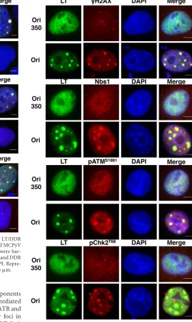
incompetent for LT-mediated replication (9). A construct with the Ori of SV40, which cannot be replicated by MCPyV LT, served as an additional negative control. MCPyV LT and most of the host DDR proteins had a diffuse nuclear pattern in the cells cotrans-fected with the plasmid carrying the replication-defective Ori350 or the SV40 Ori (Fig. 3and4and data not shown). However, when the wild-type MCPyV Ori was present, LT formed distinct foci in the nucleus in⬃20% of LT-positive cells (Fig. 3and4; also seeFig. 7C). This is consistent with our previous data showing that
MCPyV LT formed replication foci in the nucleus in the presence of the viral Ori (12). Interestingly, multiple components of the ATM-mediated DDR were also localized at these LT foci (Fig. 3). Approximately 50 cells displaying LT foci were quantified from each of the three independent experiments.␥H2AX, Nbs1, pATMS1981, and pChk2T68were localized at LT foci in 85.5%⫾
4.0%, 87.7%⫾3.3%, 54.2%⫾13.6%, and 91.5%⫾4.4% of cells displaying LT foci, respectively.
We next investigated the other arm of the host DDR, the ATR-FIG 1MCPyV LT colocalizes with␥H2AX, ATR, and pChk2T68in MCPyV-infected cells. U2OS cells were infected with native MCPyV virions (A) or MCPyV
pseudovirus with HPV capsid carrying the MCPyV genome (B). Infected and uninfected control cells were fixed at 5 days postinfection and stained for MCPyV LT (green) and DDR proteins (red), as indicated. The cells were counterstained with DAPI. Representative pictures from at least three experiments are shown. Bars, 5m. Tsang et al.
on November 7, 2019 by guest
http://jvi.asm.org/
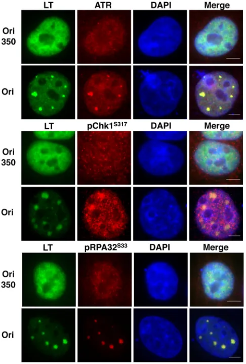
[image:4.585.138.450.63.573.2]mediated pathway. Similar to what we observed with components of the ATM-mediated DDR, components of the ATR-mediated pathway also colocalized with LT in nuclear foci (Fig. 4). ATR and pChk1S317 were observed to colocalize with LT nuclear foci in
95.8%⫾0.4% and 80.2% ⫾7.8% of cells displaying LT foci, respectively. In response to DNA damage, RPA32, the 32-kDa subunit of RPA, is also hyperphosphorylated by ATM, ATR, and DNA-dependent protein kinase (DNA-PK) to contribute to repair DNA synthesis (37). We also detected the signal of RPA32S33phosphorylation at the LT nuclear foci in 100% of
cells displaying LT foci (Fig. 4). Because the factors from both ATM- and ATR-mediated DDR pathways did not accumulate in distinct foci in cells cotransfected with an MCPyV LT-en-coding construct and a control vector carrying the SV40 Ori (data not shown), these data suggest that the DDR factors are associated with MCPyV LT at nuclear foci in an MCPyV Ori-dependent manner. C33A cells cotransfected with wild-type LT and Ori350 also displayed a diffuse pattern for both LT and various DDR factors (Fig. 3 and 4), further supporting the suggestion that the formation of DDR protein/LT foci depends on the LT-mediated replication of MCPyV DNA.
We also performed Western blot analysis to examine the acti-vation status of the ATM- and ATR-mediated DDR pathways in C33A cells transfected with MCPyV LT together with or without MCPyV Ori (Fig. 5). Consistent with our previous observations in U2OS cells (14), activation of the ATR pathway (as indicated by the induction of Chk1S345 phosphorylation) was observed in
C33A cells expressing MCPyV LT, either in the presence or in the absence of MCPyV Ori (Fig. 5A). MCPyV LT expression also ap-peared to moderately induce ATMS1981phosphorylation in C33A FIG 2The MCPyV genome alone is sufficient to reproduce the LT/DDR
colocalization phenotype. U2OS cells were transfected with religated MCPyV genome. Nontransfected control (NTC) cells and transfected cells were har-vested at 4 days posttransfection and stained for MCPyV LT (green) and DDR proteins (red), as indicated. The cells were counterstained with DAPI. Repre-sentative pictures from at least three experiments are shown. Bars, 5m.
FIG 3ATM-mediated DDR machinery colocalizes with MCPyV LT in nu-clear foci. C33A cells were cotransfected with a plasmid carrying MCPyV LT and with a plasmid carrying either the wild-type MCPyV origin (Ori) or the replication-defective origin (Ori350). At 48 hpt, cells were stained for MCPyV LT (green) and DDR proteins (red), as indicated. The cells were counter-stained with DAPI. Representative pictures from at least three experiments are shown. Bars, 5m.
on November 7, 2019 by guest
http://jvi.asm.org/
[image:5.585.46.523.64.462.2] [image:5.585.256.537.71.541.2]cells, indicating the activation of ATM kinase in these cells
(Fig. 5A). In addition, C33A cells transfected with MCPyV LT
either with or without MCPyV Ori also showed increased RPA32S33phosphorylation and an increased␥H2AX signal (Fig.
5). These results demonstrate that the ATM- and ATR-mediated DDR pathways are activated in C33A cells expressing MCPyV LT.
Host DDR machinery and MCPyV LT colocalize at actively
replicating viral origins in the nucleus.Previously, we have
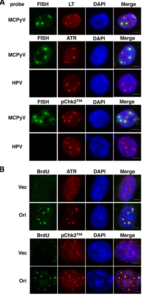
de-tected DNA damage in U2OS cells upon ectopic expression of full-length MCPyV LT or C-terminus-only LT truncation mu-tants (14). It is possible that the DDR machinery is recruited to the sites of DNA breaks on the host genome. To ensure that the DDR factor/LT nuclear foci that we observed in C33A cells are, in fact, at the MCPyV Ori and not sites of repair on the host genome, we performed immuno-FISH to detect the localization of MCPyV Ori with respect to the host DDR machinery. C33A cells were cotransfected with a plasmid carrying MCPyV LT and a different plasmid carrying the MCPyV Ori. We then detected the localiza-tion of LT and DDR proteins by immunofluorescent staining and used a specific probe to detect the MCPyV Ori-containing plasmid by FISH (Fig. 6A). The FISH signal was detected only with the
MCPyV Ori-specific probe and not with a negative-control probe recognizing the HPV16 genome, which is absent from the HPV-negative C33A cells (Fig. 6A). Consistent with our previous obser-vation (12), MCPyV LT and MCPyV Ori colocalized in nuclear foci (Fig. 6A, top), which we believe to be MCPyV replication factories containing actively amplifying viral DNA and not single copies of the origin (see below). In addition, the various compo-nents of the host DDR machinery, such as ATR, pChk2T68, and
␥H2AX, also colocalized with the MCPyV Ori foci (Fig. 6Aand data not shown). These data demonstrate the recruitment of DDR proteins to the MCPyV Ori complex, confirming that the nuclear foci that we observed are not the result of LT-induced DNA dam-age on the host chromosomes. Our results also suggest that the DDR proteins may be involved in MCPyV DNA replication.
To rule out the possibility of nuclear aggregation and to ensure that the nuclear foci that we observed in the presence of MCPyV Ori are, in fact, the sites of viral replication, we tested for BrdU incorporation at these foci. Previously, we have used this tech-nique to demonstrate the incorporation of BrdU specifically in MCPyV LT/Ori foci (12). Consistently, in cells cotransfected with MCPyV LT and the Ori, the BrdU signal was observed in distinct nuclear foci, whereas in the control samples, the BrdU signal was minimal (Fig. 6B). Interestingly, both ATR and pChk2T68were found to colocalize with the BrdU signal (Fig. 6B), suggesting that the DDR factor/LT foci that we observed are indeed sites of active, robust viral DNA replication. Nevertheless, it is also possible that cells without these replication foci are maintaining low levels of MCPyV replication that are undetectable by immuno-FISH and BrdU incorporation analysis.
Formation of DDR protein/MCPyV LT nuclear foci is viral
replication dependent.To confirm that the nuclear foci at which
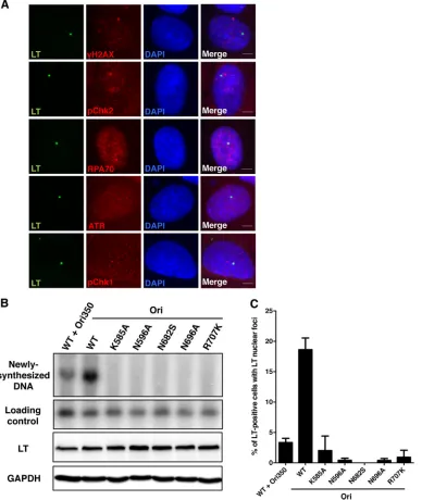
MCPyV LT and DDR factors colocalize are, in fact, dependent on viral DNA replication, we employed the U2OS 2-6-3 system, in which a LacO array has been integrated into the cellular genome of U2OS cells (38). U2OS 2-6-3 cells were transfected with a plasmid FIG 4ATR-mediated DDR machinery colocalizes with MCPyV LT in nuclear
foci. C33A cells were transfected as described in the legend toFig. 3. At 48 hpt, cells were stained for MCPyV LT (green) and DDR proteins (red), as indicated. The cells were counterstained with DAPI. Representative pictures from at least three experiments are shown. Bars, 5m.
FIG 5MCPyV LT activates a host DDR in C33A cells. C33A cells were cotransfected with a plasmid carrying either MCPyV LT or the control vector (Vec 1) and with a plasmid carrying either the MCPyV origin (Ori) or an empty vector (Vec 2). At 48 hpt, nuclear proteins were harvested for Western blotting with the indicated antibodies for ATM/ATR activation markers (A) or for␥H2AX (B). PCNA was used as a loading control for nuclear proteins. Tsang et al.
on November 7, 2019 by guest
http://jvi.asm.org/
[image:6.585.297.539.64.256.2] [image:6.585.42.285.67.426.2]carrying LacR-fused MCPyV LT. Consequently, the LT expressed in these cells was tethered to the LacO array due to the tight bind-ing of LacR to LacO, which was seen as a sbind-ingle LT-positive focus in each cell (Fig. 7A, LT). We immunostained LacR-LT-trans-fected U2OS 2-6-3 cells for various DDR proteins to see if LT itself physically interacts and brings these DDR factors to the LacO array. However, we observed no colocalization of LT and DDR proteins in these cells, suggesting that LT itself, even though it is concentrated in a single focus within the nucleus, cannot tether host DDR proteins to the LacO site. Considering the high affinity of LacR to LacO and the fact that the genome of U2OS 2-6-3 cells transfected with LacR alone cannot replicate normally (data not shown), we believe that the binding of LacR to LacO is so tight that this particular locus cannot be unwound/replicated. The absence of DDR factor/LT colocalization in this system suggests that the focus formation seen inFig. 1to4and6is replication dependent. In addition, we constructed a number of MCPyV LT mutants on the basis of homology to SV40 LT helicase mutations that abol-ish SV40 replication. When we cotransfected C33A cells with wild-type LT and the wild-wild-type viral Ori, we could robustly detect LT-mediated replication of the Ori plasmid by Southern blotting (Fig. 7B, Newly synthesized DNA). However, when MCPyV LT helicase mutants were transfected in place of the wild-type LT, replication of the Ori plasmid was completely abolished, confirming that these LT mutants were, in fact, replication defective. The mutants were expressed at levels comparable to those of wild-type LT (Fig. 7B, LT), indicating that the failure to replicate the wild-type Ori plasmid was not due to the gross instability of the mutants. DNA digested with only BamHI showed comparable hybrid-ization of the Southern blot probe to the Ori-containing plas-mid (Fig. 7B, Loading control), demonstrating comparable transfection efficiency and loading. The Southern blotting re-sults correlated with a dramatic reduction in LT focus forma-tion (quantified inFig. 7C). This provides additional evidence for the notion that the formation of LT foci depends on pro-ductive replication of viral DNA.
Moreover, wild-type LT cotransfected with Ori350 showed a much more attenuated ability to replicate the plasmid carrying the mutated Ori (Fig. 7B), and this also correlated with the decrease in the number of LT foci quantified (Fig. 7C). These data from LT mutants and the Ori350 mutant support the suggestion that the DDR protein/LT foci observed in our system are, in fact, viral replication dependent.
Treatment with DDR inhibitors reduces MCPyV DNA
repli-cation.Next, we tested the functional importance of the host DDR
to MCPyV by studying the effects of DDR inhibitors on autono-mous MCPyV DNA replication. Wortmannin was used in this study because it is a potent, covalent inhibitor of PI3Ks (39), which include ATM, ATR, and DNA-PK. However, it has been reported that wortmannin inhibits DSB repair and not SSB repair (40) and that at a higher concentration (20M) it efficiently in-hibits ATM activationin vivo(41). Therefore, we also tested the effects of NU6027, which specifically inhibits ATR activity but not ATM or DNA-PK activity (42), on MCPyV DNA replication. We first confirmed by Western blot analysis that wortmannin can reduce etoposide-induced ATMS1981 phosphorylation and that NU6027 can inhibit UVC-induced Chk1S345 phosphorylation
(data not shown). We then tested these drugs in C33A cells trans-fected with the pT⫹Ori plasmid carrying the sequences of MCPyV LT, sT, and the viral Ori, which has been shown to sup-FIG 6DDR proteins localize at the replicating MCPyV origin in the nucleus.
(A) C33A cells were cotransfected with a plasmid carrying MCPyV LT and a plasmid carrying the MCPyV origin (Ori). At 60 hpt, cells were stained for MCPyV LT and DDR proteins (red), as indicated. A specific probe recognizing the MCPyV Ori plasmid and a nonspecific probe recognizing the HPV genome were used for FISH (green). The cells were counterstained with DAPI. Repre-sentative pictures from at least three experiments are shown. Bars, 5m. (B) C33A cells were cotransfected with a plasmid carrying MCPyV LT and with a plasmid carrying either the MCPyV origin (Ori) or an empty vector (Vec). At 44 hpt, cells were pulsed with BrdU for 2 h. The cells were incubated in regular medium for another 2 h before they were fixed and stained for BrdU (green) and DDR proteins (red), as indicated. The cells were counterstained with DAPI. Representative pictures from at least three experiments are shown. Bars, 5m.
on November 7, 2019 by guest
http://jvi.asm.org/
[image:7.585.44.286.70.572.2]port autonomous replication in our previous study (12). In un-treated and DMSO-un-treated C33A cells, robust pT⫹Ori replication was detected (Fig. 8). In contrast, in the DDR inhibitor-treated samples, we noticed a decrease in pT⫹Ori replication (Fig. 8Aand
B). This reduction was particularly drastic in wortmannin-treated C33A cells (Fig. 8AandB), possibly due to wortmannin’s multi-targeting of phosphatidylinositol 3-kinases (see Discussion). Both wortmannin and NU6027 treatments also efficiently inhibited the
replication of the religated MCV genome (data not shown). Con-sistent with the Southern blotting results, we observed a decrease in replication focus formation in drug-treated cells by immuno-fluorescent staining (data not shown). We believe that the de-crease in autonomous replication is a consequence of DDR inhi-bition, as drug treatment had a minimal effect on MCPyV LT expression (Fig. 8A).
We also performed flow cytometry analysis to test how wort-FIG 7Formation of DDR protein/LT nuclear foci is dependent on MCPyV DNA replication. (A) U2OS 2-6-3 cells were transfected with a plasmid carrying LacR-fused MCPyV LT. At 36 hpt, cells were stained for MCPyV LT (green) and DDR proteins (red), as indicated. The cells were counterstained with DAPI. Representative pictures from at least three experiments are shown. Bars, 5m. (B) C33A cells were cotransfected with a plasmid carrying wild-type (WT) or mutant MCPyV LT (LT) and with a plasmid carrying either the wild-type MCPyV origin (Ori) or the replication-defective origin (Ori350). At 48 hpt, total cellular DNA was extracted for Southern blotting. Fifteen micrograms of DNA was digested with BamHI and DpnI to detect replicated origin plasmid, while 2 g of DNA was digested with only BamHI to show equal loading. Total protein extractions were used in Western blotting to detect MCPyV LT and GAPDH (glyceraldehyde-3-phosphate dehydrogenase). (C) C33A cells were transfected as described for panel B. At 48 hpt, cells were stained for MCPyV LT as described in the legend toFig. 3. About 150 LT-positive cells were quantified for the presence of LT nuclear foci. Data represent means and standard deviations calculated from three independent experiments.
Tsang et al.
on November 7, 2019 by guest
http://jvi.asm.org/
[image:8.585.103.491.69.529.2]mannin and NU6027 affect the host cell cycle, which could in turn affect pT⫹Ori replication. Although wortmannin treatment had a minimal effect on the cell cycle profile, NU6027 treatment led to an increase in the G1population compared to that for the
pT⫹Ori-transfected, DMSO-treated control (Fig. 8C). Even though we detected an⬃70% decrease in pT⫹Ori replication upon NU6027 treatment (Fig. 8B), part of this reduction could be a consequence of G1arrest in C33A cells. We further tested if drug
treatment affects cellular proliferation by immunostaining drug-treated C33A cells for Ki-67, which is an established marker for cell proliferation detected in all active phases of the cell cycle (G1,
S, and G2/M) but not in resting cells (G0). As shown inFig. 8D,
neither DDR inhibitor had much of an effect on Ki-67 positivity. This demonstrates that the drug-treated cells were, in fact, actively proliferating.
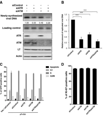
siRNA knockdown of ATR and/or ATM inhibits MCPyV
DNA replication.To rule out the possibility that wortmannin and
NU6027 have off-target and/or cell cycle effects on C33A cells that could affect MCPyV DNA replication, we performed siRNA knockdown of ATR and/or ATM to see if we could recapitulate observations with the DDR inhibitors. siRNA knockdown of ATR or ATM inhibited the activation of Chk1S345and Chk2T68 phos-FIG 8Treatment with wortmannin or NU6027 reduces viral DNA replication. C33A cells were transfected with pT⫹Ori carrying MCPyV LT, sT, and the viral origin. At 20 hpt, cells were treated with DMSO, 20M wortmannin, or 20M NU6027. DMSO and the DDR inhibitors were refreshed every 16 h. (A) At 52 hpt, total cellular DNA was extracted for Southern blotting. Thirty micrograms of DNA was digested with XhoI and DpnI to detect replicated viral DNA, while 2g of DNA was digested with only XhoI to show equal loading. Quantification was normalized to the amount for the nontreated control (NTC). Total protein extractions were used in Western blotting to detect MCPyV LT and actin. (B) Quantification of replicated viral DNA bands. The value from the nontreated control was arbitrarily set to 1. Data represent means and standard deviations calculated from three independent experiments. ***,P⬍0.001. (C) C33A cells were transfected with pT⫹Ori or an empty vector (Vec). Cells were drug treated as described for panel A. At 50 hpt, cells were pulsed with BrdU for 2 h and then fixed and stained with BrdU antibody and 7-aminoactinomycin D (7-AAD). About 12,000 GFP-positive cells were analyzed by flow cytometry. A representative gating strategy for each phase of the cell cycle is shown. Data represent means and standard deviations calculated from three independent experiments. (D) C33A cells were transfected and drug treated, as described for panel A. At 52 hpt, cells were stained for Ki-67. About 200 cells were quantified for Ki-67 positivity. Data represent means and standard deviations calculated from three independent experiments.
on November 7, 2019 by guest
http://jvi.asm.org/
[image:9.585.111.474.63.479.2]phorylation, respectively, in cells treated with UVC (data not shown). We cotransfected pT⫹Ori with control siRNA, siRNA against ATR (siATR), and/or siRNA against ATM (siATM) into C33A cells and performed Southern blotting to detect newly syn-thesized pT⫹Ori. At 48 hpt, both ATR and ATM were effectively knocked down (Fig. 9A). Similar to what we have observed with the DDR inhibitors, ATR and ATM knockdown reduced autono-mous pT⫹Ori replication in C33A cells (Fig. 9AandB). This decrease was even more apparent when both ATR and ATM were knocked down (Fig. 9AandB). We were also able to observe a decrease in replication focus formation in ATR/ATM double-knock-down cells by immunofluorescent staining (data not shown). The fact that ATR/ATM double-knockdown cells showed a slightly
greater reduction in pT⫹Ori replication than ATR or ATM single-knockdown cells could be explained by the functional redundancy of these two kinases. The decrease in pT⫹Ori replication is attributed to the knockdown of ATR and ATM, as siRNA treatment had a minimal effect on MCPyV LT expression (Fig. 9A), host cell cycle (Fig. 9C), and cellular proliferation (Fig. 9D) in C33A cells. These data suggest that both ATR- and ATM-mediated DDR is important for optimal MCPyV DNA replication.
DISCUSSION
Since its discovery, MCPyV has provided a new model for study-ing the oncogenic potential of polyomaviruses in humans. Al-though there is evidence suggesting that the interactions between FIG 9siRNA knockdown of ATR and ATM reduces viral DNA replication. C33A cells were cotransfected with pT⫹Ori and control siRNA (siControl), siATR, and/or siATM. (A) At 48 hpt, total cellular DNA was extracted for Southern blotting. Thirty micrograms of DNA was digested with XhoI and DpnI to detect replicated viral DNA, while 2g of DNA was digested with only XhoI to show equal loading. Quantification was normalized to the amount for control siRNA. Total protein extractions were used in Western blotting for ATR, ATM, MCPyV LT, and actin. (B) Quantification of replicated viral DNA bands. The value from control siRNA was arbitrarily set to 1. Data represent means and standard deviations calculated from three independent experiments. ***,P⬍0.001. (C) C33A cells were cotransfected with the indicated siRNA and either pT⫹Ori or an empty vector (Vec). At 46 hpt, cells were pulsed with BrdU for 2 h and then fixed and stained with BrdU antibody and 7-aminoactinomycin D. About 12,000 GFP-positive cells were analyzed by flow cytometry. Data represent means and standard deviations calculated from three independent experiments. (D) C33A cells were transfected as described for panel A. At 48 hpt, cells were stained for Ki-67. About 200 cells were quantified for Ki-67 positivity. Data represent means and standard deviations calculated from three independent experiments.
Tsang et al.
on November 7, 2019 by guest
http://jvi.asm.org/
[image:10.585.111.472.64.469.2]LT and host cell cycle-regulatory proteins contribute to tumor development (14,15), the exact oncogenic mechanism of MCPyV has not yet been established. Currently, we have limited under-standing of the MCPyV life cycle, including viral entry, replica-tion, and propagation processes. Recent reports on MCPyV recep-tor usage (10,36,43) and tropism (11) have begun to shed light on MCPyV basic biology. Our study of the host factors required for MCPyV replication has also started to tease out the mechanistic details of viral genome amplification (12). Using our established system for the study of MCPyV DNA replication (12), we investi-gated additional host machinery that is important for this process. In this study, we showed that, in cells infected with MCPyV, components of the ATM and ATR kinase pathways accumulate in MCPyV LT-positive nuclear foci. Colocalization of these DDR factors and MCPyV LT in nuclear foci was also observed in cells either transduced with pseudotyped virions composed of the HPV capsid and MCPyV genome or transfected with the MCPyV nome. This suggests that gene expression from the MCPyV ge-nome and not incoming virion-associated proteins is responsible for inducing the formation of DDR factor/LT nuclear foci. Using our previously established immuno-FISH method to visualize MCPyV LT/Ori replication complexes in cells (12), we showed that the DDR proteins and MCPyV LT colocalize at complexes that contain actively replicating MCPyV DNA. We believe that focus formation represents the presence of robust viral DNA rep-lication, where the newly synthesized DNA has accumulated to a level detectable by immuno-FISH/BrdU staining. It is possible that cells without these replication factories are maintaining low levels of MCPyV replication. While the immunofluorescent stain-ing revealed the formation of MCPyV DNA replication factories in the presence of robust replication, Southern blot analysis was used to demonstrate the overall level of MCPyV replication main-tained in cells with and without replication factories. Abrogation of the host DDR, using either chemical inhibitors (wortmannin and NU6027) or ATR/ATM siRNA knockdown, led to a decrease in MCPyV DNA replication, supporting the conclusion that these components of the host DDR are essential for robust viral genome amplification.
To date, it has been technically challenging to study the MCPyV life cycle in cultured cell lines due to the lack of a known natural host cell and the inefficient replication of the native viral genome in culture (reviewed in references44and45). In our ini-tial experiments, we were able to detect accumulation of DDR factors in the MCPyV LT-positive nuclear foci in a limited num-ber of cells infected with native MCPyV virions. To further inves-tigate the involvement of host DDR in MCPyV replication, our current study relied on an MCPyV DNA replication system that we previously established (12), in which MCPyV LT and Ori plas-mids were transiently transfected into C33A cells. Although this system suggests that the DDR machinery is important for MCPyV replication, it is essential to investigate how DDR activation con-tributes to viral replication in more natural MCPyV replication systems once they are established. Currently, there is some uncer-tainty about the nature of the precursor cells that give rise to MCC tumors (46). Understanding the behavior of MCPyV in MCC pre-cursor cell types will be important for further elucidation of the oncogenic effects of the virus.
In our study, we used the PI3K inhibitor wortmannin and ATR inhibitor NU6027 to show that the DDR response contributes to MCPyV replication. Since wortmannin not only inhibits ATM
and ATR but also inhibits other members of the PIKK family, such as DNA-dependent protein kinase (DNA-PK), we also tested a number of other DDR inhibitors, including NU7441 (a DNA-PK inhibitor), KU55933 (an ATM inhibitor), and AZD7762 (a Chk1 inhibitor). However, none of these drug treatments showed as dramatic of an effect as wortmannin and NU6027 (data not shown). This suggests that ATR may be more important for MCPyV replication than other DDR mediators. In line with the findings of the experiments performed with drug inhibitors (Fig. 8
and data not shown), assays with siRNA showed a more dramatic reduction in LT-mediated replication when ATR was knocked down than when ATM was knocked down, while ATR and ATM double knockdown decreased replication to the level similar to that obtained with ATR single knockdown (Fig. 9AandB).
Many groups have previously reported the activation of ATR-and/or ATM-dependent DDR pathways upon viral infection or viral protein expression. HPV, adenovirus, herpes simplex virus, Epstein-Barr virus, and retroviruses all induce a DDR (47,48). So far, research on polyomaviruses has also shown similar phenom-ena. However, despite the large amount of information on the activation of host DDR by many viruses, little is known about the mechanism by which these viruses trigger such a response in the host. Previously, SV40 LT alone has been shown to induce both ATR- and ATM-mediated DDR, and this DDR activation is dependent on LT’s interaction with the mitotic spindle check-point kinase Bub1 (30). In addition, for JCPyV, there is evidence suggesting that its LT-mediated G2cell cycle arrest is dependent
on LT’s ability to associate with cellular DNA (25). This observa-tion leads to the speculaobserva-tion that perhaps the viral origin-binding domain and the nonspecific DNA-binding domains on LT could tether this viral helicase to host chromosomes, allowing it to un-wind host DNA, which in turn would trigger DDR complex re-cruitment to the cellular DNA. This idea is in line with a recent report on BKPyV that showed severe chromosomal damage upon BKPyV infection in the absence of ATR/ATM (26). Moreover, studies have shown that SV40 and BKPyV infection leads to the accumulation of a cell population with⬎4N DNA content (26). For MCPyV, it is possible that the DNA intermediates generated during viral DNA replication are recognized by the host cells as damaged DNA, which could trigger the DDR activation that we observed upon viral infection/replication.
One of the most interesting questions remains: how exactly do viruses benefit from the activation of DDR? As mentioned above, ATR- and ATM-mediated DDR can help repair chromosomal damage caused by polyomavirus infection (26). This repair mech-anism not only allows the host to sustain virus-induced DNA damage but also permits the virus to propagate without killing the host. For SV40 and JCPyV, there is evidence suggesting that the activation of ATM- and ATR-mediated checkpoint signaling leads to cell cycle arrest in S and G2phases, which are conducive to viral
replication (24,25). We have also observed that MCPyV LT-in-duced ATR activation can lead to a modest G2arrest (14),
suggest-ing that the response may contribute to viral DNA replication by inducing a cellular environment that is beneficial for viral DNA replication. This model is consistent with a recent report from Cheng and colleagues showing that full-length LT can trigger growth inhibition in cultured cells (49). On the other hand, our immunofluorescent staining showed little ATR/ATM accumula-tion outside viral replicaaccumula-tion factories, so the virus-induced host DNA damage and repair are likely minimal. Furthermore, because
on November 7, 2019 by guest
http://jvi.asm.org/
the components of the ATM and ATR pathways localize to the sites of MCPyV replication, which contain replication factors and actively replicating viral DNA (12; this study), the DDR proteins are likely playing a more direct role in MCPyV DNA replication. For example, the host DDR factors may promote viral DNA rep-lication by repairing the viral reprep-lication-induced damage on its own DNA (27).
How these DDR proteins contribute to MCPyV DNA replica-tion will be the focus of our future study. Activareplica-tion and recruit-ment of DDR factors have also been observed during HPV repli-cation (50–53). For HPV, it is thought that the host machinery used for homologous recombination may be required for the cir-cularization of the viral genome for virion packaging (51). Inter-estingly, our recent studies have demonstrated that the cellular protein Brd4 is recruited to both HPV and MCPyV replication complexes to contribute to viral DNA replication (12,33). Brd4 has been shown to interact with the DNA damage response pro-tein ATAD5 (54,55), suggesting that it may play a role in DNA damage repair-associated viral DNA replication. Future studies will investigate whether Brd4 is involved in recruiting DDR factors and DNA damage-specific polymerases to the HPV and MCPyV origin to support DDR-mediated viral replication.
Most of the studies on polyomavirus replication have been performed using Southern blotting, quantitative PCR, orin vitro
assays. The study presented in this report combined immunoflu-orescent staining, immuno-FISH, and BrdU incorporation to vi-sualize the MCPyV replication complexes in cells. Building upon this platform, our study demonstrates that the host’s DDR pro-teins are important for robust MCPyV DNA replication. This sys-tem will be useful for further investigation of the mechanistic role of DDR factors in MCPyV replication. For example, excessive UV exposure is a major risk factor for MCC. Future study will inves-tigate whether overstimulation of host DDR by sunlight exposure may cause abnormal viral DNA replication, leading to viral DNA integration and oncogenic progression. Collectively, not only do these studies on the relationship between the host DDR and MCPyV provide insight into the host machinery required for MCPyV genome amplification, but also they may shed light on virus-associated oncogenesis. Therefore, research on the compo-nents of the host DDR may have important clinical implications for MCPyV-associated MCC.
ACKNOWLEDGMENTS
We thank Matthew D. Weitzman (The Children’s Hospital of Philadel-phia) for critical review and insightful critiques of the manuscript, Susan M. Janicki (The Wistar Institute) for the U2OS 2-6-3 cell line, and Thomas G. Magaldi for providing MCPyV native virions and reporter pseudovirions. We thank the members of our laboratories for helpful discussion and critical review of the manuscript.
This work was supported by the HIV-Associated Malignancies Pilot Project Award (National Cancer Institute), National Institutes of Health (NIH) grants R01CA148768 and R01CA142723, tumor virology training grant 2-T32-CA-115299-07, and the Intramural Research Program of the NIH, National Cancer Institute, Center for Cancer Research.
REFERENCES
1.Feng H, Shuda M, Chang Y, Moore PS.2008. Clonal integration of a polyomavirus in human Merkel cell carcinoma. Science319:1096 –1100.
http://dx.doi.org/10.1126/science.1152586.
2.Tolstov YL, Pastrana DV, Feng H, Becker JC, Jenkins FJ, Moschos S, Chang Y, Buck CB, Moore PS.2009. Human Merkel cell polyomavirus infection. II. MCV is a common human infection that can be detected by
conformational capsid epitope immunoassays. Int. J. Cancer125:1250 – 1256.http://dx.doi.org/10.1002/ijc.24509.
3.Foulongne V, Sauvage V, Hebert C, Dereure O, Cheval J, Gouilh MA, Pariente K, Segondy M, Burguière A, Manuguerra J-C, Caro V, Eloit M. 2012. Human skin microbiota: high diversity of DNA viruses identified on the human skin by high throughput sequencing. PLoS One7:e38499.http: //dx.doi.org/10.1371/journal.pone.0038499.
4.Heath M, Jaimes N, Lemos B, Mostaghimi A, Wang LC, Peñas PF, Nghiem P.2008. Clinical characteristics of Merkel cell carcinoma at di-agnosis in 195 patients: the AEIOU features. J. Am. Acad. Dermatol.58: 375–381.http://dx.doi.org/10.1016/j.jaad.2007.11.020.
5.Hodgson NC.2005. Merkel cell carcinoma: changing incidence trends. J. Surg. Oncol.89:1– 4.http://dx.doi.org/10.1002/jso.20167.
6.Bouvard V, Baan RA, Grosse Y, Lauby-Secretan B, El Ghissassi F, Benbrahim-Tallaa L, Guha N, Straif K.2012. Carcinogenicity of malaria and of some polyomaviruses. Lancet Oncol.13:339 –340.http://dx.doi.org /10.1016/S1470-2045(12)70125-0.
7.Gjoerup O, Chang Y.2010. Update on human polyomaviruses and can-cer. Adv. Cancer Res. 106:1–51.http://dx.doi.org/10.1016/S0065-230X (10)06001-X.
8.Harrison CJ, Meinke G, Kwun HJ, Rogalin H, Phelan PJ, Bullock PA, Chang Y, Moore PS, Bohm A.2011. Asymmetric assembly of Merkel cell polyomavirus large T-antigen origin binding domains at the viral origin. J. Mol. Biol.409:529 –542.http://dx.doi.org/10.1016/j.jmb.2011.03.051. 9.Kwun HJ, Guastafierro A, Shuda M, Meinke G, Bohm A, Moore PS,
Chang Y.2009. The minimum replication origin of Merkel cell polyoma-virus has a unique large T-antigen loading architecture and requires small T-antigen expression for optimal replication. J. Virol.83:12118 –12128.
http://dx.doi.org/10.1128/JVI.01336-09.
10. Schowalter RM, Pastrana DV, Buck CB.2011. Glycosaminoglycans and sialylated glycans sequentially facilitate Merkel cell polyomavirus infec-tious entry. PLoS Pathog.7:e1002161.http://dx.doi.org/10.1371/journal .ppat.1002161.
11. Schowalter RM, Reinhold WC, Buck CB.2012. Entry tropism of BK and Merkel cell polyomaviruses in cell culture. PLoS One7:e42181.http://dx .doi.org/10.1371/journal.pone.0042181.
12. Wang X, Li J, Schowalter RM, Jiao J, Buck CB, You J.2012. Bromodo-main protein Brd4 plays a key role in Merkel cell polyomavirus DNA replication. PLoS Pathog.8:e1003021.http://dx.doi.org/10.1371/journal .ppat.1003021.
13. Demetriou SK, Ona-Vu K, Sullivan EM, Dong TK, Hsu SW, Oh DH. 2012. Defective DNA repair and cell cycle arrest in cells expressing Merkel cell polyomavirus T antigen. Int. J. Cancer131:1818 –1827.http://dx.doi .org/10.1002/ijc.27440.
14. Li J, Wang X, Diaz J, Tsang SH, Buck CB, You J.2013. Merkel cell polyomavirus large T antigen disrupts host genomic integrity and inhibits cellular proliferation. J. Virol.87:9173–9188.http://dx.doi.org/10.1128 /JVI.01216-13.
15. Houben R, Adam C, Baeurle A, Hesbacher S, Grimm J, Angermeyer S, Henzel K, Hauser S, Elling R, Bröcker E-B, Gaubatz S, Becker JC, Schrama D.2012. An intact retinoblastoma protein-binding site in Merkel cell polyomavirus large T antigen is required for promoting growth of Merkel cell carcinoma cells. Int. J. Cancer130:847– 856.http: //dx.doi.org/10.1002/ijc.26076.
16. Shuda M, Feng H, Kwun HJ, Rosen ST, Gjoerup O, Moore PS, Chang Y.2008. T antigen mutations are a human tumor-specific signature for Merkel cell polyomavirus. Proc. Natl. Acad. Sci. U. S. A.105:16272–16277.
http://dx.doi.org/10.1073/pnas.0806526105.
17. Ciccia A, Elledge SJ.2010. The DNA damage response: making it safe to play with knives. Mol. Cell 40:179 –204. http://dx.doi.org/10.1016/j .molcel.2010.09.019.
18. Dupre A, Boyer-Chatenet L, Gautier J. 2006. Two-step activation of ATM by DNA and the Mre11-Rad50-Nbs1 complex. Nat. Struct. Mol. Biol.13:451– 457.http://dx.doi.org/10.1038/nsmb1090.
19. Cimprich KA, Cortez D.2008. ATR: an essential regulator of genome integrity. Nat. Rev. Mol. Cell Biol.9:616 – 627.http://dx.doi.org/10.1038 /nrm2450.
20. Shiotani B, Zou L.2009. Single-stranded DNA orchestrates an ATM-to-ATR switch at DNA breaks. Mol. Cell33:547–558.http://dx.doi.org/10 .1016/j.molcel.2009.01.024.
21. Olson E, Nievera CJ, Lee AY-L, Chen L, Wu X.2007. The Mre11-Rad50-Nbs1 complex acts both upstream and downstream of ataxia telangiectasia mutated and Rad3-related protein (ATR) to regulate the S-phase check-Tsang et al.
on November 7, 2019 by guest
http://jvi.asm.org/
point following UV treatment. J. Biol. Chem.282:22939 –22952.http://dx .doi.org/10.1074/jbc.M702162200.
22. Myers JS, Cortez D.2006. Rapid activation of ATR by ionizing radiation requires ATM and Mre11. J. Biol. Chem.281:9346 –9350.http://dx.doi .org/10.1074/jbc.M513265200.
23. Adams KE, Medhurst AL, Dart DA, Lakin ND.2006. Recruitment of ATR to sites of ionising radiation-induced DNA damage requires ATM and components of the MRN protein complex. Oncogene25:3894 –3904.
http://dx.doi.org/10.1038/sj.onc.1209426.
24. Dahl J, You J, Benjamin TL.2005. Induction and utilization of an ATM signaling pathway by polyomavirus. J. Virol.79:13007–13017.http://dx .doi.org/10.1128/JVI.79.20.13007-13017.2005.
25. Orba Y, Suzuki T, Makino Y, Kubota K, Tanaka S, Kimura T, Sawa H. 2010. Large T antigen promotes JC virus replication in G2-arrested cells by
inducing ATM- and ATR-mediated G2checkpoint signaling. J. Biol.
Chem.285:1544 –1554.http://dx.doi.org/10.1074/jbc.M109.064311. 26. Jiang M, Zhao L, Gamez M, Imperiale MJ.2012. Roles of ATM and
ATR-mediated DNA damage responses during lytic BK polyomavirus in-fection. PLoS Pathog.8:e1002898.http://dx.doi.org/10.1371/journal.ppat .1002898.
27. Sowd GA, Li NY, Fanning E.2013. ATM and ATR activities maintain replication fork integrity during SV40 chromatin replication. PLoS Pathog.9:e1003283.http://dx.doi.org/10.1371/journal.ppat.1003283. 28. Boichuk S, Hu L, Hein J, Gjoerup OV.2010. Multiple DNA damage
signaling and repair pathways deregulated by simian virus 40 large T an-tigen. J. Virol.84:8007– 8020.http://dx.doi.org/10.1128/JVI.00334-10. 29. Zhao X, Madden-Fuentes RJ, Lou BX, Pipas JM, Gerhardt J, Rigell CJ,
Fanning E.2008. Ataxia telangiectasia-mutated damage-signaling kinase-and proteasome-dependent destruction of Mre11-Rad50-Nbs1 subunits in simian virus 40-infected primate cells. J. Virol.82:5316 –5328.http://dx .doi.org/10.1128/JVI.02677-07.
30. Hein J, Boichuk S, Wu J, Cheng Y, Freire R, Jat PS, Roberts TM, Gjoerup OV. 2009. Simian virus 40 large T antigen disrupts genome integrity and activates a DNA damage response via Bub1 binding. J. Virol. 83:117–127.http://dx.doi.org/10.1128/JVI.01515-08.
31. Shi Y, Dodson GE, Shaikh S, Rundell K, Tibbetts RS. 2005. Ataxia-telangiectasia-mutated (ATM) is a T-antigen kinase that controls SV40 viral replication in vivo. J. Biol. Chem.280:40195– 40200.http://dx.doi .org/10.1074/jbc.C500400200.
32. Yan J, Li Q, Lievens S, Tavernier J, You J. 2010. Abrogation of the Brd4-positive transcription elongation factor b complex by papillomavi-rus E2 protein contributes to viral oncogene repression. J. Virol.84:76 – 87.http://dx.doi.org/10.1128/JVI.01647-09.
33. Wang X, Helfer CM, Pancholi N, Bradner JE, You J.2013. Recruitment of Brd4 to the human papillomavirus type 16 DNA replication complex is essential for replication of viral DNA. J. Virol.87:3871–3884.http://dx.doi .org/10.1128/JVI.03068-12.
34. Schowalter RM, Pastrana DV, Pumphrey KA, Moyer AL, Buck CB. 2010. Merkel cell polyomavirus and two previously unknown polyomavi-ruses are chronically shed from human skin. Cell Host Microbe7:509 – 515.http://dx.doi.org/10.1016/j.chom.2010.05.006.
35. Feng H, Kwun HJ, Liu X, Gjoerup O, Stolz DB, Chang Y, Moore PS. 2011. Cellular and viral factors regulating Merkel cell polyomavirus rep-lication. PLoS One 6:e22468. http://dx.doi.org/10.1371/journal.pone .0022468.
36. Neumann F, Borchert S, Schmidt C, Reimer R, Hohenberg H, Fischer N, Grundhoff A.2011. Replication, gene expression and particle produc-tion by a consensus Merkel cell polyomavirus (MCPyV) genome. PLoS One6:e29112.http://dx.doi.org/10.1371/journal.pone.0029112. 37. Zernik-Kobak M, Vasunia K, Connelly M, Anderson CW, Dixon K.
1997. Sites of UV-induced phosphorylation of the p34 subunit of replica-tion protein A from HeLa cells. J. Biol. Chem.272:23896 –23904.http://dx .doi.org/10.1074/jbc.272.38.23896.
38. Janicki SM, Tsukamoto T, Salghetti SE, Tansey WP, Sachidanandam R, Prasanth KV, Ried T, Shav-Tal Y, Bertrand E, Singer RH, Spector DL. 2004. From silencing to gene expression: real-time analysis in single cells. Cell116:683– 698.http://dx.doi.org/10.1016/S0092-8674(04)00171-0. 39. Wymann MP, Bulgarelli-Leva G, Zvelebil MJ, Pirola L, Vanhaesebroeck
B, Waterfield MD, Panayotou G.1996. Wortmannin inactivates
phos-phoinositide 3-kinase by covalent modification of Lys-802, a residue in-volved in the phosphate transfer reaction. Mol. Cell. Biol.16:1722–1733. 40. Boulton S, Kyle S, Yalçintepe L, Durkacz BW.1996. Wortmannin is a potent inhibitor of DNA double strand break but not single strand break repair in Chinese hamster ovary cells. Carcinogenesis17:2285–2290.http: //dx.doi.org/10.1093/carcin/17.11.2285.
41. Goldstine JV, Nahas S, Gamo K, Gartler SM, Hansen RS, Roelfsema JH, Gatti RA, Marahrens Y.2006. Constitutive phosphorylation of ATM in lymphoblastoid cell lines from patients with ICF syndrome without downstream kinase activity. DNA Repair (Amst.)5:432– 443.http://dx .doi.org/10.1016/j.dnarep.2005.12.002.
42. Peasland A, Wang LZ, Rowling E, Kyle S, Chen T, Hopkins A, Cliby WA, Sarkaria J, Beale G, Edmondson RJ, Curtin NJ.2011. Identification and evaluation of a potent novel ATR inhibitor, NU6027, in breast and ovarian cancer cell lines. Br. J. Cancer105:372–381.http://dx.doi.org/10 .1038/bjc.2011.243.
43. Neu U, Hengel H, Blaum BS, Schowalter RM, Macejak D, Gilbert M, Wakarchuk WW, Imamura A, Ando H, Kiso M, Arnberg N, Garcea RL, Peters T, Buck CB, Stehle T.2012. Structures of Merkel cell polyomavi-rus VP1 complexes define a sialic acid binding site required for infec-tion. PLoS Pathog.8:e1002738. http://dx.doi.org/10.1371/journal.ppat .1002738.
44. White MK, Gordon J, Khalili K.2013. The rapidly expanding family of human polyomaviruses: recent developments in understanding their life cycle and role in human pathology. PLoS Pathog.9:e1003206.http://dx .doi.org/10.1371/journal.ppat.1003206.
45. Dalianis T, Hirsch HH.2013. Human polyomaviruses in disease and cancer. Virology437:63–72.http://dx.doi.org/10.1016/j.virol.2012.12.015. 46. Zur Hausen A, Rennspiess D, Winnepenninckx V, Speel EJ, Kurz AK.
2013. Early B-cell differentiation in Merkel cell carcinomas: clues to cel-lular ancestry. Cancer Res.73:4982– 4987.http://dx.doi.org/10.1158/0008 -5472.CAN-13-0616.
47. Chaurushiya MS, Weitzman MD. 2009. Viral manipulation of DNA repair and cell cycle checkpoints. DNA Repair (Amst.)8:1166 –1176.http: //dx.doi.org/10.1016/j.dnarep.2009.04.016.
48. Lilley CE, Schwartz RA, Weitzman MD.2007. Using or abusing: viruses and the cellular DNA damage response. Trends Microbiol.15:119 –126.
http://dx.doi.org/10.1016/j.tim.2007.01.003.
49. Cheng J, Rozenblatt-Rosen O, Paulson KG, Nghiem P, DeCaprio JA. 2013. Merkel cell polyomavirus large T antigen has growth-promoting and inhibitory activities. J. Virol. 87:6118 – 6126.http://dx.doi.org/10 .1128/JVI.00385-13.
50. Fradet-Turcotte A, Bergeron-Labrecque F, Moody CA, Lehoux M, Laimins LA, Archambault J.2011. Nuclear accumulation of the papillo-mavirus E1 helicase blocks S-phase progression and triggers an ATM-dependent DNA damage response. J. Virol.85:8996 –9012.http://dx.doi .org/10.1128/JVI.00542-11.
51. Gillespie KA, Mehta KP, Laimins LA, Moody CA.2012. Human papil-lomaviruses recruit cellular DNA repair and homologous recombination factors to viral replication centers. J. Virol.86:9520 –9526.http://dx.doi .org/10.1128/JVI.00247-12.
52. Sakakibara N, Mitra R, McBride AA.2011. The papillomavirus E1 heli-case activates a cellular DNA damage response in viral replication foci. J. Virol.85:8981– 8995.http://dx.doi.org/10.1128/JVI.00541-11.
53. Reinson T, Toots M, Kadaja M, Pipitch R, Allik M, Ustav E, Ustav M. 2013. Engagement of the ATR-dependent DNA damage response at the human papillomavirus 18 replication centers during the initial amplifica-tion. J. Virol.87:951–964.http://dx.doi.org/10.1128/JVI.01943-12. 54. Rahman S, Sowa ME, Ottinger M, Smith JA, Shi Y, Harper JW, Howley
PM.2011. The Brd4 extraterminal domain confers transcription activa-tion independent of pTEFb by recruiting multiple proteins, including NSD3. Mol. Cell. Biol.31:2641–2652. http://dx.doi.org/10.1128/MCB .01341-10.
55. Ishii H, Inageta T, Mimori K, Saito T, Sasaki H, Isobe M, Mori M, Croce CM, Huebner K, Ozawa K, Furukawa Y.2005. Frag1, a homolog of alternative replication factor C subunits, links replication stress surveil-lance with apoptosis. Proc. Natl. Acad. Sci. U. S. A.102:9655–9660.http: //dx.doi.org/10.1073/pnas.0504222102.