Determines Its Functions during Virus Maturation Independently of a
Direct pp150 Interaction
Teal M. Brechtel,a* Edward S. Mocarski,bRitesh Tandona
Department of Microbiology, University of Mississippi Medical Center, Jackson, Mississippi, USAa
; Department of Microbiology and Immunology, Emory University School of Medicine, Atlanta, Georgia, USAb
ABSTRACT
Tegument proteins pp150 and pUL96 function at a late step in cytomegalovirus (CMV) maturation. Here, we show that pp150
interacts directly with pUL96; however, the N-terminal region of pp150 and the C-terminal region of pUL96, which are critical
for these proteins to function, are not required for this interaction. Moreover, the largely dispensable C-terminal region of
pp150 is critical for pp150-pUL96 interaction. To further study the role of pUL96, several point and clustered mutations were
engineered into the CMV Towne bacterial artificial chromosome (Towne-BAC) genome, replacing the conserved negatively
charged C-terminal residues of pUL96. Although individual point mutations (E
122A, D
124A, and D
125A) reduced virus growth
slightly, the clustered mutations of
122EVDDAV
127significantly reduced virus growth, produced small syncytial plaque
pheno-types, and impacted a late stage of virus maturation. When the UL96 C-terminal alanine conversion mutant (B6-BAC) virus was
serially passaged in cell culture, it gained a plaque size comparable to that of Towne-BAC, displayed an altered restriction
frag-ment length pattern, and replicated with increased growth kinetics. Whole-genome sequencing of this passaged virus (UL96P10)
and the similarly passaged Towne-BAC virus revealed major differences only in the RNA4.9 and UL96 regions. When one of the
mutations in the UL96 coding region was engineered into the B6-BAC virus, it significantly increased the plaque size and rescued
the virus growth rate. Thus, accumulation of compensatory mutations only in UL96 in this revertant and the specific
involve-ment of functionally dispensable regions of pp150 in the pUL96-pp150 interaction point toward a role for pUL96 in virus
matu-ration that does not depend upon pp150.
IMPORTANCE
Human cytomegalovirus causes significant medical problems in newborns, as well as in people with low immunity. In this study, we
investigated the functions of two essential virus proteins, pp150 and pUL96, and determined the impact of their mutual interaction on
virus replication. These studies provide valuable information that is critical for the development of targeted antiviral therapies.
T
he proteinaceous layer between the capsid and the envelope in
a herpesvirus virion, known as the tegument, is analogous to
the matrix layer found in some RNA viruses. Tegument proteins
function in at least four different ways; they (i) are delivered into
host cells upon virus entry as prepackaged transactivation factors
that can promote infection, e.g., cytomegalovirus (CMV) pp71 (
1
,
2
); (ii) are critical for the acquisition of the virus envelope and act
as a bridge between the nucleocapsids and the envelope layer (
3
,
4
); (iii) provide capsid stability during virus maturation and
traf-ficking, e.g., CMV pp150 and pUL96 (
5
,
6
); and (iv) help in the
trafficking of virus in the cell by engaging the host cytoskeleton,
e.g., herpes simplex virus (HSV) UL36 (
7–9
). Herpesvirus
tegu-ment has been thought to be amorphous in nature (
3
,
4
,
10
),
although a more ordered structure has been assigned to the inner
tegument layer, which is in close proximity to the nucleocapsid
(
11–13
). In CMV, this inner tegument includes pp150, the
prod-uct of the UL32 gene that binds to the capsids directly (
13
,
14
) and
plays a major role in maintaining the integrity of capsids during
maturation (
5
,
6
,
15
). More than 20 tegument proteins are
pack-aged in a CMV virion (
10
), but very few functional interactions
among these proteins have been reported. Moreover, it is
hypoth-esized that intact capsids are required for the visualization of
cap-sid-tegument interactions in CMV, a proposition strengthened by
the detection of pp150 binding to intact capsids but not with
in-dividual capsid proteins (
14
).
Studies of bacteriophages, which have a capsid structure very
similar to that of herpesviruses, have shown that the viral genome
is packaged in an energy-driven process to almost liquid
crystal-line density and thus the interior walls of the capsids sustain
sig-nificant positive pressure from inside (
16
). A changing
environ-ment during the translocation of nucleocapsids from the cell
nucleus through the cytoplasm to the outside of cell can
compro-mise the protective capabilities of the capsid shell in the absence of
any stabilizing factors. Herpesvirus tegument proteins include
important structural components that serve this
capsid-stabiliz-ing function (
3–6
). The size, shape, and structure of
betaherpes-virus capsids are very similar to those of alphaherpesbetaherpes-virus capsids
(
11
,
12
,
17
). Evidently, CMV DNA is packed at a significantly
higher pressure than HSV DNA because the CMV genome is
ap-Received27 December 2013Accepted30 January 2014
Published ahead of print5 February 2014
Editor:L. Hutt-Fletcher
Address correspondence to Ritesh Tandon, [email protected]. * Present address: Teal Brechtel, Arizona Biological & Biomedical Sciences Program, University of Arizona, Tucson, Arizona, USA.
Copyright © 2014, American Society for Microbiology. All Rights Reserved.
doi:10.1128/JVI.03784-13
on November 7, 2019 by guest
http://jvi.asm.org/
proximately 1.5 times as large as that of HSV despite their similar
capsid sizes (
18
). Thus, CMV may need additional accessory
fac-tors to stabilize the nucleocapsids. The CMV pp150 and pUL96
tegument proteins (
19
,
20
) are critical for virus growth in cell
culture (
21
,
22
) but lack obvious functional or sequence homologs
in either alpha- or gammaherpesviruses (
4
), indicating that these
proteins have important betaherpesvirus-specific functions. Both
of these proteins are essential for efficient virus maturation (
4–6
),
and they impact the accumulation of intact enveloped virus in the
cytoplasmic virus assembly compartment (AC) (
5
,
6
). pp150
binds to nucleocapsids through its amino-terminal region (
14
),
and two small conserved regions (CR-1 and-2) located in this
region determine the role of pp150 in virus maturation (
5
). The
interactions of pUL96 with capsids have not yet been established.
Deletion of the UL96 gene from the virus, insertion of stop codons
at different positions in this gene, or conditional degradation of
the pUL96 protein renders the virus unable to replicate (
6
). The
defects in UL96 mutant viruses are reflected at a late stage of the
virus life cycle, despite the fact that pUL96 is an early protein (
6
).
The phenotype of these viruses is comparable to but less severe
than that of a pp150 mutant virus (
5
). During wild-type infection,
pp150 and pUL96 colocalize in infected cells. Also, cells show
increased accumulation of pp150 in the nucleus following
infec-tion with a UL96 mutant virus (
6
). We hypothesized that pUL96
could either enhance pp150-capsid association or act as a
chaper-one to assist in the relocation of pp150-associated nucleocapsids
from the nucleus to the cytoplasm.
In this study, we explored the interaction between pp150 and
pUL96 and its impact on virus maturation. While full-length
pp150 and pUL96 interacted with each other, mutation of amino
acid residues in both pp150 and pUL96 that determine the
func-tions of these two proteins in virus maturation did not abolish this
interaction. Specific negatively charged amino acids within the
C-terminal region of the protein were found to be critical for
pUL96 function. When a replication-deficient UL96 mutant virus
was passaged in fibroblasts, it accumulated compensatory
muta-tions in UL96 but not in any other gene. These data indicate that
although pUL96 and pp150 are required at similar steps in the
virus life cycle and manifest similar mutant phenotypes, their
functions during virus maturation may not be dependent on each
other.
MATERIALS AND METHODS
Sequences and alignments.The human CMV (Merlin) UL96 amino acid sequence (YP_081543) was aligned with the amino acid sequences of pUL96 homologs from rhesus CMV (RhCMV;YP_068222), chimpanzee CMV (CCMV;AAM00734), human herpesvirus 6 (HHV6;CAA58360), and HHV7 (YP_073808). The alignments were done in VectorNTI (ver 11; Invitrogen Corporation, Carlsbad, CA) with the ClustalW algorithm. Predictprotein 2.0 (23) and Protein Investigator (ver. 3.0.2) (http://intro .bio.umb.edu/PI/) were used to predict the protein folding and secondary structure of pUL96 and its variants. Cloning of green fluorescent protein (GFP) and bacterial artificial chromosome (BAC) sequences into the CMV Towne strain (American Type Culture Collection) genome has been reported previously (21). For sequencing of the viral genomes, viral DNA was purified from pelleted virions from the cleared cell culture medium with the DNeasy Tissue kit (Qiagen, Inc.) and the resultant double-stranded DNA was quantified with a Qubit fluorometric assay (Invitro-gen). Multiplexed paired-end libraries (2⫻150 bp) were prepared with the Nextera XT DNA Sample Preparation kit (Illumina). Whole-genome sequencing was done with an Illumina MiSeq. CLC Genomics
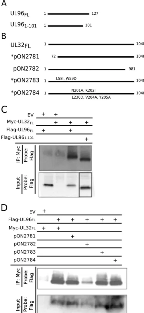
Work-FIG 1pp150 and pUL96 directly interact with each other; the N-terminal region of pUL96 and the C-terminal region of pp150 determine this interac-tion. Shown are line diagrams representing the Flag epitope-tagged UL96 ex-pression plasmids (A) and Myc epitope-tagged UL32 exex-pression plasmids used in this study (B). N-terminal pp150 mutants are denoted with an asterisk. (C) Full-length pp150 (UL32FL) interacts with full-length pUL96 (UL96FL), as well
as the C-terminal truncated pUL96 (UL961-101), but not with the empty vector
(EV) in an IP-IB assay after expression in 293T cells. (D) UL96FLinteracts with
UL32FL, as well as with the N-terminal mutants of pp150 (pON2781,
pON2783, and pON2784), but only slightly with the C-terminal mutant of pp150 (pON2782) in an IP-IB assay after expression in 293T cells. The empty vector (EV) was used as a control.
on November 7, 2019 by guest
http://jvi.asm.org/
[image:2.585.299.539.69.591.2]bench v6.0 software (CLCbio) was used for quality trimming of the reads andde novoassembly. An average of 20.5 million paired-end reads per virus were collected. The fastq files were imported into CLC Genomics Workbench 6.0. The reads were trimmed for quality with a limit of 0.05 (P
of error, equivalent to Q13) and a maximum of two ambiguities, and reads of⬍15 bp were discarded. Thede novoassembly used a word size of 45, a bubble size of 90, and a minimum contig length of 1,000; paired distances were autodetected; and reads were mapped back onto contigs. All other FIG 2pUL96 is a conserved betaherpesvirus protein critical for CMV maturation. Shown is an amino acid sequence alignment of pUL96 homologs from human CMV (HCMV;YP_081543), RhCMV (YP_068222), CCMV (AAM00734), HHV6 (CAA58360), and HHV7 (YP_073808). Identical amino acids (black) and similar amino acids (gray) are highlighted. The locations of the mutations introduced into the C-terminal region of pUL96 in this study are depicted. The last six amino acids of HCMV_UL96 are termed the EVDDAV cluster.
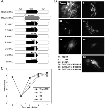
FIG 3Generation of UL96 mutant viruses by recombineering of the Towne-BAC genome. (A) Schematic of the UL95-UL97 region in Towne-BAC (top line) with other bacmid constructs shown below.␦UL96BAC is depicted with a KanSacB insert replacing UL96. In B1-BAC, glutamate 122; in B2-BAC, aspartate 124; and in B3-BAC, aspartate 125 have been replaced with an alanine residue. In B4-, B5-, and B6-BAC, a negatively charged cluster of pUL96 C-terminal residues (EVDDAV) has been replaced with positively charged residues KRKKRR, the negative charge alteration DEEEDE, or an alanine conversion of all six residues, respectively. R-BAC, constructed from␦UL96BAC, is depicted with the repaired UL96. (B) Typical eGFP⫹plaque phenotypes of indicated BAC constructs at 7 dpt in HF. Original magnification,⫻400. (C) Single-step growth curves of Towne-BAC and UL96 mutant viruses. HF were infected (MOI of 3.0), the total viruses (cells plus medium) were harvested at the indicated times postinfection, and their titers were determined on HF monolayers. The viral titers represent the averages of triplicate experiments, and the standard errors are within the symbols. The datum point at 0 dpi indicates the input virus dose.
on November 7, 2019 by guest
http://jvi.asm.org/
[image:3.585.98.490.67.176.2] [image:3.585.115.471.284.646.2]parameters were default. The number of scaffolds, scaffold N50, and total sequence length, respectively, for each virus were as follows: Towne-BAC, 4, 49 kb, and 232 kb; UL96P10, 4, 49 kb, and 232 kb. Each scaffold from each virus had an average coverage of⬎7,000. Gaps in the genome se-quence were filled by PCR and DNA sequencing of resultant amplicons. Open reading frames were identified by NCBI-BLAST search and anno-tated in Sequin 12.30.
Cells.Primary human foreskin-derived fibroblasts (HF) or 293T cells were cultured in Dulbecco’s modified Eagle’s medium (Invitrogen Cor-poration, Carlsbad, CA) containing 4.5 g/ml glucose, 10% fetal bovine serum (S1245OH; Atlanta Biologicals, Lawrenceville, GA), 1 mM sodium pyruvate, 2 mM L-glutamine, and 100 U/ml penicillin-streptomycin (Cellgro, Manassas, VA) at 37°C with 5% CO2. HF between passages 5 and 15 were used for transfections and infections. The cell culture medium was changed (for transfections) or additional medium was added (for infections) every other day.
BAC mutagenesis and recombinant viruses. BAC recombineer-ing protocols (24) were followed to mutate regions of UL96 with a KanSacB cassette as reported earlier (6). From the selected BAC clone (␦UL96BAC), additional clones were derived following the introduc-tion of a PCR fragment containing wild-type UL96 (rescued BAC [R-BAC]), E122A (B1-BAC), D124A (B2-BAC), D125A (B3-BAC), a
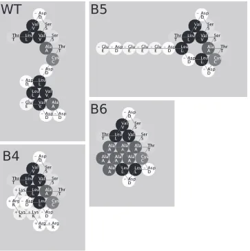
122EVDDAV127-to-KRKKRR conversion (B4-BAC), a122EVDDAV127 -to-DEEEDE conversion (B5-BAC), or a 122EVDDAV127-to-AAAAAA conversion (B6-BAC) and subsequent screening for the loss of kanamycin resistance, as well as sucrose sensitivity. Insertion of the desired mutation, as well as the absence of any undesired changes in the genome, was con-firmed by restriction fragment length pattern (RFLP) analysis and further by DNA sequencing of the UL96 region of these viruses. At 10 days post-transfection (dpt), supernatants from HF cultures transfected with repli-cation-competent BACs were harvested for recovery of recombinant vi-ruses. Single-step growth curves were obtained by infecting confluent HF at a multiplicity of infection [MOI] of 3.0 in triplicate and harvesting cells plus supernatants at designated time points postinfection. Each sample was sonicated (Sonicator 3000; Misonix Incorporated, Farmingdale, NY) on ice for 30 s before freezing or direct titration. The samples were diluted 10- to 100,000-fold for titration. At 10 days postinfection (dpi), cells were fixed in methanol, stained with Giemsa stain (Sigma-Aldrich, St. Louis, MO), and allowed to dry before enumeration of PFU under an optical microscope. The R-BAC construct does not differ in its properties from Towne-BAC, as dem-onstrated earlier (6). For passaging of the mutant viruses, BAC-transfected cells from the six-well tissue culture dishes were split (1:2) at 10 dpt into new six-well tissue culture dishes and incubated with a change of medium every alternate day. The mutant virus was allowed to grow, and its growth was FIG 4Protein Investigator (ver. 3.0.2)-based prediction of folding of the C-terminal 20 amino acids of wild type (WT) pUL96 and predicted changes in protein folding as a result of the clustered mutations (B4, B5, and B6) that were introduced in this study. The above program folds the protein on the basis of the interactions between the side chains only and does not model secondary or quaternary structure. The circles are colored to indicate the relative hydrophobicities of the side chains. The most hydrophilic amino acids are white, the most hydrophobic amino acids are black, and gray amino acids are intermediate. Amino acids with positively charged side chains have a small plus sign on their symbol; those with negatively charged side chains have a small minus sign.
on November 7, 2019 by guest
http://jvi.asm.org/
[image:4.585.113.476.66.431.2]monitored by enhanced GFP (eGFP) fluorescence. When eGFP was spread to the entire monolayer, the cells were split (1:2) into a T75 flask. Growth of virus was monitored in a similar manner, and the cells were then split into a T175 flask maintaining the approximately 1:2 split ratio. Virus was harvested when the cytopathic effect reached 100% and then passaged 10 times in T175 flasks before virus stocks were prepared and the growth rates of viruses were com-pared as described above.
Antibodies and IB analyses.293T cells were used for plasmid trans-fections. Cells were harvested at 48 h posttransfection for analysis by im-munoprecipitation (IP) and immunoblotting (IB). Anti-c-Myc affinity gel (Sigma-Aldrich, St. Louis, MO) was used for IP. Anti-FLAG M2 anti-body (Agilent Technologies, Santa Clara, CA) was used as the primary antibody, and peroxidase-labeled horse anti-mouse IgG (Vector Labora-tories, Burlingame, CA) was used as the secondary antibody for IB analy-ses. Blots were detected with ECL Western blotting detection reagents (GE Healthcare, Buckinghamshire, United Kingdom).
Microscopy.Samples for transmission electron microscopy (TEM) were prepared by transfecting HF with B4-BAC or Towne-BAC bacmids and passaging the HF once at 10 dpt and fixing them at 3 days postseeding. TEM of UL96P10, UL96ATG, or Towne-BAC virus-infected HF was per-formed by infecting HF at an MOI of 3.0 and fixing the cells at 5 dpi. In
each case, cells were fixed at the endpoint in 2.5% glutaraldehyde in 0.1 M cacodylate buffer (pH 7.2) for 2 h at room temperature. Cells were then washed with the same buffer and postfixed with buffered 1.0% osmium tetroxide at room temperature for 1 h. Following several washes with 0.1 M cacodylate buffer, cells were dehydrated with ethanol, infiltrated, and embedded in Eponate 12 resin (Ted Pella Inc., Redding, CA). Cell culture plates were cracked with a hammer to release the resin after it had solidi-fied, and ultrathin sections (60 to 70 nm) of monolayer cells were cut and counterstained with uranyl acetate and lead citrate. Examination of ultra-thin sections by TEM was carried out on a Hitachi H-7500 operated at 75 kV, and images were captured with a Gatan BioScan charge-coupled de-vice camera (Gatan Inc., Pleasanton, CA). The images were acquired and analyzed with the DigitalMicrograph software (Gatan Inc.).
Nucleotide sequence accession numbers.The whole-genome se-quences of the UL96P10 virus and the Towne-BAC passaged virus have been deposited in the GenBank database and assigned accession numbers
KF493876andKF493877, respectively (25,26).
RESULTS
pUL96 interacts with pp150.
C-terminal truncations of pUL96
arrest virus growth similarly to that of a UL96-null virus,
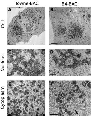
indicat-FIG 5Transmission electron microscopy (TEM) of infected fibroblasts. HF transfected with Towne-BAC (A, C, E) or B4-BAC (B, D, F) were passaged once at 10 dpt and fixed for TEM at 3 days postseeding. The infected cell (A, B), the nucleus (C, D), and the cytoplasm (E, F) are shown. Bars, 5m (A, B) or 1m (C to F).on November 7, 2019 by guest
http://jvi.asm.org/
[image:5.585.134.449.63.464.2]ing the functional importance of the UL96 C-terminal region (
6
).
We generated UL96 expression clones that were either full length
(UL96
FL) or truncated by the conserved C-terminal region
(UL96
1-101) (
Fig. 1A
). For pp150, several expression clones that
either truncate or mutate this protein (
Fig. 1B
) have been
charac-terized earlier (
15
). pp150 mutations located in the N-terminal
capsid-binding region of the protein (pON2781, pON2783, and
pON2784) (
Fig. 1B
) affect virus growth to various degrees (
5
,
15
);
however, the C-terminal mutant protein (pON2782) functions
similarly to the wild-type protein (
15
). Interactions between Myc
epitope-tagged pp150 and Flag epitope-tagged pUL96 clones were
investigated by IP and IB (
Fig. 1C
and
D
). pp150 interacted with
full-length, as well as C-terminally truncated, pUL96 (
Fig. 1C
). In
a different experiment, pUL96 interacted with all of the
N-termi-nally mutated forms of pp150; however, a C-terminal mutation
significantly reduced pp150-pUL96 interaction (
Fig. 1D
). Thus,
interaction between pp150 and pUL96 is determined by the C
terminus of pp150 and the N terminus of pUL96. Surprisingly, the
N-terminal region of pp150 and the C-terminal region of pUL96,
which are critical for these two proteins to function, are not
essen-tial for their direct interaction. Moreover, the C-terminal region
of pp150, which is largely dispensable for function (
15
), appears to
be important for pp150-pUL96 interaction. These data suggest
independent roles of pp150 and pUL96 in virus maturation
de-spite their direct interaction.
Conserved UL96 C-terminal residues are critical for virus
maturation.
A cluster of negatively charged amino acids in the
pUL96 C-terminal region is highly conserved among
betaherpes-viruses (
Fig. 2
). A virus truncated by this region of pUL96 is
ar-rested for growth in cell culture (
6
). To map the amino acids that
determine pUL96 function in virus maturation, we engineered
several point and clustered mutations replacing these amino acids
in Towne-BAC by genetic recombineering (
5
,
6
,
24
) (
Fig. 2
and
3
).
Individual amino acids E122, D124, and D125 were converted to
alanine residues (B1-, B2-, and B3-BAC, respectively) (
Fig. 3A
).
On the basis of the protein folding prediction of pUL96 and the
degree of change in predicted protein folding upon the
introduc-tion of these mutaintroduc-tions (
Fig. 4
), the entire amino acid cluster
(
122EVDDAV
127) was converted to positively charged residues
(KRKKRR [B4-BAC]), alternative negatively charged residues
(DEEEDE [B5-BAC]), or neutral alanines (AAAAAA [B6-BAC])
(
Fig. 3A
and
B
). While converting the
122EVDDAV
127cluster to
positively charged or neutral residues makes the UL96 C terminus
more compact, changing the positions of negative charges makes
it more linear (
Fig. 4
). Resulting mutant BACs were transfected
into HF and analyzed for growth by monitoring the eGFP
⫹plaque
size (
Fig. 3B
) and single-step growth curves of harvested viruses
(
Fig. 3C
). Individual point mutations (E
122A, D
124A, and D
125A)
reduced the virus plaque size (
Fig. 3B
) but did not inhibit the
production of infectious virus (
Fig. 3C
). While the B1, B2, and B3
viruses grew more slowly than Towne-BAC, all of these point
mu-tant viruses had approximately equivalent final virus yields (
Fig.
3C
). In contrast, all three-cluster mutant viruses (B4, B5, and B6)
showed impaired growth in fibroblasts, produced small syncytial
plaque phenotypes (
Fig. 3B
), and failed to release infectious virus
into the supernatant of transfected cells. In these mutant
infec-tions, pUL96 expression was not impaired (data not shown).
To study the stage of viral infection impacted by clustered
mutations in pUL96, we performed TEM of infected HF that
were passaged once after transfections (
Fig. 5
). This single
pas-saging was done to eliminate the possible secondary effects of
transfection on cell morphology before TEM.
Towne-BAC-in-fected cells manifested a kidney-shaped nucleus and a large
single juxtanuclear virus AC in the cytoplasm, where mature
and immature virions, as well as dense bodies (
27–29
),
accu-mulated (
Fig. 5A
). B4-BAC-transfected fibroblasts (
Fig. 5B
)
had multiple nuclei that were round or oval, and these nuclei
surrounded a central cytoplasmic region where mature and
immature virions, morphologically abnormal particles, and
dense bodies accumulated. Less than 25% of the virus particles
report.
FIG 6Criteria for the enumeration of CMV particle types in the cytoplasm of infected cells.
on November 7, 2019 by guest
http://jvi.asm.org/
[image:6.585.134.450.66.301.2]observed in the AC of B4-BAC-infected cells appeared regular
(
Fig. 5F
,
6
, and
7B
), compared to the almost 80% of the
parti-cles in the AC of Towne-BAC-infected cells that appeared
in-tact and regularly shaped (
Fig. 5E
,
6
, and
7B
). However, nuclear
stages and the proportions of A, B, and C capsids (
Fig. 5C
and
D
) were comparable in both infections. Thus, pUL96 cluster
mutations affect a late stage of virus maturation in the
cyto-plasm of infected cells, similar to the phenotype of a UL96
deletion virus described earlier (
6
).
Virus containing a UL96 C-terminal cluster mutation gains
replication competency on repeated passaging.
One of the UL96
cluster mutants (B6-BAC) was allowed to accumulate
compensa-tory mutations by repeated passage on fibroblasts. This was
ac-complished by passaging the BAC-transfected cells repeatedly into
new tissue culture flasks until a 100% cytopathic effect was
ob-served. Infectious virus was then harvested and passaged in tissue
culture a total of 10 times. Genomic DNA harvested from this
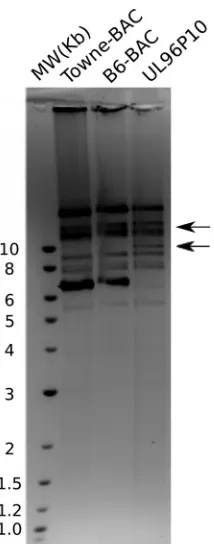
passaged virus (UL96P10) showed an RFLP different from that of
the parent B6-BAC or Towne-BAC virus (
Fig. 8
). This UL96P10
virus replicated with a higher growth rate (
Fig. 7A
) than the
non-replicating parent B6-BAC virus. To identify the changes in the
genome that accounted for a changed RFLP and the gain of
repli-cation competency in B6-BAC, we performed whole-genome
se-quencing of UL96P10 and a similarly passaged Towne-BAC virus
on the Illumina platform. No major insertions or deletions were
observed in the passaged Towne-BAC (
KF493877
) and UL96P10
(
KF493876
) genomes. However, several nucleotide differences
be-tween the Towne-BAC and UL96P10 viruses were noticed (
Table
1
). Most of these differences were located in the nonessential or
noncoding regions of the genome, which are known to
accumu-late mutations during passaging (
30
). These changes did not
over-lap the known microRNA and noncoding RNAs that are present
in the HCMV genome (
31–33
). When the UL96P10 and
Towne-BAC viruses were compared, most noticeable were two point
mu-tations in the UL96 region of the genome at positions 171699 and
171819 (
Table 1
).
Compensatory mutations in UL96 are responsible for the
gain of replication competency in the UL96 C-terminal cluster
mutant.
To study the potential impact of UL96 mutations
discov-ered in UL96P10, we enginediscov-ered the C-to-T conversion (position
171819,
Table 1
) in the B6-BAC mutant background in
Towne-BAC by recombineering as described above. This particular
mu-tation was chosen for its proximity to the original mumu-tations that
we engineered. The resultant UL96ATG BAC produced infectious
virus, unlike the parent B6-BAC, and grew with kinetics
compa-rable to those of the UL96P10 virus (
Fig. 7A
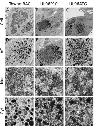
). Ultrastructural
anal-ysis of fibroblasts infected with the UL96ATG virus revealed a
proportion of regularly shaped virus particles in the cytoplasm of
infected cells that was comparable to that of the UL96P10 virus
infections, although it did not quite reach the level of Towne-BAC
virus (
Fig. 7B
and
9
). This correlates well with the differences in
final yield between the UL96P10 and UL96ATG viruses and the
Towne-BAC virus (
Fig. 7A
). In summary, a mutation discovered
in the UL96 coding region of the UL96P10 virus accounts for the
gain of replication competency in this virus.
DISCUSSION
Tegument proteins in herpesviruses are known to interact among
themselves, as well as with the capsid and envelope components of
the virion and also with a variety of host proteins (
14
,
34–50
).
These interactions have important consequences in terms of virus
tegumentation, envelopment, cellular trafficking, virion stability,
and initiation of virus infection. In the present study, we show that
CMV tegument proteins pp150 and pUL96 directly interact with
each other and that the N-terminal region of pUL96 and the
C-terminal region of pp150 determine this interaction (
Fig. 1
). These
FIG 7Reengineering of the UL96ATG (171819 C-to-T) mutation in theB6-BAC genome leads to recovery of virus growth and an increase in regularly shaped virus particles in the cytoplasm of infected cells. (A) Single-step growth curves of Towne-BAC, UL96P10, and UL96ATG viruses. (B) Percentages of particles that appeared irregular (IR) versus regular (R) (Fig. 6) in the cyto-plasm of cells infected with the indicated viruses at a late stage of infection on the basis of average counts from five individual infected cells. Printed images of sections of electron micrographs (Fig. 5and9) were used for visual counting by two study-blind volunteers who were supplied with the criteria listed inFig. 6. Tegument-filled dark particles (dense bodies) were excluded from counts. Approximately 150 particles were counted per sample.
on November 7, 2019 by guest
http://jvi.asm.org/
[image:7.585.44.282.73.536.2]two proteins are conserved in betaherpesviruses and are known to
be critical for virus maturation (
5
,
6
,
15
). Earlier studies suggested
that the phenotype of a UL96 mutant virus may depend on pp150
based on the similar stage of virus replication block in UL96
mu-tant, UL32 mumu-tant, and UL96 UL32 dual mutant virus-infected
cells (
6
). Also, the accumulation of pp150 in the nuclei of UL96
mutant virus-infected cells suggested that pUL96 either enhances
pp150-capsid association or functions as a chaperone to assist in
the translocation of pp150-labeled nucleocapsids from the
nu-cleus to the cytoplasm. To test the possibility of a direct interaction
between pp150 and pUL96, we used IP and IB. As expected, the
two proteins interacted with each other, strengthening the
possi-bility of a functional interaction. However, a domain analysis of
interaction revealed that the UL32 N-terminal and UL96
C-ter-minal regions are not required for this interaction. This came as a
surprise because these regions determine the function of these two
proteins in virus maturation (
5
,
6
,
15
). Thus, it appears that the
roles of pp150 and pUL96 in virus maturation do not depend on
their mutual interaction; however, it is possible that pp150 and
pUL96 rely on an alternative mechanism to function when a direct
interaction between these two proteins is abolished, explaining the
dispensable nature of the C-terminal region of pp150 (
15
).
We then focused on the role of pUL96 in virus maturation by
mapping the residues that are critical for function. Earlier, we
demonstrated that the UL96 C terminus is important for virus
maturation (
6
). An alignment of the pUL96 amino acid sequence
reveals a stretch of negatively charged conserved amino acids at
the C terminus of the protein (
Fig. 2
). These residues are
con-served among primate CMVs, and similar negatively charged
res-idues are present at the C termini of HHV6 and HHV7. We
hy-pothesized that pUL96 might use these amino acids for interaction
with a positively charged region of another viral or host protein;
thus, exploring the phenotype of viruses containing mutations in
these residues could lead to a better understanding of the
mecha-nism of pUL96 function. Mutation of these aspartate and
gluta-mate residues in pUL96 (
122EVDDAV
127) led to the arrest of virus
replication at the stage of virus maturation. Ultrastructural
anal-ysis showed that these mutations affect nucleocapsid stability in
the cytoplasm of infected cells, which is reminiscent of the
pheno-type of a UL96 null virus (
6
). We took advantage of the capability
of the
122EVDDAV
127mutant viruses to grow to some extent in
cell culture and subjected them to repeated passages. This way, we
intentionally allowed the accumulation of compensatory
muta-tions in the CMV genome. One possible outcome of this approach
would be the accumulation of compensatory mutations in a virus
protein that critically interacts with pUL96. In that case, it would
be possible to define the interactions between pUL96 and the
other candidate protein. One of the clustered mutants (B6-BAC)
gained replication competency in 10 passages (UL96P10) and
showed an altered RFLP on analysis of the genome. Interestingly,
the B6-BAC virus was able to overcome the block at the
multinu-cleate cell (syncytium) stage. Glycoprotein H (gH), gL, and gB are
the main fusogenic proteins in CMV and are often implicated in
syncytium formation (
51
). We hypothesized that the UL96P10
virus may be able to overcome this block by accumulating
com-FIG 8The passaged UL96P10 virus displays a restriction profile different [image:8.585.112.219.64.336.2]from that of the parental B6-BAC or Towne-BAC virus. HindIII digests of viral DNA harvested from Towne-BAC, B6-BAC, and UL96P10 virus-infected fi-broblasts were run on a 1% agarose gel and stained with ethidium bromide. The shift in band migration at⬃10 and⬃12 kb in the UL96P10 virus points toward genomic changes occurring in this virus during repeated passaging. Lane MW contains molecular size standards whose sizes are shown on the left in kilobases.
TABLE 1Summary of nucleotide differences between the Towne-BAC passaged virus (KF493877) and the UL96P10 virus (KF493876) revealed by whole-genome sequencing and subsequent comparison of nucleotide sequencesa
Reference location(s) Type of change Description Gene region Protein coding or noncodingb
27623 Insertion G Upstream of TRS1 NC
124412 Deletion G Upstream of RNA4.9 NC
171699 Conversion A¡G UL96 C
171819 Conversion C¡T UL96 C
171900 Conversion A¡G Upstream of UL97 NC
211946, 211947 Insertion GT Downstream of UL145 NC
232634 Conversion A¡G Downstream of eGFP NC
124724 Conversion C¡A RNA 4.9 NC
124728 Insertion C RNA 4.9 NC
aVirus DNA was purified from pelleted virions from the cleared cell culture medium. The Nextera XT DNA Sample Preparation kit (Illumina) was used to prepare the multiplexed
paired-end libraries (2⫻150 bp) before sequencing with an Illumina MiSeq. Genomic regions and open reading frames were identified by BLAST searching on the NCBI server.
bC, coding; NC, noncoding.
on November 7, 2019 by guest
http://jvi.asm.org/
[image:8.585.41.545.589.697.2]pensatory mutations in gH-gL or gB. Also, if pUL96-pp150
inter-action had a role in virus maturation, we would expect to see
compensatory changes in pp150. Hence, we sequenced the
com-plete genomes of the UL96P10 virus and a similarly passaged
Towne-BAC virus. On comparing these genomes, we discovered
differences in the RNA4.9 and UL96 regions (
Table 1
); however,
the gH, gL, gB, and pp150 regions were identical in all three
vi-ruses. Instead, two nonsynonymous mutations in UL96 were
identified as being unique to the UL96P10 virus (
Table 1
).
More-over, when the C-to-T conversion mutation (position 171819)
was reengineered into B6-BAC, it restored the virus growth rate to
the UL96P10 level and increased the stability of virus particles in
the cytoplasm (
Fig. 7
and
9
). Thus, results from this study show
the accumulation of compensatory mutations only in the UL96
gene in a revertant virus. This, combined with the involvement of
a functionally dispensable region of pp150 in the pUL96-pp150
interaction, points toward a role for pUL96 in virus maturation
that does not depend upon pp150; however, the possibility
re-mains that alternative pathways are used by pp150 and pUL96 to
function when intact forms of both proteins, instead of the
mu-tated forms, are present during infection.
ACKNOWLEDGMENTS
We are thankful to Hong Yi at the Robert P. Apkarian Integrated Electron Microscopy Core at Emory University for help with electron microscopy. Virus whole-genome sequencing was performed at the UMMC Molecular and Genomics Facility. Xiao Luo prepared the viral genome library, Ash-ley Robinson assembled the contigs, and Molly Tyner annotated the ge-nome sequences. Dongmei Yang and Dirk Dittmer at The University of North Carolina at Chapel Hill helped with the analysis of genomic data. Madeline Aylward helped with cell culture in the Tandon lab.
Public Health Service grant (RO1 AI20211 [principal investigator, E. S. Mocarski]) and an Intramural Research Support Program award from the University of Mississippi Medical Center to Ritesh Tandon sup-ported this research.
FIG 9Impact of reengineering of the UL96ATG (171819 C-to-T) mutation in the B6-BAC genome on the infected-cell phenotype and virus morphology. Shown are representative transmission electron micrographs of HF infected with Towne-BAC (A, D, G, J), UL96P10 (B, E, H, K), or UL96ATG (C, F, I, L) virus and processed for imaging at 5 dpi. The images show the whole infected cell (A, B, C), the cytoplasmic AC (D, E, F), the nucleus (Nuc) (G, H, I), or the cytoplasm (Cyt) (J, K, L). Bars, 5m (B), 2m (A, C, E), 1m (D, F), or 0.2m (G, H, I, J, K, L).
on November 7, 2019 by guest
http://jvi.asm.org/
[image:9.585.135.450.61.481.2]REFERENCES
1.Penkert RR, Kalejta RF.2012. Tale of a tegument transactivator: the past, present and future of human CMV pp71. Future Virol.7:855– 869.http: //dx.doi.org/10.2217/fvl.12.86.
2.Homer EG, Rinaldi A, Nicholl MJ, Preston CM.1999. Activation of herpesvirus gene expression by the human cytomegalovirus protein pp71. J. Virol.73:8512– 8518.
3.Britt W.2007. CMV maturation and egress, p 311–323.InArvin AM, Mocarski ES, Moore P, Whitley R, Yamanishi K, Campadelli-Fiume G, Roizman B (ed), Human herpesviruses: biology, therapy and immu-noprophylaxis. Cambridge Press, Cambridge, United Kingdom. 4.Mocarski ES, Jr, Shenk T, Pass RF.2006. Cytomegaloviruses, p 2701–
2772.InKnipe DM, Howley PM (ed), Fields virology, 5th edition. Lippin-cott Williams & Wilkins, Philadelphia, PA.
5.Tandon R, Mocarski ES.2008. Control of cytoplasmic maturation events by cytomegalovirus tegument protein pp150. J. Virol.82:9433–9444.http: //dx.doi.org/10.1128/JVI.00533-08.
6.Tandon R, Mocarski ES.2011. Cytomegalovirus pUL96 is critical for the stability of pp150-associated nucleocapsids. J. Virol.85:7129 –7141.http: //dx.doi.org/10.1128/JVI.02549-10.
7.Shanda SK, Wilson DW.2008. UL36p is required for efficient transport of membrane-associated herpes simplex virus type 1 along microtubules. J. Virol.82:7388 –7394.http://dx.doi.org/10.1128/JVI.00225-08. 8.Wolfstein A, Nagel CH, Radtke K, Dohner K, Allan VJ, Sodeik B.2006.
The inner tegument promotes herpes simplex virus capsid motility along microtubules in vitro. Traffic7:227–237.http://dx.doi.org/10.1111/j.1600 -0854.2005.00379.x.
9.Luxton GW, Lee JI, Haverlock-Moyns S, Schober JM, Smith GA.2006. The pseudorabies virus VP1/2 tegument protein is required for intracel-lular capsid transport. J. Virol.80:201–209.http://dx.doi.org/10.1128/JVI .80.1.201-209.2006.
10. Gibson W.2008. Structure and formation of the cytomegalovirus virion. Curr. Top. Microbiol. Immunol.325:187–204.http://dx.doi.org/10.1007 /978-3-540-77349-8_11.
11. Trus BL, Gibson W, Cheng N, Steven AC.1999. Capsid structure of simian cytomegalovirus from cryoelectron microscopy: evidence for teg-ument attachment sites. J. Virol.73:2181–2192.
12. Chen DH, Jiang H, Lee M, Liu F, Zhou ZH.1999. Three-dimensional visualization of tegument/capsid interactions in the intact human cyto-megalovirus. Virology 260:10 –16. http://dx.doi.org/10.1006/viro.1999 .9791.
13. Yu X, Shah S, Lee M, Dai W, Lo P, Britt W, Zhu H, Liu F, Zhou ZH. 2011. Biochemical and structural characterization of the capsid-bound tegument proteins of human cytomegalovirus. J. Struct. Biol.174:451– 460.http://dx.doi.org/10.1016/j.jsb.2011.03.006.
14. Baxter MK, Gibson W.2001. Cytomegalovirus basic phosphoprotein (pUL32) binds to capsids in vitro through its amino one-third. J. Virol. 75:6865– 6873.http://dx.doi.org/10.1128/JVI.75.15.6865-6873.2001. 15. AuCoin DP, Smith GB, Meiering CD, Mocarski ES.2006.
Betaherpes-virus-conserved cytomegalovirus tegument protein ppUL32 (pp150) con-trols cytoplasmic events during virion maturation. J. Virol.80:8199 – 8210.http://dx.doi.org/10.1128/JVI.00457-06.
16. Smith DE, Tans SJ, Smith SB, Grimes S, Anderson DL, Bustamante C. 2001. The bacteriophage straight phi29 portal motor can package DNA against a large internal force. Nature413:748 –752.http://dx.doi.org/10 .1038/35099581.
17. Homa F, Huffman J, Toropova K, Lopez H, Makhov A, Conway J.2013. Structure of the pseudorabies virus capsid: comparison with herpes sim-plex virus type 1 and differential binding of essential minor proteins. J. Mol. Biol.425:3415–3428.http://dx.doi.org/10.1016/j.jmb.2013.06.034. 18. Bhella D, Rixon FJ, Dargan DJ.2000. Cryomicroscopy of human
cyto-megalovirus virions reveals more densely packed genomic DNA than in herpes simplex virus type 1. J. Mol. Biol.295:155–161.http://dx.doi.org /10.1006/jmbi.1999.3344.
19. Gibson W.1981. Structural and nonstructural proteins of strain Colburn cytomegalovirus. Virology111:516 –537.http://dx.doi.org/10.1016/0042 -6822(81)90354-8.
20. Varnum SM, Streblow DN, Monroe ME, Smith P, Auberry KJ, Pasa-Tolic L, Wang D, Camp DG, II, Rodland K, Wiley S, Britt W, Shenk T, Smith RD, Nelson JA.2004. Identification of proteins in human cyto-megalovirus (HCMV) particles: the HCMV proteome. J. Virol.78:10960 – 10966.http://dx.doi.org/10.1128/JVI.78.20.10960-10966.2004.
21. Dunn W, Chou C, Li H, Hai R, Patterson D, Stolc V, Zhu H, Liu F. 2003. Functional profiling of a human cytomegalovirus genome. Proc. Natl. Acad. Sci. U. S. A.100:14223–14228.http://dx.doi.org/10.1073/pnas .2334032100.
22. Yu D, Silva MC, Shenk T.2003. Functional map of human cytomegalo-virus AD169 defined by global mutational analysis. Proc. Natl. Acad. Sci. U. S. A.100:12396 –12401.http://dx.doi.org/10.1073/pnas.1635160100. 23. Rost B, Yachdav G, Liu J.2004. The PredictProtein server. Nucleic Acids
Res.32:W321–W326.http://dx.doi.org/10.1093/nar/gkh377.
24. Thomason LC, Court DL, Bubunenko M, Costantino N, Wilson H, Datta S, Oppenheim A.2007. Recombineering: genetic engineering in bacteria using homologous recombination. Curr. Protoc. Mol. Biol. Chapter 1:Unit 1.16.http://dx.doi.org/10.1002/0471142727.mb0116s78. 25. Brechtel T, Tyner M, Tandon R.2013. Complete genome sequence of a
cytomegalovirus Towne-BAC (bacterial artificial chromosome) isolate maintained in Escherichia coli for 10 years and then serially passaged in human fibroblasts. Genome Announc.1:e00693–13.http://dx.doi.org/10 .1128/genomeA.00693-13.
26. Brechtel TM, Tyner M, Tandon R.2013. Complete genome sequence of a UL96 mutant cytomegalovirus Towne-BAC (bacterial artificial chromo-some) isolate passaged in fibroblasts to allow accumulation of compensa-tory mutations. Genome Announc. 1:e00901–13. http://dx.doi.org/10 .1128/genomeA.00901-13.
27. Sarov I, Abady I.1975. The morphogenesis of human cytomegalovirus. Isolation and polypeptide characterization of cytomegalovirions and dense bodies. Virology66:464 – 473.
28. Fiala M, Honess RW, Heiner DC, Heine JW, Jr, Murnane J, Wallace R, Guze LB.1976. Cytomegalovirus proteins. I. Polypeptides of virions and dense bodies. J. Virol.19:243–254.
29. Stinski MF.1976. Human cytomegalovirus: glycoproteins associated with virions and dense bodies. J. Virol.19:594 – 609.
30. Wang A, Ren L, Abenes G, Hai R.2009. Genome sequence divergences and functional variations in human cytomegalovirus strains. FEMS Im-munol. Med. Microbiol.55:23–33.http://dx.doi.org/10.1111/j.1574-695X .2008.00489.x.
31. Dunn W, Trang P, Zhong Q, Yang E, van Belle C, Liu F.2005. Human cytomegalovirus expresses novel microRNAs during productive viral in-fection. Cell. Microbiol.7:1684 –1695. http://dx.doi.org/10.1111/j.1462 -5822.2005.00598.x.
32. Meshesha MK, Veksler-Lublinsky I, Isakov O, Reichenstein I, Shomron N, Kedem K, Ziv-Ukelson M, Bentwich Z, Avni YS. 2012. The mi-croRNA transcriptome of human cytomegalovirus (HCMV). Open Virol. J.6:38 – 48.http://dx.doi.org/10.2174/1874357901206010038.
33. Gatherer D, Seirafian S, Cunningham C, Holton M, Dargan DJ, Bal-uchova K, Hector RD, Galbraith J, Herzyk P, Wilkinson GW, Davison AJ.2011. High-resolution human cytomegalovirus transcriptome. Proc. Natl. Acad. Sci. U. S. A.108:19755–19760.http://dx.doi.org/10.1073/pnas .1115861108.
34. Maringer K, Stylianou J, Elliott G.2012. A network of protein interac-tions around the herpes simplex virus tegument protein VP22. J. Virol. 86:12971–12982.http://dx.doi.org/10.1128/JVI.01913-12.
35. Omar OS, Simmons AJ, Andre NM, Wilson DW, Gross ST. 2013. Pseudorabies virus and herpes simplex virus type 1 utilize different tegu-ment-glycoprotein interactions to mediate the process of envelopment. Intervirology56:50 –54.http://dx.doi.org/10.1159/000339467.
36. Lee JH, Vittone V, Diefenbach E, Cunningham AL, Diefenbach RJ. 2008. Identification of structural protein-protein interactions of herpes simplex virus type 1. Virology378:347–354.http://dx.doi.org/10.1016/j .virol.2008.05.035.
37. Coller KE, Lee JI, Ueda A, Smith GA.2007. The capsid and tegument of the alphaherpesviruses are linked by an interaction between the UL25 and VP1/2 proteins. J. Virol.81:11790 –11797.http://dx.doi.org/10.1128/JVI .01113-07.
38. Vittone V, Diefenbach E, Triffett D, Douglas MW, Cunningham AL, Diefenbach RJ.2005. Determination of interactions between tegument proteins of herpes simplex virus type 1. J. Virol.79:9566 –9571.http://dx .doi.org/10.1128/JVI.79.15.9566-9571.2005.
39. Klupp BG, Fuchs W, Granzow H, Nixdorf R, Mettenleiter TC.2002. Pseudorabies virus UL36 tegument protein physically interacts with the UL37 protein. J. Virol.76:3065–3071.http://dx.doi.org/10.1128/JVI.76.6 .3065-3071.2002.
40. Ye GJ, Vaughan KT, Vallee RB, Roizman B.2000. The herpes simplex virus 1 U(L)34 protein interacts with a cytoplasmic dynein intermediate
on November 7, 2019 by guest
http://jvi.asm.org/
chain and targets nuclear membrane. J. Virol.74:1355–1363.http://dx.doi .org/10.1128/JVI.74.3.1355-1363.2000.
41. Phillips SL, Cygnar D, Thomas A, Bresnahan WA.2012. Interaction between the human cytomegalovirus tegument proteins UL94 and UL99 is essential for virus replication. J. Virol.86:9995–10005.http://dx.doi.org /10.1128/JVI.01078-12.
42. Cygnar D, Hagemeier S, Kronemann D, Bresnahan WA.2012. The cellular protein SPT6 is required for efficient replication of human cytomegalovirus. J. Virol.86:2011–2020.http://dx.doi.org/10.1128/JVI.06776-11.
43. Phillips SL, Bresnahan WA.2011. Identification of binary interactions between human cytomegalovirus virion proteins. J. Virol.85:440 – 447.
http://dx.doi.org/10.1128/JVI.01551-10.
44. Indran SV, Ballestas ME, Britt WJ.2010. Bicaudal D1-dependent traf-ficking of human cytomegalovirus tegument protein pp150 in virus-infected cells. J. Virol. 84:3162–3177. http://dx.doi.org/10.1128/JVI .01776-09.
45. Chevillotte M, Landwehr S, Linta L, Frascaroli G, Luske A, Buser C, Mertens T, von Einem J.2009. Major tegument protein pp65 of human cytomegalovirus is required for the incorporation of pUL69 and pUL97 into the virus particle and for viral growth in macrophages. J. Virol.83: 2480 –2490.http://dx.doi.org/10.1128/JVI.01818-08.
46. Kamil JP, Coen DM.2007. Human cytomegalovirus protein kinase UL97
forms a complex with the tegument phosphoprotein pp65. J. Virol.81: 10659 –10668.http://dx.doi.org/10.1128/JVI.00497-07.
47. Schierling K, Stamminger T, Mertens T, Winkler M. 2004. Human cytomegalovirus tegument proteins ppUL82 (pp71) and ppUL35 interact and cooperatively activate the major immediate-early enhancer. J. Virol. 78:9512–9523.http://dx.doi.org/10.1128/JVI.78.17.9512-9523.2004. 48. Hofmann H, Sindre H, Stamminger T. 2002. Functional interaction
between the pp71 protein of human cytomegalovirus and the PML-interacting protein human Daxx. J. Virol.76:5769 –5783.http://dx.doi.org /10.1128/JVI.76.11.5769-5783.2002.
49. Ogawa-Goto K, Irie S, Omori A, Miura Y, Katano H, Hasegawa H, Kurata T, Sata T, Arao Y.2002. An endoplasmic reticulum protein, p180, is highly expressed in human cytomegalovirus-permissive cells and inter-acts with the tegument protein encoded by UL48. J. Virol.76:2350 –2362.
http://dx.doi.org/10.1128/jvi.76.5.2350-2362.2002.
50. Tandon R, Mocarski ES.2012. Viral and host control of cytomegalovirus maturation. Trends Microbiol.20:392– 401.http://dx.doi.org/10.1016/j .tim.2012.04.008.
51. Kinzler ER, Compton T.2005. Characterization of human cytomegalo-virus glycoprotein-induced cell-cell fusion. J. Virol.79:7827–7837.http: //dx.doi.org/10.1128/JVI.79.12.7827-7837.2005.
on November 7, 2019 by guest
http://jvi.asm.org/