Papillary (PTC) and follicular thyroid carcinomas (FTC) are usually well differentiated, maintaining in part the characteris-tics of follicular thyroid cells, including expression of thyroid cell-specific antigens such as thyroid peroxidase (TPO), the membrane-bound enzyme essential for the biosynthesis of thyroid hormone. Recently, TPO gene and protein expression in thyroid carcinoma has been analysed, with results indicating low enzy-matic activity (Valenta et al, 1973; Valenta, 1976; Fragu and Nataf, 1977; Mizukami and Matsunaga, 1981), and impaired solubility (Neary et al, 1978). Mutated TPO genes were found in some differentiated thyroid carcinomas (Smanik et al, 1994). Although the expression of TPO mRNA was found to be the same in cancerous and benign tissue in one study (Ohta et al, 1991), others have shown suppression of TPO gene expression (Hoang-Vu et al, 1992; Fabbro et al, 1994; Tanaka et al, 1996; Umeki et al, 1996).
Some differences in the results of experiments using TPO murine monoclonal antibodies to detect protein expression may be due to methodological approaches (Yamashita et al, 1993; Mizukami et al, 1994; Tanaka et al, 1996; Umeki et al, 1996). However, differential immunohistochemical labelling of TPO with one monoclonal antibody (mAb#47) was proposed by De Micco et al (1991) as a marker distinguishing between benign and malig-nant thyroid tumours, including both PTC and FTC. Reduced immunostaining with mAb#47 was observed in more than 95% of
malignant thyroid cancers suggesting that abnormal TPO immunoreactivity occurs in malignancy (DeMicco et al, 1991, 1994). MAb #47 is one of a panel of well characterized murine monoclonal antibodies produced against native human TPO. The immunochemical properties of these mAbs have been published elsewhere (Ruf et al, 1989).
In order to clarify whether TPO in thyroid cancers is indeed qualitatively different from TPO in non-cancerous tissues, we analysed the reactivity of TPO from thyroid cancers with a panel of murine monoclonal antibodies (including mAb#47) and previ-ously described human monoclonal anti-TPO IgGκFab fragments (McIntosh et al, 1997). Both types of antibodies react with epitopes delineating the immunodominant region of TPO (Figure 1).
METHODS
Thyroid tissues
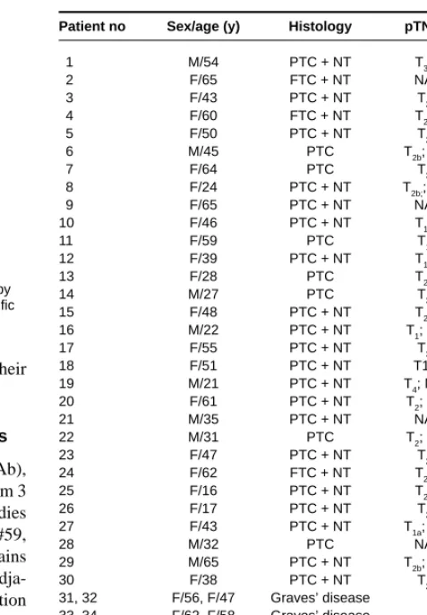
We studied 30 malignant thyroid tumours, 23 of them paired with corresponding normal tissue obtained from adjacent parts of the gland. Thyroid tissues were obtained from patients undergoing surgery for thyroid cancer by the Institute of Oncology, Gliwice, Poland. Tissues fragments were immediately frozen in liquid N2, and kept at –80˚C until use. Tumours were diagnosed histopatho-logically according to World Health Organization (WHO) criteria: 3 cases were follicular carcinomas (FTC) and 27 cases were papil-lary carcinomas (PTC). 10 thyroid samples from patients with Graves’ disease were also included in the study. Details of the patients are summarized in Table 1. The local Committees on
Is there loss or qualitative changes in the expression of
thyroid peroxidase protein in thyroid epithelial cancer?
B Czarnocka1,2, D Pastuszko1, M Janota-Bzowski1, AP Weetman3, PF Watson3, EH Kemp3, RS McIntosh4, MS Asghar4, B Jarzab5, E Gubala5, J Wloch5and D Lange5
1Department of Biochemistry, Medical Center of Postgraduate Education, Marymoncka 99,01–813 Warsaw, Poland; 2Department of Endocrinology, Medical
Research Center, Polish Academy of Science, Warsaw, Poland; 3Department of Medicine, University of Sheffield Clinical Sciences Center, Northern General
Hospital, Sheffield, United Kingdom S5 7AU; 4Clinical Immunology Unit, Queen’s Medical Center, Nottingham, United Kingdom NG7 2UH; 5Oncology Center,
M Sklodowska-Curie Memorial Institute, Gliwice, Poland
Summary There is disagreement concerning the expression of thyroid peroxidase (TPO) in thyroid cancer, some studies finding qualitative as well as quantitative differences compared to normal tissue. To investigate TPO protein expression and its antigenic properties, TPO was captured from a solubilizate of thyroid microsomes by a panel of murine TPO monoclonal antibodies and detected with a panel of anti-human TPO IgGκFab. TPO protein expression in 30 samples of malignant thyroid tissue was compared with TPO from adjacent normal tissues. Virtual absence of TPO expression was observed in 8 cases. In the remaining 22 malignant thyroid tumours the TPO protein level varied considerably from normal to nearly absent when compared to normal thyroid tissue or tissues from patients with Graves’ disease (range less than 0.5 to more than 12.5µg mg–1of protein). When expressed TPO displayed similar epitopes, to that of TPO from Graves’
disease tissue. The results obtained by the TPO capturing method were confirmed by SDS-PAGE and Western blot analysis with both microsomes and their solubilizates. The present results show that in about two-thirds of differentiated thyroid carcinomas, TPO protein is expressed, albeit to a more variable extent than normal; when present, TPO in malignant tissues is immunologically normal. © 2001 Cancer Research Campaign http://www.bjcancer.com
Keywords: differentiated thyroid cancer; TPO; mAb; IgGκFab; epitope
Received 26 October 2000 Revised 11 June 2001 Accepted 2 July 2001
Correspondence to: B Czarnocka
Medical Ethics approved these studies and all patients gave their written consent.
Murine mAb and human recombinant IgGκantibodies
A panel of 12 murine anti-TPO monoclonal antibodies (mAb), previously produced and characterized, directed to epitopes from 3 antigenic domains of TPO was used (Ruf et al, 1989). Antibodies mAb#2, #9, #47, #60 – recognize domain A; mAb#15, #18, #59, #64 – domain B, and mAb#1, #30, #40, #53 – domain D. Domains A and B, defining the immunodominant region of TPO, are adja-cent whereas domain D is topologically distant. Characterization of the human Fabs has been described elsewhere (Czarnocka et al, 1997; McIntosh et al, 1997). In this study, Fabs representative for domain A (126 TP1, 6, 7, 8, 10 and 126 TO 10), and domain B reactivity (126 TP5, 9, 13, 14, 15 and 126 TO1) were used. All experiments were carried out on Fab prepared in culture medium.
Thyroid microsomes preparation and solubilization
Thyroid microsomes were prepared and solubilized as previously described (Czarnocka et al, 1985). Briefly, the thyroid tissues were homogenized in 10 mM Tris-HCl buffer pH 7.5, containing 2µg ml–1aprotinin, 1µg ml–1leupeptin, 1µg ml–1pepstatin and 0.1 mM phenylmethylsulfonyl fluoride (all from Boehringher Mannheim, Mannheim, Germany). The plasma membranes were pelleted by centrifugation at 10 000 g for 15 min at 4˚C and the supernatants were subsequently ultracentrifuged at 100 000 g for 60 min at 4˚C. The resulting membrane fractions were solubilized with sodium deoxycholate, and stored at –80˚C until use. The protein concentration was determined by the BCA micro-method (Pierce Chemical Co, Rockford, IL, USA).
TPO capture method
The antigen capture method was used to detect solubilized TPO. Maxisorb microtitre plates (Nunc, Copenhagen, Denmark) were coated with 100µl of mAb solution diluted to 10µg ml–1in 0.1 M carbonate buffer pH 9.6, overnight at 4˚C under a humidified atmosphere. The wells were then washed, blocked with BSA, washed again, and filled with 100µl of solubilizate adjusted to 20
µg ml–1in PBS pH 7.8, containing 0.1% of Tween-20 and 3 mM sodium azide. After overnight incubation at 4˚C, unbound protein was removed by extensive washing and wells filled with 100µl of
Fab diluted 1:15 with PBS-Tween-20-azide. After 12 h at 4˚C, unbound antibodies were removed by extensive washing. Fab binding was detected using an immunoaffinity-purified antihuman Fab second antibody labelled with horseradish peroxidase (Jackson ImmunoResearch Laboratories, Inc, West Grove, PA, USA); tetramethyl benzidine was the substrate. Absorbance was read at 450 nm.
SDS-PAGE and Western blot
Thyroid microsomes as well as their solubilizates were diluted with buffer containing 0.125 M Tris-HCl pH 6.8, 10% β-mercaptoethanol, 4% SDS, and 20% glycerol, to a concentration of 1 mg ml–1, boiled and loaded (20µg lane–1) onto 10% SDS polyacrylamide mini gels. Non-denaturing sample buffer consisted of 0.3125 M Tris, 0.5 M dithiothreitol, 1.5% SDS and 50% glycerol and microsomes or solubilizates were diluted to 1 mg ml–1with the above buffer and incubated for 30 min at 37˚C and loaded (20µg lane–1) onto SDS polyacrylamide mini gels, 0.75 mm thick. Proteins were
[image:2.660.62.268.63.198.2]British Journal of Cancer (2001) 85(6), 875–880 © 2001 Cancer Research Campaign
Figure 1 Map of the epitopes on thyroid peroxidase (TPO) recognized by murine monoclonal antibodies (mAb) and recombinant human TPO specific Fabs A B D C # 2 # 60 # 9 #47 #18 #24 #59 #64 #15 #40 #30 #1 #53 TP 9 126 TP 5 TP 13 TP 14 TP 15 126 TO 1 TP 6
126 TP 1 TP 7 TP 8 TP 10 126 TO 10
Autoantibody binding domains
Table 1 Clinical data
Patient no Sex/age (y) Histology pTNM TSH
1 M/54 PTC + NT T3a NA
2 F/65 FTC + NT NA 0,15 3 F/43 PTC + NT T2 3,43
4 F/60 FTC + NT T2b 4,88 5 F/50 PTC + NT T2 1,12
6 M/45 PTC T2b; N1 NA 7 F/64 PTC T3 NA
8 F/24 PTC + NT T2b;; N1 0,7 9 F/65 PTC + NT NA NA 10 F/46 PTC + NT T1a 0,4 11 F/59 PTC T1 10,0
12 F/39 PTC + NT T1a 0,91 13 F/28 PTC T2b 1,18
14 M/27 PTC T3 0,69 15 F/48 PTC + NT T2b 0,28
16 M/22 PTC + NT T1; N1 0,99 17 F/55 PTC + NT T3 7,60
18 F/51 PTC + NT T1a NA 19 M/21 PTC + NT T4; N1b NA
20 F/61 PTC + NT T2; N1 NA 21 M/35 PTC + NT NA NA 22 M/31 PTC T2; N1 NA 23 F/47 PTC + NT T2 1,47
24 F/62 FTC + NT T2b 1,55 25 F/16 PTC + NT T2b NA
26 F/17 PTC + NT T2 NA 27 F/43 PTC + NT T1a; N1 NA
28 M/32 PTC NA NA 29 M/65 PTC + NT T2b; N1 0,89
30 F/38 PTC + NT T2 1,12 31, 32 F/56, F/47 Graves’ disease
33, 34 F/62, F/58 Graves’ disease 35, 36 F/67, M/55 Graves’ disease 37, 38 F/50, M/60 Graves’ disease 39, 40 F/42, F/39 Graves’ disease
T = primary tumour; N = lymph node metastasis; M = metastasis; NA = not available; tumour size: T1< 1 cm; T21–5 cm; T3> 5 cm (tumour without
penetration of thyroid capsule); T4= infiltration through thyroid capsule); Ta= monocentric; Tb= multicentric. N0= tumour without metastasis to lymph
nodes; N1= metastasis to lymph nodes; N1a= ipsilateral metastasis; N1b= contralateral metastasis; M0= without distant metastasis; M1= distant
[image:2.660.300.539.76.420.2]directly electro-transferred onto Immobilon P (Millipore, Bedford Corp, MA, USA), and these membranes were then blocked with 3% BSA in PBS-3 mM sodium azide for 60 min at room tempera-ture. Western blots were done by using a 1:500 dilution of anti-TPO mAb#47 antibody or a 1:1000 dilution anti-anti-TPO rabbit polyclonal antibody in PBS-0.1% Tween 20 with 3 mM sodium azide overnight at 4˚C with constant shaking. After washing 3 times for 15 minute in PBS-Tween 20, membranes were incubated for 60 min at room temperature under shaking with either immunoaffinity-purified antimouse IgG conjugated to horseradish peroxidase or immunoaffinity-purified antirabbit IgG conjugated to horseradish peroxidase (Jackson ImmunoResearch, Laboratories, Inc, West Grove, PA, USA). After additional 3 washes, the blots were developed with DAB – metal enhanced, precipitating (Boehringer Mannheim, Mannheim, Germany) as substrate.
RESULTS
TPO capture
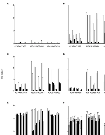
We observed 3 patterns of TPO protein expression. In 8 malignant tumours the TPO level was very low or undetectable, in 19 samples there was a variable TPO protein level from very low to almost normal and in 3 there was a high TPO level. Figure 2 shows the pattern of Fab binding to TPO captured by mAbs in tissues representative of these 3 groups, adjacent normal tissue and, as a control, Graves’ thyroid.
To assess the relative amount of TPO protein expressed in thyroid cancer tissues, the results of captured highly purified TPO at concentrations 1.0, 5.0, 10.0 and 25.0 ng well–1were compared to the data obtained for cancer tissues. The relative concentration of TPO in the 30 tumours ranged from much less than 0.5µg mg–1 in 8 cases to more than 12.5µg mg–1of the protein in 3 cases. In the majority of cancerous tissues however, the TPO amount esti-mated by the method used ranged from 2.5 to 5µg mg–1of micro-somes protein (data not shown).
Epitope mapping
To get a better insight in the characteristics of the TPO antigenic activity we analysed the relationship between Fab binding to the TPO and the mAbs used for the antigen capture. We observed a cross-competitive effect between mAbs and IgGκ Fab for the binding sites on the TPO molecule in all cases studied. The Fab binding to TPO showed cross-reactive inhibition dependent on which mAb was used for TPO capture. A positive relationship of the Fab binding to TPO was observed, especially when TPO was captured by mAbs from domain B. The binding of all Fabs reactive to domain A (126 TP 1, TP 6, TP 7, TP 8, TP 10 and 126 TO 10) was high, whereas the binding of Fabs reacting with epitopes located on domain B (126 TP 5, TP 9, TP 13, TP 14, TP 15 and 126 TO 1) was substantially reduced.
Strong inhibition of the binding of Fabs reacting with epitopes located on antigenic domain B with TPO immobilized by domain B mAbs indicated that epitopes for both mAbs and Fabs were overlapping. This effect was less pronounced when TPO was bound to mAbs from antigenic domain A. Fabs reacting with epitopes located on antigenic domain A react predominantly with antigenic domain A with some cross-reactivity to domain B. When
TPO was bound to mAbs from antigenic domain D (Figure 2) no competition between mAbs and Fabs was observed. All Fabs, whether reactive with antigenic domains A or B, bound equally, indicating that the epitopes of these monoclonal antibodies and the Fab-binding sites did not overlap each other. The observed differ-ences in absorbance values were due to variations in Fab affinity for the TPO.
The same pattern of reactivity was observed when TPO from normal and Graves’ thyroid tissues was captured (see Figure 2B and 2F). However, the cross-competitive effects between mAbs and Fabs for binding sites on the TPO was less apparent due to the high quantity of TPO present in hyperfunctioning tissue.
SDS-PAGE and Western blot analysis
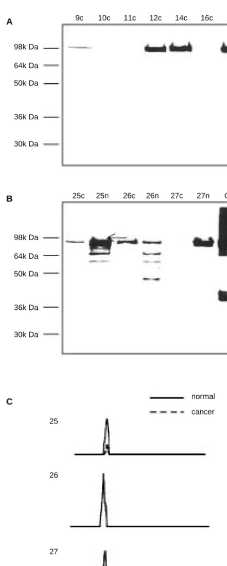
In order to confirm the presence or absence of TPO in cancerous and normal tissues observed using the antigen capture method, TPO was analysed by SDS-PAGE and Western blot. The intensity of immunostaining of the TPO bands correlated well with the TPO level detected by the antigen capture method (Figure 3A, B). In normal and cancerous tissues taken from the same individuals, TPO bands were highly immunoreactive and well visualized in the normal, but not in the corresponding cancerous tissue (Figure 3B). Only 8 of 30 cancer tissues were negative in Western blot with both antibodies (Figure 3A; patients 10 and 11). In the remaining 22 cancer tissues, the TPO band intensity varied from very slight to strong immunostaining with mAb#47 as well as with rabbit polyclonal anti-TPO antibodies. Using identical amounts of protein allowed measurement of the relative quantity of the TPO with respect to intensity and size of the TPO band (Figure 3C). Densitometric evaluation of the blots demonstrated that in cancerous tissues, the amount of TPO was 3–20 times lower than in paired normal tissues.
DISCUSSION
In the first part of this study, we evaluated the presence or absence of TPO protein in differentiated thyroid carcinomas and estimated the approximative amount of the TPO expressed. Solubilized TPO was captured by the same panel of anti-TPO murine monoclonal antibodies used by De Micco et al (1991). TPO was undetectable or barely detected in 8 of 30 differentiated carcinomas as compared with the normal paired thyroid tissues. Variable TPO protein content was found in 19 cancer tissues, and in 3 tissues TPO was present at a high level, comparable to that detected in the corresponding normal or Graves’ disease tissues. Several groups have found that one-third of papillary thyroid carcinomas studied do not express TPO mRNA, and thus lack TPO enzyme (Hoang-vu et al, 1992; Fabbro et al, 1994; Tanaka et al, 1996). These cases should not be immunoreactive with either mAbs or Fabs. The frequency (26.7%) of differentiated thyroid carcinomas with nega-tive or extremely low TPO level found in our study (albeit with 6 of them still showing a trace of TPO protein by the method used) is therefore similar to these reports. Others have produced contradic-tory data, demonstrating similar if not identical TPO mRNA levels in a series of malignant thyroid tumours and in benign tissues (Ohta et al, 1991) for reasons which are not clear.
ranged from much less than 0.5µg to more than 12.5µg mg–1of microsome proteins, but the majority was around 2.5µg mg–1to 5µg mg–1 of the protein. The presence or absence of TPO in cancerous tissues detected in antigen capture method was
confirmed by SDS-PAGE and Western blot. The immunolabelling of the TPO bands with mAb#47 demonstrate that mAb#47 epitope was expressed on the TPO molecule. Immunohistochemical techniques using monoclonal antibodies generated against native
British Journal of Cancer (2001) 85(6), 875–880 © 2001 Cancer Research Campaign
0 1 2 3 0 1 2 3 0 1 2 3
#2#9#47#60 #2 #9 #47 #60
#15 #18 #59 #64
#15 #18 #59 #64 #1 #30 #40 #53 #2 #9 #47 #60 #15 #18 #59 #64 #1 #30 #40 #53 #2 #9 #47 #60 #15 #18 #59 #64 #1 #30 #40 #53 #2 #9 #47 #60 #15 #18 #59 #64 #1 #30 #40 #53
#1 #30 #40 #53 #2#9#47#60 #15 #18 #59 #64 #1 #30 #40 #53
A
E F
C D
B
Coated mAbs
[image:4.660.85.509.59.604.2]OD 450 nm
human TPO have also been frequently used to demonstrate vari-able levels of TPO protein in thyroid follicular cells under various pathological conditions, including a large series of malignant thyroid tumours (DeMicco et al, 1991; Yamashita et al, 1993; Mizukami et al, 1994; Tanaka et al, 1996). The results of immunohistochemical methods also differ substantially. Using immunostaining with 2 (mAb#30 and mAb#47) of 13 mAbs against native human TPO, De Micco et al (1991) demonstrated differences in TPO immunoreactivity between benign and malig-nant tumours and, on this basis, mAb#47 has been proposed as a specific marker of malignancy. These and other data reported by this group suggest that although the TPO molecule is present in thyroid cancers at low levels, the epitope recognized by mAb#47 is not (DeMicco et al, 1991, 1994; Faroux et al, 1997; Garcia et al, 1998).
To detect any modifications or abnormalities of the TPO mole-cule expressed in thyroid carcinomas, we chose a structural approach of mapping the antigenic surface with a panel of 13 well characterized mAbs directed against native human TPO, and a panel of human anti-TPO IgGκ Fab fragments (Ruf et al, 1989; McIntosh et al, 1997). TPO purified from Graves’ tissues was previously used to map the interaction of a panel of 13 mouse mAbs to 4 antigenic domains of TPO (Ruf et al, 1989). Human monoclonal autoantibodies expressed as IgGκ Fab fragments recognized exclusively epitopes located in only 2 of the 4 domains, A and B (Czarnocka et al, 1997). Moreover, there was no overlap between the epitopes recognized by the panel of TPO-specific IgGκFab and the epitopes of mAbs reacting with anti-genic domains C and D (Czarnocka et al, 1997). We observed that the TPO protein present in cancer tissues did not show any differ-ences from normal in its antigenic activity, as TPO was captured by all murine anti-TPO monoclonal antibodies used. Immobilized TPO showed similar reactivity towards human IgGκ Fabs, with clearly pronounced steric hindrance dependent on which mAb was used to capture the TPO.
The occupancy of the TPO surface by the mAbs reactive to anti-genic domains A or B inhibited the reactivity of the Fabs with captured TPO. This was clearly seen when TPO was bound via mAbs directed to epitopes located on antigenic domain B, indi-cating that both mAbs and Fabs recognize overlapping part of TPO. No cross-inhibition of Fab binding to TPO was observed when TPO was captured by mAbs reactive with antigenic domain D. This TPO region is not recognized by autoantibodies, and none of the binding of the recombinant human IgGκFabs used was influ-enced by the occupancy of those mAb epitopes on the TPO mole-cule (Czarnocka et al, 1997; McIntosh et al, 1997). This result therefore indicates that, when expressed, TPO in differentiated thyroid cancer tissues does not differ from TPO in normal thyroid tissue or Graves’ tissue. TPO in the majority of the cancer tissues studied was also efficiently captured by mAb#47 with the exception of the 8 negative cases. From our present study, it is evident that although the TPO protein in the majority of differentiated thyroid cancer cases is lower than normal it has unchanged antigenic activity, when compared to that observed in normal and Graves’ disease tissues. Recently, the correlation of TPO immunostaining by mAb#47 with the differentiation and proliferative potential of follicular tumours was examined and a significant correlation between TPO mAb#47 staining and the proliferating cell nuclear antigen index with malignancy was found, suggesting that an alter-ation of TPO antigenicity is an early marker of thyroid follicular tumours, closely related to tumour growth in the first stages o
30k Da
9c 10c 11c 12c 14c 16c 18c
25c 25n 26c 26n 27c 27n G 98k Da
64k Da 50k Da
36k Da
30k Da 98k Da 64k Da 50k Da
36k Da
normal cancer
26
27 25
A
B
[image:5.660.53.282.55.626.2]C
Figure 3 Western blot analysis of TPO in thyroid cancer tissue after SDS-PAGE separation of microsome proteins (20µg lane–1) under reducing
malignant transformation (Garcia et al, 1998). We found in our series of malignant thyroid carcinoma tissues a graded expression of TPO protein from negative to almost normal. However, regardless of any differences in TPO content, its activity towards both mAbs and Fabs was similar to observed in normal or Graves’ disease tissues.
ACKNOWLEDGEMENTS
This study was supported by Medical Center of Postgraduate Education No CMKP-501-2-1-01-01/98 grant to BC, DP, MJ-B.
REFERENCES
Czarnocka B, Ruf J, Ferrand M, Lissitzky S and Carayon P (1985) Purification of the human thyroid peroxidase and its identification as the microsomal antigen involved in autoimmune thyroid diseases. FEBS Lett 190: 147–152
Czarnocka B, Janota-Bzowski M, McIntosh RS, Suhail Asghar M, Watson PF, Kemp HE, Carayon P and Weetman AP (1997) Immunoglobulin Gκantithyroid peroxidase antibodies in Hashimoto’s thyroiditis: epitope-mapping analysis.
J Clin Endocrinol Metab 82: 2639–2644
De Micco C, Ruf J, Chrestian MA, Gros N, Henry JF and Carayon P (1991) Immunohistochemical study of thyroid peroxidase in normal, hyperplastic, and neoplastic human thyroid tissues. Cancer 67: 3036–3041
De Micco C, Zoro P, Garcia S, Skoog L, Tani EM, Carayon P and Henry J-F (1994) Thyroid peroxidase detection as a tool to assist diagnosis of thyroid nodules on fine-needle aspiration biopsy. Eur J Endocrinol 131: 474–479
Fabbro D, Di Loreto C, Beltorami CA, Belfiore A, Di Lauro R and Damante G (1994) Expression of thyroid-specific transcription factors TTF-1 and Pax-8 in human thyroid neoplasm. Cancer Res 54: 4744–4749
Faroux MJ, Theobald S, Pluot M, Patey M and Menzies D (1997) Evaluation of the monoclonal antibody anti-thyroperoxidase MoAb47 in the diagnostic decision of cold thyroid nodules by fine-needle aspiration. Pathol Res Pract 193: 705–712 Fragu PN and Nataf BM (1977) Human thyroid peroxidase activity in benign and
malignant thyroid disorders. J Clin Endocrinol Metab 45: 1089–1096 Garcia S, Vassko V, Hanry J-F and De Micco C (1998) Comparison of thyroid
peroxidase expression with cellular proliferation in thyroid follicular tumors.
Thyroid 9: 745–749
Hoang-Vu C, Dralle H, Scheumann G, Horn R, von zur Mühlen A and Brabant G (1992) Gene expression of differentiation and dedifferentiation markers in normal and malignant human thyroid tissues. Exp Clin Endocrinol 100: 51–56 Mizukami Y and Matsunaga F (1981) Correlation between thyroid peroxidase
activity and histopathological and ultrastructural changes in various thyroid diseases. Endocrinol Jpn 28: 381–389
Mizukami Y, Nonomura A, Michigishi T, Noguchi M, Nakamura S, Arai Y, Kotani T, Ohtaki S and Matsukawa S (1994) Immunohistochemical demonstration of thyroid peroxidase (TPO) in human thyroid tissues from various thyroid diseases. Anticancer Res 14: 1329–1334
McIntosh RS, Asghar MS and Kemp EH (1997) Analysis of immuno-globulin Gκ antithyroid peroxidase antibodies from different tissues in Hashimoto’s thyroiditis. J Clin Endocrinol Metab 82: 3818–3825
Neary JT, Nakamura C, Davidson B, Soodak M, Vickery AL and Maloof F (1978) Studies on the membrane-associated nature of human thyroid peroxidase: a difference in the solubility of the enzyme from benign and malignant thyroid tissues. J Clin Endocrinol Metab 46: 791–800
Ohta K, Endo T and Onaya T (1991) The mRNA levels of thyrotropin receptor, thyroglobulin and thyroid peroxidase in neoplastic human thyroid tissues.
Biochem Biophys Res Commun 174: 1148–1153
Ruf J, Toubert M-E, Czarnocka B, Durand-Gorde J-M, Ferrand M and Carayon P (1989) Relationship between immunological structure and biochemical properties of human thyroid peroxidase. Endocrinology 125: 1211–1218 Smanik PA, Fithian LJ and Jhiang SM (1994) Thyroid peroxidase expression and DNA
polymorphism in thyroid cancer. Biochem Biophys Res Commun 198: 948–954 Tanaka T, Umeki K, Yamamoto I, Sugiyama S, Noguchi S and Ohtaki S (1996)
Immunohistochemical loss of thyroid peroxidase in papillary thyroid carcinoma: strong suppression of peroxidase gene expression. J Pathol 179: 89–94 Umeki K, Tanaka T, Yamamoto I, Aratake Y, Kotani T, Sakamoto F, Noguchi S and
Ohatki S (1996) Differential expression of dipeptidyl peptidase IV (CD26) and thyroid peroxidase in neoplastic thyroid tissues. J Endocrine 43: 53–60 Valenta LJ (1976) Thyroid peroxidase, thyroglobulin, cAMP, and DNA in human
thyroid. J Clin Endocrinol Metab 43: 466–469
Valenta LJ, Valenta V, Wang CA, Vickery AL, Caulfield J and Maloof F (1973) Subcellular distribution of peroxidase activity in human thyroid tissue. J Clin
Endocrinol Metab 37: 560–569
Yamashita H, Noguchi S, Murakami N, Adachi M and Maruta J (1993) Immunohistological differentiation of benign thyroid follicular cell tumors from malignant ones: Usefulness of anti-peroxidase and JT-95 antibodies. Acta
Pathol Jpn 43: 670–673