Type of the Pape: Review
1
2
Impaired cargo clearance in the Retinal Pigment Epithelium (RPE) underlies
3
irreversible blinding diseases
4
5
Eloise Keeling 1, Andrew J. Lotery 1,2, David A. Tumbarello 3 and J. Arjuna Ratnayaka 1,*
6
7
1 Clinical and Experimental Sciences, Faculty of Medicine, University of Southampton,
8
MP806, Tremona Road, Southampton, SO16 6YD, United Kingdom;
9
10
11
2 Eye Unit, University Hospital Southampton NHS Foundation Trust, Southampton,
12
SO16 6YD, United Kingdom; [email protected]
13
14
3 Biological Sciences, Faculty of Natural & Environmental Sciences, Life Science
15
Building 85, University of Southampton, Highfield Campus, Southampton, SO17 1BJ,
16
United Kingdom; [email protected]
17
18
* Correspondence: [email protected]; Tel.: +044 238120 8183
19
20
21
Abstract: Chronic degeneration of the Retinal Pigment Epithelium (RPE) is a precursor to
22
pathological changes in the outer retina. The RPE monolayer, which lies beneath the
23
neuroretina, daily internalises and digests large volumes of spent photoreceptor outer
24
segments. Impaired cargo handling and processing in the endocytic/phagosome and
25
autophagy pathways leads to the accumulation of lipofuscin and
N-retinylidene-N-26
retinylethanolamine aggregates and chemically-modified compounds such as
27
malondialdehyde and 4-hydroxynonenal within RPE. These contribute to increased
28
proteolytic and oxidative stress, resulting in irreversible damage to post-mitotic RPE cells
29
and development of blinding conditions such as Age-related Macular Degeneration,
30
Stargardt disease and Choroideremia. Here, we review how impaired cargo handling in the
31
RPE results in their dysfunction, discuss new findings from our laboratory and consider how
32
newly discovered roles for lysosomes and the autophagy pathway could provide insights
33
into retinopathies. Studies of these dynamic, molecular events have also been spurred on
34
by recent advances in optics and imaging technology. Mechanisms underpinning lysosomal
35
impairment in other degenerative conditions including storage disorders, α-synuclein
36
pathologies and Alzheimer’s disease are also discussed. Collectively, these findings help
37
transcend conventional understanding of these intracellular compartments as simple waste
38
disposal bags to bring about a paradigm shift in the way lysosomes are perceived.
39
40
41
Keywords: Retinal Pigment Epithelium (RPE); Endosomes; Phagosomes; Lysosomes,
42
Autophagy; RPE cultures; Age-related Macular Degeneration (AMD)
43
44
45
1. Introduction
46
47
The Retinal Pigment Epithelium (RPE) is a monolayer of cells which lies beneath the
48
neuroretina. Amongst its many functions, the RPE internalises photoreceptor outer
49
segments (POS) from overlying photoreceptors as part of the daily visual cycle. Each RPE
50
cell may ‘serve’ up to 45 rod or cone photoreceptors, of which the distal 10% of POS are
51
replenished daily1,2. Microvilli on the apical RPE surface interdigitate and surround these
52
photoreceptor tips in order to enhance phagocytosis3-5. On the basolateral side of RPE cells,
53
highly convoluted basal infolds increases the surface area for optimal absorption of oxygen
54
and nutrients. The substantial metabolic waste generated in the outer retina is removed via
55
the underlying choriocapillaris6,7. Sandwiched between this dense capillary network and the
56
RPE is a porous tissue of 2-4μm thickness referred to as Bruch’s membrane (BrM) which
57
supports the overlying cell monolayer (Figure 1). The daily internalisation of POS makes the
58
RPE one of the most phagocytic cells in the body. This, coupled with the fact that in-situ
59
RPE are largely post-mitotic, makes the proteolytic burden in these cells considerable. For
60
instance, it has been estimated that each cell is exposed to approximately 4000 POS per
61
day, and that each RPE cell may process up to a billion discs over a 70 year period2,8. The
62
high photo-oxidative retinal environment imposes further stresses amongst which the
63
modification of intracellular cargos within membrane bound vesicles impairs their clearance
64
and turnover in senescent RPE. Consequently, cargo handling in the RPE endocytic and
65
autophagy pathways has garnered considerable attention, as its dysregulation is associated
66
with several retinopathies. Given recent discoveries revealing new roles for lysosomes and
67
autophagosomes, and the ability to study their dynamic behaviour using novel imaging
68
technologies, we review the topic of how RPE cells cope with the high proteolytic burden in
69
the senescent retina, and why its impairment leads to irreversible blindness. We also
70
consider how lysosomal impairment in other degenerative conditions could provide insights
71
into shared pathogenic processes associated with cargo handling in the mammalian cell.
---
86
87
Figure 1: Anatomy of the eye and arrangement of cells in the retina and associated
88
tissues. [A] Schematic diagram of the eye in cross-section. [B] Enlargement of the area
89
indicated in box [A] showing relative position of the Retinal Pigment Epithelium (RPE) in
90
relation to other tissues. Sandwiched between the overlying neuroretina and the underlying
91
Bruch’s membrane/choroid, the RPE monolayer marks the important blood-retinal-barrier. A
92
black arrow indicates the pathway of light.
93
---
94
95
2. Components of the endocytic/phagosome and autophagy pathways
96
97
The endo-lysosomal pathway
98
99
The function of the endocytic pathway is to traffic and sort cargos originating from the
100
extracellular environment and the plasma membrane while autophagic vesicles capture
101
cytoplasmic-derived molecules such as large protein inclusions or damaged organelles. The
102
endocytic and autophagy pathways may be considered analogous to an elaborate pipeline
103
with various intermediary junctions for the entry/exit of cargos with a common convergence
104
point at lysosomes. During endocytosis, cargo internalisation may initiate at clathrin-coated
105
pits on the plasma membrane that bud into vesicles. Early endosomes (EEs) are relatively
106
small and range between 200-500nm with tubular/vacuolar domains and are found in the
107
peripheral cytoplasm in close proximity to the plasma membrane9. Individual EEs move
108
along microtubules in a saltatory manner10. EEs are considered to be the major sorting point
109
in the endocytic pathway; receiving cargos via the clathrin-mediated pathway as well as
110
other routes including ARF6-dependent and caveolar pathways11. Hence, EEs internalise
111
the plasma membrane as well as extracellular materials. It has been estimated that in a
112
typical mammalian cell, 50-180% of the plasma membrane surface area is recycled each
113
hour12. The Rab family of small GTPases act as molecular switches that alternate between
114
the activated GTP-bound and the inactivated GDP-bound forms. These proteins have
115
different corresponding host organelles, and are hence regarded as markers of distinct
compartments. Members of the Rab family mediate vesicle maturation through interactions
117
with various effector proteins13. For instance, EEs contain Rab-4, Rab-5 and Rab-11, which
118
provides a means for sorting to target destinations14. A noteworthy component recruited to
119
the cytosolic surface of EEs as well as maturing late endosomes (LEs) is the ‘retromer’; a
120
multimeric complex composed of sorting nexins and associated proteins that mediates the
121
retrograde retrieval of cargos from endosomes to the plasma membrane or to the
trans-122
Golgi network for re-use15. Another complex is the large, multimeric proton pump termed
123
vacuolar ATPase (vATPase), which functions as a transmembrane conduit for the
124
acidification of endosomes as well as lysosomes16. Consequently, the luminal environment
125
within EEs is weakly acidic (pH 5.9-6.8). EEs are sites at which intraluminal vesicles (ILV)
126
form; a process that occurs involving clathrin and components of the endosomal sorting
127
complex required for transport (ESCRT) which sorts ubiquitinated membrane proteins into
128
ILVs to form multivesicular bodies (MVBs). MVBs may therefore contain several ILVs17,18.
129
These MVBs and a proportion of EEs subsequently mature and become LEs.
130
131
LEs are derived from vacuolar domains of EEs, which also contain ILVs as well as other
132
incoming particles such as viruses. Nascent LEs that are positive for Rab-5 associate with
133
Rab-7, which results in Rab-5/Rab-7 hybrid endosomes. Such hybrid vesicles however are
134
short-lived, as Rab-5 appears to be lost within minutes to be rapidly replaced by Rab-719.
135
This process of LE maturation is referred to as ‘Rab conversion’20. An alternative mechanism
136
of LE formation proposes a fission event which separate portions of the hybrid vesicle
137
containing Rab-7 that serves to transport cargos to stable LE compartments prior to delivery
138
to lysosomes21. These alternative models of LE formation may not necessarily be mutually
139
exclusive, as separate mechanisms may co-exist depending on their spatial subcellular
140
localisation or the type of cargo. As endosomes mature, their saltatory movement in the cell
141
periphery change to rapid long-range oscillations with net displacement towards the
142
perinuclear zone, where lysosomes reside10,22. The formation of LEs is followed by an
143
elaborate process of maturation, the details of which are reviewed elsewhere23,24. This
144
transformation entails a complete makeover of LE, the end-product of which bears little
145
resemblance to precursor EEs. For instance, tubular extensions of EEs are lost as
146
endosomes become increasingly vesicular shaped. Mature LEs also have a larger diameter
147
(250nm-1μm) compared to EEs, whilst increasing acidification results in luminal values
148
between pH 4.9-6.0. Decreasing pH levels are important for the activity of luminal hydrolytic
149
enzymes and for acquiring an identity characteristic of LEs25. Other changes associated with
150
LE maturation includes the Rab GTPase switch, conversion to distinct phosphoinositide
151
species, association with Arf1/COP1, ILV biogenesis and acidification23. The long-range
152
movement of LEs primarily occurs along microtubules and is dependent on molecular motors
153
such as kinesin and dynein. These molecular engines, along with specific multi-subunit
154
tethering complexes, SNARE proteins, and actin-dependent myosin motors, are involved in
155
the fusion of endosomes with each other26,27. This molecular ‘refining’ of LEs may not only
156
be considered as a means by which late compartments are distinguished from Rab-4, 8, 10,
157
11, 13, and 22a positive recycling EEs that return to the plasma membrane28, but also as a
158
means to funnel specific cargoes destined for degradation to lysosomes. A similar process
of maturation may occur in autophagosomes29 and phagosomes30, prior to fusion with LEs
160
and lysosomes.
161
162
Extracellular and intracellular macromolecules trafficked via endosomes or
163
autophagosomes are eventually degraded within lysosomes31. However, degradative
164
lysosomes also arise via vesicles derived from post-Golgi traffic along with the merger of
165
membrane-derived cargo from the endocytic or autophagy pathways. The limiting outer
166
membrane of these organelles consist of a single phospholipid bilayer on which
lysosome-167
associated membrane protein (LAMP-1) and LAMP-2 constitute a majority of membrane
168
proteins32. Hence, LAMP proteins are typically used as markers of lysosomes. The limiting
169
membrane is protected from auto-digestion by resident lytic enzymes through glycosylation
170
of lysosomal integral membrane proteins LAMPs and CD36. These organelles form part of
171
a family of communicating, acidic, vesicular compartments (Figure 2), where intra-vesicular
172
pH range from 3.8 to 5.033, and their diameters can vary between 50nm-1μm34,35. To facilitate
173
cargo degradation, lysosomes contain over 50 lysosomal membrane proteins, including an
174
array of channels/transporters such as vATPase, as well as 60 different types of hydrolytic
175
enzymes36,37. This luminal environment provides ideal conditions for the activity of lysosomal
176
enzymes, which according to their preference for substrates are grouped as lipases,
177
glycosidases, acid phosphatases, proteases, sulfatases and nucleases. Internalised cargos
178
are broken down to generate monosaccharides, amino acids and free fatty acids amongst
179
other compounds37,38. The products of lysosomal digestion are eventually transported to the
180
cytoplasm for use in a variety of biosynthetic activities. Lysosomes are typically found in the
181
perinuclear region, and are transported along microtubules by aforementioned kinesin and
182
dynein motors35. Lysosomes also switch between kinesin-mediated plus-end and
dynein-183
mediated minus-end movement; hence appear to frequently alter direction in live-cell
184
microscopy39,40. Under normal physiological conditions, the population of lysosomes appear
185
to be largely stable over time, and a single cell may contain up to several hundred lysosomes
186
at any given time41,42.
187
188
The phagocytic pathway
189
190
Another method of cargo internalisation is via the formation of phagosomes. POS for
191
instance is primarily internalised via phagosomes within RPE cells, which appear as 1μm
192
diameter inclusions43,44. The process of phagosome formation/maturation has been
well-193
characterised, particularly in cells involved with immune signalling and pathogen
194
elimination45, and occurs in a series of sequential steps. The polymerisation of actin
195
molecules under the cell surface initiates plasma membrane protrusions that bring targets
196
in contact with the phagocytic surface46. Receptors such as receptor tyrosine kinase c-mer
197
(MerTK), MARCO, FcγRs and TIM4 bind targets to initiate a signalling cascade. Different
198
phagocytes may contain a distinctive repertoire of receptors depending on the cell type and
199
specificity for certain target molecules. Binding to target molecules is typically associated
200
with lateral clustering of receptors47. Membrane protrusions then coalesce at distal margins
201
to surround and seal the target molecule within the nascent phagosome48. The phagosome
then undergoes a series of fusion and fission events with the aforementioned constituents
203
of the endocytic pathway through which it matures from an early phagosome to a late
204
phagosome and eventually to a phagolysosome. Early phagosomes can fuse with EEs.
205
Although the primary aim is to degrade cargos, certain types of target molecules could also
206
be recycled back to the plasma membrane or directed to the trans-Golgi network. In this
207
respect, cargo sorting in early phagosomes appear to be analogous to events in the early
208
endocytic pathway. Recycling phagosomes are reported to be positive for Rab4, Rab10 and
209
Rab1149,50. In common with endosomes, the acquisition of Rab5 by early phagosomes is an
210
important event allowing vesicle maturation including subsequent recruitment of Rab751.
211
This ‘Rab switching’ is necessary to facilitate the maturation of early phagosomes to late
212
phagosomes and is associated with recruitment of additional vATPases, enrichment of ILVs,
213
increased luminal pH as well as the inward migration for fusion with lysosomes52,53. In this
214
final stage of forming phagolysosomes, vesicles acquire LAMP-1 and LAMP-2 positivity54.
215
Further acidification of the phagosome luminal environment may result in pH values as low
216
as ≥5.0 in certain cell types55.
217
218
The autophagy pathway
219
220
Autophagy is primarily a non-selective process through which cells degrade intracellular
221
constituents as part of a homeostatic response to nutrient and amino acid starvation.
222
Alternatively, it can function as a selective pathway to degrade misfolded or aggregated
223
proteins as well as damaged organelles and act as a quality control mechanism. Therefore,
224
autophagy is a useful mechanism through which cells cope with stress, low energy stores,
225
and associated effects of ageing. The term autophagy, meaning ‘self-eating’ has taken
226
centre-stage following the 2016 Nobel Prize award in Physiology/Medicine to Yoshinori
227
Ohsumi for his seminal work characterising components of the autophagy machinery. Three
228
types of autophagic pathways have been described referred to as (1) microautophagy, (2)
229
chaperone-mediated autophagy, and (3) macroautophagy56. Microautophagy is a process
230
through which small quantities of cytoplasm non-selectively and directly enters lysosomes
231
for degradation. In chaperone-mediated autophagy, cytosolic proteins such as the Amyloid
232
Precursor Protein (APP) containing the KFERQ motif are preferentially targeted to
233
lysosomes for degradation via interactions with the hsc70 complex57. Macroautophagy
234
involves the large-scale degradation of cytoplasmic constituents which are encapsulated by
235
a distinct double membrane-bound organelle referred to as the autophagosome. LEs and
236
MVB also fuse with autophagosomes to form hybrid amphisomes which mature to
237
lysosomes upon progressive intraluminal acidification58. Subsequently, autophagosomes
238
fuse with lysosomes to form autolysosomes (Figure 2), which results in the degradation of
239
cargos and the recycling of cellular components such as amino acids and lipids56. One of
240
the primary regulators of the autophagy pathway is the nutrient sensor mechanistic target of
241
rapamycin complex 1 (mTORC1). In response to nutrient deprivation or amino acid
242
starvation, mTORC1 becomes inhibited as a result of the modulation of various upstream
243
regulators, such as AMP-activated protein kinase (AMPK). This subsequently leads to the
244
activation of the autophagy initiating ULK1 complex, which comprises ULK1-FIP200-ATG13.
This complex triggers a cascade of events leading to the activation and recruitment of
246
primary autophagy regulators that facilitate the encapsulation of cargo by a growing
247
autophagosomal membrane. Thus formed, the autophagosome matures, in a similar
248
manner to endosomes along the endo-lysosomal pathway, leading to eventual formation of
249
an autolysosome. Intermediate compartments referred to as amphisomes may exist
250
following fusion of autophagosomes with late endosomal compartments, prior to their fusion
251
with lysosomes59,60.
252
253
---
254
255
Figure 2: Schematic diagram showing the endo-lysosomal and autophagy pathways.
256
Shed photoreceptor outer segments (POS) bind to the apical RPE surface following light
257
onset and are internalised via a series of phagocytic and endosomal compartments prior to
258
converging with lysosomes for degradation. Vesicles involved in cargo recycling as well as
259
components of the autophagy pathway are also shown. Post-mitotic RPE cells are required
260
to rapidly engulf and digest high volumes of POS daily throughout life, which results in the
261
accumulation of partially-degraded and chemically-modified cargos within mature
262
compartments in later life and the development of several binding diseases for which there
263
are no effective treatments.
264
---
3. Impaired cargo handling and proteolysis underlies several chronic degenerative
269
diseases
270
271
Dysfunctional lysosomes accumulate undigested cargo/substances leading to a group of
272
diseases known as lysosomal storage disorders (LSDs)61. These are a family of inherited
273
disorders which affect different cell-types, tissues and organs34. It appears that two-thirds of
274
LSDs are associated with brain lesions62. A majority of LSDs are caused by mutations
275
affecting genes coding for specific lysosomal hydrolases, which leads to abnormal
276
accumulation of macromolecular proteins within lysosomes63,64. However other forms of the
277
disease carry mutations in non-enzymatic proteins36, adding a further complexity and clinical
278
heterogeneity to LSDs. In most LSDs, autophagic flux is also reduced65,66, evidenced by
279
elevation of autophagic substrates and the autophagosome-associated marker LC3b61. The
280
mechanisms underlying Niemann Pick disease, a LSD, shares many intriguing parallels with
281
neurodegenerative disorders67. In juvenile Niemann Pick type C disease, cholesterol is
mis-282
trafficked through the endocytic pathway, resulting in an accumulation within both LEs and
283
lysosomes. This is associated with Alzheimer’s disease-like pathology including formation
284
of neurofibrillary tangles, increased processing of APP as well as endosomal
285
abnormalities68,69.
286
287
Impairment of the autophagy and ubiquitin-proteasome pathways also underlies conditions
288
such as Parkinson’s disease (PD), Alzheimer’s disease (AD) and Huntington’s disease as
289
well as other neurological disorders such as amyotrophic lateral sclerosis. PD for instance
290
is characterised by abnormal accumulation of α-synuclein in the shape of Lewy bodies and
291
Lewy neurites. These neuronal inclusions are observed in the frontal and parietal cortex,
292
para-hippocampal and cingulate gyri, the insula, basal nucleus of Meynert and in the
293
diencephalon70. PD is also associated with mutations in lysosomal ATPase as well as parkin
294
which is an essential component for the autophagic clearance of damaged mitochondria,
295
called mitophagy71. In addition, mutant α-synuclein fails to be translocated to lysosomes for
296
degradation72. This failure of clearance may explain the exceptionally high affinity of α
-297
synuclein for lysosomal membrane receptors, which are required for the autophagic
298
pathway. This interaction is thought to block lysosomal uptake, inhibiting the degradation of
299
mutant α-synuclein as well as other autophagy substrates61,72. Cathepsin D has been shown
300
to be a major protease involved in lysosomal clearance of α-synuclein in cellular and animal
301
models of PD. High levels of Cathepsin D has been shown to reduce α-synuclein
302
aggregation and toxicity73. However, Cathepsin D activity decreases as a result of
303
insufficient endosomal sorting and protease trafficking to lysosomes, resulting in reduced α
-304
synuclein clearance74. Elsewhere, an inability to complete autophagy leads to the
305
accumulation of ubiquitinated and aggregate-prone polypeptides in the cytoplasm, including
306
p62/SQSTM1, α-synuclein as well as the Huntingtin protein in Huntington’s disease75-77.
307
Although α-synuclein is cleared by autophagy78, it also contributes to disease by reducing
308
the efficiency of autophagosome formation79. AD brains in contrast are characterised by
309
aggregation of the APP-derived cytotoxic Amyloid beta proteins (Aβ). The endo-lysosomal
and autophagy pathways in AD neurons have garnered considerable attention as their
311
impairment is linked with dementia80-82. Following secretion to the extracellular environment,
312
Aβ accumulates in senile plaques which also contains many lysosomal enzymes including
313
Cathepsin D83. Enlarged EEs is one of the earliest known neuropathological features of AD,
314
reported decades before clinical symptoms develop84. Aβ has been identified in
intra-315
neuronal sites within the cargo-sorting pathway; in Rab-5 positive endosomes85, in
316
autophagic vacuoles86,87 and in MVBs88 within AD neurons. Dystrophic neurons in AD brains
317
also contain increased numbers of autophagic vacuoles89, which includes autophagosomes,
318
autolysosomes and lysosomal dense bodies90; indicating a gross dysfunction of the
319
autophagic pathway. This accumulation of vacuoles is thought to arise from a defect in
320
autophagy vacuole clearance rather than due to an increase in autophagy induction83. The
321
inefficient fusion between constituent vesicular compartments may be the underlying cause
322
as there is evidence of immature autophagic vacuoles and lysosomal dense bodies in the
323
cytoplasm. The accumulation of cathepsin-positive autophagic vacuoles containing LC3 (a
324
membrane associated autophagic protein which is normally degraded rapidly after
325
autolysosome formation), further suggests a defect in protein degradation within
326
autolysosomes91. Moreover, the ε4 allele of apolipoprotein E (known to be the strongest
327
genetic risk factor for AD) exacerbates dysregulation of the endosomal pathway92 and
328
induces leakage of lysosomes93. Impaired endosomes/lysosomes can also be observed in
329
congenital disorders such as in Down syndrome94, demonstrating that problems in cargo
330
trafficking and proteolytic processing can underpin a wide spectrum of neurological
331
conditions. A noteworthy feature of neurons is the presence of ~40nm diameter sized
332
synaptic vesicles (SVs) within pre-synaptic terminals and axons. SVs also contain molecules
333
such as Rab proteins, ATPase and SNAREs95, and are acidified and regulated in a manner
334
analogous to larger endocytic compartments. Here, their cargos consist of neurotransmitters
335
instead of cell-surface receptors and other molecules. Our studies have revealed how SVs
336
are shared between adjacent pre-synaptic terminals96, signal at extra-synaptic sites97, and
337
how distinct populations of SVs can be harnessed to modulate synaptic plasticity98. The
338
aforementioned findings suggest that their larger counterparts in the endocytic pathway are
339
also regulated with high precision and hence dysregulation of cargo handling may represent
340
some of the earliest stages in congenital as well as chronic degenerative diseases.
4. RPE impairment and retinal disease
353
354
Of all the tissues in the outer retina, the relative positioning of the RPE monolayer places
355
these cells under considerable mechanical and physiological stress (Figure 1B), a process
356
that becomes exacerbated with increasing age. Gradual dysfunction of the RPE is therefore
357
considered to be a key driver of disease leading to conditions such as Age-related Macular
358
Degeneration (AMD)99-101, Stargardt disease102-104 and Choroideremia105 in which
359
impairment of the endocytic/autophagy-lysosomal pathways are implicated. Collectively,
360
retinopathies are responsible for a large proportion of chronic degenerative diseases,
361
contributing to diminished quality of life and increased morbidity. AMD for instance is the
362
leading cause of irreversible sight-loss amongst adults in developed societies. Globally,
363
early AMD is estimated to affect ˃150 million individuals, whilst sight-threatening late-stage
364
forms are thought to affect ~10 million individuals106. Early disease is typified by the
365
appearance of sub-RPE protein/lipid deposits known as drusen. This asymptomatic phase
366
may progress to intermediate and advanced stages in which two broadly defined phenotypes
367
are recognised; geographic atrophy (GA) and neovascular AMD (nvAMD)107. In GA,
368
progressive RPE degeneration results in the death of overlying photoreceptors, whilst
369
nvAMD is characterised by formation of new/leaky vessels which exude fluids to damage
370
the retina. The latter is also associated with breaks in the blood-retinal-barrier, subretinal
371
fluid accumulation and formation of scar tissue. AMD is a multifactorial disease driven by a
372
combination of genetic as well as non-genetic/environmental risk factors107. Although our
373
work has contributed significantly to identifying the genetic risks of AMD108,109, the
374
mechanisms driving disease at the level of cells and tissues in the outer retina remains to
375
be fully understood. Impairment of the RPE is regarded to be a major feature of AMD, as
376
multiple disease pathways converge to disrupt this important monolayer107,110. Amongst
377
pathogenic events targeting the RPE, damage to lysosomal-mediated pathways, originating
378
from both the endocytic and autophagy routes feature prominently as triggers/drivers of
379
retinopathy, and has been the focus of considerable attention.
380
381
5. Impairment of lysosomes and other components of cargo handling in RPE cells
382
383
To cope with the aforementioned high proteolytic burden, the RPE have a highly active
384
lysosomal system which constitutes a large proportion of the cell’s cytoplasmic volume111.
385
There are three well-recognised receptors critical for regulating the internalisation of POS;
386
MerTK, αvβ5 integrin, and the macrophage phagocytosis receptor CD36. These operate in
387
a circadian rhythm where αvβ5 integrin is required for mediating POS binding to RPE112. By
388
contrast, CD36 causes the internalisation of POS6 whilst MerTK activates
389
phagocytosis113,114. Upon ingestion, phagosomes carrying POS fuse with lysosomes to form
390
phagolysosomes. Of the several lysosomal enzymes mediating proteolytic breakdown of
391
POS, Cathepsin D appears to be prominent in RPE cells115. This process of POS
392
internalisation and cargo degradation can be studied using in-vitro cultures such as those
393
utilised in our laboratory (Figure 3)116,117. ARPE-19 grown on transwell inserts for 2 months
allowed cells to form confluent, hexagonal, pigmented monolayers expressing the
cell-395
specific marker RPE65 and displaying apical and basolateral structural features of native
396
RPE. ARPE-19 is a widely utilised cell-line shown to readily internalise isolated POS
397
prepared from bovine or porcine sources112. Desirable structural features and functional
398
specialisation was similarly observed in our primary mouse RPE monolayers117 cultured
399
from postnatal day 10-12 C57BL/6 mice as reported by others118,119. Isolated POS were
400
fluorescently labelled with Alexa Fluor FITC 488, which bound to MerTK and αvβ5 integrin
401
receptors on the apical RPE surface117. Phagocytic ligands including MFG-E8 have been
402
shown to be important for POS binding via the αvβ5 integrin receptor44. These opsonisation
403
ligands may either be secreted apically by RPE or provided through heat-inactivated foetal
404
calf/bovine serum used in cultures119. Macular RPE cells, which are particularly prone to
405
damage in conditions such as AMD, phagocytose significantly more POS compared to
406
surrounding RPE as the mean ratio of photoreceptors per RPE cell is higher in the macula
407
compared to the peripheral retina120. Although this cannot be recapitulated with any accuracy
408
under culture conditions, we followed a well-established protocol of feeding 4μg/cm2 of
POS-409
FITC to cultures as described previously121. We also used a pulse-chase assay that involved
410
lowering the culture temperature to 170C for 30 minutes, prior to POS feeding, following
411
which cells were maintained at this lower temperature for a further 17 minutes to allow
412
maximal POS binding with minimal cargo internalisation. Following this, the POS-FITC
413
containing medium was aspirated and cultures washed in complete media to remove any
414
unbound molecules122. Conditions of our pulse-chase assay also conformed to ideal
415
conditions in which POS binds to the αvβ5 integrin receptor with minimal internalisation123,124.
416
Cultures were immediately returned to the physiological temperature of 370C to initiate the
417
internalisation of cargo.
437
---
438
439
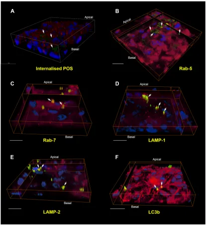
Figure 3: The internalisation pathway of photoreceptor outer segment cargo in RPE
440
cells. [A] A pulse-chase assay in cultured RPE monolayers showed bound FITC-POS in
441
green (arrows) at zero hours. Nuclei are stained with DAPI (blue) whilst Rab-5 positive
442
vesicles appear in red. Note the lack of POS co-localising with early compartments at this
443
initial stage. Scale bar in confocal orthogonal cross-section corresponds to 20μm. [B] 4 hours
444
following pulse-chase, POS may be observed in Rab-5 positive early compartments and
445
appear as discrete yellow vesicles. [C] By 6 hours, cargos had been trafficked to Rab-7
446
labelled late endosomes/phagosomes. [D] After 12 hours, POS appear in vesicles labelled
447
with the lysosomal marker LAMP-1. [E] At 24 hours, POS were found in vesicles positive for
448
the mature/late lysosomal marker LAMP-2. Note the perinuclear distribution typical of late
449
compartments. [F] POS persisted in vesicles labelled with LC3b, a marker of autophagy
bodies, as late as 48 hours. The trafficking and processing of POS cargo through distinctive
451
compartments can thus be visualised in cultured ARPE-19 cells. In all experiments, each
452
compartment was labelled with a specific antibody (red), whilst POS-FITC and nuclei appear
453
in green and blue respectively. Areas of co-localisation between POS and the
vesicle-454
specific marker appear yellow and denoted by white arrows. POS that are trafficked through
455
other compartments at a given time appear green, whilst areas of red indicate vesicles
456
devoid of any cargos. Scale bars in B-F corresponds to 20μm. Images of three-dimensional
457
RPE monolayers were captured using a Leica SP8 confocal microscope (LAS X) and
458
reconstructed using Amira 6.1 software.
459
---
460
461
The molecular events involved in POS recognition and internalisation is better understood
462
compared to those associated with cargo degradation. POS are internalised within RPE
463
phagosomes, which can be observed morphologically in toluidine blue stained sections125,
464
by cryo-immuno electron microscopy126, fluorescent labelling44,119 and more recently by
465
correlative light-electron microscopy127. Nascent phagosomes are enriched with annexin A2,
466
whilst siRNA knock-down of annexin A2 results in impaired POS internalisation128. POS
467
phagosomes have also been shown to associate with myosin-7a and kinesin-1 as they traffic
468
away from the apical RPE surface. Impaired phagosome localisation and degradation in
469
aged mice lacking kinesin-1 light chain 1 resulted in a phenotype resembling features of
470
AMD129. Our studies demonstrated labelling of internalised POS cargos with 5 and
Rab-471
7 markers for the first time117. However, we are unable to comment with any certainty on
472
whether these POS cargos were trafficked through phagosomes or endosomes. It is likely
473
that both pathways may be used by RPE cells. Quantification of POS-positive vesicles in
474
confocal z-stacks revealed vesicle diameters of 318nm ± 69.2 for Rab-5, 421nm ± 19.4 for
475
Rab-7, 677nm ± 32.1 for LAMP-1, 712nm ± 59 for LAMP-2 and 990nm ± 23.9 for LC3b117.
476
The diameter of cargo-carrying vesicles in each compartment was consistent with the
477
reported sizes of endosomes (200-500nm)9 and lysosomes (500nm-1μm)23. The increasing
478
diameter of POS vesicles also indicated trafficking from early to more mature compartments,
479
although this does not exclude the possibility of trafficking in Rab-5 and Rab-7 positive POS
480
phagosomes. Indeed, the internalisation route of cargos during early stages may largely be
481
determined by the initial size of POS. For instance, it appears that POS may be trafficked
482
through endosomes as smaller particles119. Hence, studies using POS assays necessitates
483
a step to maximise single POS molecules or produce smaller POS particles prior to feeding
484
cultures, as large POS aggregates remain bound to the apical RPE surface119. We also
485
recoded the timing of POS trafficking through distinctive compartments in primary mouse
486
RPE and ARPE-19 cells. Cargo internalisation and degradation in RPE cell-lines such as
487
d407 and RPE-J are reportedly slower130 compared to primary mouse/rat RPE119,131. Our
488
timing experiments of POS internalisation and degradation in ARPE-19 cells also revealed
489
slower kinetics compared to primary rodent RPE. Use of the pulse-chase assay enabled the
490
initiation of POS internalisation to be synchronised following receptor binding. We observed
491
the co-localisation of POS in Rab-5 compartments from 2 hours with co-localisation in equal
numbers within Rab-5 and Rab-7 positive vesicles by 6 hours. POS in Rab-7 compartments
493
diminished after 12 hours whilst those in LAMP-1 and LAMP-2 vesicles appeared at 6-12
494
hours and peaked between 12-24 hours. POS co-localised in lysosomes characteristically
495
accumulating in perinuclear regions (Figure 3E). POS eventually co-localised with LC3b
496
positive autophagy bodies after 12 hours, which reached a peak at 48 hours117. The
497
development of novel imaging technologies as well as powerful new data-handling software
498
provide the opportunity to reconstruct trafficking organelles in 3D (Figure 3). Others have
499
also used a wide range of technologies to study cargo handling in the RPE99,119,127,129.
500
Studies of this kind had once posed considerable technical challenges in the past.
501
502
The end-products of POS degradation is absorbed by the RPE, recycled back to
503
photoreceptors or removed from the cell. However, incomplete degradation of such waste
504
material accumulates within RPE lysosomes as lipofuscin. Senescent post-mitotic macular
505
RPE cells filled with lipofuscin is a characteristic feature of the ageing retina and accounts
506
for as much as ~20% of cell cytoplasmic volume by the eighth decade of life132. Lipofuscin
507
may disrupt the phagocytic mechanisms of RPE cells133, impair lysosomal proteases134and
508
inhibit vATPase so that lysosomes become less acidic and cause leakage of contents into
509
the cytosol133. Our work in neurons have revealed similar lysosomal damage leading to
510
conditions such as AD135. Healthy macular RPE cells have higher levels of lysosomal
511
enzyme acid phosphatase and cathepsin D relative to lysosomes from RPE in the
nasal/mid-512
zone and peripheral retina136. However, the presence of lipofuscin causes lysosomal
513
enzyme activity to decrease by up to 50% indicating the selective vulnerability of the macula
514
to disease137. Lipofuscin also generates reactive oxygen species, which modifies lipids and
515
forms high molecular weight components that are stable within lysosomes138-140.
Lipofuscin-516
containing compartments also contain molecules such as
N-retinylidene-N-517
retinylethanolamine (A2E)141, malondialdehyde (MDA) and 4-hydroxynonenal (HNE). A2E,
518
a derivative of vitamin A111,142, has the ability to irreversibly inhibit lysosomal cathepsin
519
activity upon exposure to light143. MDA and HNE by contrast is generated as a result of lipid
520
peroxidation of lipofuscins, and capable of forming covalent bonds with adjacent proteins144.
521
The accumulation of lipofuscin is thought to be partially caused by modification or
522
crosslinking of proteins by MDA or HNE, thus reducing susceptibility to proteolysis145.
523
Consequently, MDA and HNE modifications reduce POS clearance contributing to lipofuscin
524
accumulation within lysosomes146. With respect to mechanisms through which protein
525
aggregation may be dealt with, there appears to be no universal consensus as to whether
526
autophagy plays a role, and whether its activity increases or declines with age and disease.
527
The answer may lie in the cell-type, disease or indeed the specific stage of disease. Analysis
528
of RPE or RPE/choroid in two AMD mouse models revealed an increase in autophagy
529
markers LC3 I/II, SQSTM1/p62, ATG7, ATG9A as well as autophagosomes. Analysis of
530
donor AMD tissues also showed an upregulation of LC3, ATG7 and ATG9 in the RPE147.
531
Furthermore, drusen in AMD donor eyes also contained markers of autophagy100.
532
Interestingly, the actin motor protein myosin VI is expressed in the RPE layer and its deletion
533
in a mouse model mimics an AMD-like phenotype, as characterised by an accumulation of
basal-laminar deposits between the RPE and BrM148. Importantly, our earlier work revealed
535
that myosin VI is required for autophagosome-lysosome fusion mediated by direct
536
interactions with both autophagy and endocytic adaptor proteins149. This provides some
537
corroborating evidence that targeted disruption of key autophagic regulators, for example
538
the machinery essential for endosome and lysosome fusion, which results in
539
autophagosome accumulation can lead to AMD-like disease phenotypes. Nonetheless,
540
autophagy flux may increase or decrease depending on the capacity of RPE cells to cope
541
with the elevated proteolytic stress in different stages of disease, which may further
542
contribute to pathology. Readers are directed to several elegant reviews describing various
543
mechanisms involved in these events99,150.
544
545
6. New roles for lysosomes
546
547
New discoveries reveal lysosomes to be more than just end-points for cargo degradation.
548
For instance, it has been shown that lysosomes play an important role in nutrient sensing
549
and cellular metabolism. Lysosomes appear to have the ability to accumulate stores of
550
cationic amino acids, polyphosphates, ions as well as other building blocks which can be
551
released on demand151. The aforementioned multi-subunit mTORC1 complex is found
552
diffuse in the cell cytoplasm under nutrient-poor conditions. The presence of amino acids
553
and other nutrients causes a rapid translocation of mTORC1 to the lysosomal surface152.
554
mTORC is also capable of sensing amino acids both within and outside the organelle153.
555
Nutrient deprivation not only inhibits mTORC-mediated growth but also increases the
556
formation of autophagosomes42. Such findings may provide new insights into how cargo
557
handling/proteolytic degradation in RPE cells could be influenced by nutrition. Such
558
experiments are underway in our laboratory. Another discovery is the process of
559
transcriptional regulation by the transcription factor EB (TFEB)154 and members of the
560
microphthalmia-transcription factor E (MiT/TFE)155. These DNA-binding proteins bind
561
elements within promoters referred to as co-ordinated lysosomal expression and regulation
562
(CLEAR) to up-regulate genes encoding lysosomal and autophagy proteins154,156. TFEB is
563
recruited to lysosomal surfaces under nutrient-rich conditions and is phosphorylated by
564
mTORC1. Under starvation conditions, TFEB is dephosphorylated and subsequently
565
translocates to the nucleus to activate CLEAR genes. The Zinc-finger transcription factor
566
ZKSCAN3 functions in an antagonist manner, which under nutrient-rich conditions
567
translocates to the nucleus to repress several lysosomal and autophagy genes157.
568
Transcriptional regulators associated with lysosomes and autophagy thus play important
569
roles in cellular energy balance158 that may provide new insights into retinopathies.
570
571
The sophistication of in-vitro RPE models have grown considerably such that they represent
572
an excellent system to study how the endocytic and autophagy pathways become disrupted
573
with disease. The RPE is also a good model for epithelial/barrier studies due to the
574
availability of primary, stem cell-derived and immortalised/transformed cell-lines which have
575
been extensively characterised159. These cultures also enable a high degree of experimental
manipulation and control, such that specific pathogenic conditions in the retina can be
577
recapitulated for studies that would otherwise be difficult to undertake in whole mouse or
578
donor eye tissues. Advances in creating adult induced pluripotent stem cells such as those
579
generated in our laboratory enable studies of RPE cells from patients160, and the possibility
580
of directly studying effects on the endocytic and lysosomal-autophagy pathways. Recent
581
discoveries revealing new roles for lysosomes and autophagy are starting to provide insights
582
into how factors such as energy metabolism and nutrition, that has hitherto garnered limited
583
attention, could influence RPE cells in the senescent retina. These, coupled with advances
584
in optics and novel imaging technology has created new possibilities in studying how cargo
585
handling becomes impaired in RPE. Collectively, these have the potential to reveal
586
fundamental new insights into a range of irreversible blinding conditions.
587
588
Acknowledgments: This work was supported by grants to JAR from the Macular Society
589
UK, National Centre for the Replacement, Refinement and Reduction of Animals in
590
Research (NC3R: # NC/L001152/1), RP Fighting Blindness (GR590), National Eye
591
Research Centre (SAC 020), Fight for Sight/Alzheimer’s Research UK (Ref: 24AZ172) and
592
the Gift of Sight Appeal. AJL is a NIHR Senior Investigator. DAT is supported by a Wellcome
593
Trust Seed Award (205909/Z/17/Z). We thank our colleagues Dr David A. Johnston and Dr
594
David S. Chatelet at the Biomedical Imaging Centre (University of Southampton, UK) for
595
expertise in confocal imaging and assistance with Amira software.
596
597
Conflicts of Interest: The authors declare no conflict of interest.
598
599
References