A Study on
LAPAROSCOPIC vs. OPEN APPENDECTOMY –
A Prospective Comparative Case Series
A DISSERTATION
IN GENERAL SURGERY
Submitted in partial fulfillment
of the Requirements for the Degree of
M.S General Surgery (Branch I)
THE TAMILNADU DR.M.G.R. MEDICAL UNIVERSITY
KILPAUK MEDICAL COLLEGE
CHENNAI
CERTIFICATE
This is to certify that this dissertation entitled ‘Laparoscopic vs
Open Appendectomy – a Prospective Comparative Case Series
Study’ is the bonafide work of Dr. D. MADHU submitted as partial
fulfilment of the requirements for the degree of M.S. General Surgery at
Govt. Kilpauk Medical College – Govt. Royapettah hospital, Chennai.
Prof. P.N.Shanmugasundaram M.S.
Professor and Head of the department, Dept. of General Surgery,
Govt. Kilpauk Medical College
Chennai
Prof. K.Kuberan M.S.,
Dissertation guide and Unit Chief Dept. of General Surgery,
Govt. Royapettah Hospital
Chennai
Prof .P . RAMAKRISHNAN MD DLO DEAN,
DECLARATION
I Dr. D. MADHU solemnly declare that the dissertation submitted
on the topic “LAPAROSCOPIC VS. OPEN APPENDECTOMY –
A PROSPECTIVE COMPARATIVE CASE SERIES ” is a bonafide work done
by me from May 2010 to November 2012, towards partial fulfillment of
the requirements of M.S Degree examinations, General Surgery,
April 2013.
Chennai Dr. D. MADHU
ACKNOWLEDGEMENTS
I am pleased to acknowledge Prof. Dr. P. RAMAKRISHNAN
M.D. DLO, Dean, Govt. Kilpauk Medical College and Hospital for the
opportunity to conduct this study in the Department of General Surgery,
at Government Royapettah Hospital, Govt. Kilpauk Medical College,
Chennai.
My sincere gratitude to my guide and mentor;
Prof. Dr. K. KUBERAN M.S., Professor of Surgical unit II,
Government Royapettah Hospital, who has inspired and sculpted me as a
surgeon during my training as a postgraduate student under him and
guided through my dissertation.
I am most thankful to Prof. Dr. P.N. SHANMUGASUNDARAM,
M.S., Professor & Head of the Department of General Surgery, Govt.
Kilpauk Medical College for the encouragement and permission to avail
all the study materials in the Department of General Surgery for my
dissertation.
I also acknowledge the invaluable advice and guidance in various
aspects of my dissertation given by Prof. Dr. R.A. PANDYARAJ M.S.
This study would have not been possible without the support of my
Assistant professors, Dr. S.MANISELVI M.S.,D.G.O.
Dr. S.THIRUNAVUKKARASU M.S., Dr. S. SELVAKUMAR M.S.,
to whom I owe my initiation in surgical training.
I also thank my fellow post-graduates and interns who have been a
source of immense help in many aspects.
I am indebted to all my patients for the co-operation and patience
they showed to all the ordeal. I am most thankful to them to have helped
TABLE OF CONTENTS
S.
No.
Title Page
No.
1. INTRODUCTION 1
2. OBJECTIVES 5
3. REVIEW OF LITERATURE 6
4. MATERIALS & METHODS 71
5. PROFORMA 78
6. DATA ANALYSIS AND RESULTS 81
7. STATISTICAL ANALYSIS 95
8. DISCUSSION 96
9. CONCLUSION 101
10. FIGURE INDEX i
11. CHART INDEX ii
12. TABLE INDEX iii
13 BIBLIOGRAPHY iv
FIGURE INDEX
Fig No. Name Page No.
1 REGINALD FITZ 2
2 KURT SEMM 4
3 Cut section of Normal Appendix-Histology 10
4 Normal anatomy and relations of the appendix 11
5 Variations in the position of the appendix 12
6 Blood supply of the Appendix 13
7 Charles McBurney 15
8 The McBurney’s Point 15
9 USG showing appendicular lumen in
appendicitis
27
10 CT scan showing inflamed appendic with periappendiceal fat stranding and fluid collection.
30
11 Position of Appendix in Pregnancy 34
12 Open appendectomy - Stump inversion 51
13 Open Appendectomy showing the delivered appendix with the caecum
74
CHART INDEX
Chart No.
Name Page
No
1a Age distribution in L.A 81
1b Age distribution in O.A 81
1c Age distribution comparison between L.A &
O.A
81
2 Sex distribution between L.A and O.A 82
3a Pathology Report in L.A 83
3b Pathology Report in O.A 83
3c Pathology Report of L.A & O.A 83
4 Operating time in mins between L.A & O.A 85
5a Parenteral analgesic between L.A & O.A 86
5b Oral Analgesic between L.A & O.A 86
6 Return of Bowel function in hours 87
7a Wound infection between L.A & O.A 88
7b Intra-abdominal abscess between L.A &
O.A
88
8 Resumption of Liquids and Solids in L.A 89
9 Resumption of Liquids and Solids in O.A 90
10a Length of stay in hospital in days in L.A 91 10b Length of stay in hospital in days in O.A 91
10c Length of stay in hospital in days between L.A & O.A
92
11a Return to work in days in L.A 93
TABLE INDEX
Table No.
Name Page No.
1 Alvarado Score 47
2 Comparison of Sex Distribution btw. L.A and
O.A
82
3 Comparison of Pathology Report bte L.A and
O.A
84
4 Comparison of Operating time btw. L.A and O.A 85
5 Comparison of Parenteral and Oral Analgesic
dose btw L.A and O.A
86
6 Comparison of Bowel function return btw L.A.
& O.A
87
7 Comparison of Liquid & Solid Diet in L.A 89
8 Comparison of Liquid & Solid Diet in O.A 90
9 Comparison of Length of Stay btw L.A & O.A 92
10 Comparison of Return to Work btw L.A & O.A 94
BIBLIOGRAPHY
1.
Laparoscopic vs open appendectomy A prospective comparative study of 227 patients M. Marzouk, M. Khater, M. Elsadek, A. Abdelmoghny Department of Surgery, Saudi German Hospitals Group—Jeddah, Post Office Box 2550, Jeddah 21461, Saudi Arabia Received: 1 April 2002/Accepted: 17 October 2002/Online
publication: 6 March 2003
2.
Outcomes of Laparoscopic versus Open AppendectomyALFREDO
M. CARBONELL, D.O., JUSTIN M. BURNS, M.D., AMY E. LINCOURT, PH.D., KRISTI L. HAROLD, M.D. From the
Carolinas Laparoscopic and Advanced Surgery Program, Department of General Surgery,Carolinas Medical Center, Charlotte, North Carolina; and the Department of General Surgery, MayoClinic-Scottsdale, Scottsdale, Arizona Scientific Meeting in
Atlanta, GA, January 31-February 3, 2004
3.
Laparoscopic Versus Open Appendectomy A Prospective Randomized Double-Blind Study Namir Katkhouda, MD, Rodney J. Mason, MD, Shirin Towfigh, MD, Anna Gevorgyan, MD, and
Rahila Essani, MD Posted: 09/15/2005; Annals of Surgery. 2005;242(3):439-450. © 2005 Lippincott Williams & Wilkins
4. ECAB Clinical Update Surgical Gastroenterology and Liver
6. Schwartz’s Principles Of Surgery 9th edition F.Charles Brunicardi
7. Sabiston Textbook of Surgery 18th edition Volume 2
8. Bailey & Love’s Short Practice Of Surgery 25th edition
9. Grading and Staging in Gastroenterology Guido N.J.Tytgat; Stefaan H.A.J. Tytgat
10. Evaluation of Laparoscopic Appendectomy vs. Open Appendectomy: A Retrospective Study Aga Khan University Hospital, Karachi, Pakistan Yasmin Vellani, Shaheena Bhatti, Ghina Shamsi, Yasmin Parpio, Tazeen Saeed Ali Department of
Nursing Services, Department of Surgery, School of Nursing, Department of Community Health Sciences, The Aga Khan University Hospital, Karachi, Pakistan.
11. Laparoscopic versus Open Appendectomy: Is There a Difference?
Long KH, Bannon MP, Zietlow SP, et al. A prospective randomized comparison of laparoscopic appendectomy with open appendectomy: clinical and economic analyses. Surgery 2001;129:390–400.
Yongfeng Liu BMC Gastroenterology 2010, 10:129 doi:10.1186
/1471-230X-10-129
13. Laparoscopic versus open appendicectomy: prospective randomised trial J.J.T. Tate FRCS, J.W. Dawson FRCS, S.C.S. Chung FRCS, W.Y. Lau FRCS, A.K.C. Li FRCS The Lancet,
Volume 342, Issue 8872, Pages 633 - 637, 11 September 1993 doi:10.1016/0140-6736(93)91757-D
14. Laparoscopic Versus Open Appendectomy: Outcomes Comparison Based on a Large Administrative Database Ulrich Guller, MD,
MHS; Sheleika Hervey, BA; Harriett Purves, MPH, BS; Lawrence H. Muhlbaier, PhD; Eric D. Peterson, MD, MPH; Steve Eubanks, MD; Ricardo Pietrobon, MD Posted: 01/21/2004; Annals of Surgery. 2004;239(1) © 2004 Lippincott Williams & Wilkins
15. Laparoscopic versus open appendectomy in females with a clinical diagnosis of appendicitis Abdulrahman S. Al-Mulhim, FRCSI, FICS, Faisal M. Mulhim, FRCSI, Abdulmohsen A. Al-Suwaiygh, MBBS, Nabil A. Al-Masaud, MBBS Saudi Med J 2002;
INTRODUCTION
HISTORY :
The appendix is considered historically as a vestige with no
discernible function. However its inflammation produces one of the most
common causes of acute abdomen presenting to the emergency
department, the appendicitis leading to appendectomy the most
commonly performed general surgical procedure till date.
The appendix described first by 1521by the physician-anatomist
Berengario DeCarpi. However as were many things that came to
knowledge from the great Leonardo da Vinci so was the appendix which
was illustrated by him in his drawings in 1492 published later in the 18th
century. In 1824, Louyer-Villermay identified appendiceal inflammation
in 2 autopsied cases. In 1827, Francois Melier was the first to report an
antemortem case of appendicitis. Dupuytren discounted by arguing only
inflammation of the caecum, ‘typhilitis’ was the primary cause of right
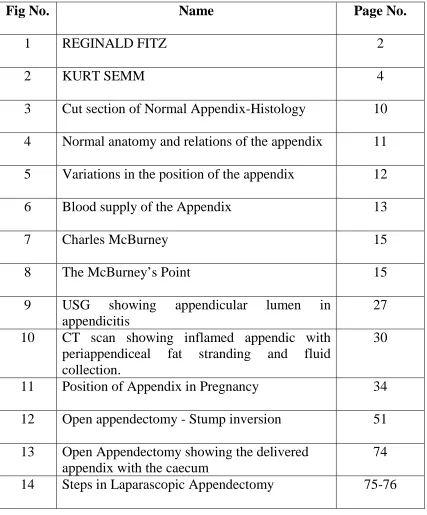
lower quadrant pain. Reginald H. Fitz, a Surgical pathologist and
pathologic anatomist at Harvard has the credit for coining the term
‘appendicitis’. In humans the appendix has been long considered to be a
appendix secretes enzymes to degrade cellulose into carbohydrates and
[image:14.595.233.385.155.374.2]functions as an important adjunct to digestion and absorption.
Fig. 1 Reginald H.Fitz
VESTIGIALITY AND EVOLUTION :
It has long been known that the human appendix and the terminus
of the caecum of mammals are structurally homologous in the vertebrate
comparative anatomy. A blind pouch, another name for the appendix is
in fact the "true caecal apex". In primates particularly, the termination of
the caecum and the vermiform appendix share the same relative position,
both have a similar structure and form, both are blind ending structures
From the analysis of anatomy systematically and comparatively, it
is known that in primates a large caecum with a small or absent appendix
is the ancestral, primitive state. In general, the length of the caecum,
relative to that of the colon, decreases as one travels down the
phylogenetic tree of evolution from the monkeys to humans.
Concurrently, the size of the appendix increases.. The human appendix
has lost a major and previously essential function, which is cellulose
digestion. Though it has decreased in size to a become a rudimentary
organ, the appendix retains a structural originality specifically adapted for
containing the cellulose digesting bacteriae and extending the time course
of digestion. The appendix in humans though vestigial, functions in the
development of the immune system in children as a source of antigen
presenting lymphocytes in the gastrointestinal tract inducing memory
cells for further defence against similar pathogens in the future. In
humans the cellulose being a component of the diet is not actually
digested but is useful as a bulk forming agent that ensures colonic transit
and functional integrity with mucosal protection from the deleterious
effects of refined foods and other pathogens. Thus the human appendix is
a rudiment of the caecum that is useless as a normal mammalian,
cellulose-digesting caecum. Thus to say, the topography of the appendix
on a non-evolutionary basis being at the end of the caecum has lost its
SURGICAL HISTORY :
The first recorded appendicectomy was performed by Claudius
Amyand (1681 – 1740), surgeon at St.George Hospital, London and
Sergeant Surgeon to Queen Ann, King George I and II. A 11 yr old lad
with scrotal hernia with a fecal fistula and containing a perforated
appendix was operated by him in 1736. The initial surgical therapy for
appendicitis was right lower quadrant drainage alone. The first surgical
therapy for appendicitis without abscess was done by Hancock in 1848,
which was peritoneal incision and drainage without appendectomy. The
first elective appendectomy performed by Fergus in Canada in 1883. The
first published account of emergency appendectomy for appendicitis was
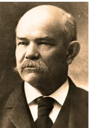
by Kronlein in 1886.The first laparoscopic appendectomy was performed
in 1982, by Kurt Semm, a Professor of Gynaecology at the University of
[image:16.595.191.425.512.707.2]OBJECTIVES OF THE STUDY
To compare the operative difficulties in terms of operating time
between laparoscopic and open appendectomies.
To prove the effectiveness of either of the procedures in reducing the
postoperative morbidity in terms of
Postoperative pain and analgesic use
Return of bowel function
The resumption of liquid and subsequent solid diet
Rate of infection, both surgical site and intra-abdominal infections
Number of days of hospital stay
REVIEW OF LITERATURE
EMBRYOLOGY :4,5
The caecal bud arises as a diverticulum from the post-arterial
segment of the midgut loop from which the caecum and the appendix are
formed. The proximal part of this bud grows rapidly to form the caecum
while the distal part remains narrow giving rise to the appendix.
The rapid, asymmetric growth of the lateral wall more than the
medial wall of the caecum results in medial displacement of the point of
attachment of the appendix leading to the medial position of the same. In
some cases the right colon and the caecum rotate about their long axis
leading to the common position of the appendix retrocaecally.
The development occurs at around 8th week of gestation; at 4th / 5th
month villi are found which disappear before birth. At about 7th month
the lymphoid nodules develop in the lamina propria and remain active
until puberty when they gradually decrease thereafter. At birth the
appendix is short and broad, and by the differential caecal growth it
becomes the typical tubular narrow structure by the end of two years of
Congenital anomalies and Duplication : 4
Eventhough it is a vestigial organ the frequency of anomalies is
very low.
Duplications of appendix can be of three types :
1. Type A : Partial duplication of the tip with a single base to the
caecum
2. Type B : Two completely separate appendices from a single
caecum which can be one of the 2 subtypes :
Type B1 : one on either side of the ileo-caecal valve
Type B2 : one in normal location and other on one of
the taenia coli – the ‘taenia-coli type’
3. Type C : Double caecum each with an appendix.
It is important to understand these duplications to look for a
duplicate when u find a normal appendix during appendectomy and it
Left-sided appendicitis rarely can occur with
1. Situs inversus viscerum
2. Midgut malrotation
3. Caecum with long mesentery
4. Long appendix
Interestingly in situs inversus and midgut malrotation the pain of
the left sided appendicitis is felt in the right side in 30% of the cases
The advent of laparoscopy has obviated the need in these
circumstances where one cannot find the appendix that is left sided by a
classical McBurney’s incision to convert into a laparotomy.
Rarer forms of congenital anomalies like total absence of appendix
and ectopic appendix are also reported.
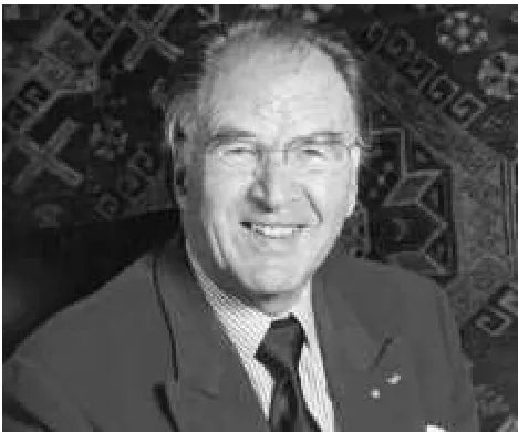
HISTOLOGY : 6
The appendix similar to the colon has four layers namely
3. Submucosa
4. Mucosa
a. Muscularis mucosa
b. Lamina propria
c. Epithelial layer
The muscularis propria consists of two layers of smooth muscles
namely the outer longitudinal and inner circular layers
The submucosa and the lamina propria of the mucosa contain
numerous lymph nodules as follicles and aggregations containing both B
and T lymphocytes akin to the Peyer’s patches as a part of the Gut
Associated Lymphoid tissues (GALT) which are responsible for the
mucosal immune function of the Gastrointestinal Tract.
The epithelium is columnar cells of the intestinal mucosa of
colonic type.
Fig. 3 Cut section of normal appendix - histology
ANATOMY : 6,7,8
The vermiform (Ln : worm-like) appendix is a narrow tubular
structure of varying size and shape arising from the caecum located in a
posteromedial position around 2 cm inferior to the opening of the ileum.
The length of the appendix is highly variable and can be from 2 to
20 cm and average being 9 to 11 cm long.
The lumen is irregular being encroached upon by longitudinal folds
of mucous membrane. A few crypts are present.
The base of the appendix can vary in location relative to the
variable position of the caecum to posture, respiration and intestinal
[image:23.595.127.494.199.403.2]distension.
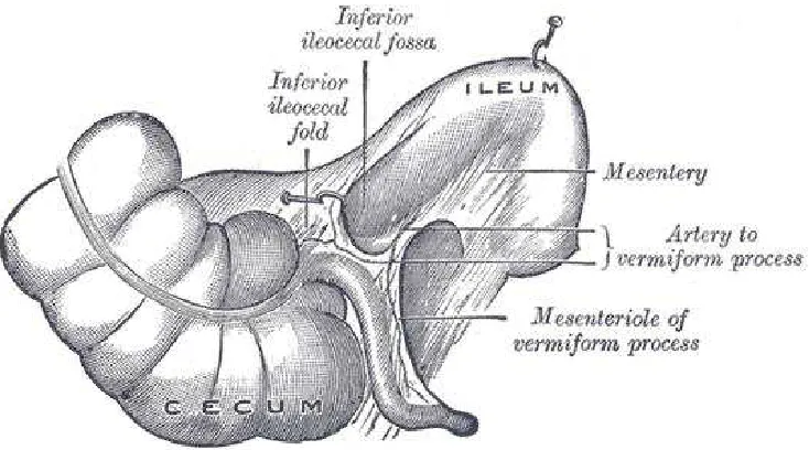
Fig. 4 Normal anatomy and relations of the appendix
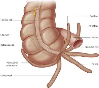
The position of the tip of the appendix is highly variable and can be
one of the following:
1. Retrocaecal – 75% being the commonest
2. Paracolic – 2%
3. Pre-ileal – 1%
4. Post-ileal – 0.5%
5. Subcaecal -1.5%
The appendix is usually intraperitoneal while in some rare
[image:24.595.100.421.159.446.2]circumstances its tip may be extraperitoneal also.
Fig. 5 Variations in the position of the appendix
The mesentery of the appendix / Mesoappendix :
It is a triangular fold derived from the mesentery of the terminal
ileum, which is attached to the caecum and appendix. It contains the
appendicular vessels, nerves and lymphatics.
Arterial Supply :
The appendicular artery is a branch of the ileocolic artery which is
derived from the superior mesenteric artery. It is an end artery with no
other anastomosing vessel and gangrene and perforation are early in acute
appendicitis. Occasionally 2 appendicular arteries are present.
An accessory atypical appendicular artery derived from the caecal
artery supplies the appendix.
The appendicular veins accompany the artery and drain into the
[image:25.595.148.473.435.637.2]superior mesenteric vein.
Lymphatic drainage :
They drain directly into the ileo-colic nodes or into the
appendicular nodes in the mesoappendix.
Innervation :
The sensory innervation of the appendix is carried by 8th, 10th, and
11th thoracic nerves.
The parasympathetic originates from the vagus.
The sympathetic originates from the celiac and superior mesenteric
ganglia.
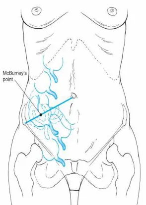
SURFACE MARKING :
The point of maximum tenderness in acute appendicitis is 1.5 to
2 inches from the right anterior superior iliac spine along a straight line
from it to the umbilicus, the McBurney’s point ( Charles McBurney US,
Fig. 7 Charles McBurney
‘ maximum tenderness, when one examines with finger tips is, in
adults, one half to two inches inside the right anterior spinous process
of the ilium on a line drawn to the umbilcus’
[image:27.595.235.382.443.650.2]Otto Lanz description of the surface marking of appendicular base is:
‘The base of appendix being located at the point one-third distance
from the right anterior superior iliac spine along a line joining the two
anterior superior iliac spines’ (Otto Lanz Amsterdam, 1908)
ACUTE APPENDICITIS
The inflammation of the appendix is called appendicitis or
formerly called ‘epityphilitis’
INCIDENCE : 6
The lifetime incidence of appendicitis is around 12% for men and
25% for women with average rate of appendectomy for appendicitis
around 7-8%in a lifetime. It is usually seen in second through fourth
decade of life and peaking in 20-30 yrs of age. There is a slight male :
female preponderance. (1.2 – 1.3:1) Appendectomy for appendicitis is the
most commonly performed emergency operation in the world. The
incidence is however lower in the regions of the world where there is a
high dietary fibre intake as in Africa and Asia. The misdiagnosis of
more in case of women with a bimodal distribution, one at 40 – 49 yrs of
age and the other at >80 yrs of age leading to a similar percentage of
negative appendectomies.
The rate of appendiceal rupture has also remained relatively
constant.
AETIOLOGY : 6,7,8
Fecaliths are the most common causes of appendiceal obstruction.
Other less common causes are :
1. Lymphoid hypertrophy
2. Inspissated barium from previous radiologic studies
3. Tumors
4. Vegetables
5. Fruit seeds
PATHOPHYSIOLOGY :6
There is a predictable sequence of events leading to the
characteristic pathophysiology of appendicitis and its course.
There is a proximal obstruction of the appendiceal lumen leading
to a closed loop obstruction, while the distal mucosa continues to secrete
mucous leading to intraluminal distension.
The luminal capacity of the normal appendix is just around 0.1 ml
and distension of the lumen by 0.5ml or more of secretions raises the
intraluminal pressure to 60 cm of H20.
The distension increases from continued mucosal secretions and
from rapid multiplication of the resident bacteria. The venous pressure is
exceeded and the capillaries and venules become occluded. The arterial
inflow continues and results in engorgement and vascular congestion. The
inflammatory process soon involves the serosa and the parietal
peritoneum eventually gets inflamed.
As the vascular compromise occurs the areas with poorest blood
supply i.e. concentric ellipsoidal infarcts of the anti-mesenteric border
The three ominous components of distension, vascular compromise
and bacterial invasion coexist and act in a vicious cycle, infarction
progresses and perforation is the end point.
The area of perforation is just beyond the point of obstruction
rather than the tip according to the physics of luminal / diametric
proportions.
However perforation is not the only eventuality of appendiceal
obstruction. This process can rather be discoherent and discordant and the
sequence could be disrupted at any point and the inflammation may
burnout leading to chronic and recurrent appendicitis.
BACTERIOLOGY : 6
The bacteriology of the normal appendix is same as that of the
normal colon. The appendiceal flora remains constant throughout life
except Porphyromonas gingivalis which is seen only in adults. The
bacteria cultured in cases of appendicitis are therefore similar to those
seen in other colonic infections such as diverticulitis. The principal
organisms seen in the normal appendix, in acute appendicitis, and in
perforated appendicitis are Escherichia coli and Bacteroides fragilis. A
may be present. Appendicitis is a polymicrobial infection, with different
organisms found in cases of perforation. Hence all the patients must be
started on broad spectrum antibiotics that cover the normal colonic flora.
Peritoneal culture should be reserved for patients who are
immunocompromised as a result of either illness or medication, and for
patients who develop an abscess after the treatment of appendicitis.
Antibiotic coverage is limited to 24 to 48 hours in cases of
non-perforated appendicitis. For perforated appendicitis, 7 to 10 days is
recommended. Intravenous antibiotics are usually given until the white
blood cell count is normal and the patient is afebrile for 24 hours. The use
of antibiotic irrigation of the peritoneal cavity and transperitoneal
drainage through the wound are controversial.
CLINICAL FEATURES : 6,7,8
The classic patient presents with periumbilical pain 1 to 2 days
prior and which has migrated to the right lower quadrant consequent to
the involvement of parietal peritoneum leading to somatic visceral
efferent irritation from the initial visceral afferent fibre stimulation
There is a low grade fever owing to the inflammatory ongoing
pathology, few episodes of vomiting which is self-limiting and the patient
feels nauseated and early onset anorexia. Diarrhoea may occur but is
unusual and some patients may have constipation also and hence the
bowel function is of little significance in the diagnosis.
The vomiting is neither prolonged or prominent and is due to the
presense of ileus. The anorexia is constant with appendicitis and a patient
who feels hungry should be suspected of a different diagnosis.
The sequence is peri-umbilical pain followed by nausea vomiting
and then fever.
The acute phase lasts for 2 to 3 days by which either the
inflammation is burning down or has progressed to form an
abscess/phlegmon or perforated complicating the clinical course.
On examination:
Vital signs are minimally changed. Temperature is elevated by
rarely more than 1°C (1.8°F) and the pulse rate is normal or slightly
elevated. Changes of greater magnitude usually indicate that a
complication has occurred or that another diagnosis should be considered.
particularly the right thigh, drawn up, because any motion increases pain.
If asked to move, they do so slowly and with caution.
Focal tenderness over the right lower quadrant due to the parietal
peritoneal irritation, usually at the McBurney’s point but may be variable
depending on the location of the appendix.
If an abscess has formed, a vague mass can be palpable.
If the appendix has perforated, then guarding and rigidity localized
to the right lower quadrant can be made out.
There is tachycardia, fever, and signs of peritonism according to
the stage of presentation.
Few classical signs of appendicitis are :
Unsolicited pain on
1. Palpation of the left lower quadrant - ROVSING sign
2. On cough - DUNPHY sign
3. Internal rotation of the flexed right thigh- OBTURATOR sign
4. Extension of the right hip - ILIOPSOAS sign
Cutaneous hyperesthesia in the area supplied by the spinal nerves
on the right at T10, T11, and T12 frequently accompanies acute
appendicitis.
LABAROTORY INVESTIGATIONS :
Mild leukocytosis 10000-18000 cells/cu.mm with left shift to
polymorphonuclear predominance in uncomplicated appendicitis is the
only remarkable laboratory abnormality.
The elevation of leukocytes to more than 18000 cells/cu.mm is
suggestive of onset of a complication either an abscess or perforation
C- reactive protein may be raised in cases of perforated appendix
Urinalysis may demonstrate pyuria when possibility of a pelvic
appendicitis or a different diagnosis of urinary tract pathology.
RADIOLOGICAL INVESTIGATIONS : 4,6
The routine use of imaging modalities to confirm a suspected
appendicitis in a patient with obvious clinical symptoms and signs is
discouraged.
In certain high risk patients and in the extremes of age when the
and signs necessitate the use of radiological imaging to help in
establishing a diagnosis. In the event of operation without the aid of
imaging modalities in the diagnosis the incidence of 20% negative
appendectomies is accepted. The incidence of negative appendectomies is
higher without imaging, especially in the females of reproductive age
group which is up to 42%. In this specific group of patients, imaging can
help reduce the incidence of a negative appendectomy. But the use of
imaging delays the surgical procedure thereby increasing the risk of
appendiceal perforation.
The two imaging modalities mainly used are the ultrasound and
computerised tomography of the abdomen and pelvis
ULTRASOUND EXAMINATION:
Graded compression sonography has a highly variable sensitivity
and specificity due to the operator dependent outcome. It becomes most
useful in female patients when the gynaecological causes of their
symptoms mask the causes of appendicitis.
It is also the imaging of choice in pregnant patients in whom
The appendix whether normal or abnormal is often not found by
the study which is why it is most often unhelpful and inconclusive
The variability in the position of the tip of the appendix makes it
difficult to localize.
The bowel mass interpositioning and the body mass index of the
patient makes the image quality and resolution poorer.
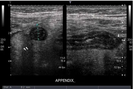
FINDINGS on USG in Appendicitis :
1. thickening of the appendiceal wall
2. loss of wall compressibility
3. increased echogenicity of the surrounding fat signifying
inflammation and loculated pericaecal fluid
The appendix is identified sonographically as a blind-ending,
nonperistaltic bowel loop originating from the cecum. With maximal
compression, the diameter of the appendix is measured in the
anteroposterior dimension. A scan is considered positive if a
noncompressible appendix 6 mm or greater in the antero-posterior
Oedema and free fluid are usually present. The compressibility of
the appendix reduces with increasing inflammation and there may be air
within the lumen.
The complications of appendicitis like mass formation or abscess
can be visualized well with the ultrasound. In certain cases, free fluid
alone in the right iliac fossa, right paracolic gutters or the pouch of
Douglas suggest a positive diagnosis with clinical correlation. The
presence of particulate matter especially pus or blood in the fluid may
help preclude surgery and resort to conservative modality of treatment
with ultrasound guided drainage of the pus in cases of appendiceal
abscess or postoperative collections.
The probe tenderness elicited during the ultrasound examination in
the right iliac fossa especially in a female may suggest either appendiceal
inflammation or gynaecological pathology.
The adnexal structures such as Fallopian tubes and ovaries can be
the cause of right iliac fossa pain. Often physiological changes in the
ovaries like corpus luteum may explain the pain. In the instances of
gynaecological pathologies a transvaginal probe may be more useful in
Fig. 9 USG showing appendicular lumen in appendicitis
COMPUTERISED TOMOGRAPHY :
The quality and reliability of CT scan has made it a reliable
diagnostic tool when the ultrasound findings are inconclusive with
sensitivity and specificity approaching 100%.
It allows better visualization of other intrabdominal organs
particularly with oral / iv contrast. It is especially useful in the elderly
when the likelihood of other conditions like diverticulitis, inflammatory
bowel disease are more. It can also pick up small bowel pathology which
Findings on a CT scan in Appendicitis :
- An inflamed appendix is thickened with surrounding free fluid
and inflammatory periappendiceal fat stranding seen as
streaking and poorly defined increased attenuation.
- PHLEGMON
- Appendicolith (not pathognomonic)
- Arrow-head sign - This is caused by thickening of the cecum,
which funnels contrast toward the orifice of the inflamed
appendix.
- If free or localized gas is seen it may indicate the perforation of
the appendix
- Small bowel ileus may be present with appendiceal perforation.
- Appendiceal abscesses are seen as a large inflammatory
phlegmon.
- Features such as portal gas and soft tissue gas usually suggest a
- CT is useful tool to guide percutaneous drainage of an abscess
or collections and useful in assessing the postoperative
complications like pelvic collections, stump leak, bleeding etc.,
LIMITATIONS :
• Lack of availability
• Not cost effective
• Higher dose of ionizing radiation harmful to the patient especially
the younger and pregnant.
The use of a CT scan must thus be limited to high risk patients for
surgery and anaesthesia where the clinical suspicion of appendix is low
and other pathologies need be ruled out. It is though useful in reducing
the negative appendectomy rate and reducing needless admissions and
diagnostic errors it is best used judiciously so that it doesn’t prolong or
Fig. 10 CT scan showing inflamed appendix with periappendiceal fat stranding and fluid collection.
OTHER DIAGNOSTIC MODALITIES :
X-RAY ABDOMEN PLAIN :
Dilated bowel loops suggest bowel obstruction or ileus from
appendicitis.
BARIUM Studies :
Not useful unless a mechanical distal bowel obstruction is
suspected. If the appendix is filled with barium appendicitis is ruled out,
if not filled no determination can be made.
MRI:
RADIOISOTOPIC SCANS :
Tc99 labelled leucocyte radioisotope scans may localize
inflammation to the appendix but the sensitivity is less (82%) for acute
appendicitis compared to CT scan and is maybe of diagnostic value in
chronic appendicitis.
DIAGNOSTIC LAPAROSCOPY :
The advent of laparoscopy for diagnosis in routine use makes the
imaging modalities less useful for appendicitis.
To clearly emphasize, the diagnosis of appendicitis is always based
primarily on clinical findings and physical examination and imaging
should be used in specially warranted clinical situations.
COMPLICATIONS OF ACUTE APPENDICITIS : 4
The primary adverse outcome of appendicitis is perforation and its
sequelae. The early exploration minimizes the chance of perforation but
increases the incidence of negative appendectomies.
The rate of negative appendectomies is 10-15%
The rate of perforation ranges from 12% to 35% with more
The complications are related to the frequency of infection of the
peritoneal cavity either by direct perforation or by bacterial translocation
through the appendicular wall.
The pathology can either be obstructive or non-obstructive
(catarrhal) inflammation. The inflammatory process beginning in the
mucosa rapidly extends outwards once it reaches the loose submucosal
tissues.
The vascular compromise occurs distally as the vessels are
intramural toward the tip and jeopardized by the pressure and occluded.
The non-obstructive type progresses slowly and the protective
adhesions form causing the peritonitis to be localized.
As gangrenous changes proceeds there are small infarcts occur
permitting the escape of bacteria into the peritoneal cavity and
accumulation of pus occurs.
When this occurs early as in obstruction induced gangrene the
peritonitis is diffuse but when it occurs prolonged over a time the
1. Gangrenous appendicitis
2. Appendicular mass
3. Perforated appendicitis with Localized abscess formation
4. Perforated appendicitis with Diffuse Peritonitis.
5. Cecal Gangrene
6. Pylephlebitis / Portal Pyemia
7. Intestinal obstruction due to contact with the inflamed appendix.
APPENDICITIS IN SPECIAL CIRCUMSTANCES :4
APPENDICITIS IN PREGNANCY :
Acute appendicitis is the most common non-obstetric indication for
surgical intervention in pregnant women. The appendicitis occurring in
pregnancy is difficult to diagnose due to atypical presentations and vague
lower abdominal pain. The appendix and the caecum is displaced by the
gravid uterus and come to lie in the right upper quadrant. The symptoms
of nausea, vomiting and abdominal pain are attributable to the state of
pregnancy and surgery for appendicitis carries risk of spontaneous
abortion and miscarriage during pregnancy. The anaesthetic
which is the safe time to embark on an appendectomy. Diagnostic
dilemma of misdiagnosing appendicitis and missing the diagnosis of
appendicitis and leaving it to progress to complications is a great concern.
Ultrasound helps delineate the appendix, its location and its involvement.
CT scan should be avoided. Of the complications related to appendicitis
in pregnancy, the most dreaded is the progressing sepsis which is most
detrimental to the fetus and the pregnancy itself which outweighs the
[image:46.595.198.419.353.585.2]anaesthetic and operative risks involved.
Fig. 11 Position of Appendix in Pregnancy
decade of life. The diagnosis is difficult in the young patient. The
etiology of appendicitis in children can be varied and the luminal
obstruction by the well-formed lymphoid follicular hypertrophy induced
by a viral infection and worm infestations are two different causes of
appendicitis in these group of patients especially. The pathophysiology of
the disease however is the same. However the incidence of complications
of appendicitis is early and severe in children compared to the adults
which stresses the need for prompt diagnosis and initiation of treatment.
The pain of appendicitis in children is not typically migrating and
can be varied with increased micturition and loose stools, spasm of the
psoas. The vomiting follows the onset of pain and can be differentiated
from the more common disease of gastroenteritis in children where the
vomiting is the presenting complaint. The elicitation of clinical signs in
children is most difficult and rovsing’s sign and psoas spasm are easily
elicitable compared to rebound tenderness which would deter further
examination. The investigations are the same as for adults and the
ultrasound is diagnostic in most instances. CT should not be used
unnecessarily due to the attendant risks. The management of appendicitis
whether complicated or uncomplicated is essentially same as for adults
with the use of antibiotics according to the level of complications and
inflammatory masses, percutaneous drainage by image guidance and
antibiotic cover is indicated. Appendicitis in neonates is extremely rare
and very difficult to diagnose as the presenting feature is irritability alone.
Imaging is essential for the diagnosis. The cause of appendicitis in the
neonates has to be identified and can be Hirschsprung disease,
meconium-plug syndrome or cystic fibrosis.
DIFFERENTIAL DIAGNOSIS : 4,6
The differential diagnosis of acute appendicitis is the same as for
the acute abdomen. The symptomatology is due to a perturbation of the
physiological function rather than a specific organ induced. So clinically
identical picture can result from a wide variety of acute conditions within
or near the peritoneal cavity that produce the same alterations of function
as acute appendicitis.
The rate of preoperative diagnosis of appendicitis in any particular
centre should be around 85 – 90% and if more or less it indicates the need
for a look into differential diagnosis.
There are a few conditions in which operation is contraindicated.
The most common erroneous preoperative diagnoses accounting
for more than
75% in descending order of frequency are
• acute mesenteric lymphadenitis,
• pelvic inflammatory disease,
• twisted ovarian cyst or
• ruptured graafian follicle, and
• acute gastroenteritis.
The differential diagnosis of acute appendicitis depends upon four
major factors:
• The anatomic location of the inflamed appendix;
• The stage of the process (i.e., simple or ruptured);
• The patient's age; and
• The patient's sex.
ACUTE MESENTERIC ADENITIS :
Acute mesenteric adenitis is the disease most often confused with
respiratory tract infection is notable. The pain is usually diffuse and not
localized.
Laboratory procedures indicate although a relative lymphocytosis,
when present suggests mesenteric adenitis. Often Campylobacter or
Yersinia is implicated in the causation. Observation for several hours
likely points the diagnosis, because mesenteric adenitis is a self-limited
disease. However, if the diagnosis remains in doubt, immediate
exploration is clearly indicated.
ACUTE GASTROENTERITIS :
Acute gastroenteritis is common in childhood but can usually be
easily differentiated from appendicitis. Viral gastroenteritis, an acute
self-limited infection of diverse causes is characterized by profuse watery
diarrhea, nausea, and vomiting. Abdominal cramps with tenesmus
precede the watery stools. The abdomen is relaxed between cramps, and
more importantly there are no localizing signs of peritonism. Laboratory
values are normal. Salmonella gastroenteritis results from ingestion of
contaminated food. Abdominal findings are usually similar to those in
viral gastroenteritis, but in some cases, the abdominal pain is intense,
organisms can be isolated from nearly 100% of patients. However,
cultures may take too long for differentiation to assist the clinician in
making a timely differential diagnosis. Similar attacks in other persons
eating the same food as the patient greatly strengthen the presumptive
diagnosis of salmonella gastroenteritis. Because typhoid fever is now a
rare disease, its diagnosis is frequently missed. The onset is less acute
than in appendicitis, with a prodrome of several days. Differentiation is
usually possible because of prostration, maculopapular rash,
inappropriate bradycardia, and leukopenia. Diagnosis is confirmed by
culture of Salmonella typhosa from stool or blood. Intestinal perforation,
usually in the distal ileum, develops in 1% of cases and requires
immediate surgical therapy.
GENITOURINARY INFECTIONS:
The diseases of male urogenital tract form a most important
differential diagnosis for appendicitis including torsion of the testis and
acute epididymitis, because epigastric pain may overshadow local
symptoms early in these diseases. Seminal vesiculitis may also mimic
appendicitis, but can be diagnosed by palpating the enlarged, tender
MECKEL’s DIVERTICULITIS :
The Meckel's diverticulum is located within the distal 2 feet of the
ileum and is true diverticulum of the intestinal tract representing the
junction of the midgut and hindgut namely the prearterial and postarterial
segment of the embryologic gut. It can be the cause of variety of
abdominal conditions but when present as a diverticulum arising out of
the intestinal tract it may mimic appendicitis . Meckel's diverticulitis is
due to the inflammation of the ectopic mucosa of the diverticulum and the
nearby intestinal mucosa usually acid secreting gastric mucosa causing
ulcerationof the normal nearby intestinal mucosa. Resection of the
segment of ileum bearing the the diverticulum with end-to-end
anastomosis can nearly always be done through a McBurney incision,
extended if necessary, as well as laparoscopically.
INTUSSUSCEPTION :
In contrast to Meckel's diverticulitis, it is extremely important to
differentiate intussusception from acute appendicitis as the treatment is
entirely different. The age of the patient is important: appendicitis is very
uncommon in children younger than age 2 years, whereas nearly all
suddenly doubled up by apparent colicky pain. Between attacks of pain,
the infant appears well. After several hours, the patient usually passes a
bloody mucoid stool. A sausage-shaped mass may be palpable in the right
lower quadrant. As the intussusception progresses distally, the right lower
quadrant feels abnormally empty. The preferred treatment of
intussusception, if seen before signs of peritonitis supervene, is reduction
by barium enema, but treatment of acute appendicitis by barium enema
may be catastrophic.
CROHN’s ENTERITIS :
Then manifestations of Crohn’s regional enteritis of the distal
ileum mainly causes symptoms of right lower quadrant pain with fever,
vomiting and diarrhea. Without a antecedent diagnosis of inflammatory
bowel disease the symptoms cannot be differentiated from that of
appendicitis and if on operating, the appendix and caecum found to be
normal with an inflamed distal ileum, appendectomy must be proceeded
with.
PERFORATION OF A PEPTIC ULCER :
In a sealed perforation the contents that spilled from a
right lower quadrant through the paracolic gutter and the upper abdominal
symptoms minimized due to no more spillage, may mimic a appendicular
pathology.
COLONIC LESIONS :
Diverticulitis and perforation of caecal or right side lying sigmoid
carcinoma can sometimes mimic appendicitis in the elderly with vague
symptoms. Imaging modalities like CT are particularly suited for these
groups of patients to clearly identify the pathology.
EPIPLOIC APPENDAGITIS :
The epiploic appendages of the colonic wall may get torsed and
infarcted producing inflammation and pain localized to the site and
without the systemic upset or the sequence of events of appendicitis. The
only symptom will be continuous pain at the location correspondingly
with rebound tenderness but no rigidity. It is selflimiting and resolves, in
around 25% patients exploration is undertaken for the continuous or
recurrent pain and removal of the infarcted appendage will typically
URINARY TRACT INFECTION :
Acute upper urinary tract infection – pyelonephritis of the right
kidney can present clinically with symptoms suggesting acute
appendicitis of retrocaecal or postileal type, the accompanying renal
angle tenderness, fever with chills and urinalysis abnormalities are
sufficient enough to make a diagnosis.
URINARY CALCULI :
A ureteral calculi most often mimics appendicitis particularly when
lodged around the region of the appendix on the right side. The radiating
pain from loin to groin, absence of leukocytosis and fever and the
sequence of symptoms or clinical signs of rebound tenderness/rigidity
points to the diagnosis. CT scan usually helps clinch the diagnosis.
PRIMARY PERITONITIS :
In nephrotic syndrome patients, a primary peritonitis caused by
gram-positive cocci may resemble the peritonitis of a perforated
appendix, and the cultures fro the peritoneal fluid may clearly point the
YERSINIA INFECTION :
Yersenia species like Y.enterocolitica and Y.pseudotuberculosis is
spread by the feco-oral route and they predominantly cause infection of
the terminal ileum, caecum, mesentery and appendix. Most are
self-limiting and some may cause systemic sepsis if unrecognized. The
clinical scenario of appendicitis must be intervened with appendectomy.
The Yersinia species are responsible for 5% of the cases of acute
appendicitis. Campylobacter jejuni induced diarrhea and abdominal pain
may mimic appendicitis and Salmonella typhi can cause mesenteric
adenitis mimicking appendicitis. Stool cultures and serology help to
delineate the etiology.
GYNAECOLOGIC DISORDERS :
The gynaecological disorders of young females are the most
common differential diagnosis of appendicitis for which appendectomies
are performed and they are the most common reason of high rate of
false-negative appendectomies. Some of the commonest causes of the female
reproductive tract pathologies that mimic appendicitis in the descending
PELVIC INFLAMMATORY DISEASE :
The pelvic inflammatory disease which is most of the time bilateral
when predominant on the right side may mimic appendicitis. The
characteristic absence of symptoms and clinical signs of appendicitis and
lower abdominal tenderness lower than that in appendicitis with cervical
motion tenderness on per vaginal examnination points to the diagnosis.
The symptoms of PID might mimic appendicitis more so in the luteal
phase of the menstrual cycle and vaginal smear will help rule out
appendicitis.
RUPTURED GRAAFIAN FOLLICLE :
Mittlesmerchz is the midcycle pain and tenderness in the lower
abdomen caused by the rupture of a matured graafian follicle with the
release of the ovum see in young ovulating women. If it is from the right
ovary and the amount of fluid released into the peritoneal cavity is
sufficient enough it causes pain mimicking appendicitis. Ultrasound will
aid in the diagnosis.
RUPTURED ECTOPIC PREGNANCY :
The rupture of an ectopic pregnancy either tubal or ovarian
the right lower quadrant. The history may reveal abnormal or missed
menstrual cycles. The presence of a pelvic mass with leukocytosis, pallor
and falling hematocrit along with tenderness on cervical motion in per
vaginal examination indicates the diagnosis. Culdocentesis and fluid
analysis will confirm the diagnosis. The clinical diagnosis is sufficient to
warrant immediate exploration and it not prudent to wait for imaging
modalities to confirm the diagnosis, as it is a gynaecological emergency.
TWISTED OVARIAN CYST :
The ovarian cysts most commonly benign serous cyst may undergo
torsion particularly when on the right side may mimic acute appendicitis.
The presence of an abdominal mass and tenderness in a young women
warrant the search for ovarian pathology and imaging modalities are
sufficient to clinch the diagnosis. There may be pain, rebound tenderness,
leukocytosis and fever. If the torsion has caused gangrene of the ovary,
immediate resection is the treatment of choice.
OTHER CAUSES :
Other diseases which may rarely mimic appendicitis are small
chest wall, acute cholecystitis, acute pancreatitis, abdominal wall
hematomas, etc.,
[image:59.595.115.513.259.531.2]SCORING SYSTEMS OF APPENDICITIS :
9Table 1. ALVARADO SCORE
ALVARADO SCORE :
SYMPTOM / SIGN / TEST SCORE
Migration of Pain 1
Anorexia 1
Nausea-vomiting 1
Tenderness at right iliac fossa 2
Rebound pain 1
Raised temperature ( >= 37.3 deg C) 1
Leucocyte count >= 10*109/L 2
Differential WBC count with
neutrophils >= 75%
1
Total score 10
A score that is more than 6 is highly predictive of acute
appendicitis and scores between 5 and 6 are prudent to be kept in
MANAGEMENT OF ACUTE APPENDICITIS:
4,6The management of acute appendicitis is essentially and primarily
surgical. The patient must be prepared for surgery with adequate
hydration and correction of electrolyte abnormalities. The use of
antibiotics preoperatively has been largely studied and proved to be
effective in reducing the postoperative infection rates.
For simple appendicitis without any complication, the antibiotic
therapy is a single agent third generation cephalosporin for 24 hours
alone. For appendicitis with complications, the antibiotic need to be
continued till the fever subsides and the leukocyte count normalizes. For
severe infections higher antibiotics like carbapenems, monobactums with
aminoglycoside and metronidazole must be used.
TYPES OF SURGERY :
OPEN APPENDECTOMY
OPEN APPENDECTOMY :
INCISIONS :
• McBurney’s incision :
It is a gridiron incision obliquely at right angles to a line joining the
right anterior superior iliac spine to the umbilicus and the center of the
incision lies on the McBurney’s point. The external oblique is cut and the
internal oblique and transversus are split (the muscle-splitting incision),
and retracted to reach the peritoneum.
The advantage of the incision is that regardless of the position of
the appendix, the access to the caecum and appendix are superior and the
incision also permits easy extension if necessary
• Lanz incision :
This is a low incision for appendectomy than the classic gridiron
incision, though this is cosmetic, the incision is difficult to extend. It is a
transverse incision 1 inch above and medial to the anterior superior iliac
spine extending to the lateral border of the rectus medially. This also is a
muscle splitting incision.
• Other incisions:
• RUTHERFORD-MORRISON’s incision : oblique muscle cutting
the McBurney’s point with right paramedian incision of the rectus
sheath vertically and retraction of the rectus muscle medially.
• ROCKEY-DAVIS incision; FOWLER WEIR incision
o These are transverse incisions for better cosmesis and with
extension medially when necessary.
• A lower midline laparotomy incision is considered when the
diagnosis is in doubt particularly in elderly patients in whom
carcinoma or diverticulitis is suspected and in selected group of
female patients also.
PROCEDURE :
The incision is made and layers opened to reach the peritoneum.
The peritoneum on opening, any intraperitoneal fluid assessed, if pus
encountered it is taken for microscopy, culture and sensitivity. The taenia
are traced along the caecum to their confluence to reach the base of the
appendix. The lateral to medial movement helps to deliver the tip of the
appendix. The delivery of the appendix out of the incision is the most
tricky and difficult step of open appendectomy as the position of the tip is
variable and may have adhesions to the neighbouring structures due to the
appendix particularly a retrocaecally placed one, the caecum can be
delivered out by a rocking motion and then appendix can be traced. The
appendix is mobilized by dividing the mesoappendix. Care must be taken
to carefully ligate the appendicular artery. Once the appendix is freed
from the mesoappendix. The base is double ligated and cut. An adequate
stump must be left to ensure a secure ligation. The stump can be just
ligated and left or ligated and inverted into the caecum by a purse-string
[image:63.595.178.442.357.621.2]or Z suture.
However this step is not followed nowadays because it does not
confer any advantage in preventing stump leaks. The peritoneum is
irrigated and hemostasis verified. All layers closed with absorbable
sutures. Skin sutured with non-absorbable sutures.
In the event of encountering pus or fecal matter on opening the
peritoneum, the need for a laparotomy or the extension of the incision
should be assessed. The appendectomy must be proceeded with, and a
careful handling of the friable tissues must be borne in mind. Once the
appendectomy is completed, the distal ileum and caecum must be
inspected to confirm their integrity, peritoneal irrigation with normal
saline and metronidazole is indicated. The need for drainage of the
peritoneal cavity must be assessed and a closed non-suction drain must be
placed. If grossly infected or contaminated, the wound must be left open
to heal by secondary intention or a delayed primary closure contemplated
after 4 to 5 days when the infection is reduced.
If the appendix is found to be normal, then a thorough search for
other pathologies must be made starting from the caecum, mesentery, the
whole of small intestine must be checked as far as possible in a retrograde
them. In the event of no other pathology being found the appendectomy
must be proceeded with and the peritoneal fluid must be sent for
microscopy, culture and sensitivity in the prospect of finding other
pathologies.
LAPAROSCOPIC APPROACH :
PREOPERATIVE WORKUP AND PREPARATION :
The patient selection is very important in laparoscopic
appendectomy as the role of LA in complicated appendicitis is
controversial however in experienced hands they are forming valid
indications these days. The main contraindications are in patients with
peritonitis and those with comorbidities precluding general anaesthesia.
The preoperative preparation with resuscitation and investigations
is the same as for open appendectomy. The patient must be informed of
the likelihood of conversion of the procedure into an open approach if
conditions warranted. A nasogastric tube may be used in the event of
ileus complicating the appendicitis and a urinary catherization to drain the
PROCEDURE :
The surgeon stands to the left of the patient and the camera
assistant on the right of the surgeon near the left shoulder of the patient.
The trolley and monitor on the right of the patient in front of the surgeon
at the level of the umbilicus.
The patient is placed supine and the arms tucked by the sides, and
patient secured to the table facilitating the rotation of the table to a
Trendelenburg and right side up position for better visualization by
allowing the abdominal viscera to gravitate to the left away from the right
lower quadrant.
A 10 mm trocar is sited at the umbilicus or supraumbilical position
by either an open method or close Veress needle insufflation (12 mm
Hg). Two more working ports are established, one along the suprapubic
region and the other along the left iliac fossa both being directed to the
right iliac fossa. One 5 mm port along the right side of the abdomen
lower down and the other on the lower midline also is used by some
surgeons but this causes the surgeon’s right hand to cross the
A 10 mm 30 degree telescope is introduced through the umbilical
port and a diagnostic inspection of the entire abdomen is carried out in a
step wise fashion. Using grasping forceps the appendix is located and if
found normal, rest of the caecum, small intestine and pelvic organs in the
female are visualized to delineate the pathology.
The omentum adhesed in the region of the right iliac fossa being
involved in the inflammatory process is gradually teased off and the
appendix is traced if not readily located, the caecum is grasped and the
taeniae are traced to the base of the appendix.
Mesoappendix coagulation :
The mesoappendix is coagulated by the use of bipolar coagulation
in short bursts and teased with the grasper or cut with scissors. If the
mesoappendix is bulky and inflamed then it must be completed
coagulated before dividing to avoid bleeding of the appendicular artery. If
in doubt the divided mesoappendix should be ligated with an endoloop.
The mesoappendix is separated completely from the appendix till the base
and the base clearly delineated to the caecum to avoid leaving behind a
Appendix excision :
Pre-readied endoloops of chromic catgut introduced through the 5
mm port closer to the surgeon and the appendix is guided through the
loop and the knot secured around the base. The appendix is crushed and
another endoloop is placed just above the previous one by the same
method. The appendix is cut between the two endoloops. By preference
one endoloop alone is sufficient and the appendix cut distally. There is no
difference of outcome proven between the use of number of endoloops.
The use of stapler for division of the appendicular base is not
widely practiced because of the cost and the need for a bigger 12 mm
port. It is useful in special occasions where there is no healthy base to
ligate or the caecal pole is also involved, wherein a stapler can resect that
part of the caecum without compromising the ileo-caecal valve.
Saline lavage of the region or the whole of the peritoneal cavity
may be carried out accordingly to minimize the postoperative infection
rates.
Extraction of the divided appendix :
Usually, the left handed trocar with the appendix specimen is
introduced retrogradely through the umbilical port and the laparoscope
gradually withdrawn. As soon as it reached the flap valve, the
laparoscope is removed and the appendix is caught with the hemostat
releasing from the left hand grasper and the specimen is delivered. In case
of bulky and grossly infected appendicular specimen, the same is placed
in a plastic bag before extraction to avoid wound infection.
The placement of a drain is not always indicated. If the appendix
was perforated and contamination was significant and the closure of the
stump was precarious, small closed drain is placed in a separate incision
in a dependent and direct contact to the right iliac fossa intraperitoneally.
The fascial closure at the 10 mm port is a must with a
non-absorbable suture material to avoid port site herniation in the future. The
skin is closed with non-absorbable simple or subcuticular sutures.
DIFFICULT SITUATIONS :
• Inflammation of the base of the appendix and involvement of the
caecum by the inflammation may preclude the feasibility of
endoloop ligature of the base and the need for partial resection of