A DISSERTATION ON
DEGREE, DURATION AND CAUSES OF
VISUAL LOSS IN UVEITIS
M.S. DEGREE BRANCH ( III )
OPHTHALMOLOGY
THE TAMILNADU
DR.M.G.R. MEDICAL UNIVERSITY
CHENNAI, TAMILNADU
DECLARATION
I, Dr. C. VIDHYA solemnly declare that the dissertation titled
“DEGREE, DURATION AND CAUSES OF VISUAL LOSS IN
UVEITIS”
has been prepared by me.
This is submitted to The Tamil Nadu Dr. M.G.R. Medical
University, Chennai, in partial fulfilment of the requirement for the
award of M.S., degree (Branch III Ophthalmology) Examination to
be held in MARCH 2007.
Place : Madurai
ACKNOWLEDGEMENT
I am grateful to The Dean, Madurai Medical College, Madurai
for permitting me to do the study.
I am extremely grateful to Professor Dr. R. GeethaRamani.
M.S. D.O., Professor and HOD of Ophthalmology, Madurai Medical
College, Madurai for the able guidance, inspiration and
encouragement she rendered at every stage of the study.
I take this opportunity to express my deep sense of gratitude to
Professor Dr. R. Unnamalai M.S. D.O. for her guidance and help for
executing my study.
I am grateful to Dr.G.S.SRINIVASAN. M.S.,D.O., Asst.
Professor, and Dr. A.R. ANBARASI. M.S., D.O., Asst. Professfor,
Department of Ophthalmology for their valuable guidance, support
I am extremely grateful to all the Assistant professors,
Department of Ophthalmology for having helped me during the
study.
I thank my study subjects who formed the back bone of the
study and without whom this work would not have been possible.
Last but not the least, I thank “God, the Almighty” for being
CONTENTS
S.No. Page
No.
PART – I
ABBREVIATION
1.
INTRODUCTION
1
2.
ANATOMY
2
3.
CLASSIFICATION
4
4. CLINICAL
FEATURES
15
5. CAUSES OF VISION LOSS
26
6.
INVESTIGATIONS
27
7.
TREATMENT
31
PART - II
8.
AIMS OF THE STUDY
37
9.
MATERIALS
&
METHODS
38
10. REVIEW
OF
LITERATURE
44
11.
RESULTS & COMPARATIVE ANALYSIS
48
12.
SUMMARY
63
13.
DISCUSSION
66
14.
CONCLUSION 69
15.
BIBLIOGRAPHY
16. PROFORMA
INTRODUCTION
ANATOMY
ANATOMY OF THE UVEAL TRACT
The term ‘Uvea’ is derived from the greek word ‘UVA’ i.e. grape. Uveal tract is the middle coat of the eyeball. It is the most vascular layer of the eye.
It comprises of three continuous parts namely, iris, ciliary body and choroid.
IRIS
It is the most anterior part of the uveal tract. It is a thin mobile diaphragm ,dark brown to light blue in colour. It has two zones – pupillary and ciliary zones separated by collarette. Histologically , the iris consists of the following layers –
1. Anterior limiting membrane
2. Stroma – composed of collagenous connective tissue, sphincter and dilator muscles, blood vessels and nerves
3. Anterior epithelial layer
4. Posterior pigmented epithelium
CILIARY BODY
It forms a girdle, 6mm in width, extending from ora serrata to scleral spur anteriorly. It has 2 parts namely,
(i) pars plicata – anterior 2mm of ciliary body, with ciliary processes
Histologically, it is composed of 4 parts inside outwards :
(i) non pigmented ciliary epithelium (ii) pigmented ciliary epithelium (iii) stroma
(iv) ciliary muscles
CHOROID
It extends from ora serrata to optic nerve head. It is comosed of the following layers from outside inwards:
(i) suprachoroidal lamina of fusca
(ii) vascular layer - Haller’s layer, sattler’s layer and choriocapillaries
(iii) Bruch’s membrane
Uveitis is one of the vision threatening ocular disorder responsible for 10% of legal blindness.
CLASSIFICATION OF UVEITIS
Uveitis may be classified on the basis of (a) anatomy
(b) aetiology
ANATOMICAL CLASSIFICATION
iritis
(i) anterior anterior cyclitis
iridocyclitis
posterior cyclitis
(ii) intermediate hyalitis
basal retinochoroiditis
chorioretinitis (iii) posterior retinochoroiditis neurouveitis
choroiditis focal multifocal
AETIOLOGICAL CLASSIFICATION ( DUKE ELDER’S) (1)uveitis wherein the infective element is dominant:
(a)exogenous – 1.wound infection
2. parasitic entry
(b)from neighbouring structures by direct continuity 1.extraocular
2.ocular
(c) endogenous – metastatic / occurring in the course of general infection – bacterial , viral , rickettsiae , mycotic , parasitic
(2)uveitis wherein the element of hypersensitivity is dominant: (a)anaphylactic / atopic uveitis
(b)uveitis due to bacterial (delayed) allergy (c)autoimmune uveitis
(d)focal infections (3)toxic uveitis :
(a)endogenous toxin
(i) auto-intoxication (ii)organismal toxins
(b)endocular toxin – hemorrhagic / neoplastic (c)exogenous chemical irritants
(4)traumatic uveitis
(a)sarcoidosis
(b)collagen related diseases (c)diseases of CNS
(d) diseases of skin (6)uveitis of unknown etiology
(a)sympathetic ophthalmitis
(b)heterochromic iridocyclitis CLINICAL CLASSIFICAION active On activity resolved Acute On duration and onset chronic
Recurrent
mild
On threat to vision Severe
PATHOLOGICAL CLASSIFICATION (1) Suppurative or purulent uveitis
• ANTERIOR UVEITIS :
→ Iritis – Inflammation predominantly involving the iris
→ cyclitis – inflammation predominantly involving the ciliary body
→ iridocyclitis – inflammation affecting both the iris and ciliary body , usually to the same degree
• INTERMEDIATE UVEITIS :
Inflammation largely involving the parsplana and post oral uveal tract.
• POSTERIOR UVEITIS :
Inflammation limited to the posterior segment of the eye, particularly the retina and choroid
• PANUVEITIS
Inflammation involving all the segments of the uvea, typically with a severe sight reducing inflammatory response.
• ACUTE UVEITIS:
Uveitis lasting for a period of less than two weeks
• CHRONIC UVEITIS:
Uveitis lasting for a period of greater than four to six weeks
• PURULENT UVEITIS:
It includes endophthalmitis and panophthalmitis
Endophthalmitis
:Panuveitis with inflammation of and exudation into the vitreous cavity.
Panophthalmitis
: It is spread of inflammation in endophthalmitis acrossthe sclera to involve the extraocular tissues
CAUSES OF UVEITIS
ANTERIOR UVEITIS19 INTERMEDIATE UVEITIS19
POSTERIOR UVEITIS FOCAL RETINITIS FOCAL CHOROIDITIS
toxoplasmosis toxocariasis onchocerciasis tuberculosis cysticercosis nocardiosis
masquerade syndromes masquerade syndromes
MULTIFOCAL RETINITIS MULTIFOCAL CHOROIDITIS Syphilis histoplasmosis
Herpes simplex sympathetic ophthalmitis Cytomegalovirus VKH syndrome sarcoidosis sarcoidosis
PANUVEITIS Syphilis
Sarcoidosis VKH syndrome
Infectious endophthalmitis Behcet’s disease
ACUTE UVEITIS CHRONIC UVEITIS Idiopathic juvenile rheumatoid arthritis Ankylosing spondylitis birdshot choroidopathy Reiter’s syndrome serpiginous choroidopathy Fuch’s uveitis tuberculous uveitis
VKH syndrome post surgical uveitis Toxoplasmosis intraocular lymphoma White dot syndromes sympathetic ophthalmia Acute retinal necrosis sarcoidosis
GRANULOMATOUS UVEITIS Tuberculosis
Syphilis and other infectious agents Sarcoidosis
Sympathetic ophthalmia Lens induced uveitis VKH Syndrome
AGE RELATED CAUSES OF UVEITIS19
AGE (years) DIAGNOSTIC CONSIDERATIONS < 5 juvenile rheumatoid arthritis
toxocariasis
post viral neuroretinitis retinoblastoma
leukemias
5 – 15 JRA
Parsplanitis Toxocariasis
Postviral neuroretinitis Sarcoidosis
Leukemia
ankylosing spondylitis idiopathic anterior uveitis toxoplasmosis
sarcoidosis
acute retinal necrosis
25 – 45 ankylosing spondylitis idiopathic anterior uveitis fuch,s uveitis
idiopathic intermediate uveitis toxoplasmosis
behcet’s disease sarcoidosis
white dot syndromes VKH syndrome AIDS , syphilis
serpiginous choroidopathy acute retinal necrosis
> 65 idiopathic anterior uveitis idiopathic intermediate uveitis idiopathic retinal vasculitis serpiginous choroidopathy masquerade syndromes
SYSTEMIC ASSOCIATIONS IN UVEITIS19
Symptoms or signs Possible associated conditions Headache Sarcoidosis , VKH syndrome Deafness Sarcoidosis , VKH syndrome Vitiligo /poliosis VKH syndrome
Paresthesia Multiple sclerosis , Behcet’s disease
Alopecia VKH syndrome
Skin rash Behcet’s , Sarcoidosis , Herpes Zoster , Lyme disease
Skin nodules Sarcoidosis , Onchocerciasis Erythema nodusum Behcet’s , Sarcoidosis
Oral ulcers Behcet’s , inflammatory bowel Disease
Gland swelling
Diarrhea Whipple’s disease , inflammatory bowel disease
Cough , Dyspnea Tuberculosis , sarcoidosis Sinusitis Wegener’s granulomatosis Systemic vasculitis Behcet’s , sarcoidosis
Arthritis Behcet’s , Reiter’s , Sarcoidosis , JRA , Rheumatoid arthritis ,
Lyme disease , inflammatory bowel
Disease
CLINICAL FEATURES OF UVEITIS
SYMPTOMS
The following are the symptoms of anterior , intermediate and posterior uveitis
ANTERIOR INTERMEDIATE POSTERIOR Pain floaters impaired vision Redness blurred vision floaters
Photophobia metamorphopsia Dimness of vision micropsia Lacrimation macropsia
SIGNS OF UVEITIS
1. Lid and surrounding skin
• edema
• vitiligo
• poliosis VKH
• alopecia
• herpetic lesions
2. lacrimal gland enlargement – as occurs in sarcoidosis 3. conjunctiva
• circumcorneal congestion
• associated scleritis and episcleritis
4. corneal signs :
• corneal edema - due to toxic endothelitis and increased intraocular pressure if present
• keratic precipitates – they are conglomerates19 of inflammatory
cells attached to the back of cornea . They are arranged in a triangular fashion (Arlt’s triangle) occupying the inferior part of cornea due to convection currents in aqueous humour. In conditions like Fuch’s uveitis , KPs are distributed throughout the back of cornea.
fresh
On the basis of
Duration old
Keratic precipitates
Endothelial dusting
fine On the basis of
Size medium sized Mutton fat
→ Old KPs – these are signs of healed uveitis. The KPs with
the healing process shrink , fade , become pigmented and has crenated margins
→ Fine and medium sized KPs ( granular KPs ) – are
pathognomonic of non-granulomatous uveitis .They are small, discrete, dirty white KPs arranged irregularly at the back of cornea. They are composed of neutrophils and lymphocytes
→ Mutton-fat KPs – they typically occur in granulomatous
uveitis . They are large , thick, fluffy, lardaceous KPs. They are greasy white or waxy in appearance. They are composed of epitheloid cells and plasma cells.
• corneal dendrites - seen in herpetic infections
• interstitial keratitis with uveitis - in cases syphilis
• band keratopathy – in chronic uveitis
5. anterior chamber
• Flare
→ when the slit beam is obliquely aimed across the
anterior chamber , the ability to visualize the path of beam is
→ Since the inflammatory cells donot occur in the aqueous , the
presence of cells or increased proteins in the anterior chamber is evidence of spillover from the inflamed iris or ciliary body.
→ Increased proteins in the anterior chamber is seen as flare . This
is a manifestation of break-down of blood-ocular barrier.
→ There are approximately 7 g of proteins / 100 ml of blood , but
only 11 mg / 100 ml of aqueous.
→ Grading of flare19
Grade Schlaegel Hogan et al 0 - - ½ faint (normal) - 1 very slight very slight 1 ½ mild -
2 mild – moderate moderate (iris / lens clear) 3 moderate marked (iris / lens hazy)
4 severe intense (fibrin / plastic aqueous)
→ flare and anterior chamber cells are seen with slit lamp set to
maximum intensity and magnification
• cells
→ anterior chamber cells are primarily lymphocytes in most cases
→ the size of individual cells decrease as the inflammation
begins to resolve
→ red blood cells , iris pigment cells and malignant cells may be
mistaken for inflammatory cells.
→ Grading of anterior chamber cells19 Hogan
Grade cells / field 0 0 rare cells 1 – 2 occasional cells 3 – 7 1+ 7 – 10 1 – 2+ 10 – 15 2+ 15 – 20 3+ 20 – 50
4+ > 50
• depth of anterior chamber
→ irregular - posterior uveitis
→ funnel shaped - iris bombe
6.anterior chamber angle
→ Glaucoma is a frequent complication of uveitis
→ neovascularisation - Fuch’s uveitis
→ in patients with uniocular uveitis , the angle should be looked
for occult foreign body or ciliary body malignancy 7.Iris
→ muddy iris
→ iris nodules - They are accumulations of inflammatory cells.
They are commonly seen in cases of
Granulomatous uveitis. They are of two types - (i) Koeppe nodules – occurs at the pupillary border (ii) Busacca’s nodules - near the collarette of iris
→ synechiae - They are the adhesions between the iris and the
lens capsule (posterior synechiae) or the iris and the cornea near the anterior chamber angle ( peripheral anterior synechiae )
→ patients with severe chronic inflammation can develop
adhesions of the entire posterior iris surface to the anterior lens surface - total posterior synechiae
→ seclusio pupillae - ring synechiae i.e., 360° adhesion of the
→ occlusio pupillae - fibrovascular membrane may form in
longstanding and recurrent uveitis which remains adherent to the surface of lens and occludes the pupil
→ ectropion uvea
→ heterochromia – Fuch’s uveitis
→ rubeosis iridis – occurs in chronic uveitis , Fuch’s uveitis
8. pupils
→ miotic
→ Sluggish or non reacting pupil
→ Irregular and festooned pupil
9. lens
→ Pigments over the anterior lens capsule
→ Cataractous changes –
(i) complicated cataract – has polychromatic lustre and bread-crumb appearance
(ii) cataract due to the use of steroids in the treatment
of uveitis POSTERIOR SEGMENT Posterior segment examination can be done with : ~ Hruby lens
~ Fundus contact lens
~ Direct ophthalmoscopy
~ Indirect ophthalmoscopy with scleral indentation 10. vitreous
→ fine opacities - Individual inflammatory cells
→ coarse opacities - Result of severe tissue destruction
→ stringy opacities - Due to alteration of the vitreous itself
→ snowball opacities - Sarcoidosis
- Pars planitis
→ GGrraaddiinngg ooff ooppaacciittiieess wwiitthh ddiirreecctt oopphhtthhaallmmoossccooppee1111 Grade description 0 Clear vitreous + Few , fundus view unimpaired + + Moderate scattered opacities , fundus view somewhat obscured + + + Many opacities , blurring of fundus details + + + + Dense opacities – no fundus view → wwiitthh iinnddiirreecctt oopphhtthhaallmmoossccooppyy1111
+ Better definition of optic nerve head and retinal blood vessels + Blurring of retinal nerve fiber striations 0 Nerve fiber striations well-defined
11. Fundus lesions (i) Retinitis →Retina white , cloudy , indistinct margins
- Retinal vessels pass over the choroiditis lesions Without interruption → Vasculitis - Perivascular cellular cuffing → Snow banking - inferior oral region - pars planitis
(iii) Neovascularisation
→ periphery - parsplanitis
→ macular region – histoplasmosis
→ periphery and optic nerve head – sarcoidosis
(iv) macular edema → parsplanitis
→ birdshot retinochoroidopathy
→ any chronic uveitis (v) exudative retinal detachment
→ Harada’s disease
→ Sympathetic ophthalmia
→ severe toxoplasmic retinochoroiditis 12. Optic nerve head
→ papillitis - in VKH syndrome , sarcoidosis → granuloma
→ edema - due to hypotony
COMPLICATIONS AND SEQUELAE OF UVEITIS : ¾ Band keratopathy in cases of chronic uveitis ¾ Complicated cataract
¾ Secondary glaucoma - it is caused by -
~ open angle glaucoma is due to the clogging
inflammatory cells and plasmoid aqueous in the trabeclar meshwork , trabeculitis and steroid induced.
~ angle closure glaucoma – ring synechiae and iris bombe - occlusio pupillae leading to peripheral anterior synechiae and secondary angle closure
¾ Cyclitic membrane – due to the fibrosis of exudates present behind the lens
¾ Retinal complications – cystoid macular edema , exudative retinal detachment and secondary periphlebitis
¾ Papillitis
¾ Phthisis bulbi - final end stage of any chronic uveitis
CAUSES OF VISION LOSS IN UVEITIS
:
Corneal edema – occurs in all cases of uveitis as a mild or a severe form
Aqueous turbidity – if severe, obscures vision Induced myopia due to ciliary spasm
Pupillary block due to exudates
Complicated cataract6 – develops in chronic or recurrent cases, due to both the inflammation itself and the corticosteroids used to treat it.
Cyclitic membrane
Vitreous haze6 – permanent vitreous opacification affecting vision occurs in eyes with toxoplasma retinitis and parsplanitis.
Macular edema – cystoid macular edema is a common cause of visual loss in uveitis
Papillitis
Retinal detachment6 – parsplanitis and posterior uveitis(CMV Retinitis and ARN) cause a rhegmatogenous or tractional detachment
INVESTIGATIONS IN UVEITIS
Reasons to investigate in a case of uveitis are :• To confirm a clinical diagnosis
• To commence antimetabolite / immunosuppressive therapy
• To identify complications
• To explain the causes of poor vision
• To rule out masquerade syndromes
The following are investigations recommended : (1) Routine tests
- Hemoglobin , total and differential lymphocyte count , ESR , routine urine analysis
- These are important when the patient is to be started on antimetabolite / immunosuppressive therapy
(2) Immunological tests
- VDRL and FTA-ABS tests to detect syphilis
- rheumatoid factor estimation – done by immunoturbimetry
- anti nuclear antibody (ANA) - detected by immunofluorescence / ELISA - it is positive in some cases of pauciarticular juvenile rheumatoid arthritis with uveitis
- ELISA and PCR (polymerase chain reaction ) – done to look for HIV , Toxoplasma , Toxocara , etc.
- Anti – DNA antibody - positive in SLE
- Angiotensin converting enzyme (ACE) – increased in sarcoidosis - Serum globulins - increased in sarcoidosis
- Serum lysozyme - increased in sarcoidosis and tuberculosis - Serum C- reactive protein - nonspecific indicator of the
inflammatory activity of the body
(3) Radiological tests
- chest x-ray – tuberculosis and sarcoidosis
- skull x-ray – to rule out congenital toxoplasmosis - sacroiliac and spinal x- ray - ankylosing spondylitis - gallium scan - done in sarcoidosis
(4) HLA typing
- HLA antigens are now considered to be the genetic markers for disease susceptibility
- As it is an expensive test , it is reserved for those patients in whom it would make a therapeutic or prognostic difference
- The following diseases are associated with HLA antigens :
~ Behcet’s disease – HLA B-5 ~ Birdshot choroidopathy - HLA A-29
~ Sympathetic ophthalmia - HLA A-11 ~ VKH syndrome - MT-3/HLA BW22J
(5) Skin tests
- Mantoux test : Tuberculosis - Histoplasmin test : Histoplamosis - Kveim test : Sarcoidosis
- Anergy : Sarcoidosis / Leprosy (6) Ultrasonography
As such it doesnot help in diagnosing uveitis , but helpful to diagnose a masquerading anterior uveitis with hazy media.
- It can rule out long standing retinal detachment , any intraocular
tumour or coats disease .
- It can help in planning surgery in patients with hazy media or complicated cataract due to uveitis.
- Helpful in diagnosing Sympathetic ophthalmia by analyzing the retinochoroidal thickening
(7) Fundus fluorescein angiography
- anterior segment angiography has little role except to detect or prove suspected iris neovascularization
- it not only helps in diagnosing certain conditions like birdshot retnochoroidopathy , VKH syndrome or macular edema but also helps to regulate treatment and detection of sequelae or complications
- Any case of anterior uveitis or intermediate uveitis with unexplained loss of vision must be investigated by fluorescein angiography to rule out cystoid macular edema
(8) Diagnostic surgical procedures
- surgical procedures are frequently the only method of distinguishing a disease of presumed auto-immune etiology from an infectious or malignant ocular process , since the clinical appearance may not always be diagnostic
- There are different invasive procedures , each with its own merit and disadvantages . They include :
Paracentesis - Anterior chamber paracentesis provides a small amount of fluid , that can be used for immuno-histology , cytology , antibody estimation and for culture , depending upon the merit of the case
Vitreous aspiration - It provides undiluted specimen that can be sent for culture and a portion of it is processed for cytology or antibodies
Vitrectomy - If time and technical skills are available vitrectomy is preferred method as compared to aspiration due to the following factors : collection of more material , better follow-up due to clear media , less complications due to controlled traction on vitreous base or avoidance of traction and because chorioretinal biopsy can also be taken
TREATMENT OF UVEITIS
MEDICAL TREATMENT
Treatment of uveitis can be undertaken in a rational manner only by first categorizing the nature of uveitis and then defining the
treatment objectives.
The objectives of treatment
- to preserve macular acuity - to preserve visual fields - to provide symptomatic relief
- to prevent complications
In general , anterior uveitis may be managed in most cases
by topical medication ; intermediate uveitis by periocular injections , posterior uveitis by specific or non-specific systemic medications and panuveitis by a combination of the above routes of drug administration . Also , greater the threat to vision , more intense and
more rapid must be the treatment initiated .
Steroid Resistant(non-response) :
No clinical improvement or worsening despite 2 weeks of treatment with maximum dose of oral corticosteroids . Also called steroid non-responsiveness.
Immunosuppressive Resistant : No clinical improvement despite a trial of atleast 3 months.
(i) Non – specific treatment
1. Mydriatic – cyclopleigic drugs :
Spasm of the ciliary muscle is thought to play an important role in the genesis of these symptoms. Hence, an important and necessary intervention in patients with uveitis ( mainly anterior uveitis ) is to ameliorate these symptoms by the use of not only anti-inflammatory agents but also cycloplegics . Commonly used drug is Atropine sulphate 1% eye oinment . In case of atropine allergy , other cycloplegics like 2% Homatropine or 1% cyclopentolate eyedrops may be instilled 3-4 times per day . For more powerful cycloplegic effect , a subconjunctival injection of 0.25 ml mydricaine ( a mixture of atropine , adrenaline , procaine ) should be given. The cycloplegics should be continued for at least 2 – 3 weeks after the eyes become quiet ,otherwise relapse may occur .
2. Corticosteroid Therapy
For the management of uveal disorders corticosteroids have
been administered by three routes : systemic ( oral and parenteral ) , peri - ocular ( subconjunctival and subtenon ) and topical ( drops and
ointments ) .
The usual frequency of using topical steroids is every 1 – 2 hourly in acute cases and 6 hourly to once daily for maintenance in chronic cases . Topical steroids once commenced must be tapered only gradually as otherwise there could be a flare up in disease activity . For maintenance the therapy should be individualized by trial and error . The end point is normally less than 1+ cells and flare .
Periocular delivery of corticosteroids by injections is indicated whenever the response to topical therapy is less than anticipated , patient compliance for frequent administration cannot be assured , and in intermediate and posterior uveitis .
The various routes of periocular injections are subconjunctival , anterior subtenon , posterior subtenon , combined anterior and posterior subtenon and peribulbar. For intermediate uveitis the most widely recommended route is posterior subtenon injection . The usual dose delivered is 0.5- 1.0 ml. In patients with intermediate uveitis , injection of depot corticosteroids into the posterior subtenon space has been found effective .
Systemic administration of corticosteroids is recommended in the following situations :
Bilateral intermediate uveitis
Posterior uveitis and panuveitis with
~ severe inflammation
~ associated exudative detachment
~ rapid loss of vision
~ lesion threatening the macula , papillomacular bundle
or the optic nerve
3. Immunosuppresive therapy
In the pathogenesis of several uveitic entities , immunological mechanisms play a significant role. Hence , the drugs that have an ability to suppress the immune drive would also decrease the tissue damage resulting from the inflammatory mediators .
The objectives of immunosuppressive treatment in uveitis are:
~ to decrease the inflammation when there is steroid resistance , high steroid dependence or steroid induced complications
~ Help the patients to maintain atleast ambulatory vision.
Pre-requisites before commencing immunosuppressive therapy inorder to decrease the systemic risks and achieve the treatment objectives :
~ The uveitis must be bilateral with best corrected visual acuity below 6 / 12 in the better eye
~ There must be steroid resistance , high steroid dependence or steroid complications
~ No systemic contraindications
~ Patient must be compliant and provide an informed consent
~ There must be availability of laboratory monitoring
These drugs are mainly useful in Bencet’s disease, Sympathetic ophthalmia , Pars planitis and VKH syndrome .
A few available immunosuppressive drugs include Azathioprine , Cyclosporine , Cyclophosphamide , Chlorambucil and Methotrexate .
Some newer immunosuppressive drugs include – Tacrolimus and Monoclonal antibodies.
5. Physical measures :
~ Hot fomentation - it is very soothing , diminishes pain and increases circulation , and thus reduces venous stasis . As a result more antibodies are brought and toxins are drained. It can be done by dry heat or wet heat.
~ Dark goggles - it gives a feeling of comfort , especially when used in sunlight , by reducing photophobia , lacrimation and blepharospasm .
6. Treatment of endophthalmitis
It includes all the above said measures along with –
~ Antibiotics - administered topically , subconjunctivally , intravitreally and systemically . Antibiotics are selected according to the culture and sensitivity report of vitreous aspirated fluid .
7. Treatment of Panophthalmitis
~ anti-inflammatory drugs and analgesics to relieve pain
~ Broad spectrum antibiotics
~ Evisceration operation should be performed to avoid the risk of intracranial extension.
(ii) Specific treatment of the cause
The non-specific treatment described above is very effective and usually eats away the uveal inflammation , but it does not cure the
disease process , resulting in relapses. Therefore all possible efforts should be made to find out and treat the underlying cause . so a full course of anti-tubercular drugs for underlying Koch’s disease , adequate treatment of syphilis , toxoplasmosis , etc when detected should be carried out.
(iii) Treatment of complications
~ inflammatory glaucoma – in such cases , drugs to lower intraocular pressure should be added
~ complicated cataract – requires lens extraction under steroid cover, with guarded prognosis inspite of all precautions
~ retinal detachment - of exudative type usually resolves by itself if uveitis is treated aggressively . A Tractional detachment requires vitrectomy and management of complicated retinal detachment
AIMS OF THE STUDY
1.
To study the clinical patterns of uveitis in the patients
referred to a tertiary care hospital
2.
To evaluate the complications associated with uveitis
3.
To investigate the degree, duration and causes of visual
loss in these patients and to compare the same with
MATERIALS AND METHODS
A randomized prospective study was performed on consecutive uveitis patients referred to the Uvea clinic, Department of Ophthalmology, Government Rajaji Hospital, Madurai. The study was started in the month of September 2004 and 100 patients were selected and followed up for a period of 2 years.
Inclusion Criteria :
The patients included in this study had - Age > 15 years
- Uveitis new case Exclusion Criteria :
The following patients are excluded from the study - Age < 15 years
- Intraocular inflammation secondary to bacterial /
fungal keratitis
- Onset of uveitis was < 3 months of intraocular surgery - Patients treated outside with periocular,
Systemic steroids / Immuno suppressives
- Patients unable / unwilling to come for follow up The questionnaire included
1. Age 2. Sex 3. Address 4. Laterality
5. Presenting complaints in details – onset, severity, etc 6. Leading questions regarding various etiological factors
a. Tuberculosis – family history b. HLA – B27 related uveitis c. Leptospirosis
d. Pet (Toxoplasmosis / Toxocariasis) e. Leprosy
f. Cytomegalovirus
g. Infective foci elsewhere in the body
h. Symptoms related to genito-urinary system, gastro intestinal system, CNS, respiratory system and skin diseases.
7. Past history of Trauma, eye inflammation, prior similar episodes, prior visual loss.
8. Treatment history
b. Intraocular pressure at presentation
c. Detailed slit lamp examination (Either eye) i. Lids – vitiligo, alopecia, herpetic lesions
ii. Conjunctiva & sclera – Sarcoid nodules, scleritis, episcleritis, circumciliary congestion
iii. Cornea - superficial lesions - deep stromal lesions - Band keratopathy - Ulceration (dendritic)
- interstitial keratitis (Disciform keratitis) - Corneal edema, scarring
- Keratic precipitates - Size, appearance & distribution
iv. Anterior chamber
- Flare
- Cells and their grading
- Depth
v. Iris
- Muddy iris
- Koeppe’s and Bussaca nodules
- Atrophy
- Synechiae
- Ectropion uvea
- Rubeosis
vi. Pupils - Size, shape and reaction vii. Lens - Pigments on anterior capsule
- Cataractous changes
d) Posterior segment examination
The posterior segment is examined with direct, indirect and three mirror contact lens.
i) Vitreous - Opacities
- Hemorrhage
- Snow banking
ii) Retina - Retinitis, choroiditis & choroid - Vasculitis
- Macular edema
- Retinal detachment
iii)Optic nerve head - Papillitis
- optic disc edema from hypotony
- optic atrophy – secondary to retinal damage e) Detailed Systemic examination – done by a physician to R/o any
associated systemic diseases
Subsequently a tailored laboratory investigations was carried out. The investigations included.
3. Erythrocyte sedimentation rate 4. Mantoux test
5. VDRL
6. ELISA for HIV I & II 7. Rheumatoid factor
8. Ig M assay for leptospirosis 9. Fundus fluorescein angiography 10.X – ray chest / sacroiliac spine 11.Ultrasound B – Scan
The final aetiological diagnosis was based on the clinical features, relevant investigations and systemic evaluation.
HLA B 27 related uveitis was diagnosed mostly on the basis of clinical presentation with features of low back ache, history of significant joint pain i.e., joint pain involving larger joints lasting for a period of time, hypopyon uveitis with multiple posterior synechiae and the other eye showing evidence of old uveitis and those who were diagnosed as HLA B 27 patients by the physician.
Endogenous endophthalmitis was diagnosed mostly on the acute presentation with hypopyon, USG evidence of vitreous exudates and culture positivity.
For the purpose of study, visual loss was defined as best corrected vision of worser than 6/18 patients. The visual morbidity was subdivided into two groups.
- Moderate visual loss – defined as visual acuity of 6/18 to 6 / 60 - Severe visual reduction – visual acuity of < 3/60
The patients were followed up for a period of 2 years. Best corrected visual acuity, Intraocular pressure measurements and slit-lamp examination was done at each visit.
REVIEW OF LITERATURE
1. Durrani OM, Tehrani NN, Mar JE, et al 5. Br J Ophthalmol 2004 ; 88 : 1159 – 1162 Degree, duration and causes of visual loss in uveitis
The retrospective, non-interventional observational survey was conducted with 315 consecutive patients attending a tertiary referral uveitis service. The mean duration of follow up was 36.7 months. Reduced vision (less than or equal to 20/60) was found in 220 of 315 patients (69.95%) with a subset of 120 patients having vision less than or equal to 20/200 unilateral visual loss occurred in 109 patients (49.54%) while 111 patients (50.45%) had bilateral visual loss. The mean duration of visual was loss was 21 months. Of the 148 patients with panuveitis, 125 (84.5%) had reduced vision, with 66(53%) having vision < 20/200. Main causes of visual loss was cystoid macular edema (CMO) in 59 of 220 patients (26.8%), cataract in 39 patients (17.7%), and a combination of CMO and cataract in 44 patients (20%). The following were predictors of a poorer visual prognosis - panuveitis, bilateral inflammation, increasing duration, Indian or Pakistani ethnic background and increasing patient age.
2. Rothova A 25, Suttorp – VanSchulten MS, Frits Treffers W, Kijlstra A. Br J Ophthalmol 1996 Apr 80 (4) : 332-6
Causes and frequency of blindness in patients with intraocular inflammatory diseases.
A cross sectional and retrospective study of 582 patients, 203 (35%) exhibited blindness or visual impairment, bilateral legal blindness developed in 22 (4%) patients, 26 (4.5%) had one blind eye with visual impairment of the other and nine (1.5%) had bilateral visual impairment. Unilateral blindness developed in 82 (14%) patients, whereas 64 (11%) exhibited unilateral visual impairment. The most important causes of both blindness and visual impairment was cystoid macular edema (29% and 41% respectively). Complications of uveitis were encountered in more than half of the patients and 23% underwent one or more surgical procedures. Patients with panuveitis had worst visual prognosis.
3. Jesus Merayo8 – Lloves, William J. Power, et al. Ophthalmologica 1999 ; 213 : 300 -304. Secondary glaucoma in patients with uveitis
The hospital records of patients with uveitis referred to immunology service of the Massachusetts Eye and Ear infirmary for a decade were reviewed for cases of secondary glaucoma (SG).
uveitis (13%) and pars planitis (4%). Herpetic kerato uveitis (22%), Fuch’s iridocyclitis (19%), Juvenile Rheumatoid arthritis – associated iridocyclitis (16%), Syphilis (14%) and sarcoidosis (12%) were the leading types of uveitis associated with SG. Despite aggressive medical and surgical therapy, SG was associated with progressive visual field loss and optic nerve head damage in 39 patients (33%). So SG is an under appreciated vision threatening complication in patients with uveitis.
4. Gritz DC, Wong IG. Ophthalmology 2004, Mar ; 111 (B) : 491-500, Incidence & prevalence of uveitis in Northern California ; the Northern California Epidemiology of uveitis study.
The patient database of a large health maintenance organization was searched for all patients who, during a 12 month period, had the potential diagnosis of uveitis.
satisfactory significant. The study also showed that women had higher prevalence of uveitis than men, and the largest differences were in old age groups.
5. Guex - Crosier Y
Rev Prat 1999 Nov 15 ; 49 (18) : 1989 – 94
Epidemiology of Uveitis
RESULTS AND COMPARATIVE ANALYSIS
A prospective study involving 100 cases of uveitis
presenting at the eye department of Govt. Rajaji Hospital was done
[image:53.612.95.526.318.459.2]and the following results obtained.
Table – 1:
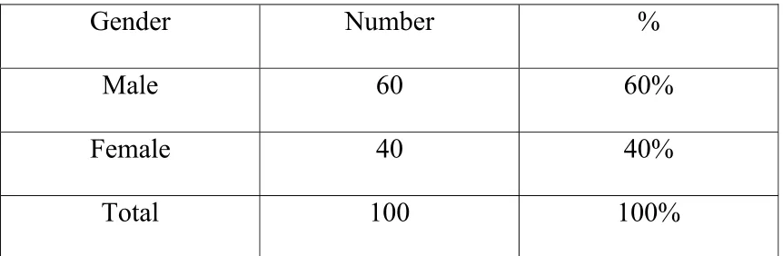
Gender Distribution
Gender Number %
Male 60 60%
Female 40 40%
Total 100 100%
Table – 2:
Course of Diseases
Course Number %
Acute 60 60%
Chronic 40 40%
Table – 3:
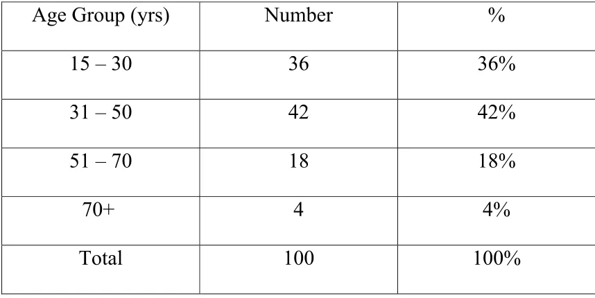
Age Distribution
Age Group (yrs)
Number
%
15 – 30
36
36%
31 – 50
42
42%
51 – 70
18
18%
70+
4
4%
Total 100 100%
In this study, 36 cases were in 15-30 yrs age group, 42 cases
(42%) were in 31-50 years age group, 18 cases (18%) were in 51-70
years age group and 4 patients were in 71+ age group. Most cases of
uveitis were in 31-50 years age group. This is in accordance with the
Table – 4:
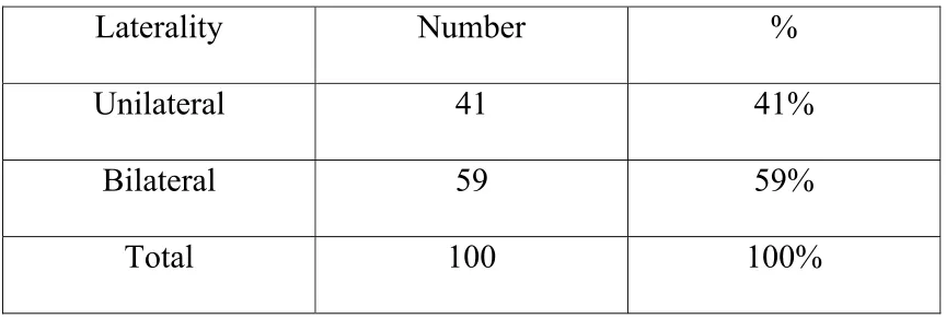
Laterality
Laterality Number
%
Unilateral 41 41%
Bilateral 59 59%
Total 100 100%
41 cases were unilateral and remaining 59 cases were bilateral.
Both eyes are equally affected. There is no right – left
Table – 5:
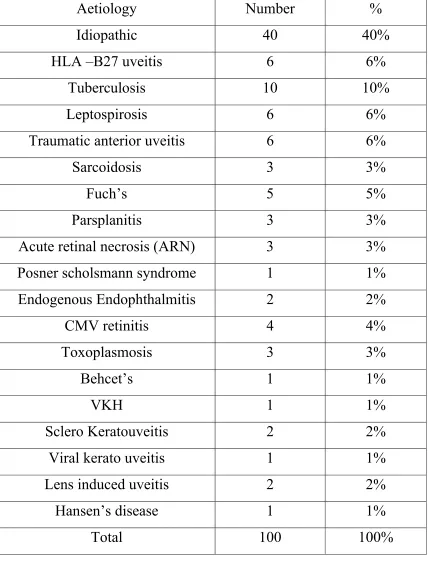
Aetiology of Uveitis
Aetiology Number
%
Idiopathic 40
40%
HLA –B27 uveitis
6
6%
Tuberculosis 10
10%
Leptospirosis 6
6%
Traumatic anterior uveitis
6
6%
Sarcoidosis 3
3%
Fuch’s 5
5%
Parsplanitis 3
3%
Acute retinal necrosis (ARN)
3
3%
Posner scholsmann syndrome
1
1%
Endogenous Endophthalmitis
2
2%
CMV retinitis
4
4%
Toxoplasmosis 3
3%
Behcet’s 1
1%
VKH 1
1%
Sclero Keratouveitis
2
2%
Viral kerato uveitis
1
1%
Lens induced uveitis
2
2%
Hansen’s disease
1
1%
The most common cause of uveitis in this study was idiopathic
(40%), followed by tuberculosis (10%). The other cases were HLA
B-27 associated uveitis 6 cases (6%), Leptospirosis 6 cases
(6%),Traumatic anterior uveitis 6 cases (6%), Sarcoidosis 3 cases
(3%), Fuch’s heterochromic uveitis 5 cases (5%), Parsplanitis 3
cases (3%), Acute Retinal necrosis 3 cases (3%) , Posner
scholssman syndrome 1 case (1%), Endogenous endophthalmitis 2
cases (2%), CMV retinitis 4 cases (4%), Toxoplasmosis 3 cases
(3%), Behcet’s disease 1 case (1%), Vogt Koyanagi Harada’s
syndrome 1 case (1%), Sclerokerato uveitis 2 cases (2%), viral
kerato uveitis 1 case (1%), Lens induced uveitis 2 cases (2%) and
Table – 6:
Anatomic Location of Uveitis
Location Number
%
Anterior 60 60%
Intermediate 10
10%
Posterior 14 14%
Panuveitis 16
16%
Total 100 100
In this study, 60 cases (60%) presented with anterior
uveitis, out of this 60 cases, 36 cases were males and rest 24 were
females. 10 cases presented as intermediate uveitis, with 6 cases as
males and 4 cases as females. Pan uveitis occurred in 16 cases,
where 10 were in males and 6 in females. 14 cases presented with
posterior uveitis where 8 were in males and 6 in females. Anterior
uveitis is the most common presentation and this is in accordance
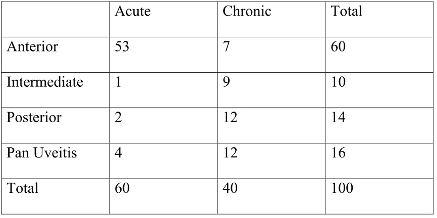
Table – 7
: Course Vs Anatomic Location of uveitis
Acute
Chronic
Total
Anterior 53
7
60
Intermediate 1
9
10
Posterior 2
12
14
Pan Uveitis
4
12
16
Total 60
40 100
Table – 8:
Glaucoma in Uveitis
Uveitis No.of
patients
Percentage
Anterior 4 80
Panuveitis 1 20
Total 5 100
Of the 100 patients of uveitis, 5 patients developed
glaucoma. It occured in 4 patients of anterior uveitis (80%) and 1
patient (20%) of pan uveitis. SG is common in anterior uveitis and is
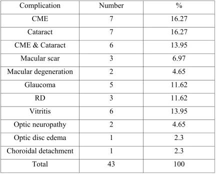
[image:59.612.98.526.454.559.2]Table – 9:
Complications of Uveitis
:
Complication Number
%
CME 7 16.27
Cataract 7 16.27
CME & Cataract
6
13.95
Macular scar
3
6.97
Macular degeneration
2
4.65
Glaucoma 5 11.62
RD 3
11.62
Vitritis 6 13.95
Optic neuropathy
2
4.65
Optic disc edema
1
2.3
Choroidal detachment
1
2.3
Total 43 100
Table – 10:
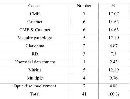
Causes of Visual Loss
:
Causes Number
%
CME 7
17.07
Cataract 6
14.63
CME & Cataract
6
14.63
Macular pathology
5
12.19
Glaucoma 2
4.87
RD 3
7.3
Choroidal detachment
1
2.43
Vitritis 5
12.19
Multiple 4
9.76
Optic disc involvement
2
4.88
Total 41
100
%
Table – 11:
Causes Vs Degree of visual loss
Causes <
3/60
>6/60 - 6/18
%
CME 1
6
7
Cataract 0
6
6
CME & Cataract
0
6
6
Macular pathology
1
4
5
Glaucoma 1
1
2
RD 1
2
3
Choroidal detachment
0
1
1
Vitritis 0
5
5
Multiple 0
4
4
Optic disc pathology
2
0
2
Total 6
35
41
(100%). Durrani
5and Rothava
25et al study found CME to be the
commonest cause of visual loss as in the present series.
Table – 12:
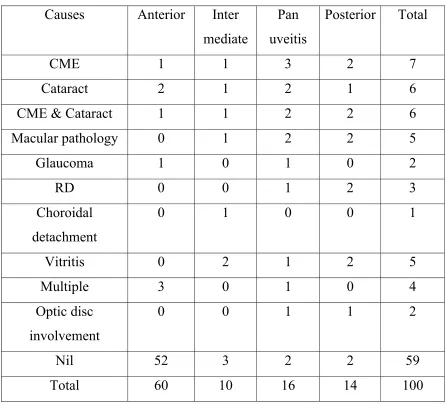
Causes of Visual loss vs Anatomic Location
Causes Anterior
Inter
mediate
Pan
uveitis
Posterior Total
CME 1
1
3
2
7
Cataract 2
1
2
1
6
CME & Cataract
1
1
2
2
6
Macular pathology
0
1
2
2
5
Glaucoma 1 0
1
0
2
RD 0
0
1
2
3
Choroidal
detachment
0 1 0 0 1
Vitritis 0
2
1
2
5
Multiple 3
0
1
0
4
Optic disc
involvement
0 0 1 1 2
Nil 52
3
2
2
59
Total 60
10
16
14
100
Table – 13:
Visual loss Vs Gender distribution
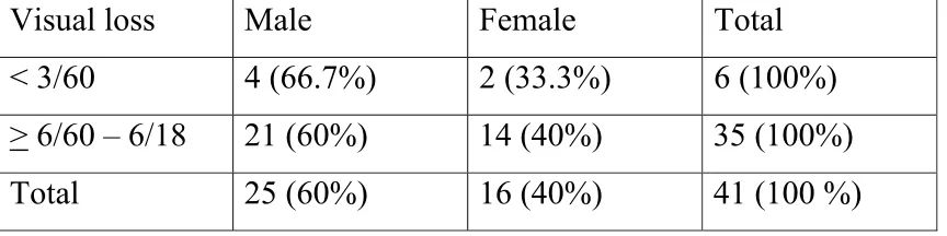
Visual loss
Male
Female
Total
< 3/60
4 (66.7%)
2 (33.3%)
6 (100%)
> 6/60 – 6/18
21 (60%)
14 (40%)
35 (100%)
Total
25 (60%)
16 (40%)
41 (100 %)
Table – 14:
Aetiology Vs Degree of visual loss
Aetiology <
3/60
> 6/60-6/18
Total
Idiopathic 0
10
40
Tuberculosis 0
8
10
Leptospirosis 0
1
6
Traumatic uveitis
1
0
6
Sarcoidosis 0
3
3
Fuch’s uveitis
0
1
5
Parsplanitis 0
3
3
ARN 1
2
3
Endogenous endophthalmitis
0
2
2
CMV Retinitis
1
2
4
Toxoplasmosis 1
2
3
Behect’s disease
1
0
1
VKH syndrome
0
1
1
Hansen’s disease
1
0
1
Total 6
35
41
parsplanitis (100%), 2 cases of acutue retinal necrosis (66.7%), 2
cases of endogenous endophthalmitis (100%), 2 cases of CMV
retinitis (50%), 2 cases of Toxoplasmosis (66.71%) and 1 case of
vogt Koyangai Harada’s disease (100%).
Table – 15
: Visual Loss Vs Laterality
Visual Loss
Unilateral
Bilateral
Total
< 3/60
1 (16.7%)
5 (83.3%)
6 (100%)
> 6/60 – 6/18
14 (40%)
21 (60%)
35 (100%)
Total
15 (36.6%)
26 (63.4%)
41 (100%)
Table – 16:
Visual loss Vs Course of disease
Course Visual
loss(<6/18)
Total
Acute
10 (16.6%)
60 (100%)
Chronic
31 (77.5%)
40 (100%)
Total 41
100
10 patients (16.6%) of acute uveitis developed visual loss,
while 31 patients (77.5%) of chronic uveitis had visual loss.
Table – 17:
Mean Duration of visual loss
Visual loss
Mean Duration (mon)
Severe 24
Moderate 17.25
[image:67.612.92.527.413.514.2]SUMMARY
In this study, uveitis commonly occurred in males (60% of
cases)
60% of uveitis had acute course, while 40% had chronic
course. i.e., Acute uveitis is commonest in this study.
Most of the patients, 42% were in the age group of 31-50 yrs
who are economically productive to the society and have a
socioeconomic impact. In this age group idiopathic (40.47%)
was the common cause followed by Tuberculosis 5 cases
(11.9%) and HLA B 27 uveitis 4 cases (9.53%).
59% of patients had bilateral inflammation
In 40% cases, the cause could not be identified and hence,
certified as idiopathic. It is the commonest cause of uveitis
followed by Tuberculosis in 10% cases.
60% cases had anterior uveitis followed by panuveitis in 16%
cases, posterior uveitis in 14% cases and intermediate uveitis in
10% cases.
Most common complication of uveitis in this study are Cystoid
Secondary glaucoma is common in anterior uveitis (80%)
Most common causes of visual loss was cystoid macular edema
(17.07%) followed by cataract (14.63%) and a combination of
cataract and CME in 14.63 % cases.
CME, macular pathology, glaucoma, retinal detachment and
optic disc involvement were the causes of severe visual loss.
The common causes of moderate visual loss are CME, cataract,
combination of cataract and CME and multiple factors.
CME commonly occurs in panuveitis (42.9%) and posterior
uveitis (28.6%). Cataract commonly occurs in anterior
(33.3%) and pan uveitis (33.3%). The combination of CME &
cataract occurs commonly in panuveitis (33.3%) and posterior
uveitis (33.3%).
Visual loss commonly occurs in panuveitis (87.5%) followed
by posterior uveitis (85.71%), intermediate uveitis (70%) and
anterior uveitis (13.3%). So it can be concluded that visual
prognosis was better in anterior uveitis and a guarded
prognosis has to be given in pan uveitis and posterior uveitis
The diseases associated with severe visual loss were Traumatic
anterior uveitis(CME), ARN(optic neuropathy) , CMV
retinitis(retinal detachment) ,Toxoplasmosis(macular scar),
Behect’s disease(optic atrophy) and Hansen’s
disease(glaucomatous optic atrophy)
Bilateral visual loss (63.4%) was common in this study.
3 patients developed blindness as per WHO definition
Visual loss was common in cases of chronic intraocular
inflammation (77.5%).
Permanent visual damage leading to severe visual loss occurred
in 6 patients leading to the mean duration of 24 months of
visual loss. The mean duration of visual loss in cases with
DISCUSSION
This study provides information about the uveitis pattern in
a tertiary referral eye centre, causes of visual loss and duration of it.
However there may be limitations due to its short duration and
referral bias of cases in a tertiary hospital.
This study was compared to OM Durrani, Tehrani et al
5study and Aniki Rothova et al
25study.
Anterior uveitis was the most common presentation which
is in agreement with the studies by Rothova et al
25(42%).
Secondary glaucoma is most common in anterior uveitis and
is in accordance with Jesus Merayo et al
8study.
In this study, 41% had visual loss which is comparable to
Rothova et al
25study (35%). In Durrani et al
5study the visual loss
occurred in 69.97%. but they have included the patients with visual
acuity of 6/18 in the category for defining visual loss whereas in this
study, patients with visual acuity of < 6/18 were categorized under
visual loss.
Panuveitis (87.5%) had worst prognosis in this study as that
CME was the most common cause (17.07%) of visual loss
in this study, comparable to Rothova et al
25study (26%) and Durrani
et al
5study (26.8%).
Main Causes of visual loss :
Present
Series
Durrani
study
CME 17.07
%
26.8%
Cataract
14.63 %
17.7 %
Combination 14.63% 20%
The difference between these 2 studies could be due to
large number of patients longer duration of follow-up. Bilateral
visual loss is common in this series comparable to Durrani et al
5study.
The commonest systemic disorder associated with visual
loss in uveitis was due to sarcoidosis as per Rothava
25study whereas
in the present study the commonest systemic disorder associated
Bilateral legal blindness as per Rothava’s
25study was 4% and
in this study was 3%.
Behcet’s uveitis leads to unilateral blindness as per Rothava’s
25study and comparable to this study.
Mean duration of vision loss in uveitis was 24 months for
severe visual loss and 17.25 months for moderate visual loss.
According to Durrani et al
5study, it was 22.8 months for
patients with severe visual loss and 20.35 months for patients
CONCLUSION
Visual loss is common in patients with uveitis. This
prospective study was basically a descriptive study analyzing 100
cases of uveitis that presented to the eye department of Govt. Rajaji
Hospital.
In out study, the commonest cause of uveitis was idiopathic
followed by tuberculosis. There was a male preponderance. This
may be due to the fact that males tend to seek more medical care, as
they are wage earners in a developing country.
Acute uveitis was the common presentation. Anterior
uveitis was commoner. Majority of the patients were in the
economically productive age group of 31-50 years. So visual loss
made socio economic impact on the community.
The common complications of uveitis were cystoid macular
edema, cataract and a combination of these both. The most common
Male patients with uveitis had a higher risk of visual loss.
Pan uveitis had the worst prognosis of vision. Bilateral chronic
visual loss usually result in significant permanent visual damage.
The results were comparable with other studies.
Uveitis is one of the major causes of visual loss in working
population
The patients should be insisted on regular follow up and
educated about the warning symptoms of complications
A routine and periodic ophthalmic screening is necessary in all
the systemic disorders found associated with uveitis
The patients who have been adequately treated should also be
educated about the possibility of recurrence and advised to
come for follow up if any symptoms recur.
With more specific and sophisticated investigations and
treatment modalities, the sight threatening complications and
the resulting effect on socio economic performance of the
BIBLIOGRAPHY
1. A Ablose et al : Distribution and etiology of blindness and visual impairment in mesoendemic onchocercal communities, Kaduna state, Nigeria. Br J Ophthalmol 1994 : 78 : 8-13.
2. Chia EM, Wang JJ, et al. Impact of visual impairment on health related quality of life : the blue mountains Eye study. Invest Ophthalmol Vis Sci 2004 : 45 : 71-6.
3. Dandona L, Dandona R, John RK, et al. Population based assessment of uveitis in an urban population in Southern India. Br J Ophthalmol 2000 ; 84 : 706-9.
4. Darrell RW, Wagner HP, Kurland LT, Epidemiology of uveitis ; incidence and prevalence in a small urban community. Arch Ophthalmol 1962 : 68 : 502-14
5. Durrani OM, Tehrani NN, Mar JE, et al. Br J Ophthalmol 2004 ; 88 : 1159 – 1162 Degree, duration and causes of visual loss in
uveitis
7. Gardiner AM, Armstrong RA, Dunne MC, et al. Correlation between visual function and visual ability in patients with uveitis. Br J Ophthalmol 2002 ; 86 : 993-6.
8. Jesus Merayo – Lloves, William J. Power, et al. Secondary glaucoma in patients with uveitis. Ophthalmologica 1999 ; 213 : 300 -304.
9. Kearney et al. Clinical features and associated features of HLA B 27 uveitis. Am J Ophthalmol 1997 ; 121 : 47-56.
10. Kotaniemi K, Ahok, Kotaniemi A, Uveitis as a cause of visual loss in arthrides and comparable conditions. J. Rheumatol 2001 ; 28 : 309 – 12.
11. L C Dutta, Nittin K Dutta et al : Modern Ophthalmology, Third edition – volume 3 , 2005 : 1249 - 1324
12. Lightman S. Uveitis : Management. Lancet 1991 ; 338 : 1501-4. 13. Malinowski SM, Folk JC, et al. Pars Planitis. Curr Opin
Ophthalmol 1994 ; 5 : 72-82.
14. Mamo JG, The rate of visual loss in Behcets disease. Arch ophthalmol 1970; 84 : 451-2.
16. Narsing AR, Blackman HJ, Basic and clinical Science course Intraocular inflammation and uveitis (section 9), American academy of ophthalmology, 1997-98 : 41-58.
17. Nussenblatt RB. The natural history of uveitis. Int ophthalmol 1990 ; 14 : 303 – 8.
18. Rathinam et al. Leptospirosis. Journ of TN 0A 1997 Dec : 1-4
19. Robert B. Nussenblatt : Uveitis – fundamentals and clinical practise – second edition
20. Robert MS et al. Leptospiral uveitis. Arch ophthalmol 1959 ; 61 : 633-639
21. Ronday MJH, Stilma JS, et al : Blindness from uveitis in a hospital population in Sierra leone : Br J Ophthalmol 1994 : 78 : 690-693.
22. Rosner RS, Uveitis and blindness. Med Trail Tech Q 1967 : 14 : 39-42.
23. Rothova A et al, Clinical features of acute anterior uveitis. Am J Ophthalmol 1987 ; 103 : 137-145.
24. Rothova A, Meenken C et al. Uveitis and Systemic diseases. Br J. Ophthalmol 1992 ; 76 : 137 – 41.
26. Suttorp – Schulten MSA, Rothova A. The possible impact of uveitis in blindness : A literature survey. Br J Ophthalmol 1996 ; 80 : 844-8.
27. Taylor HR, Keeflee JE, World Blindness : a 21st century perspective. Br J ophthalmol 2001 : 85 : 261-6.
28. Wirostrko et al. Lens induced uveitis. Arch ophthalmol 1967 ; 78 : 1-7.