J
OURNAL OFV
IROLOGY, Oct. 1996, p. 7004–7012
Vol. 70, No. 10
0022-538X/96/$04.00
1
0
Copyright
q
1996, American Society for Microbiology
JC Virus Infection of Hematopoietic Progenitor Cells, Primary B
Lymphocytes, and Tonsillar Stromal Cells: Implications
for Viral Latency
MARIA CHIARA G. MONACO,
1,2* WALTER J. ATWOOD,
1† MANETH GRAVELL,
1CARLO S. TORNATORE,
1ANDEUGENE O. MAJOR
1Laboratory of Molecular Medicine and Neuroscience, National Institute of Neurological Disorders and Stroke,
Bethesda, Maryland, 20892,
1and Laboratorio di Immunologia e Genetica, Istituto di
Ricerca Codivilla Putti, Instituti Ortopedici Rizzoli, Bologna, Italy
2Received 15 May 1996/Accepted 26 June 1996
The human polyomavirus JC virus (JCV) infects myelin-producing cells in the central nervous system,
resulting in the fatal demyelinating disease progressive multifocal leukoencephalopathy (PML). JCV-induced
PML occurs most frequently in immunosuppressed individuals, with the highest incidence in human
immu-nodeficiency type 1-infected patients, ranging between 4 and 6% of all AIDS cases. Although JCV targets a
highly specialized cell in the central nervous system, infection is widespread, with more than 80% of the human
population worldwide demonstrating serum antibodies. A number of clinical and laboratory studies have now
linked the pathogenesis of PML with JCV infection in lymphoid cells. For example, JCV-infected lymphocytes
have been suggested as possible carriers of virus to the brain following reactivation of a latent infection in
lymphoid tissues. To further define the cellular tropism associated with JCV, we have attempted to infect
immune system cells, including CD34
1hematopoietic progenitor cells derived from human fetal liver, primary
human B lymphocytes, and human tonsillar stromal cells. Our results demonstrate that these cell types as
well as a CD34
1human cell line, KG-1a, are susceptible to JCV infection. JCV cannot, however, infect KG-1,
a CD34
1cell line which differentiates into a macrophage-like cell when treated with phorbol esters. In
addi-tion, peripheral blood B lymphocytes isolated by flow cytometry from a PML patient demonstrate JCV
infection. These results provide direct evidence that JCV is not strictly neurotropic but can infect CD34
1hematopoietic progenitor cells and those cells which have differentiated into a lymphocytic, but not monocytic,
lineage.
JC virus (JCV), a DNA virus of the
Papovaviridae
family, is
the etiologic agent of progressive multifocal
leukoencephalop-athy (PML), a fatal demyelinating disorder of the central
ner-vous system associated with compromised immunity. On the
basis of the detection of JCV-specific antibodies, it is estimated
that more than 80% of the human population has been
in-fected by JCV (23, 34). Seroconversion generally occurs during
childhood, since 65% of adolescents produce JCV-specific
an-tibodies (23). Although primary infection appears to be
asymp-tomatic, JCV may persist in a latent state for an extended
period, perhaps for life, even in immunocompetent individuals.
JCV reactivation, however, may occur if immune function is
compromised.
Reactivation of JCV, with concomitant development of
PML, has been found in immunosuppressed patients receiving
bone marrow or renal transplants (5, 15). JCV viruria is
de-tected in bone marrow transplant patients (5), but only in those
who have tested positive for JCV antibodies by enzyme-linked
immunosorbent assay prior to immunosuppression. Thus,
viru-ria most likely results from a JCV infection which occurred
prior to the time of immunosuppression (5). Moreover, PML
patients have immunoglobulin G antibodies to JCV even
though their immunity is compromised, providing further
evi-dence that PML results from reactivation of latent JCV
infec-tion. Neither the cell types harboring JCV nor the sites of its
reactivation from latency have been well characterized.
Previously, B-cell lines were shown to be susceptible to JCV
infection in vitro (6). We have also found JCV
DNA-associ-ated kappa-light-chain-positive lymphocytes from bone
mar-row and spleens of two PML patients (18). Additionally, using
PCR assays, we and others have amplified JCV-specific
se-quences from peripheral blood lymphocytes obtained from
both PML (31) and immunosuppressed (11, 28) patients.
These findings clearly demonstrate that cells of the peripheral
lymphoid system carry the JCV genome. In the event of either
induced or acquired immunodeficiency, these latently infected
cells would potentially initiate JCV replication, with the
re-lease of viral particles.
It remains unclear whether peripheral B cells are the
prin-cipal targets of early JCV infection in lymphoid tissue or
whether other cells within the hematopoietic
microenviron-ment may also serve as potential virus reservoirs. In particular,
it is of interest to determine if early hematopoietic progenitor
(CD34
1) cells and the stromal elements with which lymphoid
cells interact are also susceptible to infection. Accordingly, we
have compared the in vitro susceptibilities of human
hemato-poietic progenitor (CD34
1) cells, primary B lymphocytes, and
human tonsillar stromal cells to JCV infection. These studies
provide a better understanding of the cellular tropism of JCV
and identify additional viral targets that may contribute to
clinical viremia and the mechanisms of PML induction.
* Corresponding author. Mailing address: Laboratory of Molecular
Medicine and Neuroscience, National Institute of Neurological
Disorders and Stroke, Building 36, Room 5W21, Bethesda, MD 20892.
Phone: (301) 496-2043. Fax: (301) 594-5799. Electronic mail address:
[email protected].
† Present address: Department of Molecular Microbiology and
Im-munology, Brown University, Providence, RI.
7004
on November 9, 2019 by guest
http://jvi.asm.org/
MATERIALS AND METHODS
Hematopoietic progenitor cells.Hematopoietic CD341cell lines (KG-1 and KG-1a), originating from bone marrow of an acute myelogenous leukemia pa-tient, were obtained from the American Type Culture Collection, Rockville, Md. KG-1a cells, which are morphologically, cytochemically, and functionally less mature than the parental KG-1 cells (20), were propagated in RPMI 1640 with 20% fetal bovine serum. The parental KG-1 line, when cultured with phorbol 12-myristate 13-acetate (PMA) (19), differentiated into a more mature macro-phage-like state. For this reason, KG-1 cells were cultured with 1027
M PMA for 3 days prior to use.
Primary CD341progenitor cells were collected from human fetal liver at 16 to 18 weeks of gestation. To separate mononuclear cells from hepatocytes, the liver tissue was fragmented and passed through sieves (Thomas Scientific, Swedes-boro, N.J.) of different pore sizes (3 in. [7.62 cm], 60 mesh, and 30 mesh). The mononuclear cells were recovered after stratification on a Ficoll-Hypaque den-sity gradient (M.A. Bioproducts, Walkersville, Md.) and washed twice in RPMI 1640–10% fetal bovine serum. Mononuclear cells (23107
in a 25-cm2
flask coated with anti-CD34 antibody; Applied Immune Sciences Inc., Santa Clara, Calif.) were then incubated for 1 h at room temperature. The nonadherent cells were discarded, and the adherent CD341cells were dissociated from the flask by agitation. Both adherent and nonadherent cells were analyzed by flow cytometry (FACSort; Becton Dickinson, Mansfield, Mass.). The adherent cells were labeled with a panel of monoclonal antibodies including CD2, CD3, CD10, CD13, CD14, CD15, CD19, CD33, CD34, CD38, and CD45 (Becton Dickinson, San Jose, Calif.) to test their purity. The adherent population, containing.96% CD341 cells, was used for in vitro infection experiments.
Primary B cells.Mononuclear cells were isolated from tonsils of patients (4 to 20 years old) after tonsillectomy or from peripheral blood of normal donors by centrifugation on Ficoll-Hypaque gradients. T lymphocytes were removed from the mononuclear preparations by rosetting with sheep erythrocytes treated with aminoethylisothio-uronium bromide (Sigma, St. Louis, Mo.) (7), and the B lymphocyte population was recovered. The percentage of B cells was determined by fluorescence-activated cell sorting (FACS) analysis. Only cell preparations having.95% CD19 cells were used in the experiments. The purity of these preparations was further tested with CD3, CD4, CD8, and anti-CD14 monoclonal antibodies (Becton Dickinson).
Human primary stromal cells.The method used to obtain human tonsillar stromal cells has previously been described in detail (21). Briefly, human tonsils were obtained from 20 tonsillectomized children ranging in age from 3 to 8 years. Tonsillar parenchyma was separated from tissue, dissected into small pieces, and
placed in 20 ml of calcium- and magnesium-free Hanks saline solution containing 150 U of collagenase (GIBCO BRL, Grand Island, N.Y.) per ml, 0.1% trypsin (ICN Biochemicals, Cleveland, Ohio), and 2% heat-inactivated chicken serum (GIBCO BRL). The tissue was then incubated in a water bath for 1 h at 378C. After enzymatic digestion, the tissue pieces and cells were washed twice with RPMI 1640 with 10% fetal bovine serum, resuspended in medium, and seeded into T75 flasks (Costar, Cambridge, Mass.). Nonadherent cells were subse-quently removed, and the adherent stromal cells were expanded by several passages. The medium was changed twice weekly.
In vitro JCV infection.The following nonadherent human cells, primary CD341hematopoietic precursors, primary B lymphocytes from tonsils or pe-ripheral blood, and cells of the line KG-1, KG-1a, or U937 were each seeded at 105cells per well in six-well plates. Adherent stromal cells were also seeded at the
same density into similar vessels containing glass coverslips. All cell types were inoculated with 300 hemagglutination (HA) units (HAU) of JCV (MAD-4 strain). Two hours after infection, the cells were washed and new medium was added to the cultures. Five days later, the cells were harvested and fixed for in situ hybridization and immunofluorescence analysis. Human fetal glial (HFG) cells were used as a positive control of JCV infection.
Immunofluorescence staining for T and V viral antigens.Detection of JCV T antigen was considered a measure of early viral transcription, while detection of V antigen (capsid protein) denoted late viral transcription and potential virion formation.
After harvesting, all infected nonadherent cell types were washed twice in phosphate-buffered saline (PBS), resuspended at 106cells per ml, dried onto
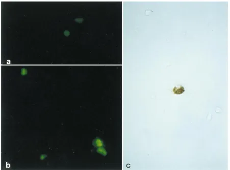
[image:2.612.110.508.68.354.2]glass coverslips, and fixed for 15 min at2208C with 100% acetone and then with 100% methanol. Adherent stromal cell cultures on coverslips were similarly fixed. After rehydration of cells in PBS, all cell types were treated with an antibody (PAB 416; Oncogene Science, Inc., Cambridge, Mass.) to simian virus 40 T antigen diluted 1:10 in PBS. This antibody cross-reacts with the amino-terminal sequence of the JCV T antigen (23). Cells were also tested for the presence of V antigen by treatment with an antibody to simian virus 40 capsid protein, which also cross-reacts with JCV capsid proteins (PAB 597; a gift from L. Norkin, University of Massachusetts, Amherst), diluted 1:500 in PBS. After 30 min of incubation at room temperature, the coverslips were washed twice in PBS and incubated for 30 min at room temperature in the dark with a secondary fluorescein isothiocyanate-conjugated goat anti-mouse antibody (Jackson Immu-noresearch, West Grove, Pa.) diluted 1:50 in PBS. The coverslips were washed twice in PBS and mounted on glass slides with 2% antifade (1,4-diazabicyclo-[2.2.2.]octane; Sigma) in 90% glycerol. By using a Zeiss ICM 405 epifluorescence FIG. 1. JCV infection of KG-1a, a human hematopoietic cell line. The infected KG-1a cells were by immunofluorescence examined 5 days postinfection. T-antigen antibody PAB 416 (a) and V-antigen antibody PAB 597 (b) were used for detection. Positive cells were visualized with a secondary goat anti-mouse fluorescein isothiocyanate-conjugated antibody. (c) JCV-infected KG-1a cells were examined by in situ hybridization with a JCV biotin-labeled DNA probe. Magnification,340.
V
OL. 70, 1996
JCV IN LYMPHOID TISSUE
7005
on November 9, 2019 by guest
http://jvi.asm.org/
microscope, the percentage of T- and V-antigen-positive cells was calculated from 25 different fields from each experiment.
In situ hybridization. A positive signal in the in situ hybridization assay indicates de novo viral DNA replication (1, 2). Replication of JCV DNA was analyzed by in situ DNA hybridization in all cell types, using a full-length JCV biotinylated DNA probe (ENZO Diagnostic Inc., Farmingdale, N.Y.). The in situ DNA hybridization protocol used was described previously (17). This method does not require protease digestion and acid hydrolysis. These steps are not necessary for entry of the biotin-labeled DNA probe into lymphocytes dried on coverslips.
The hybridization reaction mixture contained 50% (vol/vol) deionized form-amide, 10% dextran sulfate, 0.4mg of herring sperm DNA per ml, and 2 ng of biotinylated probe DNA per ml in 23SSC (13SSC is 0.15 M NaCl plus 0.015 M sodium citrate). A 25-ml aliquot of this mixture was applied to the coverslips; a larger coverslip was placed on top, and the edges were sealed with rubber cement. Both the cells and the DNA probe were denatured in an oven set at 858C for 10 min, and hybridization was performed at 378C for 48 h. The hybridization signal was detected with a streptavidin-biotin-horseradish peroxidase kit (Detek I-hrp; ENZO Diagnostic). Diaminobenzidine tetrahydrochloride was used as the chromogen.
Assay of HFG cell culture harvests for infectious JCV.Harvests of supernatant medium were made from all cultures 1 week after inoculation with JCV. A small aliquot was used to measure JCV replication by HA of human type O erythro-cytes (26, 35, 36). HA was used to measure production of JC virions by the various cell types studied after passage onto highly susceptible human fetal brain cultures. The remainder of the harvests were filtered and added to cultures of uninfected primary HFG cells to determine whether infectious JCV was con-tained in the harvests. Glial cell cultures were harvested 5 days after inoculation and assayed by both in situ hybridization and immunofluorescence analysis (as described above).
Flow cytometry from in vivo lymphocytes.Peripheral blood mononuclear cells (PBMC) from two AIDS patients were examined for the presence of the JCV genome. One patient was clinically diagnosed with PML. This diagnosis was confirmed by histopathology of brain biopsy specimens. In situ hybridization of these brain biopsy specimens with a JCV probe also confirmed the diagnosis. The second patient did not have PML. Blood (20 ml) was collected in heparinized
tubes from each patient by venipuncture. PBMC were isolated from each blood sample by centrifugation on Ficoll-Hypaque gradients by a previously described protocol (31). Cells were counted, adjusted to 106/ml in PBS, and incubated for
30 min on ice with 200ml of Simultest B- and T-cell reagent containing anti-CD3 and anti-CD19 antibodies in the same tube (Becton Dickinson) or with the Simultest negative control antibodies (Becton Dickinson) diluted 1:5 in PBS. The cells were then washed twice in cold PBS and once in sheath fluid (Baxter Diagnostics Inc., Deerfield, Ill.) and fixed in 1 ml of sheath fluid containing 1% paraformaldehyde. Equal numbers of B and T cells were collected from each patient by cell sorting into 50-ml Falcon tubes. PBMC from a JCV-negative individual were also stained and sorted by the same protocol to control for possible virus contamination of the cell sorter. After sorting, the cells were pelleted and a fraction was reanalyzed on the flow cytometer to assess purity. The remaining sorted cells and an equivalent number of unsorted PBMC from these patients were lysed in 0.5 ml of 10% sodium dodecyl sulfate and 55 ml of proteinase K (10mg/ml). DNA from lysed cells was extracted twice with phenol, twice with a 1:1 mixture of phenol-chloroform, and twice with a 29:1 mixture of chloroform-isoamyl alcohol and precipitated in 2.5 volumes of cold ethanol containing 0.1 volume of 3 M sodium acetate.
Nested PCR and Southern analysis.Nested PCR was performed with primers from the T-antigen coding region of the JCV genome. Primers were purchased from GIBCO BRL (Gaithersburg, Md.). The primer sites and their locations within JCV genome were as follows. The external primer set was 59-GAATAG GGAGGAATCCATGG-39(nucleotides [nt] 4999 to 4979) and 59-GGAATGC ATGCAGATCTACAGG-39 (nt 4231 to 4252). The internal primer set was: 59-GGTGGGGACGAAGACAAGATG-39(nt 4878 to 4858) and 59-GTGTTG GGATCCTGTGTTTTC-39(nt 4301 to 4321).
PCRs were performed in a total volume of 100ml with 2.5 U ofTaq polymer-ase and 2 mM MgCl2as instructed by the manufacturer (Perkin-Elmer,
[image:3.612.105.511.70.380.2]Branch-burg, N.J.). After an initial period of 5 min at 958C, reaction products were amplified for 30 cycles, using the following program: 1 min at 958C, 1 min at 558C, and 2 min at 728C. Amplification was completed with a 7-min extension period at 728C. One positive and two negative controls were included in all PCR experiments. Negative controls consisted of the reaction mixtures with no DNA template or 1mg of B-cell DNA from a JCV-negative individual. The positive control was 1 ng of DNA from the JCV plasmid pM1Tc.
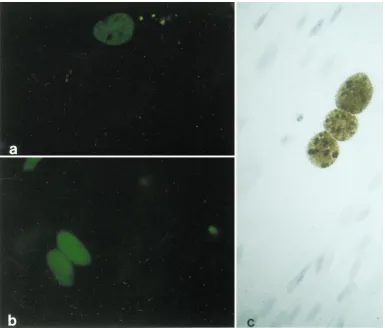
FIG. 2. Primary CD341cells infected in vitro with JCV. Five days postinfection, the expression of T and V antigens was detected by indirect immunofluorescence. (a) Representative field of CD341progenitor cells positive for viral T protein; (b) cells positive for JCV V antigen. (c) In situ hybridization with a biotinylated DNA probe for JCV genome was performed in primary human CD341cells 5 days postinfection. A single cell in the lower center of the field contains JCV DNA, as demonstrated by the intranuclear brown reaction product (no hematoxylin counterstain). Magnification,340.
7006
MONACO ET AL.
J. V
IROL.
on November 9, 2019 by guest
http://jvi.asm.org/
The reaction mixture with external primers contained 1mg of cellular DNA. Nested PCR was performed with 10ml of the reaction product from the first amplification.
Southern blot analysis was performed with 20ml of reaction mixture from the nested primer amplification as previously described (31).
RESULTS
Human CD34
1cell infection with JCV.
Previous clinical and
cell culture data suggest that JCV can infect lymphoid cells (6,
11, 18, 31), particularly those cells expressing the B-cell
phe-notype (6, 18). If lymphoid tissue is an initial site of limited
infection and also a potential site for viral latency, then human
hematopoietic precursor cells may also be susceptible to JCV
infection. Although human stem cells do not express cell
sur-face markers associated with lineage cell commitment (4, 8, 24,
32), they do express the CD34 antigen, which is a stem cell
marker (4, 8, 24, 30, 32, 33).
Two cell lines, KG-1 and KG-1a, are available for studying
hematopoietic progenitors and their differentiation. The KG-1
cell line was derived from a patient who presented with
eryth-roleukemia but eventually developed acute myelogenous
leu-kemia (20). Both KG-1 and KG-1a cells exhibit the CD34
antigen marker on cytofluorographic analysis. After treatment
with phorbol esters, KG-1 cells differentiate into mature
mac-rophages (19), whereas KG-1a cells are unaffected by this
treatment. The KG-1a cell line is composed of undifferentiated
blast cells, which are morphologically, cytochemically, and
functionally less mature than the KG-1 cells (20). For these
reasons, untreated KG-1 and KG-1a cells and PMA-treated
KG-1 cells were inoculated with JCV to determine their in
vitro susceptibility to infection.
Untreated KG-1 and KG-1a cells were infected by JCV, as
demonstrated by both immunocytochemistry and in situ
hy-bridization (Fig. 1). However, when parental human KG-1 cells
were treated with PMA and differentiated into cells with
mac-rophage-like characteristics, they were no longer susceptible.
Immunocytochemistry and in situ hybridization assays also
in-dicated that the promonocytic cell line U937 was not
suscep-tible to JCV infection (data not shown). These data provide
evidence that cells of monocytic lineage are not susceptible to
JCV infection in vitro. The susceptibility of the less mature
KG-1a cells to JCV infection prompted us to test the
suscep-tibility of primary CD34
1cells. Therefore, CD34
1cells were
isolated from human fetal liver, the main hematopoietic organ
in the fetus; an immunofluorescence assay was used to test
their susceptibility to JCV infection. T (Fig. 2a) and V (Fig. 2b)
antigens were detected in primary CD34
1cells 5 days after in
vitro infection with JCV. JCV DNA in primary CD34
1cells
was also detected by in situ hybridization (Fig. 2c).
[image:4.612.83.534.68.402.2]JCV infection of primary human B lymphocytes.
Previously
we demonstrated that cells of B-cell lineage (18) and
B-lym-phoblastoid cell lines (6) were susceptible to JCV infection.
We have now extended this work by testing the susceptibility of
primary B lymphocytes from peripheral blood and tonsillar
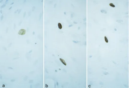
FIG. 3. Primary human tonsillar B lymphocytes infected in vitro with JCV. Immunofluorescence analysis was performed to detect T (a) and V (b) viral proteins. Five days postinfection, JCV-positive B lymphocytes were detected by in situ hybridization (c) with a biotinylated JCV probe. Cells positive for JCV have a brown precipitate in their nuclei, as seen in the B lymphocyte in the center of the field (no hematoxylin counterstain). Magnification,340.
V
OL. 70, 1996
JCV IN LYMPHOID TISSUE
7007
on November 9, 2019 by guest
http://jvi.asm.org/
tissue of normal donors (as described in Materials and
Meth-ods) to JCV infection. We used only cells found to be CD19
1by flow cytometric analysis. Five days after inoculation with
JCV, primary tonsil and peripheral B lymphocytes were tested
by immunofluorescence for the presence of viral T and V
proteins. As shown in Fig. 3a and b, primary B cells were
positive for JCV T and V proteins. B lymphocytes from
pe-ripheral blood or tonsil were positive for JCV DNA when
tested by in situ hybridization with a biotin-labeled JCV DNA
probe. JCV DNA was found in approximately 2% of the
hu-man B lymphocytes studied (Fig. 3c).
JCV infection of primary human stromal cells.
Stromal cells
contribute to the architecture and function of lymphoid organs
and are in direct contact with circulating B lymphocytes. For
this reason, stromal cells were tested for the capacity to
sup-port JCV replication and ability to serve as a reservoir for the
infection of other cell types with which they interact. Stromal
cell cultures were inoculated with JCV and 5 days later were
monitored by immunofluorescence analysis. In these cultures,
T antigen was detected in 25% of the cells (Fig. 4a) and V
antigen was detected in 10% (Fig. 4b). Furthermore, primary
human stromal cells were shown by in situ hybridization to be
JCV DNA positive (Fig. 4c).
In Table 1, we compared the percentage of immune system
cells positive for both JC viral proteins and JCV DNA with the
percentage of control HFG cells. Glial cells are currently
rec-ognized as the most susceptible to JCV infection. The
percent-age of stromal cells positive for T and V antigens and viral
DNA was only 2.5 times lower than the percentage of HFG
cells. However, the percentage of CD34
1cells positive for JCV
was about 15 times lower than the percentages of HFG cells as
well as primary human B lymphocytes.
Virion production from KG-1a, CD34
1, and B cells.
Al-though we have demonstrated the susceptibility of KG-1a cells,
CD34
1cells, and B lymphocytes to JCV infection, the
percent-age of JCV-infected cells was low. Furthermore, only about 4
HAU of JCV per 50
m
l was present in the supernatant medium
of infected KG-1a cells, primary CD34
1cells, and B
lympho-cytes. To verify that infectious virus was produced by these cell
types, cell lysates of infected cultures were inoculated onto
highly susceptible HFG cells. After inoculation of the HFG
cells with the cell lysate inoculum, the HA titer increased to
2,048 HAU/50
m
l. In situ hybridization also confirmed the HA
results (Fig. 5). Conversely, the cell lysate inoculum from KG-1
PMA-treated cells and the U937 cell line was not able to infect
HFG cells (data not shown).
B lymphocytes from PML patients contain JCV DNA.
Our in
vitro experiments show that CD19
1lymphocytes are infected
by JCV but to a much lower degree than HFG cells. These
results might be expected if such cells serve as a site of JCV
latency. Detection of JCV DNA in peripheral lymphocytes of
immunosuppressed patients also supports our findings (11, 28,
31). Since we found that different B-cell lines are also
suscep-tible to JCV infection (6), we evaluated which lymphocyte
subpopulation carried the JCV genome.
[image:5.612.116.501.69.397.2]PBMC from a non-PML patient (patient 1) and a PML
FIG. 4. In vitro infection of human tonsillar stromal cells. (a) Representative field of viral T-antigen-positive cells; (b) representative field of viral V-antigen-positive cells. (c) In situ hybridization was performed with a biotinylated DNA probe for the JCV genome. Positive intranuclear staining (brown reaction product) appears in three cells in the center of the field (hematoxylin counterstain). Magnification,316.
7008
MONACO ET AL.
J. V
IROL.
on November 9, 2019 by guest
http://jvi.asm.org/
patient (patient 2) were stained with monoclonal antibodies to
CD3 antigen for T cells and CD19 antigen for B cells. The
stained B- and T-cell populations were then sorted, as
de-scribed in Materials and Methods, with a FACSort flow
cytom-eter to
.
99% purity (Fig. 6). DNA was extracted from the
unsorted and sorted cell populations and subjected to nested
PCR. JCV DNA was detected only in the unsorted PBMC
population and the sorted B-cell subpopulation of the PML
patient (Fig. 7). There was no JCV DNA in either the T-cell
population from the PML patient or the T-cell, B-cell, or
unsorted populations from the non-PML patient. Lymphocytes
which were sorted to control for possible contamination of the
cell-sorting apparatus were negative by PCR for JCV DNA
(Fig. 7).
DISCUSSION
[image:6.612.64.558.82.171.2]The human polyomavirus JCV is responsible for the
demy-elinating disease PML in the human brain. It is a lytic infection
of oligodendrocytes, which are myelin-producing differentiated
cells. While PML occurs in patients who are
immunocompro-mised, primary exposure to JCV is not associated with any
known clinical manifestations. JCV was detected in the kidneys
of healthy individuals after primary infection, suggesting that
[image:6.612.92.523.397.691.2]FIG. 5. HFG cells infected with supernatants from infected CD341progenitor cells (a), KG-1a cells (b), and B lymphocytes (c). CD341cells, KG-1a cells, and B lymphocytes were infected in vitro with 300 HAU of JCV (Mad-4 strain) per ml, and the supernatants were collected, filtered, and added to fresh cultures of uninfected HFG cells as described in Materials and Methods. After 5 days, the inoculated cultures of HFG cells were harvested for in situ DNA hybridization analysis. JCV-positive cells show a brown precipitate. Cells were subsequently counterstained with hematoxylin. Magnification,316.
TABLE 1. Detection of JCV products in immune system cells
Samplea
Mean of 5 expts6SD
T-antigen-positive cellsb (%)
V-antigen-positive cellsb (%)
Cells replicating viral DNAc (%)
Viral multiplication (HAU/50ml)d
HFG cells
60.2
6
8.5
25.3
6
5.4
15.4
6
2.3
4,096
Human tonsillar stromal cells
25.3
6
3.3
10.1
6
2.4
5.9
6
1.5
1,024
Primary CD34
1cells
5.2
6
0.8
3.6
6
0.4
1.2
6
0.4
,
4
eKG-1a cells
3.1
6
0.5
3.4
6
0.4
1.7
6
0.6
,
4
ePrimary tonsillar B lymphocytes
4.1
6
0.7
3.1
6
0.6
1.5
6
0.5
,
4
ea
All cell types were infected in vitro with 300 HAU of JCV (MAD-4 strain) per ml and cultivated for 5 days. b
Immunofluorescence detection of viral antigens. c
Replication of viral DNA was determined with a biotin-labeled JCV DNA probe and in situ DNA hybridization (17). d
Hemagglutination of human type O erythrocytes. e
Supernatants from initial infection were used as an inoculum on the more susceptible HFG cells (Fig. 5).
V
OL. 70, 1996
JCV IN LYMPHOID TISSUE
7009
on November 9, 2019 by guest
http://jvi.asm.org/
this virus may establish a latent infection in renal tissue (10).
Several reports have also shown that lymphocytes may be the
site of primary infection and subsequent virus latency (11, 18,
28). Virus reactivation can result in cytolytic invasion of the
nervous system, which occurs most often in immunosuppressed
individuals. However, the site of viral reactivation from latency
is unknown.
To gain insight into the range of cell types susceptible to
JCV and their potential roles in latency and reactivation, we
tested the susceptibility to JCV infection in primary CD34
1hematopoietic progenitor cells, the CD34
1cell lines KG-1 and
KG-1a, primary B lymphocytes, and stromal cells of the tonsil.
Our results show that all cells are infectable by JCV. The viral
T protein, the first protein to be expressed, plays a critical role
in both viral replication and late structural protein (V antigen)
expression. Our data show that in both CD34
1and B cells, the
T and V viral proteins are present, although at low levels.
The low percentage of B lymphocytes infected by JCV in
vitro correlates with the in vivo data. Dorries et al. (11)
re-ported that the concentration of virus-specific DNA in
periph-eral blood lymphocytes of immunocompetent individuals was
low, estimated in the range of less than one viral genome
equivalent per 20 cells, compared with thousands of copies of
the JCV genome in cytolytically infected CNS tissue of PML
patients. This observation raises the possibility that only a
specific subpopulation of B lymphocytes is susceptible to JCV
infection. We have begun to address the question of the
sus-ceptibility of putative subpopulations of B lymphocytes by use
of flow cytometry and cell sorting analysis. However, the
ob-servation that the relatively homogeneous B-cell lines BJA-B
and Namalwa show only limited susceptibility to JCV infection
suggests that a low-level infection occurs in B lymphocytes.
However, in vivo studies have reported a high incidence of
polyomavirus DNA in lymphoid cells in leukemic patients,
both before and after bone marrow transplantation (28).
Our results are analogous to the studies of other viruses that
are carried in lymphocyte populations. For example, human
EpsteBarr virus, a ubiquitous human DNA herpesvirus,
in-fects more than 90% of the population worldwide. Infected
lymphocytes from tonsillar tissue are a reservoir for
Epstein-Barr virus infection, and these cells participate in spreading
virus between individuals (3). Moreover, group C human
ad-enoviruses (serotypes 1, 2, 5, and 6) often become latent in
lymphoid tissue, especially in the tonsils (14). Both DNA
rep-lication and progeny virion production are extremely low (1
and 2%) in peripheral blood lymphocytes (16). Moreover,
lym-phoid organs have a central role in the pathogenesis of HIV-1
infection and can be a reservoir for the virus. Localization of
HIV-1 in these organs triggers a series of events representing
the normal immune response against pathogens and may
con-tribute to the initial spreading of HIV-1.
The significance of these comparisons is that although only
a low percentage of hematopoietic CD34
1cells are infectable
with JCV, they could be essential in disseminating virus
throughout the body. Because CD34
1cells are a stable
popu-lation of cells, they can be a continual source of virus. In
contrast, stromal cells are more susceptible to JCV infection
than CD34
1cells and B lymphocytes, being only about 2.5
[image:7.612.60.298.72.340.2]times less susceptible than HFG cells (Table 1). Stromal cells
are a major constituent of the lymphoid tissues. They are
involved in lymphopoiesis in the bone marrow (12) and
inter-act with recirculating lymphocytes. Previously, we described
the interactions between human tonsillar stromal cells and B
lymphocytes, and we demonstrated that B cells adhere to
stro-mal cells through various adhesion molecules (21). Therefore,
because of their function and location, we evaluated the role of
these cells in polyomavirus infection. If JCV infection occurs
by the respiratory route, tonsillar stromal cells, because of their
relatively high susceptibility and natural interactions with
lym-phoid progenitors and lymphocytes, are ideally positioned to
FIG. 6. Postsort FACS analysis of CD191B cells and CD31T cells from an AIDS patient without PML (patient 1) and an AIDS patient with PML (patient 2). CD191B cells were sorted to 99.76% purity from patient 1 and 99.71% purity from patient 2. CD31T cells were sorted to 99.17% purity from patient 1 and 99.57% purity from patient 2.
FIG. 7. Nested PCR amplification and Southern blot analysis of peripheral lymphocytes from two AIDS patients. Patient 1 did not have PML, while patient 2 was diagnosed with PML. Lanes marked patient 1 and patient 2 contain DNA extracted from unfractionated PBMC or B- and T-cell subpopulations isolated by the cell-sorting technique as shown in Fig. 6. The first lane isHaeIII-digestedfX DNA molecular weight markers. The negative control is PCR amplification without template. The lane marked normal B cells contains DNA extracted from the B subpopulation sorted from PBMC of a JCV-negative individual. It served as a control for virus contamination of the cell sorter. The last lane was a JCV plasmid (pM1Tc), used as a positive control.
7010
MONACO ET AL.
J. V
IROL.
on November 9, 2019 by guest
http://jvi.asm.org/
[image:7.612.349.514.477.641.2]be an initial site of infection and possibly disseminate virus. In
preliminary PCR experiments, we tested tonsillar tissue from
children and adult donors for JCV genomic DNA. The JCV
genome was detected in one of the eight tonsils examined. We
have not determined in which cell type the JCV DNA was
sequestered, but these preliminary results are additional
evi-dence that the tonsils and other lymphoid organs could be a
reservoir for JCV or a site for primary infection. Another
member of the human papovavirus family, BK virus (BKV),
was demonstrated in tonsils of children with acute respiratory
disease (13). The cell type harboring BKV DNA was not
de-termined in that study. However, it is possible that tonsil tissue
is susceptible to infection by either BKV or JCV.
The fact that cells of monocytic lineage (KG-1 and U937)
were not susceptible to JCV infection in vitro supports our
hypothesis of lymphoid cell susceptibility. Orenstein and
Jan-notta (25) demonstrated the presence of virions in the
cyto-plasm of phagocytes, which can be attributed to a specific
internalization of cellular debris from the lytic areas. Boldorini
et al. (9), however, demonstrated the presence of virions inside
the nuclei of macrophages in only one of eight brain specimens
from AIDS patients.
We also demonstrated that the JCV genome can be
ampli-fied by PCR from the DNA of B lymphocytes of a PML
patient. These data emphasize that infection of B lymphocytes
with JCV occurs in vivo. It is unclear whether the JCV found
in B cells in vivo can replicate. Experiments using reverse
transcription-PCR have recently shown mRNA for T protein
in peripheral blood lymphocytes of PML patients (27).
Whether the presence of JCV DNA in B lymphocytes is an
accurate indication of productive virus infection has not been
determined. However, in support of this contention, we
previ-ously demonstrated that both HFG cells and B lymphocytes
have nuclear DNA-binding proteins that interact with specific
nucleotide sequences in the regulatory region of the JCV
ge-nome (22). Additionally, we recently reported that HFG cells
express high level of a specific class D gene for nuclear factor
1 (29). HeLa cells, which are not permissive to JCV infection,
express much lower levels of this class D gene than do HFG
cells. Furthermore, our preliminary data obtained through
Northern (RNA) analysis also clearly show high levels of class
D gene expression by stromal cells, the cell type which is
slightly less susceptible to JCV infection than HFG cells. These
findings suggest that nuclear factor 1 proteins could also be
important for expression of JCV in lymphoid tissue.
These studies report an expanded host range for JCV
infec-tion to include immune system cells. We are presently
inves-tigating the effects of different cytokines and chemokines on
the multiplication of JCV in both progenitor cells and B
lym-phocytes to clarify the possible role of cell differentiation in
JCV infection.
ACKNOWLEDGMENTS
We gratefully acknowledge K. Conant for critical review of the
manuscript. We thank S. Houff, D. Goletti, D. Hilbert, P. Jensen, A.
Riva, and R. W. Crowley for helpful discussion. We also thank B.
Curfman and R. Traub for providing excellent technical assistance. We
thank L. Norkin and W. Breau for providing anti-V-antigen antibody
(PAB 597) and J. Barton and G. Fullmer, Department of Pathology,
Shady Grove Hospital, Rockville, Md., for supplying tonsils. We thank
D. Schoenberg for editorial assistance and P. Ballew for preparation of
the manuscript.
Maria Chiara G. Monaco was funded by a fellowship for AIDS
research from Istituto Superiore di Sanita
´, Rome, Italy.
REFERENCES
1.Aksamit, A. J., P. Mourrain, J. L. Sever, and E. O. Major.1985. Progressive multifocal leukoencephalopathy: investigation of three cases using in situ
hybridization with JC virus biotinylated DNA probe. Ann. Neurol.18:490– 496.
2.Aksamit, A. J., J. L. Sever, and E. O. Major.1986. Progressive multifocal leukoencephalopathy: JC virus detection by in situ hybridization compared with immunocytochemistry. Neurology36:499–504.
3.Anagnostopoulos, I., M. Hummel, C. Kreschel, and H. Stein.1995. Mor-phology, immunophenotype, and distribution of latently and/or productively Epstein-Barr virus-infected cells in acute infectious mononucleosis: implica-tions for the interindividual infection route of Epstein-Barr virus. Blood
3:744–750.
4.Andrews, R. G., J. W. Singer, and I. D. Bernstein.1990. Human hematopoi-etic precursors in long term culture: single CD341cells that lack detectable T cell, B cell and myeloid antigens produce multiple colony forming cells when cultured with marrow stromal cells. J. Exp. Med.172:355–358. 5.Arthur, R. R., K. V. Shah, P. Charache, and R. Saral.1988. BK and JC virus
infections in recipients of bone marrow transplants. J. Infect. Dis.158:563– 569.
6.Atwood, W. J., K. Amemiya, R. Traub, J. Harms, and E. O. Major.1992. Interaction of the polyomavirus, JC virus, with human B-lymphocytes. Vi-rology190:716–723.
7.Bach, J. F., and M. Dardenne.1972. Antigen recognition by T lymphocytes. Thymus and marrow dependence of spontaneous rosette forming cells in the mouse. Cell. Immunol.3:1–10.
8.Baum, C. M., I. L. Weissman, A. S. Tsukamoto, A. M. Buckle, and B. Peault.
1992. Isolation of a candidate human hematopoietic stem-cell population. Proc. Natl. Acad. Sci. USA89:2804–2808.
9.Boldorini, R., S. Cristina, L. Vago, A. Tosoni, S. Guzzetti, and G. Costanzi.
1993. Ultrastructural studies in the lytic phase of progressive multifocal leukoencephalopathy in AIDS patients. Ultrastruct. Pathol.17:599–609. 10. Chesters, P. M., J. Heritage, and D. J. McCance.1983. Persistence of DNA
sequences of BK virus and JC virus in normal human tissues and in diseased tissues. J. Infect. Dis.147:676–682.
11. Dorries, K., E. Vogel, S. Gunther, and S. Czub.1994. Infection of human polyomaviruses JC and BK in peripheral blood leukocytes from immuno-competent individuals. Virology198:59–70.
12. Dorshkind, K.1990. Regulation of hematopoiesis by bone marrow stromal cells and their products. Annu. Rev. Immunol.8:111–137.
13. Goudsmit, J., P. Wertheim-van Dillen, A. van Strien, and J. van der Noordaa.1982. The role of BK virus in acute respiratory tract disease and the presence of BKV DNA in tonsils. J. Med. Virol.10:91–99.
14. Green, M., W. S. M. Wold, J. K. Mackey, and P. Ridgen.1979. Analysis of human tonsil and cancer DNAs and RNAs for DNA sequences of group C (serotypes 1, 2, 5, and 6) human adenoviruses. Proc. Natl. Acad. Sci. USA
76:6606–6610.
15. Hogan, T. F., E. C. Borden, J. A. McBain, B. L. Padgett, and D. L. Walker.
1980. Human polyomavirus infections with JC virus and BK virus in renal transplant patients. Ann. Intern. Med.92:373–378.
16. Horvath, J., L. Palkonyay, and J. Weber.1986. Group C adenovirus DNA sequences in human lymphoid cells. J. Virol.59:189–192.
17. Houff, S. A., D. Katz, C. Kufta, and E. O. Major.1989. A rapid method for in situ hybridization for viral DNA in brain biopsies from patients with AIDS. AIDS3:843–845.
18. Houff, S. A., E. O. Major, D. A. Katz, C. V. Kufta, J. L. Sever, S. Pittaluga, J. R. Roberts, J. Gitt, N. Saini, and W. Lux.1988. Involvement of JC virus infected mononuclear cells from the bone marrow and spleen in the patho-genesis of progressive multifocal leukoencephalopathy. N. Engl. J. Med.
318:301–305.
19. Koeffler, H. P., M. Bar-Eli, and M. C. Territo.1981. Phorbol ester effect on differentiation of human myeloid leukemia cell lines blocked at different stages of maturation. Cancer Res.41:919–926.
20. Koeffler, H. P., R. Billing, A. J. Lusis, R. Sparkes, and D. W. Golde.1980. An undifferentiated variant derived from the human acute myelogenous leuke-mia cell line (KG-1). Blood56:265–273.
21. Lisignoli, G., M. C. G. Monaco, A. Facchini, S. Toneguzzi, L. Cattini, D. M. Hilbert, S. Lavaroni, O. Belvedere, and A. Degrassi.1996. In vitro cultured stromal cells from human tonsils display a distinct phenotype and induce B cell adhesion and proliferation. Eur. J. Immunol.26:17–27.
22. Major, E. O., K. Amemiya, G. Elder, and S. A. Houff.1990. Glial cells of the human developing brain and B cells of the immune system share a common DNA binding factor for recognition of the regulatory sequences of the human polyomavirus, JCV. J. Neurosci. Res.27:461–471.
23. Major, E. O., K. Amemiya, C. S. Tornatore, S. A. Houff, and J. R. Berger.
1992. Pathogenesis and molecular biology of progressive multifocal leukoen-cephalopathy, the JC virus-induced demyelinating disease of the human brain. Clin. Microbiol. Rev.5:49–73.
24. Miller, J. S., C. M. Verfaillie, and P. B. McGlave.1992. The generation of natural killer cells from CD341/DR2primitive progenitors in human long term bone marrow culture. Blood80:2182–2187.
25. Orenstein, J. M., and F. Jannotta.1988. Human immunodeficiency virus and papovavirus infections in acquired immunodeficiency syndrome: an ultra-structural study of three cases. Hum. Pathol.19:350–361.
26. Padgett, B., and D. Walker.1973. Prevalence of antibodies in human sera
V
OL. 70, 1996
JCV IN LYMPHOID TISSUE
7011
on November 9, 2019 by guest
http://jvi.asm.org/
against JC virus, an isolate from a case of progressive multifocal leukoen-cephalopathy. J. Infect. Dis.127:467–470.
27. Pietzuch, A., M. Bodemer, S. Frye, P. Cinque, C. Trebst, W. Lueke, and T. Weber.1996. Expression of JCV specific cDNA in peripheral blood lympho-cytes of patients with PML, HIV-1 infection, multiple sclerosis, and in healthy controls. J. Neurovirol.2:46.
28. Schneider, E. M., and K. Dorries.1993. High frequency of polyomavirus infection in lymphoid cell preparations after allogeneic bone marrow trans-plantation. Transplant. Proc.25:1271–1273.
29. Sumner, C., T. Shinohara, L. Durham, R. Traub, E. O. Major, and K. Amemiya.1996. Expression of multiple classes of the nuclear factor-1 family in the developing human brain: differential expression of two classes of NF-1 genes. J. Neurovirol.2:87–100.
30. Terstappen, L. W. M. M., S. Huang, M. Safford, P. Landsdorp, and M. Loken.1991. Sequential generation of hematopoietic colonies derived from single non-lineage-committed CD341CD382progenitor cells. Blood77:
1218–1227.
31. Tornatore, C. S., J. R. Berger, S. A. Houff, B. Curfman, K. Meyers, D.
Winfield, and E. O. Major.1992. Detection of JC virus DNA in peripheral lymphocytes from patients with and without progressive multifocal leukoen-cephalopathy. Ann. Neurol.31:454–462.
32. Verfaillie, C., K. Blakolmer, and P. McGlave.1990. Purified primitive human hematopoietic progenitor cells with long-term in vitro repopulating capacity adhere selectively to irradiated BM stroma. J. Exp. Med.172:509–520. 33. Verfaillie, C. M.1994. Can human hematopoietic stem cells be cultured ex
vivo? Stem Cells12:466–476.
34. Walker, D. L., and R. J. Frisque.1986. The biology and molecular biology of JC virus, p. 327–377.InN. P. Salzman (ed.), The Papovaviridae. Plenum Press, New York.
35. Walker, D. L., and B. L. Padgett.1983. Progressive multifocal leukoenceph-alopathy. Comp. Virol.18:161–193.
36. Warren, J.1979. Serologic diagnosis of BKV and JCV, p. 1087–1102.In
E. H. Lennette and N. J. Schmidt (ed.), Diagnostic procedures for viral, rickettsial and chlamydial infections, 5th ed. American Public Health Asso-ciation, Inc., Washington, D.C.