0022-538X/97/$04.00
1
0
Copyright © 1997, American Society for Microbiology
Activation of Gene Expression by Herpes Simplex Virus Type 1
ICP0 Occurs at the Level of mRNA Synthesis
ROBERT JORDAN
ANDPRISCILLA A. SCHAFFER*
Division of Molecular Genetics, Dana-Farber Cancer Institute, and Department of Microbiology and Molecular
Genetics, Harvard Medical School, Boston, Massachusetts 02115
Received 14 April 1997/Accepted 13 June 1997
ICP0 is a nuclear phosphoprotein involved in the activation of herpes simplex virus type 1 (HSV-1) gene
expression during lytic infection and reactivation from viral latency. Although available evidence suggests that
ICP0 acts at the level of transcription, definitive studies specifically addressing this issue have not been
reported. In the present study we measured the ability of ICP0 to activate gene expression (i) from promoters
representing the major kinetic classes of viral genes in transient expression assays and (ii) from the same
promoters during viral infection at multiplicities of infection ranging from 0.1 to 5.0 PFU/cell. The levels of
synthesis and steady-state accumulation of mRNA, mRNA stability, and levels of protein synthesis were
compared in cells transfected with a reporter plasmid in the presence and absence of ICP0 and in cells infected
with wild-type HSV-1 or an ICP0 null mutant,
n
212. In transient expression assays and during viral infection
at all multiplicities tested, the levels of steady-state mRNA and protein were significantly lower in the absence
of ICP0, indicating that ICP0 activates gene expression at the level of mRNA accumulation. In transient
expression assays and during infection at low multiplicities (<1 PFU/cell) in the presence or absence of ICP0,
marked increases in the levels of viral mRNAs accompanied by proportional increases in the levels of protein
synthesis were observed with increasing multiplicity. At a high multiplicity (5 PFU/cell) in the presence or
absence of ICP0, mRNA levels did not increase as a function of multiplicity and changes in the levels of protein
were no longer related to changes in the levels of mRNA. Collectively, these tests indicate that transcription of
viral genes is rate limiting at low multiplicities and that translation is rate limiting at high multiplicities,
independent of ICP0. Consistent with the lower levels of mRNA detected in the absence of ICP0, the rates of
transcription initiation measured by nuclear run-on assays were uniformly lower in cells infected with the ICP0
null mutant at all multiplicities tested, implying that ICP0 enhances transcription at or before initiation or
both. No evidence was found of posttranscriptional effects of ICP0 (i.e., effects on the stability of mRNA,
nuclear-cytoplasmic distribution, polyribosomal mRNA distribution, or rates of protein synthesis). Taken
together, these results suggest that ICP0 activates gene expression prior to or at the level of initiation of mRNA
synthesis in transient expression assays and during viral infection. Based on these findings, we hypothesize
that the exaggerated multiplicity-dependent growth phenotype characteristic of ICP0 null mutants reflects the
requirement for ICP0 under conditions where the steady-state level of mRNA is rate limiting, such as during
low-multiplicity infection and reactivation from latency.
The temporal regulation of herpes simplex type 1 (HSV-1)
gene expression is coordinated primarily by the activities of
viral immediate-early (IE) proteins and cellular factors (7, 10,
12, 17, 28, 40, 46, 51, 62). Of the more than 75 viral genes
expressed during lytic infection, only 5 encode IE proteins.
These proteins are designated infected cell polypeptides (ICP)
0, 4, 22, 27, and 47. The functions of the IE proteins were
initially inferred from the phenotypes of viruses with mutations
in IE genes (11, 35, 38, 55, 56, 59, 66). Transient gene
expres-sion assays and analysis of the properties of IE proteins and
peptides have corroborated many of the functions defined by
genetic studies (12, 16, 43, 44, 61). Two of the five IE proteins,
ICP4 and ICP27, are essential for viral replication (10, 55).
ICP4 is the major transcriptional regulator of viral gene
ex-pression, repressing synthesis of its own mRNA and that of
other IE genes and activating transcription of early (E) and
late (L) viral genes (10–12, 24, 34, 44, 53). ICP27 controls 3
9
mRNA processing and may also regulate gene expression at
the transcriptional level (25, 38, 39, 48, 51, 57, 61). ICP22,
which is dispensable for viral replication, alters the
phosphor-ylation state of the host cell RNA polymerase II, which in
certain cell types is correlated with efficient viral gene
expres-sion during lytic infection (52, 59). ICP47, although not directly
involved in the regulation of HSV-1 gene expression, has been
implicated in blocking viral antigen presentation on the surface
of the infected cell, thus facilitating immune evasion (71).
ICP0 is a nuclear phosphoprotein required for efficient viral
gene expression during lytic infection (7, 8, 18, 27, 66). In
transient expression assays, ICP0 activates expression of any
gene, viral or cellular, whose promoter exhibits basal activity
and functions cooperatively with ICP4 to activate viral gene
expression (16, 43, 44). Because ICP0 activates expression of
all three major classes of viral genes, ICP0 null mutants exhibit
significantly reduced levels of gene expression and virus yields,
especially at low multiplicities of infection (7, 8, 18, 66). ICP0
is also required for efficient reactivation from latency in mouse
and rabbit models (4, 9, 21, 54). The reduced reactivation
frequency of ICP0 null mutants appears to be due in part to the
reduced efficiency with which latency is established in these
mutants (4). In an in vitro model of HSV-1 latency, ICP0 is
both necessary and sufficient for reactivation (26, 54, 72). ICP0
is also required for efficient de novo viral gene expression
following transfection of cultured cells with infectious HSV
* Corresponding author. Present address: Department of
Microbi-ology, University of Pennsylvania School of Medicine, Philadelphia,
PA 19104. Phone: (215) 573-9863. Fax: (215) 573-5344. E-mail: pschfr
@mail.med.upenn.edu.
6850
on November 9, 2019 by guest
http://jvi.asm.org/
DNA (5). Initiation of gene expression from transfected DNA
resembles reactivation from latency in that it occurs in both
instances in the absence of preexisting viral proteins.
The growth of ICP0 null mutants is highly multiplicity
de-pendent (7, 8, 18, 66). Thus, following low multiplicity
infec-tion, gene expression and replication are severely impaired in
ICP0 null mutant-infected cells relative to wild-type
virus-in-fected cells, whereas at high multiplicities, replication and gene
expression are similar in mutant- and wild-type virus-infected
cells (7, 8, 18, 66). The dependence of ICP0 mutant replication
efficiency on multiplicity is not simply a function of genome
copy number, since ICP0 null mutants replicate significantly
less efficiently than wild-type virus at multiplicities of 0.1 PFU/
cell and lower, well below the multiplicity required for more
than one genome to enter the same cell (7, 18). Furthermore,
it is unlikely that virion-associated ICP0 contributes to the
multiplicity dependence of ICP0 null mutants, since these
mu-tants, which contain no virion-associated ICP0, exhibit marked
multiplicity-dependent replication (7, 8, 18, 66). The near
wild-type replication efficiencies of ICP0 null mutants at high
mul-tiplicities implies that some factor other than ICP0, but
asso-ciated with the process of infection, activates gene expression.
Consistent with this hypothesis, following low multiplicity
in-fection with an ICP0 deletion mutant, replication was
en-hanced by superinfection with varicella-zoster virus and human
cytomegalovirus (HCMV) but not adenovirus (65). Although
varicella-zoster virus contains a gene homologous to ICP0
(gene 61), an HCMV homolog of ICP0 has not been identified.
Although multiplicity is a determinant of the efficiency of gene
activation in the absence of ICP0, the factor(s) that
compen-sates for the absence of ICP0 at high multiplicities of infection
has not been identified.
Some insight into the mechanism of gene activation by ICP0
has been provided by the observation that cellular activities can
complement the growth of ICP0 null mutants. In Vero cells, a
cell-cycle-dependent activity expressed 6 to 8 h after release
from growth arrest in G
0/G
1enhances the plating efficiency of
the ICP0 null mutant 7134 (6). A similar activity expressed with
similar kinetics in both Vero cells and cells of neural lineage
(NB41A3) can activate HSV-1 IE promoters in transient gene
expression assays (50). An activity constitutively expressed by
the osteosarcoma cell line U2OS enhances the plating
effi-ciency of the ICP0 null mutant
n
212 200-fold relative to the
plating efficiency of
n
212 on Vero cell monolayers (70). These
observations indicate that cell-cycle-regulated or
cell-type-de-pendent cellular activities can substitute for the gene-activating
activity of ICP0.
Since both ICP0 and the ICP0-complementing cellular
ac-tivities promote gene expression, it is possible that the function
of ICP0 is to “turn on” these cellular activities. If this were the
case, ICP0 and the cellular activities would activate gene
ex-pression by the same mechanism. Because the levels at which
ICP0 and the cellular activities activate gene expression
(pre-or posttranscriptionally (pre-or translationally) have not been
de-termined definitively, it is not known whether they function by
the same or different mechanisms. In the present study, we
attempted to identify the level at which ICP0 activates gene
expression. Specifically, the ability of ICP0 to activate gene
expression was measured (i) from promoters representing the
major kinetic classes of viral genes in transient expression
assays and (ii) from the same promoters during viral infection
at various multiplicities of infection. The results of these tests
indicate that in both transient expression assays and during
viral infection at all multiplicities tested, the level of
steady-state mRNA was higher in the presence than in the absence of
ICP0 and that the reduction in steady-state levels of mRNA in
the absence of ICP0 was due to lower rates of initiation of
mRNA synthesis and not to posttranscriptional effects.
Nota-bly, the reduced levels of E and L gene expression seen in ICP0
mutant-infected cells relative to wild-type-infected cells
be-came progressively less marked with increasing multiplicities of
infection, such that the levels of E and L gene expression
correlated directly with the multiplicity-dependent growth of
the mutant. Moreover, changes in the levels of IE, E, and L
mRNA correlated directly with changes in the levels of
re-porter enzyme activity or protein accumulation in transient
expression assays and during infection at low multiplicities,
suggesting that the efficiency of mRNA translation was not
affected by ICP0. Taken together, these findings demonstrate
that ICP0 activates gene expression at the level of mRNA
synthesis.
MATERIALS AND METHODS
Cells and viruses.Vero, 0-28, and U2OS cells were grown and maintained in Dulbecco’s modified Eagle’s medium supplemented with 5% fetal calf serum and 5% newborn bovine serum as previously described (55, 56, 70). The 0-28 cell line was constructed by stable transfection of the KOS ICP0 gene in combination with the gene encoding G418 resistance into Vero cells as previously described (56). The U2OS cell line was derived from a human osteosarcoma and constitutively expresses an activity that complements ICP0 mutant viruses as efficiently as 0-28 cells (70).
Wild-type HSV-1 strain KOS and the ICP0 nonsense mutantn212, derived from KOS, were propagated as previously described (5, 58). Virus P32, contain-ing the gene encodcontain-ing chloramphenicol acetyltransferase (CAT) under the con-trol of the HSV-1 ICP8 promoter inserted into the ICP0 locus, was constructed and propagated as described below. Viral titers were measured on Vero cell monolayers for strain KOS and on 0-28 or U2OS cells for ICP0 mutant viruses
n212 and P32. Genome numbers were determined for all virus stocks by slot blot analysis and by quantitating the number of ICP4-expressing cells after low-multiplicity infection in Vero cells by indirect immunofluorescence. The genome numbers and the numbers of ICP4-expressing cells correlated with the titers of KOS measured by plaque assay on Vero cell monolayers andn212 measured on U2OS cell monolayers.
Plasmids.Plasmid pWRICP0, which expresses CAT under control of the wild-type ICP0 promoter-regulatory region, was constructed as previously de-scribed (50). Plasmid pWRICP8, which expresses CAT under control of the KOS ICP8 promoter-regulatory region, was constructed by removing the ICP8 pro-moter-regulatory sequences (2335 to191) within a 435-bpBstYI fragment of the ICP8 promoter-CAT expression vector p8CAT and inserting it into the uniqueBamHI site of pGem7Zf1(Promega Inc., Madison, Wis.) (67). The ICP8 promoter, flanked by SacI and HindIII restriction sites, was removed as a
SacI-HindIII fragment and cloned into theSacI-HindIII site of pWR-CAT. Plasmid pWR-CAT was derived from pGemCAT (Promega Inc.) and contains three tandem copies of the simian virus 40 polyadenylation signal in an 810-bp fragment inserted immediately 59of the ICP8 promoter (50). The polyadenyla-tion signal reduces read-through transcrippolyadenyla-tion originating from cryptic promoters contained within vector sequences (50).
Plasmids prp4, prpTK, and prpgC, used to generate probes for RNase pro-tection assays, were constructed as follows. Plasmid prp4 was derived from p4Sma which contains aSmaI fragment of the HSV-1 strain KOS ICP4 gene cloned into theSmaI site of pGEM3Zf1. Plasmid p4Sma was cleaved with
HindIII immediately downstream of the SP6 promoter and the ends made blunt by treatment with T4 DNA polymerase and deoxynucleoside triphosphates. In a separate reaction, plasmid pGEM7Zf1was cleaved withHaeIII, and the 142-bp
HaeIII fragment was isolated, treated with T4 DNA polymerase and deoxynucle-otides, and cloned into theHindIII site of p4Sma to generate prp4. Plasmid prp4 was linearized withXcmI. Runoff RNA transcripts generated by transcription with SP6 RNA polymerase produced a 441-nucleotide probe and, after RNase protection, a 299-nucleotide protected product.
Plasmid prpTK was derived from pTK. Plasmid pTK contains a Bam
HI-HindIII fragment of the HSV-1 strain KOS TK gene cloned into theBam
HI-HindIII site of pGEM3Zf1. Plasmid pTK was cleaved withSacI, and a 142-bp blunt-endedHaeIII fragment from pGEM7Zf1was cloned into theSacI site of pTK. RNase protection probes were generated by cleaving plasmid prpTK with
HindIII, and runoff transcripts, synthesized by SP6 RNA polymerase, produced a 673-nucleotide probe and, after RNase protection, a 531-nucleotide protected product.
Plasmid prpgC was constructed as follows. Plasmid prp4 was cut withSmaI, and vector DNA was separated from ICP4 DNA sequences by gel electrophore-sis and religated to generate pRNAP. Plasmid pRNAP was cut withBamHI and
XbaI, and a 901-bpEcoRI-XbaI fragment from pgCEX-pUC19 was inserted into theBamHI-XbaI site of pRNAP. Plasmid pgCEX-pUC19 contains anEco
RI-XbaI fragment of the gC gene from HSV-1 strain KOS inserted into theEco
on November 9, 2019 by guest
http://jvi.asm.org/
XbaI site of pUC19 (20). Plasmid prpgC was linearized withBsgI to give rise to a 383-nucleotide probe and, after RNase protection, a 241-nucleotide protected product.
As an intermediate in the construction of virus P32 (described below), a plasmid containing the CAT gene under control of the ICP8 promoter-regula-tory region (pWRICP8-CAT) flanked by HSV-1 DNA sequences immediately 59 and 39of the ICP0 gene was constructed by W. Ralph (Dana-Farber Cancer Institute) as follows: the 1,087-bpAatII-PstI fragment from pSH, which contains sequences 39of the ICP0 gene (positions 120,466 to 121,553) (47), was inserted into theAatII-PstI site of plasmid pMRICP8 to generate plasmid pMRICP8-39. In a separate reaction, plasmid pSG28, which contains the entireEcoRI E-K fragment, containing sequences 59 of the ICP0 gene (positions 124,814 to 128,624) (47), was inserted into theEcoRV site of pGEM 5Zf(2), resulting in plasmid p10, which contains theStuI insert flanked by aBspHI site on its 59side and aPstI site on its 39side, with respect to its natural orientation. The 4.6-kbp
BspHI-PstI fragment in p10 was isolated and inserted at theNsiI-AflIII site of plasmid pMRICP8-39, placing the originalStuI fragment immediately 59of the promoter-regulatory region of the ICP8 gene. The resulting plasmid was desig-nated pGRICP8.
Plasmid pSH containing the ICP0 gene under control of the ICP0 promoter-regulatory region was constructed as previously described (5).
Mutant virus construction.The ICP8-promoter CAT gene was transferred into the HSV-1 genome by W. Ralph in the following manner. Vero cells (53 105/60-mm-diameter dish) were transfected with 2mg of infectious 7134 DNA,
4.5mg of linearized pGRICP8, and 0.5mg of pSH. The HSV-1 mutant virus 7134, an ICP0 null virus that contains the bacteriallacZgene in place of both copies of the ICP0 gene, replicates poorly in the absence of ICP0 (5). In order to enhance viral DNA replication and recombination between linearized plasmid DNA and infectious 7134 DNA, 0.5mg of pSH was added to the transfection mixture to supply ICP0 exogenously. Four days later, when cytopathic effects were apparent, cultures were harvested, frozen at220°C, thawed, sonicated (Heat Systems-Ultrasonics Inc.) for 1 min at 60% efficiency, and used to infect Vero cell monolayers. Infected monolayers were overlaid with medium contain-ing methylcellulose. After incubation at 37°C for 4 days, 2 ml of neutral red containing 300mg of X-Gal (5-bromo-4-chloro-3-indolyl-b-D-glucuronic acid) (GIBCO/BRL, Gaithersburg, Md.)/ml was added to each 35-mm-diameter petri dish. Plaques produced by 7134 were blue. White plaques initiated by recombi-nant viral genomes were picked and plaque purified. Two plaque-purified iso-lates resulting from the cotransfection of infectious 7134 DNA and linearized pGRICP8 plasmid DNA were designated P32. P32 viral DNA was digested with restriction endonucleases and analyzed by Southern blot analysis (63). In this way the ICP8-CAT sequences were shown to be inserted in the correct position and orientation within the genome.
Transfections.Vero cells were seeded at 2.53106cells per 100-mm-diameter
dish, and medium was changed 2 h prior to transfection. DNA was added as a calcium phosphate precipitate according to the method of Graham and van der Eb (22). At 18 h posttransfection, monolayers were washed twice with 5 ml of trypsin diluent (0.4 mM sodium phosphate, 5.6 mM dextrose, 0.04 mM potassium phosphate, 140 mM sodium chloride, 2% phenol red) and 10 ml of fresh medium was added. The cells were harvested at the indicated times posttransfection by two washes with 5 ml of 13Tris-buffered saline (TBS) (25 mM Tris-HCl [pH 7.4], 140 mM sodium chloride, 5 mM potassium chloride) at 0°C and scraping into 1 ml of 13TBS at 0°C. The cells were collected by centrifugation at 8003 gfor 4 min at 4°C. The cell pellet was resuspended in 0.5 ml of 13TBS at 0°C, and 100ml was removed and stored at270°C for measurement of CAT activity. The remaining cells were used to isolate cytoplasmic RNA and nuclear DNA as described below.
Cytoplasmic RNA and nuclear DNA isolation.Vero cell pellets containing 33 106to 63106transfected or infected cells were resuspended in 0.375 ml of RNA
lysis buffer (50 mM Tris-HCl [pH 7.6], 150 mM potassium acetate, 5 mM magnesium acetate, 0.5% Nonidet P-40, 1 mM dithiothreitol) and incubated at 0°C for 5 min. Under these conditions more than 95% of the cells were lysed as measured by microscopic examination and trypan blue exclusion. The nuclei and insoluble debris were isolated by centrifugation at 14,0003gfor 2 min at 4°C. The supernatant fluids were transferred to a new microcentrifuge tube and sodium dodecyl sulfate (SDS) and proteinase K were added to final concentra-tions of 0.1% and 100mg/ml, respectively. The mixture was incubated at 55°C for 15 min, extracted twice with an equal volume of phenol-chloroform-isoamyl alcohol (25:24:1, vol/vol) and once with an equal volume of chloroform-isoamyl alcohol (24:1, vol/vol). Sodium acetate was added to the aqueous phase to a final concentration of 0.3 M, and the RNA was precipitated at220°C for 12 to 18 h after the addition of 1 ml of 95% ethanol.
Nuclei from the cytoplasmic RNA isolation procedure were resuspended in 400ml of TES (10 mM Tris-HCl [pH 7.6], 0.5 mM EDTA, 0.1% SDS, 100mg of proteinase K/ml) and incubated at 37°C for 4 to 8 h. The mixture was extracted twice with an equal volume of phenol-chloroform-isoamyl alcohol and once with an equal volume of chloroform-isoamyl alcohol and precipitated in ethanol as described above. The DNA precipitate was collected by centrifugation, dried under vacuum, and resuspended in 400ml of TE (10 mM Tris-HCl [pH 7.6], 0.1 mM EDTA) containing 100mg of RNase A/ml. The DNA solution was incubated at 37°C for 30 min. SDS and proteinase K were added to final concentrations of 0.1% and 100mg/ml, respectively. The mixture was incubated at 55°C for 15 min
and phenol-chloroform-isoamyl alcohol extracted and ethanol precipitated as described above. Slot blot hybridizations were carried out as previously described (70).
RNase protection.Cytoplasmic RNA (5 to 20mg), stored as a precipitate in ethanol at220°C, was concentrated by centrifugation at 4°C for 15 min at 14,0003g, washed once with 0.5 ml of a solution containing 75% ethanol and 25% 0.1 M sodium acetate and dried under vacuum. The RNA pellet was resuspended in 100ml of a solution containing 50 mM Tris-HCl (pH 7.6), 1 mM MgCl2, 1 mM dithiothreitol, and 5 U of RNase-free DNase I (Promega Inc.), and
the mixture was incubated at 37°C for 15 min. The RNA was extracted once with an equal volume of phenol-chloroform-isoamyl alcohol and ethanol precipitated. The RNA precipitate was concentrated by centrifugation, resuspended in 100ml of a solution containing 0.3 M sodium acetate and 4 ng of [a-32
P]GTP-labeled RNA probe (2.53105
cpm/ng), and reprecipitated in ethanol. After collecting the precipitate by centrifugation, the RNA pellet was resuspended in 30ml of 13 hybridization buffer {80% formamide, 20 mM PIPES [piperazine-N,N9 -bis(2-ethanesulfonic acid)] [pH 6.8], 200 mM sodium chloride, 0.5 mM EDTA}, heated at 85°C for 10 min, and then incubated at 56°C for 12 to 18 h. The hybridization mixture was cooled to room temperature (22°C), 0.35 ml of RNA digestion buffer (10 mM Tris-HCl [pH 7.6], 300 mM sodium chloride, 5 mM EDTA, 0.2mg of RNase A/ml, 0.5mg of RNase T1/ml) was added, and the
mixture was incubated at 30°C for 30 min. The reaction was terminated by addition of SDS and proteinase K to 0.1% and 100-mg/ml final concentrations, respectively. The reaction mixture was incubated for 15 min at 30°C and ex-tracted once with an equal volume of phenol-chloroform-isoamyl alcohol (25: 24:1). The RNA was precipitated in ethanol after the addition of sodium acetate to 0.3 M and the addition of 20mg ofEscherichia colitRNA. The precipitate was collected by centrifugation, and the RNA pellet was resuspended in 10ml of RNA loading buffer (95% formamide, 10 mM EDTA, 0.1% bromphenol blue, and 0.1% xylene cyanol). The RNA sample was heated for 5 min at 90°C prior to size separation by denaturing polyacrylamide gel electrophoresis (PAGE).
A technical problem routinely encountered in quantitative measurement of viral mRNA levels in HSV-1-infected cells is the lack of suitable cellular mRNA controls to normalize for recovery. Both cellular actin and rRNA probes, when present in excess, detected decreased signal intensity as a function of increasing multiplicity of infection or time postinfection. Consequently, these probes were not used to normalize for viral mRNA recovery. With this in mind, all RNA and protein isolations from KOS- and n212-infected cells were performed at the same time and processed together to minimize variability in recovery and anal-ysis. Comparisons were made from samples collected from cells infected at the same multiplicity and harvested at the same times postinfection in multiple tests. Because the differences in RNA recovered were similar, we can assume that the differences detected between KOS- andn212-infected cells are due to the ab-sence of ICP0 and not to differences in recovery of RNA and protein from KOS-andn212-infected cells.
CAT assay.Cell suspensions from transfection or infection experiments were thawed at room temperature and sonicated for 20 s at 40% power in a W-380 sonicator. The cell lysate was incubated at 60°C for 10 min to inactivate deacety-lases, and the insoluble cell debris was collected by centrifugation at 14,0003g
for 10 min at 4°C. The supernatant fluid was assayed for CAT activity by the method of Seed and Sheen (60). CAT activity did not change with increasing concentrations of chloramphenicol or butyryl-coenzyme A as substrates. In ad-dition, the change in CAT activity was linear over the time of the assay and exhibited first-order dependence with respect to the concentration of enzyme extract. These control experiments indicate that CAT activity was measured under steady-state conditions and that the level of enzyme activity was propor-tional to the level of CAT enzyme in crude cell extracts.
TK assay.Thymidine kinase (TK) activity was measured in crude soluble enzyme extracts as previously described (62). Briefly, 33106infected cells were
washed twice with 5 ml of 13TBS at 0°C. The cells were scraped into 1 ml of 13 TBS and collected by centrifugation at 8003gfor 4 min at 4°C. The cell pellet was resuspended in 0.5 ml of 13TBS, 0.25 ml of the cell suspension was transferred to a separate tube for cytoplasmic RNA isolation, and the remaining cells were concentrated by centrifugation at 8003gfor 4 min at 4°C. The cell pellet was resuspended in 40ml of 13TBS containing 0.2 mM phenylmethyl-sulfonyl fluoride (PMSF) and 1mg of TPCK (tolylsulfonyl phenyalanyl chloro-methyl ketone)/ml, quick frozen in liquid nitrogen, and stored at270°C. The cells were thawed on ice and sonicated at 0°C for 20 s as described above. The insoluble cell debris was collected by centrifugation at 14,0003gfor 10 min at 4°C.
A reaction mixture containing 150 mM Tris-HCl (pH 7.6), 10 mM MgCl2, 3
mM ATP, 10 mM creatine phosphate, 0.6 U of creatine kinase, 1mg of TPCK/ml, 0.2 mM PMSF, 100mM thymidine, 5mCi of [3H]thymidine (50 Ci/mmol), and
150 to 200mg of crude cell lysate (40ml) in a 100-ml total volume was incubated at 37°C for 20 min. At 4-min intervals, 10ml of the reaction mixture was removed and spotted onto a DEAE filter disk and air dried. The filter disks were washed twice with 5 ml of 30 mM ammonium formate and once with 5 ml of H2O and
were rinsed in 95% ethanol. Radiolabeled material bound to the filters was counted by liquid scintillation counting. The rate of product formation over time was determined by linear regression of the data by using the program Enzfitter (Cambridge Biosoft, Cambridge, Mass.).
TK activity, expressed as a specific activity (rate/microgram), was linear over
on November 9, 2019 by guest
http://jvi.asm.org/
the course of the assay. In control experiments, TK activity exhibited a first-order dependence on extract concentration and did not change with increasing con-centrations of ATP or thymidine substrates. These experiments demonstrate that TK activity was measured under steady-state conditions and that changes in TK activity were proportional to changes in TK levels.
Quantitative immunoprecipitation. Vero cells (106
cells/60-mm-diameter dish) were infected with KOS orn212 at the indicated multiplicities. At 1 h postinfection the virus inoculum was removed and replaced with methionine-free Dulbecco’s modified Eagle’s medium supplemented with 20mCi of [35
S]methi-onine/ml and 2% fetal calf serum. The cells were incubated at 37°C in 5% CO2
for the indicated times. At the time of harvest, monolayers were washed twice with 2 ml of 13TBS containing 1mg of TPCK/ml and 0.2 mM PMSF at 0°C. The cells were scraped into 1 ml of 13TBS and collected by centrifugation at 8003
gfor 4 min at 4°C. The cell pellet was resuspended in 0.4 ml of radioimmuno-precipitation assay (RIPA) buffer (13TBS, 1mg of TPCK/ml, 0.2 mM PMSF, 0.1% SDS, 1% deoxycholate) and incubated on ice for 15 min, and the insoluble material was collected by centrifugation at 14,0003gfor 15 min at 4°C. The supernatant fluid (350ml) was transferred to a new tube and 0.4ml of monoclonal antibody H112 against ICP4 was added (Goodwin Institute, Plantation, Fla.). The mixture was incubated, with constant mixing, for 2 h at 4°C. A 22.5-ml slurry of rabbit anti-mouse immunoglobulin G antibody bound to protein A-Sepharose (Gibco/BRL, Grand Island, N.Y.) was added to the mixture and incubated, with constant mixing, for 1 h at 4°C. The immunoprecipitate was collected by cen-trifugation at 8003gfor 4 min at 4°C. The pellet was washed twice in 500ml of RIPA buffer and once in 500ml of 13TBS. The final pellet was resuspended in 40ml of SDS-PAGE sample buffer and boiled for 5 min, and the insoluble material was collected by centrifugation at 10,0003gfor 2 min at 22°C. The proteins in the supernatant were separated by SDS-PAGE.
The changes in the amount of radiolabeled ICP4 in the immunoprecipitate were linear with increasing concentrations of cell lysate. In addition, the amount of ICP4 recovered in the immunoprecipitate did not change with increasing concentrations of H112 antibody. Furthermore, we could not immunoprecipitate additional ICP4 protein from the supernatant fluids of the initial immunopre-cipitation reaction. These control experiments indicate that the concentration of antibody was saturating for immunoprecipitation of ICP4.
mRNA stability.Vero cells (33106
cells/100-mm-diameter dish) were in-fected with 5 PFU/cell of KOS orn212. At 6 or 9 h postinfection (hpi), medium containing actinomycin D at a concentration of 5mg/ml was added, and at 0, 0.5, 1, 2, 3, and 5 h after addition, infected cells were harvested and cytoplasmic mRNA was isolated. The mRNA was separated by gel electrophoresis and transferred to nylon membranes by Northern blotting (1). The membranes were probed for TK mRNA as described below. In experiments measuring CAT mRNA stability, cells transfected with the CAT expression vectors were treated with actinomycin D at 48 h posttransfection. Cytoplasmic mRNA was isolated at 0, 0.5, 1, 2, 3, 5, and 8 h posttreatment, and CAT message was measured by quantitative RNase protection assay.
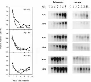
Nuclear-cytoplasmic mRNA distribution.Vero cells (33106
cells/100-mm-diameter dish) were infected with 0.5, 1.0, and 5.0 PFU/cell of KOS orn212. At the times indicated, infected cells were lysed in RNA lysis buffer and fractionated by differential centrifugation. Nuclei were resuspended in RNA lysis buffer and pelleted through a 20% sucrose cushion to remove contaminating membrane components. The RNA from cytoplasmic and nuclear fractions was isolated, separated by gel electrophoresis, and transferred to nylon membranes by North-ern blotting (1). NorthNorth-ern blots were probed with32
P-labeled antisense mRNA specific for TK message. The amount of radioactivity in the band corresponding to TK mRNA was quantitated by phosphorimager analysis. The range value for the image display was the same forn212 and KOS at each multiplicity.
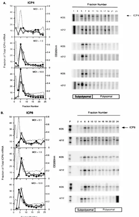
Polyribosome distribution.Vero cells (43107
per roller bottle) were infected with 0.1, 1.0 or 10.0 PFU/cell of KOS orn212, and at 6 hpi cells were harvested and cytoplasmic mRNA was isolated. Heparin sulfate was added to a final concentration of 1 mg/ml. The RNA was layered on a 0.5 to 1.1 M sucrose gradient (12 ml) and centrifuged at 180,0003gfor 110 min at 4°C. Fractions (0.5 ml) were collected from the top of the gradient, and absorbance at 260 nm was monitored. The RNA in each fraction was quantified by RNase protection assay. Fractions containing 40S, 60S, and 80S ribosomal subunits (subpolysomal pool) were determined by visual inspection of the ethidium bromide-stained agarose gels prior to RNase protection assay and from the absorbance measurements at 260 nm.
Protein synthesis rates.Protein synthesis rates in Vero cells infected with KOS orn212 were determined as previously described (41).
RESULTS
ICP0 activates gene expression at the level of mRNA
accu-mulation. (i) Cotransfection experiments.
As a first step
to-wards elucidating the level of gene activation by ICP0, we
performed transient expression assays and measured the levels
of mRNA and protein accumulation in the presence and
ab-sence of ICP0 (Fig. 1). Vero cells were cotransfected with a
constant amount of a reporter plasmid containing the CAT
gene under the control of the promoter of the ICP8 gene (an
E gene) and increasing concentrations of plasmid pSH, an
ICP0-expressing plasmid. Aliquots of the transfected cell
sus-pension were used to measure the levels of transfected DNA
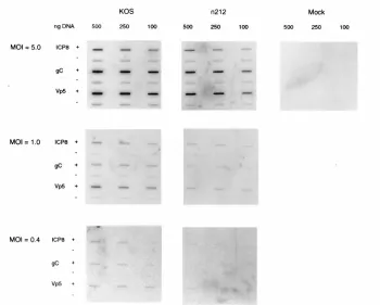
FIG. 1. Activation of CAT mRNA and CAT activity by ICP0 in transient gene expression assays. Vero cells (33106
/100-mm-diameter dish) were co-transfected with 15mg of pMRICP8-CAT and various amounts of plasmid pSH as indicated. (A) At 48 hours posttransfection the nuclei from 20% of the cells were used to prepare nuclear DNA to measure transfection efficiencies by slot blot hybridization probing for CAT DNA sequences. Nuclear DNA was applied to the slot blot apparatus in 5- and 0.5-mg amounts. (B) Cytoplasmic mRNA was purified from 80% of the cells, and the concentration of CAT mRNA was measured by quantitative RNase protection assay. (C) Lysates were prepared from the remaining cells, and CAT activity and CAT mRNA levels were mea-sured. CAT activity is expressed as picomoles/hour/microliter of extract and was measured in the linear range of the assay (less than 40% substrate utilized). Also shown are CAT mRNA levels quantitated with a PhosphorImager (Molecular Dynamics, Sunnyvale, Calif.). CAT mRNA values are expressed in PhosphorIm-ager units (PI units). The maximum range value for the image display was set so that the band having the highest range value was just within the visual linear range. The lower range value for the image display was set at the lowest setting (0.012 PI units). (D) Control experiment showing the linearity of the CAT assay. Vero cells were transfected with 0 to 30 mg of pWRICP0-CAT. Plasmid pWRICP0-CAT contains the promoter-regulatory region of ICP0 from plasmid pSH driving expression of CAT. At 48 h posttransfection the amounts of CAT mRNA, transfected DNA, and CAT activity were measured.
on November 9, 2019 by guest
http://jvi.asm.org/
(Fig. 1A), CAT mRNA (Fig. 1B and C), and CAT activity (Fig.
1C).
Transfection efficiencies of pMRICP8-CAT measured by
slot blot hybridization of nuclear DNA were similar in the
presence and absence of pSH (Fig. 1A). The amount of CAT
mRNA and CAT activity in crude enzyme extracts of cells
transfected with pMRICP8-CAT increased with increasing
amounts of pSH added to the transfection mixture, saturating
at 100 ng of pSH (Fig. 1B and C). Amounts of pSH greater
than 1,000 ng were inhibitory to the accumulation of both CAT
mRNA and CAT activity (data not shown). Similar effects were
observed when pWRICP0-CAT (a plasmid in which the CAT
gene is regulated by the ICP0 promoter) was transfected with
increasing amounts of pSH (data not shown). The saturation of
CAT activity at 100 ng of pSH was shown not to be due to
squelching of the ICP0 promoter or to the inability of
trans-fected cells to take up additional DNA. Thus, in control
ex-periments (Fig. 1D), the amounts of transfected DNA, and the
resulting levels of CAT mRNA and CAT activity expressed
from pWRICP0-CAT, were linear when 10 to 30
m
g of
re-porter plasmid was added to the transfection mixture. Finally,
no major differences were observed in the half-life of CAT
mRNA in cells transfected with pMRICP8-CAT in the
pres-ence (
t
1/25
2.9
6
0.5 h) or absence (
t
1/25
3.9
6
2.1 h) of pSH.
Taken together, these data suggest that ICP0 activates gene
expression at the level of mRNA accumulation in
transient-expression assays and that the changes in steady-state amounts
of mRNA observed in these experiments occur at the level of
mRNA synthesis. Since these experiments measured
cytoplas-mic CAT mRNA, however, we cannot rule out the possibility
that mRNA transport is affected by ICP0.
(ii) Transfection and superinfection experiments.
Although
ICP0 alone is sufficient to activate gene expression in transient
expression assays, ICP0 is known to cooperate with other viral
factors (e.g., ICP4) during infection to regulate HSV-1 gene
expression (7, 15, 16, 43, 44). The ability of ICP0 to activate
gene expression in the presence of other viral proteins was
therefore tested by transfecting Vero cells with the reporter
plasmid pMRICP8-CAT and infecting these cells with 5 PFU/
cell of either wild-type virus or the ICP0 null mutant
n
212.
Levels of cytoplasmic CAT mRNA and CAT activity were
measured at selected times postinfection.
As shown in Fig. 2, the levels of CAT activity and CAT
mRNA increased through 10 hpi in cells infected with
wild-type virus, whereas CAT activity and CAT mRNA levels
de-creased progressively over time in cells infected with
n
212,
suggesting that ICP0, when expressed together with other viral
proteins, activates expression of transfected genes at the level
of mRNA accumulation. Notably, changes in the levels of CAT
activity were roughly proportional to changes in the levels of
CAT mRNA in cells infected with either wild-type virus or
n
212, indicating that ICP0 had no major effect on the efficiency
of translation during viral infection.
(iii) Viral infection experiments.
Transient gene expression
assays have proven useful in identifying many of the
cis
-acting
elements and
trans
-acting factors involved in the regulation of
HSV-1 gene expression (2, 16, 43, 44, 51, 61). Transient
ex-pression assays can be problematic, however, in that the
reg-ulatory activities defined in these assays are not always
ob-served during viral infection (11, 19, 32). To determine
whether ICP0 activates expression of viral genes in their
nat-ural context at the level of mRNA accumulation, as observed
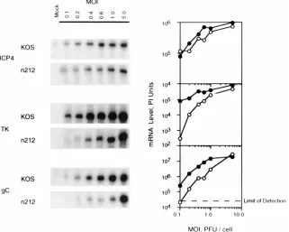
in Fig. 1 and 2, the levels of ICP4, TK, and gC mRNA were
measured in Vero cells infected with wild-type virus and
n
212
at multiplicities of infection ranging from 0.1 to 5.0 PFU/cell.
At times when the rates of RNA and protein synthesis from the
test genes were maximal (ICP4, 3 hpi; TK, 6 hpi; gC, 9 hpi)
infected cells were harvested, and cell extracts were prepared
for isolation and measurement of cytoplasmic ICP4, TK, and
gC mRNA by quantitative RNase protection assay (Fig. 3).
The results of these tests demonstrate that for selected
tran-scripts representing the major classes of viral genes, the levels
of cytoplasmic E and L (but not IE) gene-specific mRNAs in
n
212-infected cells were markedly lower than in wild-type
vi-rus-infected cells, especially at low multiplicities of infection.
The most dramatic differences in TK and gC mRNA levels in
KOS- and
n
212-infected cells were observed at multiplicities
lower than 1 PFU/cell. Similar results were obtained when
Northern blotting was used to measure levels of TK mRNA
(data not shown). In contrast to TK and gC mRNAs, the level
of ICP4 mRNA was only slightly reduced in
n
212-infected cells
relative to KOS-infected cells, suggesting that ICP0 had only a
minor effect on the level of accumulation of at least one IE
mRNA (ICP4). Collectively, these results are consistent with
the observations of Cai and Schaffer which showed that the
absence of ICP0 has a greater effect on E and L gene
expres-sion than on IE gene expresexpres-sion (7).
In order to study the effects of ICP0 on the kinetics of
expression of individual viral genes at low and high
multiplic-ities of infection, ICP4 and TK mRNA and protein
accumu-lation were measured over time in Vero cells infected with
KOS or
n
212 at multiplicities of 0.2 and 5.0 PFU/cell. As
shown in Fig. 4, the times of peak synthesis of both ICP4 and
TK mRNA in KOS- and
n
212-infected cells (4 hpi) occurred
before the times of peak protein synthesis at the higher
mul-tiplicity, suggesting that at high multiplicities translation is rate
limiting for gene expression early in infection with or without
ICP0. In contrast, at the lower multiplicity (0.2 PFU/cell) KOS
ICP4 (Fig. 4A), KOS TK (Fig. 4C), and
n
212 TK (Fig. 4D)
mRNA accumulation peaked at later times than at the higher
multiplicity and the kinetics of protein accumulation
corre-lated closely with the kinetics of mRNA accumulation,
sug-gesting that transcription is rate limiting for gene expression at
low multiplicities. Only in the case of
n
212 ICP4 did the
kinet-ics of mRNA and protein accumulation at the lower
multiplic-ity resemble those seen at the higher multiplicmultiplic-ity. The reduced
FIG. 2. Effect of ICP0 on CAT mRNA and CAT activity following viral superinfection. Vero cells (33106/100-mm-diameter dish) were transfected with
15mg of pMRICP8-CAT, and 24 h later cells were infected with 5.0 PFU/cell of either KOS orn212. At the indicated times, cells were harvested and soluble enzyme extracts were prepared from approximately 10% of the cells to measure CAT activity. The remaining cells were used to prepare cytoplasmic RNA, and CAT mRNA was measured by quantitative RNase protection assay.
on November 9, 2019 by guest
http://jvi.asm.org/
levels of
n
212 ICP4 (Fig. 4B) and
n
212 TK (Fig. 4D) mRNA
accumulation observed in cells infected with 0.2 PFU/cell
fur-ther confirm the requirement for ICP0 for wild-type levels of
transcription at low multiplicities of infection.
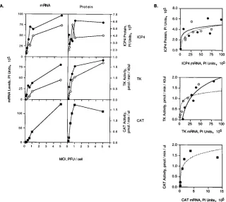
Effect of multiplicity of infection on the accumulation of
viral mRNA and protein in the presence and absence of ICP0.
To examine more closely the effect of multiplicity of infection
on gene expression in the presence and absence of ICP0, Vero
cells were infected at multiplicities ranging from 0.1 to 5.0
PFU/cell with KOS,
n
212, or P32, and the expression of ICP4,
TK, and CAT was measured. P32 is an ICP0 null mutant in
which the ICP0 locus has been replaced by the CAT gene
under control of the ICP8 promoter. The growth kinetics of
P32 and
n
212 are similar and both viruses grow more slowly
than KOS at low multiplicities of infection (49a). At 3 (ICP4)
and 6 (TK and CAT) hpi infected cells were harvested and
ICP4, TK, and CAT mRNA and protein levels or enzyme
activities were measured (Fig. 5).
Several points can be made from these experiments. (i) The
levels of ICP4 and TK mRNA and ICP4 protein and TK
activity in KOS-infected cells were higher than the
correspond-ing levels of ICP4 and TK mRNA and protein and TK activity
in
n
212-infected cells, consistent with the data shown in Fig. 3
and 4. Similar results were observed for ICP8 and gC mRNA
(data not shown). (ii) The levels of ICP4 and TK mRNA and
protein in KOS- and
n
212-infected cells, and of CAT mRNA
and CAT activity in P32-infected cells, increased roughly in
proportion to the amount of input virus at multiplicities below
1.0 PFU/cell. However, at a multiplicity greater than 1.0 PFU/
cell, the increased levels of mRNA and protein were no longer
proportional to the change in multiplicity. For example, in
KOS-infected cells the level of TK activity at 5.0 PFU/cell is
approximately 1.2-fold greater than the level of TK activity at
1.0 PFU/cell. If the level of protein correlated with the
multi-plicity of infection one would expect five times more TK
ac-tivity at 5.0 PFU/cell than at 1.0 PFU/cell. These data suggest
that independent of the presence or absence of ICP0, gene
expression is saturable at high multiplicities of infection.
Notably, the saturation of gene expression shown in Fig. 5A
at 5.0 PFU/cell was not due to saturation of the components of
the assays used to measure mRNA levels or protein expression
but rather is a characteristic of the infection process. Thus, in
control experiments changes in TK activity, and changes in
ICP4, ICP8, and gC mRNA levels, were proportional to the
amount of cell lysate or infected-cell mRNA added to the assay
(data not shown).
[image:6.612.148.469.72.329.2]Replotting the data in Fig. 5A to compare the amount of
protein detected per unit of mRNA revealed that the levels of
protein increased proportionally with changes in the levels of
mRNA for KOS and
n
212 at low concentrations of mRNA for
ICP4, TK, and CAT (Fig. 5B). At high concentrations of
mRNA, however, a saturation point was reached and changes
in the levels of protein were no longer proportional to the
changes in concentrations of mRNA. This observation was
most apparent for CAT gene expression, where at the highest
concentration of mRNA there was actually slightly less CAT
activity in three independent experiments. A similar pattern
was observed for ICP4 and TK gene expression (Fig. 5B). The
lines of best fit achieved by logarithmic regression of the data,
especially for TK and CAT, are consistent with processes
ex-hibiting saturable kinetic behavior. These observations imply
that at low mRNA concentrations, the level of mRNA is rate
limiting for gene expression and that at high, saturating mRNA
concentrations, the translational capacity of the infected cell is
FIG. 3. ICP0 effects the steady-state level of mRNA accumulation during lytic infection at several multiplicities. Vero cells (106
/100-mm-diameter dish) were infected with KOS (F) orn212 (E) at the indicated multiplicities (MOI, multiplicities of infection). Cells were harvested as described below, and cytoplasmic RNA was prepared. Twenty micrograms of infected-cell RNA was used to measure the amounts of ICP4, TK, and gC transcripts by quantitative RNase protection assay. The data are plotted to show the amount of each transcript as a function of multiplicity. The data shown are from infections harvested at 3 (ICP4), 6 (TK), and 9 (gC) hpi. The maximum range value for the image display was set so that the band having the highest range value was just within the visual linear range. The lower range value for the image display was set at the lowest setting (0.012 phosphorimager [PI] units).
on November 9, 2019 by guest
http://jvi.asm.org/
rate limiting. Therefore, gene expression can be saturated at
high multiplicities of infection (Fig. 5A) and at high
concen-trations of mRNA (Fig. 5B). Notably, the effects of multiplicity
were independent of ICP0.
ICP0 does not affect mRNA stability but activates gene
ex-pression at the level of or prior to initiation of mRNA
synthe-sis.
Steady-state accumulation of mRNA is dependent on the
rate of mRNA degradation as well as on the rates of initiation
and elongation of mRNA synthesis. To measure the stability of
TK mRNA in KOS- and
n
212-infected cells, Vero cells
in-fected with 5.0 PFU/cell of KOS and
n
212 were treated with
actinomycin D at 6 and 9 hpi, and mRNA was isolated at 0, 0.5,
1, 2, 3, and 5 h post-actinomycin D treatment. The levels of
mRNA were measured by Northern blotting using
32P-radio-labeled RNA antisense to TK mRNA as a probe. The
reduc-tion in TK mRNA signal as a funcreduc-tion of time
post-actinomy-cin D treatment was used to calculate the half-life of TK
mRNA. As shown in Table 1, no major differences in the
half-life of TK mRNA in the presence or absence of ICP0 were
observed at 6 or 9 hpi. The half-lives of these mRNAs are
consistent with previously published results (45). These
find-ings indicate that the changes in steady-state mRNA
accumu-lation shown in Fig. 3 to 5 were not due to differences in the
stability of mRNA in KOS- and
n
212-infected cells but to
changes in the rate of initiation or elongation of mRNA
syn-thesis.
To measure mRNA initiation rates, nuclear run-on
experi-ments were performed. Vero cells were infected with either
KOS or
n
212 at multiplicities of 0.4, 1.0, or 5.0 PFU/cell. (At
multiplicities lower than 0.4 PFU/cell, hybridization signals
became difficult to quantify.) At 5 hpi, nuclei were isolated and
newly initiated mRNA was labeled to high specific activity in
vitro. The radiolabeled mRNA was hybridized to immobilized
single-stranded DNA specific for either sense or antisense
transcripts of ICP8, gC, and VP5.
The results shown in Fig. 6 demonstrate that the amount of
radioactivity in newly initiated mRNA that hybridized to
im-mobilized DNA was higher in KOS-infected cells than in
n
212-FIG. 4. Kinetics of ICP4 and TK mRNA and protein accumulation in KOS- andn212-infected cells. Vero cells (33106
/100-mm-diameter dish) were infected with either 0.2 or 5.0 PFU/cell of KOS (A and C) orn212 (B and D). (A and B) At 1 hpi the medium was removed and replaced with medium containing either [35
S]methionine or unlabeled methionine. At 2-h intervals, infected radiolabeled cells were harvested, and ICP4 was immunoprecipitated with monoclonal antibody H112. The radiolabeled immunoprecipitate was separated by SDS-PAGE. At the same time intervals, cytoplasmic RNA was isolated from the remaining unlabeled cultures, and the level of ICP4 mRNA was measured by quantitative RNase protection assay. ICP4 mRNA and protein were quantitated with a phosphorimager. (C and D) Vero cells were infected as described above; however, at 2-h intervals, cells were harvested and approximately 50% of cells were used to prepare crude soluble enzyme extracts and TK activity was measured. Cytoplasmic RNA was isolated from the remaining cells, and the level of TK mRNA was measured by quantitative RNase protection assay. The data are plotted to show the amounts of mRNA and protein detected as a function of time. Note the difference in scales used to measure mRNA and protein for infections performed at 0.2 and 5.0 PFU/cell. In vitro ICP4 (panel A), in vitro-transcribed and -translated ICP4 used as a marker. The 10-h ICP4 mRNA sample (panel B) and the 6-h TK sample (panel D) were lost during isolation. The data points in the graphs representing these samples have been omitted.
on November 9, 2019 by guest
http://jvi.asm.org/
[image:7.612.63.554.69.422.2]infected cells at all three multiplicities tested. These data
in-dicate that differences in the levels of mRNA accumulation in
n
212- and KOS-infected cells occur at or before initiation of
mRNA synthesis. They do not rule out the involvement of
ICP0 in mRNA elongation, however.
ICP0 does not activate gene expression by
posttranscrip-tional mechanisms.
Although the results of nuclear run-on
assays indicated that ICP0 activates gene expression at or
be-fore initiation of mRNA synthesis, it is possible that ICP0 may
also act at a posttranscriptional level. To test this possibility the
nuclear-cytoplasmic mRNA distribution, polyribosomal
distri-bution, and protein synthesis rates were measured in KOS- and
n
212-infected cells. Vero cells were infected with KOS or
n
212
at multiplicities of 0.5, 1.0, or 5.0 PFU/cell, harvested at 1 to 2
hpi, and fractionated by differential centrifugation into nuclear
and cytoplasmic fractions. Total mRNA from each pool was
isolated, and the level of TK mRNA in each pool was
mea-sured by Northern blotting (Fig. 7). The effectiveness of the
fractionation procedure and the gel loading efficiencies were
monitored by measuring the amount of preribosomal and
ri-bosomal mRNA in ethidium bromide-stained agarose gels
prior to Northern blotting, and they were found to be
equiva-lent in all cases (data not shown). Although the overall levels
of nuclear and cytoplasmic TK mRNA were slightly lower in
n
212- than in KOS-infected cells at the lowest multiplicity, and
were slightly higher in
n
212- than in KOS-infected cells at the
highest multiplicity (Fig. 7, blots), no major differences were
observed in the levels of TK mRNA in the nucleus of KOS- or
n
212-infected Vero cells (Fig. 7, graphs).
The distribution of polyribosomes on mRNA encoding ICP4
or ICP8 was measured in KOS- and
n
212-infected cells.
Cyto-plasmic RNA from Vero cells infected with 0.1, 1.0, or 10.0
PFU/cell of either
n
212 or KOS was fractionated by velocity
sedimentation on sucrose density gradients. The RNA from
each fraction was purified, and the amount of ICP4 or ICP8
mRNA was measured by RNase protection assay (Fig. 8).
Although most of the infected-cell mRNA copurified with
monosome or low-number polysome fractions, no major
dif-ferences were observed in the polyribosomal distribution for
either ICP4 (Fig. 8A) or ICP8 (Fig. 8B) in KOS- and
n
212-FIG. 5. Multiplicity-dependent synthesis of mRNA and protein in KOS- andn212-infected cells. Vero cells (33106
/100-mm-diameter dish) were infected at the indicated multiplicities with either KOS (F),n212 (E), or P32 (■). Virus P32 contains the ICP8-CAT construct from pMRICP8-CAT inserted in place of thelacZgene in the ICP0 locus. At 3 hpi for ICP4 and 6 hpi for TK and CAT, the levels of mRNA and protein or enzyme activity were measured as described in Materials and Methods. (A) The levels of mRNA and protein or enzyme activity were plotted as a function of multiplicity. The actual ICP4 and TK mRNAs for these tests are shown in Fig. 3. (B) The data shown in panel A were replotted to show the amount of ICP4 protein or TK and CAT activity as a function of mRNA level. A logarithmic regression of the data points is shown (KOS, solid line;n212, dotted line). Thervalues for the regression are as follows: 0.444 and 0.601 for ICP4 in KOS- and
[image:8.612.147.467.67.352.2]n212-infected cells, respectively; 0.959 and 0.826 for TK in KOS- andn212-infected cells, respectively; and 0.929 for CAT in P32-infected cells. PI, phosphorimager.
TABLE 1. Half-life of TK mRNA in KOS- and
n
212-infected cells
aTime (hpi) Virus TK mRNA half-life (h)
Expt 1 Expt 2 Expt 3 Mean (SD)
6
KOS
0.5
1.0
1.8
1.1 (0.7)
n
212
1.9
0.9
1.2
1.3 (0.6)
9
KOS
1.1
3.8
ND
2.5 (1.9)
n
212
2.2
1.4
ND
1.8 (0.6)
aVero cells were infected with KOS orn212 at a multiplicity of infection of 5.0 PFU/cell. At 6 or 9 hpi actinomycin D was added to a concentration of 5mg/ml. At time zero, mRNA synthesis was inhibited by addition of actinomycin D at a concentration of 5mg/ml. At 0, 0.5, 1, 2, 3, 5, and 8 h postaddition of actinomycin D, cells were harvested and cytoplasmic mRNA was isolated. The level of TK mRNA was determined by Northern blotting analysis. The decrease in TK signal intensity as a function of time post-actinomycin D treatment was fit to a single exponential decay equation by using the program Enzfitter to calculate the rate of mRNA decay.
on November 9, 2019 by guest
http://jvi.asm.org/
[image:8.612.60.298.582.654.2]infected cells. These data indicate that ICP0 does not affect the
distribution of polyribosomes on ICP4 or ICP8 mRNA.
ICP0 may activate gene expression by multiple mechanisms
which may lead to broad effects on cellular metabolism.
Al-though our findings strongly suggest that ICP0 activates gene
expression at a transcriptional or pretranscriptional level, it is
also possible that ICP0 has an effect on translation. Indeed,
ICP0 was recently reported to interact with a component of the
translational apparatus in vitro and in cells in culture (30). No
evidence of translational effects of ICP0 during viral infection
were reported, however. To determine whether ICP0 exerts a
global effect on the regulation of protein synthesis in
virus-infected cells, the relative rates of protein synthesis were
mea-sured in Vero cells infected with 0.1, 1.0, and 10.0 PFU/cell of
either KOS or
n
212. At 3 hpi the rate of incorporation of
[
35S]methionine into trichloroacetic acid (TCA)-precipitable
material was measured. To account for differences in amino
acid pool sizes, infected Vero cells were treated with medium
containing [
35S]methionine or [
35S]methionine supplemented
with a known concentration of unlabeled methionine. The
rates of [
35S]methionine incorporation into TCA-precipitable
material following each treatment were used to calculate the
relative rates of protein synthesis in KOS- and
n
212-infected
cells (Table 2). A value of 1.0 corresponds to an equal rate of
protein synthesis. As shown in Table 2, the rate of
35S
incor-poration into TCA-precipitable material in both
n
212- and
KOS-infected cells approached 1.0, indicating that no major
differences in the relative rates of protein synthesis occurred in
KOS- or
n
212-infected Vero cells. Taken together, these
re-sults suggest that the contribution of ICP0 to
posttranscrip-tional events is not great.
DISCUSSION
ICP0 activates a wide variety of viral and cellular promoters
without apparent DNA sequence specificity. Although the
broad transactivating activity of ICP0 has been
well-docu-mented, the mechanism by which ICP0 activates gene
expres-sion is poorly understood. Because the levels at which proteins
function to regulate gene expression reflect their mechanisms
of action, we attempted to determine the level at which ICP0
functions. For this purpose, we measured the accumulation of
the mRNA and protein products of selected viral genes in
transient expression and infection assays in the presence and
absence of ICP0. In this paper we report that ICP0 activates
gene expression at the level of mRNA accumulation, and more
specifically, at or before initiation of transcription. Notably,
ICP0 had no major effects on the stability, nuclear cytoplasmic
distribution, or polyribosomal distribution of mRNA, or on the
rates of protein synthesis, indicating that ICP0 does not affect
viral gene expression at a posttranscriptional level. In both
transfections and infections, ICP0 had a greater effect on E
and L transcript accumulation than on IE transcript
accumu-lation, consistent with previous reports.
[image:9.612.133.483.72.353.2]ICP0 functions to enhance mRNA synthesis at low
multi-plicities of infection.
A unique characteristic of ICP0 null
mu-tants is the exaggerated multiplicity dependence of their
growth relative to wild-type virus (7, 8, 18, 56, 66). At low
multiplicities viral gene expression at the mRNA and protein
levels, and consequently virus yields, are severely reduced (7, 8,
18, 56). At high multiplicities of infection, however, gene
ex-pression and virus yields approach wild-type levels (7, 8, 18,
56). Interestingly, the low-multiplicity growth defect of ICP0
FIG. 6. Nuclear run-on assays. Vero cells (33106
cells/100-mm-diameter dish) were infected with 0.4, 1.0, or 5.0 PFU/cell of either KOS orn212. At 5 hpi nuclei were isolated and newly initiated mRNA was labeled to high specific activity in vitro with [32
P]GTP. The radiolabeled mRNA was hybridized to single-stranded DNA specific for either the sense (1) or antisense (2) strand of the indicated genes immobilized on nylon membranes.
on November 9, 2019 by guest
http://jvi.asm.org/
null mutants can be complemented by superinfection with
HCMV, for which no homolog of ICP0 has been identified
(65). These observations suggest that the exaggerated
multi-plicity dependence of ICP0 null mutants may reflect an activity
associated with the herpesvirus infection process that activates
viral gene expression and viral replication in the absence of
ICP0.
In order to determine whether the level (transcriptional,
translational, etc.) at which multiplicity-dependent gene
ex-pression occurs is the same or different in the presence and
absence of ICP0, the levels of mRNA and protein synthesis
from genes representing each of the major kinetic classes of
viral genes were measured as a function of multiplicity in
KOS-and
n
212-infected cells. The results of these tests demonstrate
that at low multiplicities of infection mRNA accumulation is
rate limiting and that at high multiplicities translation is rate
limiting for gene expression both in the presence and absence
of ICP0, although the levels of mRNA and protein were
uni-formly lower in
n
212- than in KOS-infected cells.
The observation that ICP0 null mutants exhibit markedly
reduced levels of mRNA at low multiplicities relative to
wild-type virus, and that translation in infected cells is a saturable
process, suggests that ICP0 serves to increase the level of
mRNA accumulation at low multiplicities of infection when
RNA is rate limiting for gene expression. By boosting the level
of mRNA during low-multiplicity infection, ICP0 exerts a
pos-itive effect on viral growth. At high multiplicities, an activity
associated with the infection process per se but independent of
ICP0 apparently raises the level of mRNA until a point is
reached where the rate-limiting step for gene expression is
independent of the level of mRNA (i.e., translation becomes
rate limiting). At this point, ICP0 is no longer required for
efficient gene expression.
How does ICP0 activate transcription?
The findings
pre-sented in this paper demonstrate that ICP0 functions at the
level of transcript accumulation and, more specifically, at or
before initiation of transcription. Available evidence
concern-ing the physical and functional properties of ICP0 supports this
hypothesis, suggesting that ICP0 is a transcriptional activator
with properties not unlike the transcriptional regulatory
pro-teins of other DNA-containing viruses.
[image:10.612.150.467.73.359.2]By definition, transcriptional activators function to increase
the rate of initiation of mRNA synthesis through binding to
specific
cis
-acting DNA sequences as well as to components of
the transcriptional complex. Although ICP0 alone increases
the rate of initiation of mRNA synthesis, as demonstrated in
these studies, it appears to do so without binding to specific
DNA sequences. Notably, bona fide viral transcriptional
acti-vators that do not themselves bind to specific DNA sequences
are well documented. For example, HSV-1 VP16, the
virion-associated activator of IE gene expression, activates
transcrip-tion through interactranscrip-tions with cellular DNA binding proteins
(2, 29, 33, 42, 49, 64). Moreover, E1A, which is incapable of
binding directly to DNA, activates gene expression indirectly
through interactions with sequence-specific DNA binding
pro-teins (14). It may be that ICP0 also activates gene expression
FIG. 7. Nuclear-cytoplasmic distribution of TK mRNA during infection with KOS orn212. Vero cells (33106
cells/100-mm-diameter dish) were infected with 0.5, 1.0, or 5.0 PFU/cell of KOS orn212. At the times indicated, infected cells were fractionated and RNA was isolated from cytoplasmic and nuclear fractions. Cytoplasmic and nuclear RNAs were separated by gel electrophoresis and transferred to nylon membranes by Northern blotting. Northern blots were probed with32
P-labeled antisense mRNA specific for TK message. The amount of radioactivity in the band corresponding to TK message was quantitated by phosphorimager analysis. The range value for the image display is the same forn212 and KOS at each multiplicity. Loading efficiency was monitored by the intensity of ethidium bromide staining of ribosomal or preribosomal mRNA (data not shown). The graphs show the fraction of mRNA relative to the total amount of TK mRNA for KOS (F) andn212 (E) at each time point.