overcome for successful gene therapy. Recent efforts to improve adenovirus vectors for in vivo use have focused on the sequential deletion of essential early genes. Adenovirus vectors have been constructed with the E1 gene deleted and with this deletion in combination with an E2a, E2b, or E4 deletion. We report here a novel vector
(Av4orf3nBg) lacking E1, E2a, and all of E4 except open reading frame 3 (ORF3) and expressing ab
-galac-tosidase reporter gene. This vector was generated by transfection of a plasmid carrying the full-length vector sequence into A30.S8 cells that express E1 and E2a but not E4. Production was subsequently performed in an
E1-, E2a-, and E4-complementing cell line. We demonstrated with C57BL/6 mice that the Av4orf3nBg vector
effected gene transfer with an efficiency comparable to that of the Av3nBg (wild-type E4) vector but that the
former exhibited a higher level ofb-galactosidase expression. This observation suggests that E4 ORF3 alone
is able to enhance RNA levels from theb-galactosidase gene when the Rous sarcoma virus promoter is used
to drive transgene expression in the mouse liver. In addition, we observed less liver toxicity in mice injected
with the Av4orf3nBg vector than those injected with the Av3nBg vector at a comparable DNA copy number per
cell. This study suggests that the additional deletion of E4 in an E1 and E2a deletion background may be beneficial in decreasing immunogenicity and improving safety and toxicity profiles, as well as increasing transgene capacity and expression for liver-directed gene therapy.
Adenovirus vectors are currently used for gene transfer in basic research and gene therapy protocols due to their high titers, ability to target a wide range of both dividing and non-dividing cells, and mediation of high-level foreign protein ex-pression without replication or integration of the viral genome (4, 18, 36, 37). In spite of this, several factors significantly limit the utility of currently used adenovirus vectors. Data obtained from murine and other animal models have shown that host immune responses to viral and transgene protein products are responsible for eliminating transduced cells and preventing readministration (9, 20–22, 41–44). Progress toward the provement of these vectors involves strategies to reduce im-munogenicity and toxicity associated with viral backbone gene expression.
Two approaches have been taken to disable adenoviral vec-tors. One is based on the concept that removal of all coding sequences for viral proteins from the vector backbone renders the vector less immunogenic and restricts the immune re-sponse to the injected viral capsid proteins. This gutted ad-enovector (7, 15, 24, 27, 30, 31, 34) is propagated in the pres-ence of helper adenovirus. A second alternative in decreasing adenovirus vector immunogenicity and toxicity is based on a gene attenuation strategy wherein the sequential removal of key early genes is anticipated to reduce expression from other essential genes. This type of vector grows in packaging cell lines that complement the deleted viral genes. Adenovirus vectors have been constructed with the E1 gene deleted (28, 36, 37, 45) and with E1 plus E2a, E2b, or E4 deleted (1, 2, 6, 10, 13, 14, 16, 17, 28, 40). Studies carried out in vitro to evaluate adenovirus vectors with double deletions consistently
feature an absence of detectable replication and late gene expression (1, 17). Recently, we extended those studies by semiquantifying the levels of early and late transcripts in an adenovector with a double deletion of E1 and E2a in vitro and in vivo. An analysis of RNA-specific PCR-amplified fragments showed that expression of adenoviral major late gene products was minimal. In contrast, early promoters such as E4, pIX, and E2b were found to actively express high levels of gene tran-scripts (25).
This molecular evaluation suggests that further elimination of early genes is required to attenuate viral gene expression. Recently, it has been demonstrated that additional deletion of the E4 region in an E1 deletion background has a major in-fluence on the stability of the adenovirus vector genome in addition to prolonging transgene expression (2, 10, 16, 40). The E4 region, which contains seven open reading frames (ORFs), is essential for virus growth, DNA replication, and particle assembly (reviewed in reference 26). In particular, the E4 region encodes E4 ORF3 and E4 ORF6, which share functions required for mRNA splicing and accumulation (5).
In this article we describe the construction of a novel vector (Av4orf3nBg) lacking E1, E2a, and all of E4 except ORF3 and expressing a b-galactosidase reporter gene. This vector was generated by transfection of a plasmid bearing the entire mod-ified vector genome into an A549-based cell line that comple-ments E1 and E2a (17). Production was subsequently per-formed in an E1-, E2a-, and E4-complementing cell line. To determine the effect of E4 viral gene deletion on vector trans-duction and liver toxicity, similar doses of either Av4orf3nBg or Av3nBg (with E1 and E2a deleted) (17) were administered to C57BL/6 mice. We demonstrate that the Av4orf3nBg vector exhibits gene transfer with an efficiency comparable to that of the Av3nBg vector but that the former exhibits a higher level of b-galactosidase expression. In addition, we observed less liver toxicity in mice injected with Av4orf3nBg than in mice
* Corresponding author. Mailing address: DNA Viral Vector Unit, Genetic Therapy, Inc., a Novartis Company, 938 Clopper Rd., Gaith-ersburg, MD 20879. Phone: (301) 258-4661. Fax: (301) 948-8034. E-mail: [email protected].
6048
on November 9, 2019 by guest
http://jvi.asm.org/
injected with Av3nBg at comparable DNA copy numbers per cell. This study suggests that the additional deletion of E4 may be useful in limiting cytopathic effects, increasing transgene capacity, and increasing expression over levels expressed by double-deletion vectors in the context of liver-directed gene therapy.
MATERIALS AND METHODS
Plasmid construction.The recombinant adenovirus vector with E1, E2a, and all of E4 except ORF3 deleted, Av4orf3nBg, expressing a nucleus-targetedb -ga-lactosidase reporter was generated with a single plasmid, pAv4orf3nBg, contain-ing the entire adenoviral genome sequence within a pBR-L backbone (Fig. 1). pBR-L is identical to pBR322, except that it is missing all nucleotide sequences betweenNdeI (position 2297) andEcoRI (position 4286). A multiple-cloning site was created betweenNdeI andEcoRI by insertion of a DNA linker. This mul-tiple-cloning site contains sites forEcoRI,NspV,ClaI,BamHI,SalI,BclI,PacI, andNdeI. Previous cloning had generated a plasmid in pBR-L that contained the 39end of the adenovirus type 5 genome (bp 21562 to 35936) lacking the E2a and E3 regions (17). This plasmid was extended further to contain an additional segment of the adenoviral genome by incorporating sequences from theBamHI site (bp 21562) back to theSalI site (bp 16746), resulting in a plasmid, pdl23. The modification to the E4 region was created by deleting theAvrII (bp
35469)-Sse8387 (bp 33289) fragment (removing all of the E4 coding region except for the E4 promoter) from a plasmid designated pREpac (Fig. 1A). This plasmid con-tains the right end of adenovirus type 5dl327 (including the E3XbaI deletion) from theSnaBI site (bp 25171) to the end of the right inverted terminal repeat (ITR), which was ligated between the SmaI andHindIII sites of pBluescript SK(1) (Stratagene). APacI site (TTAATTAA) was engineered 152 bp internal to the end of the right ITR by insertion of a 4-bp sequence, TTAA, adjacent to the TTAA sequence already present at this location. TheHindIII (bp
34936)-SapI (bp 34330) fragment containing the E4 ORF3 was then cloned into the T4 DNA polymerase blunt-endedAvrII-Sse8387 site to create pREpac1orf3. The wild-type E4 region was removed from pdl23 and replaced by anXbaI-PacI fragment from pREpac1orf3, resulting in plasmid pdl234 (Fig. 1B). An inter-mediate cloning vector, pdl234LE, was created by ligating theNotI-SpeI frag-ment from pdl234 (retaining the pBR-L backbone and the E4-modified region) to the left end of the shuttle plasmid pAvS6a (35), to yield a hybrid containing
the left ITR, packaging signal (C), Rous sarcoma virus (RSV) promoter, tripar-tite leader (TPL) nucleotide sequences, regions lacking E3 and E4, and right ITR (Fig. 1B). The resultingSpeI site was then used to insert the 25,633-bpSpeI fragment, derived from the Av3nBg vector, containing theb-galactosidase gene and the region lacking E2a (17). The final construct containing the entire Av4orf3nBg genome in a pBR-L backbone is called pAv4orf3nBg (Fig. 1B).
Generation and propagation of the Av4orf3nBg recombinant adenovirus.The Av4orf3nBg vector was generated by cotransfecting 1mg ofNspV-linearized pAv4orf3nBg and 1mg of pREpac plasmids with Lipofectamine (GIBCO-BRL, Gaithersburg, Md.) into 0.3mM dexamethasone-induced AE1-2a cells. AE1-2a is a stable cell line, derived from A549 cells, which contains the adenovirus type 5 E1 and E2a genes inducibly expressed from separate glucocorticoid-responsive promoters (17). Cell cultures were maintained in Richter’s CM medium (Bio-Whittaker) supplemented with 5% fetal bovine serum. Transfected cells were lysed after 7 days by five cycles of freezing and thawing, and the lysate was used to infect a fresh, dexamethasone-induced monolayer of A70.S54 cells (first am-plification), which became available after the initial transfection. A70.S54 is a stable cell line derived from AE1-2a cells which additionally expresses adenovi-rus type 5 E4 genes (this cell line will be described elsewhere). After 7 days, the infected cells were lysed and the crude viral lysate (CVL) was used to isolate individual plaques for analysis.
Virus production and purification.CVL from infected A70.S54 cells was used to infect a fresh, induced monolayer of A70.S54 cells. After 14 days, individual plaques representing Av4orf3nBg were recovered and virus was purified. This plaque-purified material was subsequently used to prepare large-scale stocks of virus in A70.S54 cells. The vector Av3nBg (lacking E1 and E2a) was propagated and purified as previously described (17). Both vectors used in this study lacked E3, a region not required for viral replication, and contained the sameb -galac-tosidase expression cassette.
[image:2.612.118.484.72.327.2]Structural characterization of the Av4orf3nBg vector.Adenovector DNA, obtained from CVLs of purified and amplified plaques, was analyzed byHindIII restriction enzyme digestion. The resulting fragments were fractionated by aga-rose gel electrophoresis and visualized with ethidium bromide. Additionally, DNAs obtained from CVL of A70.S54 cells infected with Av3nBg or Av4orf3nBg and DNA from plasmid pAv4orf3nBg were digested with theHindIII restriction enzyme, fractionated on a 1% agarose gel, transferred to a Zeta-Probe GT nylon membrane, and hybridized with32P-labeled DNA probes containing sequences specific for E4 ORF3, E4 ORF6, or the entire viral genome (HindIII-digested pAv4orf3nBg DNA probe).
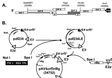
FIG. 1. Schematic representation of the adenovirus type 5 E4 region and construction of plasmids for generating the Av4orf3nBg vector. (A) ORFs of the E4 region are indicated by open boxes. ORF3 is represented by a hatched bar. (B) Construction of pAv4orf3nBg. pdl23 is a plasmid that contains the 39end of the adenovirus type 5 genome (bp 16746 to 35936) with deletions in the E2a and E3 regions (17). APacI-XbaI fragment from pdl23 was replaced with aPacI-XbaI fragment containing the E4 promoter and the ORF3 gene (bp 34936 to 34330), generating pdl234. TheNotI-SpeI fragment from pdl234 was replaced with aNotI-SpeI fragment derived from pAvS6a (35) to generate pdl234LE, which contains the left-end ITR,C, RSV promoter, TPL, regions with E3 and E4 deleted, and right ITR. The single infectious plasmid, pAv4orf3nBg, was generated by inserting anSpeI fragment of 25,633 bp flanking the nucleus-localizingb-galactosidase reporter transgene (nBg) gene and the region with E2a deleted. E4p, E4 promoter; RSV, heterologous RSV promoter.
on November 9, 2019 by guest
http://jvi.asm.org/
cessor, and embedded in paraffin, followed by hematoxylin and eosin staining. In vivo gene transfer efficiencies.Immunohistochemical staining ofb -galacto-sidase was performed by the avidin-biotin immunoperoxidase method. Briefly, sections were deparaffinized, rehydrated, and pretreated with 0.3% hydrogen peroxide solution for 30 min at room temperature (RT) to quench endogenous peroxidase activity. The pretreated sections were then incubated with 2% normal goat serum for 30 min at RT. After being blocked, the slides were incubated with a polyclonal antibody tob-galactosidase (Cortex Biochem, Inc.) diluted 1:2,000 in phosphate-buffered saline (PBS) for 1 h at RT, rinsed in PBS, and then incubated with a biotinylated goat anti-rabbit antibody for 30 min at RT. Sections were then washed in PBS and incubated with avidin DH-biotinylated horseradish peroxidase H (Vectastain Elite ABC kit) for 30 min at RT. After a PBS wash, the reaction was visualized with diaminobenzidine tetrahydrochloride as the perox-idase substrate. Sections were counterstained with hematoxylin and coverslipped. In addition, liver tissues were processed for quantification ofb-galactosidase activity by following the protocol used for in vitro samples.
Southern blotting.Total genomic DNA was isolated from mouse liver with a QIAmp tissue kit (Qiagen) and further incubated with RNase. DNA concentra-tions were determined spectrophotometrically. A total of 10mg of each DNA sample was digested withBamHI and subjected to electrophoresis on a 0.8% agarose gel, stained with ethidium bromide, and transferred to a Zeta-Probe GT membrane (Bio-Rad). The copy number control standards were prepared by adding 600 and 60 pg of pAv4orf3nBg plasmid DNA, equivalent to 10 and 1 vector copies per cell, respectively, to 10mg ofBamHI-digested genomic DNA from an uninfected animal. The32P-labeled probe, prepared by random oligo-nucleotide priming, containedb-galactosidase cDNA sequences. The relative amount of DNA was determined by PhosphorImager analysis as the ratio be-tween sample and control signals.
Northern blot analysis.Total RNA was extracted from liver tissue by the RNAzol B method (Teltest, Inc.). RNA concentration was quantified by deter-mining the A260. Total RNA (15mg) was electrophoresed through a 0.8% agarose–7% formaldehyde gel, transferred to nylon membrane, and bound to the filter by UV cross-linking. A DNA probe was radiolabeled by random oligonu-cleotide priming of alacZcDNA template. The band intensities were quantified with a Molecular Dynamics PhosphorImager SF.
RESULTS
Construction of Av4orf3nBg plasmid. Av4orf3nBg, a
new-generation vector lacking E1, E2a, and all of E4 except ORF3 and expressing a nucleus-targeted b-galactosidase reporter, was constructed in several steps. E4 ORF3 was maintained in the vector backbone for two reasons. First, we had a cell line that complemented only E1 and E2a functions (17) and tran-sient-transfection experiments with 293 cells demonstrated that expression of E4 ORF3 alone could support growth of a virus with E4 deleted,dl1011 (5), although not as well as a virus with the entire E4. Second, previous studies suggested that E4 gene products helped maintain vector transgene expression (2, 6, 10).
In developing the next phase of construction, we took ad-vantage of an observation made by other investigators, namely, that an entire adenovirus genome could be propagated as a bacterial plasmid. After release from the plasmid by restriction digestion and transfection into cells, the cloned viral genome could produce infectious virus (19, 39). Our strategy is outlined in Fig. 1. TheNotI-SpeI fragment (1,020 bp) from plasmid pAvS6a containing the 59 terminus, ITR, packaging signal, RSV promoter, and TPL was subcloned into pdl234 to produce
cells (which complement E1, E2A, and E4). When all the cells demonstrated cytopathic effects, virus was harvested and a stock was grown by reinfecting fresh A70.S54 cells. The vector growth was efficient, resulting in approximately 700 viral par-ticles/cell with purified vector stocks in the range of 231011to 431011particles/ml as quantified by determining the optical density at 260 nm.
The genome of Av4orf3nBg (32,083 bp) was structurally characterized by restriction enzyme digestion and Southern blot analysis (Fig. 2).HindIII digestion provided the expected fragment pattern for the entire genome in six independent adenovirus vector isolates (Fig. 2A, lanes 3 to 8). The same fragments were visualized by Southern blot analysis (Fig. 2B, lane 9); of these, the 4.2- and 2.3-kb fragments (lane 9, arrows) are characteristic of the adenoviral genome compared to that of the plasmid DNA and represent the ends of the adenovirus vector. In the plasmid DNA these fragments were not ob-served; instead, an expected plasmid fragment of 9.2 kb was detected (Fig. 2B, lane 7). This restriction pattern difference suggests that after transfection, the plasmid DNA was able to serve as the template and package the viral genome without carryover of extra nucleotides.
Evaluation of the E4 region of Av4orf3nBg by PCR (data not shown) and by Southern blotting confirmed the expected deletion. Thus, theHindIII restriction digestion gave the ex-pected 2.3-kb fragment with Av4orf3nBg (Fig. 2B, lane 9), ra-ther than the 2.3- and 1.0-kb fragments corresponding to the wild-type E4 region present in the Av3nBg vector (Fig. 2B, lane 8).
Because the transfection contained both the pAv4orf3nBg and the pREpac plasmid (which contains the entire E4 region) there was the potential of incorporating this entire region in the Av4orf3nBg vector by recombination. Thus, a further South-ern blot analysis of the E4 region was carried out afterHindIII digestion with probes specific for E4 ORF3 and E4 ORF6 nucleotide sequences. In agreement with the previous results, the E4 ORF3 probe hybridized with the 9.2-kb fragment of pAv4orf3nBg, the 2.9-kb fragment of Av3nBg, and the 2.3-kb fragment of Av4orf3nBg (Fig. 2B, lanes 1, 2, and 3, respective-ly). As predicted, the E4 ORF6 probe recognized only the 2.9-kb fragment of the Av3nBg vector (Fig. 2B, lane 5). Al-together, these results demonstrated that the E4 region of Av4orf3nBg contained only E4 ORF3 and that the adenovec-tor was free of any wild-type E4 sequences. In addition, HindIII fragments of 6.5 and 3.7 kb were observed in both the Av3nBg (Fig. 2B, lane 8) and Av4orf3nBg (Fig. 2B, lane 9) vectors; these fragments verified the E2a deletion (6.5 kb) and the E3 deletion (3.7 kb). PCR evaluation of the E2a and E3 regions also confirmed the expected deletions (data not shown).
Quantitative analysis of in vitrob-galactosidase expression.
A remarkable feature that emerged from infection with the adenoviral vectors with E1 and E4 deleted was that expression
on November 9, 2019 by guest
http://jvi.asm.org/
of a reporter gene under the control of the cytomegalovirus (CMV) or RSV promoter was influenced by E4 protein expression (2, 6, 10). Thus, a quantitative b-galactosidase assay was used to evaluate transgene expression in a non-complementing A549 cell background, by comparing the
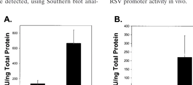
b-galactosidase expression of the Av3nBg vector (E41) to that of the Av4orf3nBg vector. Under the conditions used in this experiment (10 particles/cell) there was about fivefold more b-galactosidase detected in cells transduced with Av4orf3nBg than in those transduced with Av3nBg (Fig. 3A).
Recombinant adenovirus-mediated transduction and
trans-gene expression in mouse liver.Based on the previous results
that E4 ORF3 influences the activity of the RSV promoter in vitro, we wanted to determine if E4 ORF3 would have a similar effect in vivo. To determine the profiles of transgene expres-sion, paired low and high doses of Av4orf3nBg and Av3nBg (631010and 331011particles, respectively) were adminis-tered to C57BL/6 mice by tail vein injection. At 7 days postin-jection, animals were sacrificed and liver tissues were analyzed for vector content. We detected, using Southern blot
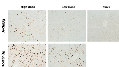
[image:4.612.147.459.72.224.2]anal-ysis that probed for theb-galactosidase gene, similar DNA copy numbers in animals receiving either the Av3nBg or the Av4orf3nBg vector (Fig. 4). Similarly, the efficiencies of trans-duction based onb-galactosidase staining of liver sections were not significantly different for any of the adenoviral vectors tested when equivalent numbers of particles were used. Only the intensity of the staining increased in cells transduced with the Av4orf3nBg vector (Fig. 5). In fact, a quantification of RSV-promotedb-galactosidase expression from the Av4orf3nBg vector showed a fivefold increase compared to the expression from cells infected with the Av3nBg vector (Fig. 3B). Using Northern blot analysis, we then examined whether theb -ga-lactosidase expression in vivo was directly correlated to the amount ofb-galactosidase mRNA present in liver tissues (Fig. 6). Mice that received the Av4orf3nBg vector showed on aver-age a fivefold increase in transgene transcription accumulation compared with that of mice receiving the Av3nBg vector. To-gether, these results support the concept that E4 ORF3 mod-ulates expression of the RSV promoter and further suggest that a gene product(s) of E4 other than ORF3 may repress RSV promoter activity in vivo.
FIG. 2. Structural characterization of the Av4orf3nBg vector. (A) Adenovector DNAs obtained from crude lysates of purified and amplified adenoviral plaques were analyzed byHindIII restriction enzyme digestion. The DNA fragment products were fractionated by agarose gel electrophoresis and visualized with ethidium bromide (lanes 3 to 8). TheHindIII-digested plasmid pAv4orf3nBg (pAv4) was included as a control (lane 2). The sizes of marker DNA fragments (1-kb ladder andl-HindIII; GIBCO-BRL) are indicated (lanes 1 and 9). Av4, cells infected with the Av4orf3nBg vector. (B) Adenovector DNA obtained from crude lysates of cells infected with the Av3nBg (Av3) or Av4orf3nBg (Av4) vector and DNA from plasmid pAv4 were digested with theHindIII restriction enzyme, fractionated on a 1% agarose gel, transferred to a nylon filter, and hybridized with a32P-labeled ORF3 DNA probe (lanes 1 to 3), a32P-labeled ORF6 DNA probe (lanes 4 to 6), or a32P-labeledHin dIII-digested pAv4 probe (lanes 7 to 9). Arrows indicate fragments representing the left and right ends of the adenovirus vector.
FIG. 3. Av4orf3nBg vector gene transfer efficiency in vitro (A) and in vivo (B). (A) Noncomplementing A549 cells were infected with either the Av3nBg (Av3) or the Av4orf3nBg (Av4) vector at equivalent doses of 10 particles/cell based on the optical density measurements of the adenoviral vectors at 260 nm.b-Galactosidase activity in cell lysates was determined 48 h postinfection by the Tropix GalactoLight chemiluminescent-reporter assay. The results are expressed as relative light units (RLU) ofb-galactosidase activity per nanogram of total cellular protein. The data are means6standard deviations of three separate determinations for three different production lots of each vector type. (B) Livers of C57BL/6 mice infected with a low dose of Av3nBg or Av4orf3nBg were evaluated quantitatively forb-galactosidase expression at 7 days postinjection by the same procedure described for panel A. The data are means6standard deviations of three separate determinations with three different animals.
on November 9, 2019 by guest
http://jvi.asm.org/
[image:4.612.148.460.532.669.2]Reduced mouse liver toxicity with the Av4orf3nBg vector.
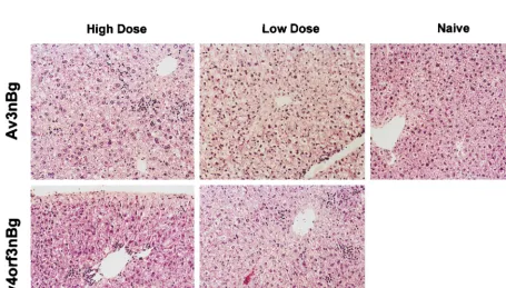
Previous analysis of early gene transcripts in vitro and in vivo indicated that the E4 promoter was functional in an Av3nBg vector lacking E1 and E2a. The hypothesis in the present study was that an additional E4 deletion in the Av3nBg vector would impart lowered expression of early and late viral proteins and further attenuate viral gene expression; the result could be to improve the in vivo utility of this vector by reducing toxicity and the host immune response and increasing the duration of transgene expression. Histochemical analysis of hematoxilin-and eosin-stained liver sections of mice 7 days postinjection of high and low doses of Av3nBg and Av4orf3nBg are shown in Fig. 7. In a dose-dependent manner, Av3nBg caused vacuol-ization, nuclear pleomorphism, and a loss of tissue architec-ture. These histopathologic changes were not observed with
Av4orf3nBg. However, both vectors produced mild inflamma-tory changes characterized by mononuclear cell infiltrates.
The toxicities of both Av3nBg and Av4orf3nBg were also quantified by determining levels of ALT in serum. Both vectors at low and high doses resulted in elevation of the ALT levels at 24 h, with a subsequent decrease by day 3 (Fig. 8). With the higher vector dose of Av3nBg the ALT levels increased signif-icantly above those of uninjected control animals at day 7 (Fig. 8B). In contrast, ALT levels in Av4orf3nBg-injected animals were not different from those in controls at day 7.
DISCUSSION
[image:5.612.146.456.74.197.2]Based on previous studies demonstrating that the E4 pro-moter is still active in an E1 and E2a deletion vector (25), we C57BL/6 mice infected with a high dose (331011viral particles) of the infectious Av3nBg (lanes 4 to 7) or Av4orf3nBg (lanes 8 to 11) vector was isolated on day 7 following gene transfer and digested withBamHI. The Southern blot was probed with aBamHI32P-labeled fragment encoding theb-galactosidase gene. Lanes 1 and 2 contain pAv4orf3nBg plasmid DNAs equivalent to 10 copies and 1 copy per cell, respectively. The control sample included liver DNA of naive mice (lane 3).
FIG. 5. Recombinant adenovirus-mediated transgene expression in mouse liver. A low or high dose of the Av4orf3nBg or Av3nBg vector (631010or 331011viral particles, respectively) were injected into the tail vein of C57BL/6 mice. Seven days postinjection animals were sacrificed and liver tissues were evaluated for
b-galactosidase expression by immunohistochemical staining.
on November 9, 2019 by guest
http://jvi.asm.org/
[image:5.612.72.530.443.700.2]reasoned that an additional E4 deletion would further atten-uate viral gene expression and toxicity compared to levels produced by double-deletion vectors. Thus, six of the seven ORFs were deleted from the E4 region, with the exception of ORF3. This ORF was chosen for its ability to functionally complement a complete E4 region deletion such as that in the dl1011 mutant virus (5). A recombinant adenovirus vector with E1, E2a, and E4 deletions and expressing a nucleus-targeted
b-galactosidase reporter gene was generated with a single plas-mid system to transfect an A549-based cell line that expresses E1 and E2a genes. Several reports have described the use of a plasmid containing a copy of the adenovirus genome to rescue the virus after transfection (e.g., see references 19 and 39). In these previous cases, plasmids were linearized before transfec-tion to release an adenovirus genome as an intact linear mol-ecule with exposed ITRs. In the present study, we also linear-ized the plasmid, but the right and left ITRs were flanked by 2.0 and 0.5 kb, respectively, of extra nucleotides. Virus
[image:6.612.54.290.73.159.2]pro-duction was subsequently performed in a triple-complementa-tion (E1, E2a, and E4) cell line, in which a viral titer similar to those obtained with Av3-type vectors (17) was achieved. DNA analysis performed on six randomly selected amplified virus plaques and on total crude lysates confirmed that the cloned viral genome was able to generate a stable and full-length genome containing the appropriate deleted regions. The fact that no rearrangements in DNA structure were ob-served between virus stock and virus plaques suggests that little or no detectable sequence instability is associated with this procedure. This and other reports clearly demonstrate that this approach to generating adenoviral vectors is faster and less tedious than the more traditional methods which rely on ho-mologous recombination in situ. The level and duration of in vivo transgene expression measured from the RSV- and CMV-promoted expression cassette in the adenovirus vectors ap-pears to correlate with the structure of the vector backbone. An interesting observation was made with adenovirus vectors lacking E1 and E4 in which the presence of E4 gene products seemed to regulate the expression from both RSV and CMV promoters (2, 6, 10). Transgene expression was observed only with a vector that had a wild-type E4 region and not with vectors that contained either a complete E4 deletion or a deletion of all of E4 except ORF6 (2). Our results with the Av4orf3nBg vector indicate that ORF3 alone is able to en-hance expression ofb-galactosidase when the RSV promoter is used to drive transgene expression in the mouse liver. We observed that the level of b-galactosidase expression was higher in animals receiving the Av4orf3nBg than in those re-ceiving the Av3nBg (wild-type E4) vector. These observations suggest that an E4 product(s) competes with ORF3 to affect expression or the accumulation ofb-galactosidase in vivo. The molecular basis for ORF3 activation must await further evaluation; however, preliminary analysis of b-galactosidase FIG. 6. In vivo detection of Av3nBg and Av4orf3nBg transgene activities.
Total liver RNA of C57BL/6 mice infected with a high dose (331011viral particles) of the infectious Av3nBg (lanes 1 to 4) or Av4orf3nBg (lanes 6 to 9) vector was isolated on day 7 following gene transfer, transferred to a nylon membrane, and hybridized with a32P-labeledlacZprobe. The control sample included liver RNA extracted from a naive mouse (lane 5).
FIG. 7. Pathological response in liver following Av3nBg or Av4orf3nBg vector administration. A low or high dose of the Av4orf3nBg or Av3nBg vector (631010 or 331011viral particles, respectively) was injected into the tail veins of mice. Morphological observation of liver sections prepared at 7 days postinjection was carried out after hematoxylin and eosin staining. For consistency, liver samples from the same mice used for the samples in Fig. 4 are shown here.
on November 9, 2019 by guest
http://jvi.asm.org/
[image:6.612.74.530.442.701.2]mRNA accumulation suggests that ORF3 may act either di-rectly or indidi-rectly on the activity of the RSV promoter. Both E1-E2a and E1-E4 double-deletion vectors have lower toxicity profiles than a first-generation vector for liver-directed gene therapy. Studies performed with murine models show reduced liver damage as determined by serum transaminase levels. Based on the results of our experiment, we speculate that the toxicity was biphasic. The first ALT elevation observed for both vectors at day 1 may simply involve the vector entering the liver, and ALT levels would be the same with both vectors, or alternatively, this early hepatocellular toxicity may not be re-lated to capsid proteins but rather to nonspecific immune mechanisms (27). The levels of ALT at day 7, which were lower with Av4orf3nBg, may be related to reduced backbone gene expression. This pattern of ALT elevation correlates well with the reduced cytopathic effect observed in animal livers follow-ing vector administration. Tail vein injection of Av3nBg in C57BL/6 mice resulted in liver vacuolization, nuclear pleomor-phism, and loss of tissue architecture; none of the mice that received the Av4orf3nBg vector demonstrated these patholog-ical changes. This result was expected by virtue of the known effects of E4 viral proteins on cell toxicity. Among the many activities of E4 proteins are oncogenesis, blocking of p53 func-tion, modulation of cellular transcription factors, induction of the cell cycle, and modification of protein phosphatase 2a activity (11, 23, 32, 33). Removal of these functions from the adenovirus vector backbone will undoubtedly have an impact on cell viability. However, since E4 ORF3 is active in the
mice (3, 16). In this study both the Av3nBg and the Av4orf3nBg vector behave similarly in C57BL/6 mice by their ability to induce mononuclear cell infiltrates. The interpreta-tion of this result is not immediately evident becauseb -galac-tosidase expression is much higher in animals injected with the Av4orf3nBg vector than in animals injected with an E4-containing Av3nBg vector. This observation may imply that transgene-directed cellular immune responses are more predominant in adenovirus vectors whose virus-specific gene expression is significantly reduced than in those whose virus-specific gene expression is stable. Interestingly, C57BL/6 mice administered an Av4nBg vector lacking E1, E2a, and E4 (at-tenuated by a complete removal of the E4 promoter and ex-pressing a b-galactosidase gene from an RSV promoter) showed a lower level of transgene expression, a lower toxicity, and a smaller amount of cell infiltrates than mice administered the Av4orf3nBg vector (data not shown). The above results ex-emplify the relationship between E4 viral proteins and pro-moter regulation but more importantly suggest that attenua-tion of E4 proteins in the vector backbone may reduce host antiviral responses to the transduced cells. The existence of an interplay between the immune responses to transgene and viral antigens was documented in this model of liver-directed gene transfer (10) and illustrates the impact that a reporter gene may harbor when adenovirus vector backbone modifications are being evaluated in vivo. Thus, a key issue in the further evaluation of a triple-deletion adenovirus vector is to demon-strate persistent transgene expression of a nonimmunogenic transgene in several mouse strains and in large-animal models. We describe in this study the construction of an adenovirus vector lacking E1, E2a, and all of E4 except ORF3. This ade-novirus vector can be propagated to a high titer in a cell line that complements the deleted viral functions, facilitating scale-up and purification steps. The vector has several clear advan-tages over a double-deletion vector: (i) additional deletion of E4 in a vector with previous E1 and E2a deletions improves the safety and toxicity profile of the vector and should decrease immunogenicity, (ii) rescue of replication-competent adenovi-rus is unlikely, and (iii) the shortened adenoviadenovi-rus vector back-bone allows for larger insertions of foreign DNA sequences. In addition, identification of ORF3 as the only E4 gene product required to increase RSV promoter activity has important im-plications for the design of other recombinant vectors. The impact of this protein in long-term gene expression should be carefully investigated.
ACKNOWLEDGMENTS
We thank Robert Jambou for critical review of the manuscript. We also thank Russette Lyons and Christoph Wey for assistance with the animal procedures and Christine Mech for preparing liver sections and staining.
FIG. 8. Comparison of levels of liver injury between mice infected with the Av3nBg vector and those infected with the Av3orf3nBg vector. Serum samples from mice infused with a low dose (A) or a high dose (B) of the Av3nBg or Av4orf3nBg vector were harvested at the indicated time points and analyzed for ALT concentrations. Numbers in parentheses are the numbers of animals per point. For reference, the average normal levels of ALT are shown.
on November 9, 2019 by guest
http://jvi.asm.org/
REFERENCES
1.Amalfitano, A., M. A. Hauser, H. Hu, D. Serra, C. R. Begy, and J. S. Chamberlain.1998. Production and characterization of improved adenovi-rus vectors with the E1, E2b, and E3 genes deleted. J. Virol.72:926–933. 2.Armentano, D., J. Zabner, C. Sacks, C. C. Sookdeo, M. P. Smith, J. A. St.
George, S. C. Wadsworth, A. E. Smith, and R. J. Gregory.1997. Effect of the E4 region on the persistence of transgene expression from adenovirus vec-tors. J. Virol.71:2408–2416.
3.Barr, D., J. Tubb, D. Fergunson, A. Scaria, A. Lieber, C. Wilson, J. Perkin, and M. A. Kay.1995. Strain related variations in adenovirally mediated transgene expression from mouse hepatocytes in vivo: comparison between immunocompetent and immunodeficient inbred strains. Gene Ther.2:151– 155.
4.Berkner, K. L.1988. Development of adenovirus vectors for the expression of heterologous genes. BioTechniques6:616–629.
5.Bridge, E., and G. Ketner.1989. Redundant control of adenovirus late gene expression by early region 4. J. Virol.63:631–638.
6.Brough, D. E., C. Hsu, V. A. Kulesa, G. M. Lee, L. J. Cantolupo, A. Lizonova, and I. Kovesdi.1997. Activation of transgene expression by early region 4 is responsible for a high level of persistent transgene expression from adeno-virus vectors in vivo. J. Virol.71:9206–9213.
7.Burcin, M. M., G. Schieder, S. Kochaneck, S. Y. Tsai, and B. W. O’Malley. 1999. Adenovirus-mediated regulable target gene expression in vivo. Proc. Natl. Acad. Sci. USA96:355–360.
8.Carvalho, T., J.-S. Seeler, K. Ohman, P. Jordan, U. Pettersson, G. Akusjarvi, M. Carmo-Fonseca, and A. Dejean.1995. Targeting of adenovirus E1A and E4orf3 proteins to nuclear matrix-associated PML bodies. J. Cell Biol.131: 45–56.
9.Dai, Y., E. M. Schwartz, D. Gu, W. W. Zhang, N. Sarvetnick, and I. M. Verma.1995. Cellular and humoral immune responses to adenoviral vectors containing factor IX gene: tolerization of factor IX and vector antigens allows for long-term expression. Proc. Natl. Acad. Sci. USA92:1401–1405. 10. Dedieu, J. F., E. Vigne, C. Torrent, C. Julien, I. Mahfouz, J. M. Caillaud, N. Aubailly, C. Orsini, J. M. Guillaume, P. Opolon, P. Delaere, M. Perricaudet, and P. Yeh.1997. Long-term gene delivery into the livers of immunocom-petent mice with E1/E4-defective adenoviruses. J. Virol.71:4626–4637. 11. Dobner, T., N. Horikoshi, S. Rubenwolf, and T. Shenk.1996. Blockage by
adenovirus E4orf6 of transcription activation by the p53 tumor suppressor. Science272:1470–1473.
12. Doucas, V., A. M. Ishov, A. Romo, H. Juguilon, M. D. Weitzman, R. M. Evans, and G. G. Maul.1996. Adenovirus replication is coupled with the dynamic properties of the PML nuclear structure. Genes Dev.10:196–207. 13. Engelhardt, J. F., X. Ye, B. Doranz, and J. M. Wilson.1994. Ablation of E2A in recombinant adenoviruses improves transgene persistence and decreases inflammatory response in mouse liver. Proc. Natl. Acad. Sci. USA91:6196– 6200.
14. Engelhardt, J. F., L. Litzky, and J. M. Wilson.1994. Prolonged transgene expression in cotton rat lung with recombinant adenoviruses defective in E2a. Hum. Gene Ther.5:1217–1229.
15. Fisher, K. J., H. Choi, J. Burda, S.-J. Chen, and J. M. Wilson.1996. Re-combinant adenovirus deleted of all DNA viral genes for gene therapy of cystic fibrosis. Virology217:11–22.
16. Gao, G. P., Y. Yang, and J. M. Wilson.1996. Biology of adenovirus vectors with E1 and E4 deletions for liver-directed gene therapy. J. Virol.70:8934– 8943.
17. Gorziglia, M. I., M. J. Kadan, S. Yei, J. Lim, G. M. Lee, R. Luthra, and B. C. Trapnell.1996. Elimination of both E1 and E2a from adenovirus vectors further improves prospects for in vivo human gene therapy. J. Virol.70: 4173–4178.
18. Graham, F. L., and L. Prevec.1992. Adenovirus based expression vectors and recombinant vaccines, p. 363–390.InR. W. Ellis (ed.), Vaccines: new approaches to immunological problems. Butterworth-Heinemann, London, United Kingdom.
19. Hanahan, D., and Y. Gluzman.1984. Rescue of functional replication origins from embedded configurations in a plasmid carrying the adenovirus genome. Mol. Cell. Biol.4:302–309.
20. Joos, K., C. J. Hildegund, and J. M. Wilson.1998. Cytotoxic T-lymphocyte target proteins and their major histocompatibility complex class I restriction in response to adenovirus vectors delivered to mouse liver. J. Virol.72: 2945–2954.
21. Kajiwara, K., A. P. Byrnes, H. M. Charlton, M. J. Wood, and K. J. Wood. 1997. Immune responses to adenoviral vectors during gene transfer in the brain. Hum. Gene Ther.8:253–265.
22. Kaplan, J. M., D. Armentano, T. E. Sparer, S. G. Wynn, P. A. Peterson, S. C. Wadsworth, K. K. Couture, S. E. Pennington, J. A. St. George, and L. R. Smith.1997. Characterization of factors involved in modulating persistence
of transgene expression from recombinant adenovirus in the mouse lung. Hum. Gene Ther.8:45–56.
23. Kleinberger, T., and T. Shenk.1993. Adenovirus E4orf4 protein binds to protein phosphatase 2A, and the complex down regulates E1A-enhanced
junBtranscription. J. Virol.67:7556–7560.
24. Kochaneck, S., P. R. Clemens, K. Mitani, H.-H. Chen, S. Chan, and C. T. Caskey.1996. A new adenoviral vector: replacement of all viral coding sequences with 28 kb of DNA independently expressing both full-length dystrophin andb-galactosidase. Proc. Natl. Acad. Sci. USA93:5731–5736. 25. Lapcevich, C., Q. Kang, and M. Gorziglia.Unpublished results.
26. Leppard, K. N.1997. E4 gene function in adenovirus, adenovirus vector and adeno-associated virus infections. J. Gen. Virol.78:2131–2138.
27. Lieber, A., C.-Y. He, I. Kirillova, and M. A. Kay.1996. Recombinant adeno-viruses with large deletions generated by Cre-mediated excision exhibit dif-ferent biological properties compared with first-generation vectors in vitro and in vivo. J. Virol.70:8944–8960.
28. Lusky, M., M. Christ, K. Rittner, A. Dieterle, D. Dreyer, B. Mourot, H. Schultz, F. Stoeckel, A. Pavarani, and M. Mehtali.1998. In vitro and in vivo biology of recombinant adenovirus vectors with E1, E1/E2a, or E1/E4 de-leted. J. Virol.72:2022–2032.
29. Maizel, J. V., D. O. White, and M. D. Scarff.1968. The polypeptides of adenovirus. II. Soluble proteins, cores, top components and structure of the virion. Virology36:126–136.
30. Mitani, K., F. L. Graham, C. T. Caskey, and S. Kochanek.1995. Rescue, propagation, and partial purification of a helper virus-dependent adenovirus vector. Proc. Natl. Acad. Sci. USA92:3854–3858.
31. Morsy, M. A., M. Gu, S. Motzel, J. Zhao, J. Lin, Q. Su, H. Allens, L. Franlin, R. J. Parks, F. L. Graham, S. Kochaneck, A. J. Bett, and C. T. Caskey.1998. An adenoviral vector deleted for all viral coding sequences results in en-hanced safety and extended expression of a leptin transgene. Proc. Natl. Acad. Sci. USA95:7866–7871.
32. Nevels, M., S. Rubenwolf, T. Spruss, H. Wolf, and T. Dobner.1997. The adenovirus E4orf6 protein can promote E1A/E1b-induced focus formation by interfering with p53 tumor suppressor function. Proc. Natl. Acad. Sci. USA94:1206–1211.
33. Nevins, J. R.1991. Utilization of cellular transcription factors for adenovi-rus-induced transcription. Virus Res.20:1–10.
34. Schiedner, G., N. Morral, R. J. Parks, Y. Wu, S. C. Koopmans, C. Langston, F. L. Graham, A. L. Beaudet, and S. Kochanek. 1998. Genomic DNA transfer with a high-capacity adenovirus vector results in improved in vivo gene expression and decreased toxicity. Nat. Genet.18:180–183. 35. Smith, T. A. G., M. G. Mehaffey, D. B. Kayda, J. M. Saunders, S. Yei, B. C.
Trapnell, A. McClelland, and M. Kaleko.1993. Adenovirus mediated ex-pression of therapeutic plasma levels of human factor IX in mice. Nat. Genet.5:397–402.
36. Trapnell, B. C.1993. Adenoviral vectors for gene transfer. Adv. Drug De-livery Rev.12:185–199.
37. Trapnell, B. C., and M. Gorziglia.1994. Gene therapy using adenoviral vectors. Curr. Opin. Biotechnol.5:617–625.
38. Tripathy, S. K., H. B. Black, E. Goldwasser, and J. M. Leiden.1996. Immune responses to transgene-encoded proteins limit the stability of gene expres-sion after injection of replication-defective adenovirus vectors. Nat. Med.2: 545–550.
39. Vrati, S., E. S. Macavoy, Z. Z. Xu, C. Smole, D. B. Boyle, and G. W. Both. 1996. Construction and transfection of ovine adenovirus genomic clones to rescue modified viruses. Virology220:200–203.
40. Wang, Q., C. Greenburg, D. Bunch, D. Farson, and M. H. Finer.1997. Persistent transgene expression in mouse liver following in vivo gene transfer with dlE1/dlE4 adenovirus vectors. Gene Ther.4:393–400.
41. Yang, Y., H. C. J. Ertl, and J. M. Wilson.1994. MHC class I-restricted cytotoxic T lymphocytes to viral antigens destroy hepatocytes in mice in-fected with E1-deleted recombinant adenoviruses. Immunity1:433–442. 42. Yang, Y., F. A. Nunes, K. Berencsi, E. E. Furth, E. Gonczol, J. F. Engelhardt,
and J. M. Wilson.1994. Cellular immunity to viral antigens limits E1-deleted adenoviruses for gene therapy. Proc. Natl. Acad. Sci. USA91:4407–4411. 43. Yang, Y., F. A. Nunes, K. Berencsi, E. Gonczol, J. F. Engelhardt, and J. M.
Wilson.1994. Inactivation of E2a in recombinant adenoviruses improves the prospect for gene therapy in cystic fibrosis. Nat. Genet.7:362–369. 44. Yang, Y., Q. Li, H. J. C. Ertl, and J. M. Wilson.1995. Cellular and humoral
immune responses to viral antigens create barriers to lung-directed gene therapy with recombinant adenovirus. J. Virol.69:2004–2015.
45. Yei, S., N. Mittereder, S. Wert, J. A. Whitsett, R. W. Wilmott, and B. C. Trapnell.1994. In vivo evaluation of the safety of adenovirus-mediated transfer of the human cystic fibrosis transmembrane conductance regulator cDNA to the lung. Hum. Gene Ther.5:731–744.