0022-538X/94/$04.00+0
Copyright © 1994,American Society for Microbiology
Analysis of AP-1 Function in Cellular Transformation Pathways
TAKEHISA SUZUKI,1 MASAO MURAKAMI,1 NOBUYUKI ONAI,' EIKOFUKUDA,'
YOSHIKO
HASHIMOTO,'
MARTHA H. SONOBE,1 TAKASHIKAMEDA,MASAOICHINOSE,2 KAZUMASA
MIKI,2
ANDHIDEOIBAI*DepartmentofTumorVirus Research, Instituteof MedicalScience, Universityof
Tokyo,
Minato-ku, Tokyo 108,1 andFirst DepartmentofIntemnal Medicine, Faculty ofMedicine, University of Tokyo, HongoBunkyo-ku, Tokyo 113,2 Japan Received20December 1993/Accepted 22 February 1994
To understand the role of endogenous AP-1activityin cellular transformation induced by oncogenes, we have made use of a fos mutant
(supfos-1)
and ajun mutant (supjun-1), either of which can function as a transdominant inhibitor of AP-1-mediated transcriptional regulation. Chicken embryo fibroblasts (CEF) infected with a series of transformingretroviruses weredoubly
infected with retrovirus carryingsupfos-1
or supjun-1,and suppression of cellulartransformation was monitored in terms of reversion to normal cellular morphology or acquisition of anchorage-dependent growth. Cellular transformation induced by several exogenously expressed transforming genes of thefos orjunfamily
wasefficientlysuppressed, as expected. CEF transformed by v-src, v-yes,v-.fps,
c-Ha-ras, andN-terminally
truncatedc-raf were also induced to revert to the normal phenotype by these transdominant mutants, suggesting that functional transcription factor AP-1 activity is essential for the cellular transformation induced by these oncogenes. The suppression is not attributabletononspecific inhibition of cellular proliferation, because CEF transformed by v-ros or v-myc were notinducedtoreverttothe normal phenotype. We next analyzed changes in all known components of chicken AP-1 induced by v-src, c-Ha-ras, or activated c-raf transformation. The levels of both Fra-2 and c-Jun expressionwereelevatedtwo- to fourfold, and hyperphosphorylationof Fra-2 was also observed. We further showedthatFra-2-c-Jun heterodimer is mainly responsible for the elevatedAP-1DNA-bindingactivity in these transformed cells,and weproposethat thisheterodimer playacrucialroleinthetransformation inducedby theseoncogenes.Thec-fos and
c-jun
proto-oncogenes were originally identi-fiedasthe cellular counterparts of the viral oncogenes carried by Finkel-Biskis-Jinkins (FBJ) murine sarcoma virus (11)and avian sarcoma virus 17 (21), respectively. c-fos belongs to a multigene family thatincludesfra-1(9),fra-2
(23,27), and fosB (42), and the fos gene family codes for nuclear proteins that dimerize with the Junfamily proteins, such as c-Jun(26),JunB (33), and JunD (16, 32), to form the transcription factor complex AP-1. Dimerization occurs specifically through a leucine zipper structure: Jun family members can form low-affinity homodimers and high-affinity heterodimers with the Fos family, whereas Fos-related proteins do not form stable homodimers(10, 25). Althoughthese hetero- and homodimers bindtosimilarDNA-bindingsites (TGACTCA, AP-1-binding sites) through the basic domains of both proteins, which are juxtaposed bythedimerization, eachdimerwasshowntohave adistincttranscriptional regulatory function,sothat transcrip-tioncanbepositively and negatively modulated (37).High-level expression of most members of thefos orjun genefamilyhas beenreportedto causecellular transformation ofchicken embryo fibroblasts(CEF) (17,27,36).JunD hasno transforming activity,but itcanacquire transforming potential byspontaneous mutation (15, 18).These results indicate that uncontrolledexpression and qualitative changeofany compo-nentofAP-1 caninduce cellulartransformation.
Logarithmically growing CEF express c-Jun and Fra-2 at relatively high levels (36, 41), and basal-level expression of
*Corresponding author. Mailing address: Department of Tumor Virus Research, Institute of Medical Science, University ofTokyo,
4-6-1 Shirokanedai, Minato-ku, Tokyo 108.Phone: 3-3443-8111, ext.
502. Fax:3-3443-6604.
JunD was also reported in these cells (14). In
c-fos-overex-pressing cells, cellular transformation seemstobe mediated by the heterodimeric complex of exogenous c-Fos and endoge-nousc-Jun (38). Inthecaseoftransforming junD mutants, it was suggested that the heterodimeric complexes of endoge-nous Fra-2 and JunD mutants play a crucial role in cellular transformation (18).
In our previous attempt to suppress specifically the tran-scriptional function of c-jun, weconstructed atransdominant mutant designated supfos-1 (28). supfos-1 was originally de-scribed as anontransformingv-fosmutant (40)which has an insertion of four amino acids between the basic domain and the leucinezipperstructure(Fig. 1),and its geneproductwas shown, both in vitro and in vivo, to forma heterodimer with c-Jun that lacks the specific AP-1 DNA binding (28). This mutant, when introduced by retrovirus vectors, efficiently suppresses c-jun transformation by sequestering the unstable c-Junhomodimer into the stable and nonfunctional supFosl-Jun heterodimer in the cells. Interestingly, nuclear extracts from the supfos-1-expressing CEF reducedendogenous AP-1 DNA-binding activity(mainlycontributedbyFra-2-Jun)to an almost nondetectable level by competing out endogenous Fra-2(27), suggestingthat thismutant canalsofunctionas an inhibitor of Fos family proteins. Severalfos orjun mutants have also been reported by other groups to function as transdominant suppressors by a mechanism similar to that used by
supfos-1
(38). Some of the transdominant mutants, however, function without the leucinezipper structure, possi-bly bya squelchingmechanism (38),while others retain both dimer-formingandDNA-bindingactivities but lackthetrans-activation domain(6, 13, 20).
Most of the fos and jun family members
belong
to the 3527on November 9, 2019 by guest
http://jvi.asm.org/
1 60
P LE R
supFosl
l
I
1
supJunl
2 4 7 3 3 1a a
v
7/I
L---a-FIG. 1. Proteinstructuresof supFos-1 andsupJun-1. supFos-1is a
derivative of v-Fos (FBJ), which has an insertion composed of four amino acids (aa) between the basic domain (striped box) and the leucine zipper motif (black box). supJun-1 isanN-terminallytruncated
mutantof c-Jun(human) andstartsfromaninternalmethionine 247.
category ofimmediate-early genes and are promptly induced
by several external stimuli aswell as transient expression of
such oncogenes as src, ras, and raf, which are believed tobe locatedupstream offos andjun family members in the signal transductionpathway (2). It is noteworthy that cellular
trans-formation induced by constitutive expression of these
onco-genesseems to require functional endogenous c-Fosorc-Jun.
Thiswassuggested by the reversion of transformedcellstothe normal phenotype following introduction of antisense RNA (19) for the fosgene, anti-Fos antibody (31), or a
transdomi-nant mutantoffos orjun. BecauseAP-1 activation by several
oncogenes has beenstudied ina number of different cell lines
by using either stableortransienttransfections, it isnoteasyto develop a general model from these observations. It seems necessary to perform extensive screening ofoncogenes which
require endogenous AP-1 for their transforming activity and detailed analysis of the induced changes in all of the AP-1 components, using thesame cellsystem.
In the first part of this report, we introduce a new
trans-dominantsuppressor,supjun-1,andshowthat both thismutant
c-Jun supJun-1
competitor + - +
c-Jun
supJun-- - ., gee
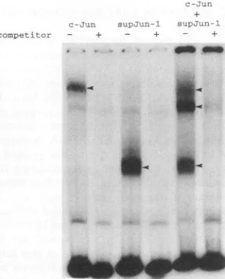
FIG. 2. supJun-1 binds to AP-1-bindingsitesas ahomodimer or heterodimerwithc-Jun. Eachproteinwassynthesized separately,and
mixtures wereincubatedfor30minat37°C. DNA-binding activitywas determinedby gelshiftanalysis using0.2ng ofa32P-labeled
oligonu-cleotidecontainingtheAP-1-bindingsite(FSE2) and4Kgof
reticu-locyte lysatemixture inthepresence (+)orabsence(-)of 20ngof
unlabeledoligonucleotideas aspecific competitor. Arrowheads
indi-catepositionsofspecificAP-1DNA-bindingactivity.
and supfos-J can function as general inhibitors ofAP-1. By introduction of the dominant negative mutants into CEF transformedby a series of oncogenes that do notbelong to the fos orjun family, we were able to categorize the oncogenesinto two groups; one group requires functional AP-1 activity for transforming activity, while the other does not. We have further analyzed all known components of theAP-1 complex in CEF transformed by oncogenes that belong to the former group in an attempt to understand the molecularmechanisms by which these oncogenes affect endogenous AP-1 activity.
MATERIALS AND METHODS
Plasmid construction. The 0.8-kbSmaI fragment containing the entire cDNA of human c-H-ras was isolated from pSPT-Hras (from the Japanese Cancer Research ResourcesBoard) (30), ligated to BamHI linkers, and inserted into the unique BglII site of pDS3 (17) to generate pHras. pND-raf was generated from the humanc-raf-1 cDNA, p627 (from JCRB) (5), by PCR using a 5' primer that covers the artificial methionine codon preceded by an artificial Kozak consensus sequence and aBamHI site (5'-AGGGATCCACCATGGAA AGAGAGCGGCACCAGT-3') and a 3' primer that contains the stopcodon (5'-CCGGATCCCTACTAGAAGACAGGCA GCCTCGG-3'). The PCR product was digested with BamHI and inserted into the unique BglII site of pDS3. pND-raf encodes the same amino acid sequence as the 20A clone constructed by Stanton et al. (35), which lacks the N-terminal 313 amino acids of human c-raf. The 0.5-kb NaeI-EcoT4I fragment of pHJ(gift from R. Tjian) (4) was filled in, ligated to BglII linkers, and inserted into the BglII site of pDS3 to generatepsupjun-1, which produces an N-terminally truncated Jun from the internal methionine 247. The truncated Jun retains the entire basic domain and the intact leucine zipper structure, as shown in Fig. 1. The 0.83-kb EcoRI-BspMI fragment of pSPfra-1 (rat) (9) was filled in and ligated with blunt-endedBglII-cut pDS3 to generate pFlR1, which carries the entire ratfra-] gene.
For RNA probesynthesis, four DNA templates were constructed. The450-bpBstXl-EcoRI fragment of pJun (gift from P. Vogt) (21) encoding the C-terminal half of the v-jun gene was cloned into pBlueskm to generate
pjunRPA,
which waslinearized atthePvuII site and used as a template. The 260-bp PstI-SacI fragment of chicken v-fos (12) was inserted into pSPT19 to generate pfosRPA, which was linearized at theNdeI site and used as a template. The 250-bpBstEI-NcoIfragment of chickenfra-2(26) was filled in and ligated to pSP64,whichwas doubly digested with EcoRI andBamHI andblunt ended, to generatepfra2RPA. The pfra2RPA was linear-ized at thePvull site and used as a template. The chicken junD construct was generated by PCR, using the published sequence (14). The245-bpSadH-TaqI fragment of the PCR product was inserted between the AccI and SacII sites of pBlueskm to generate thepjunDRPA. pjunDRPA
waslinearized at the intemal DdeI site and used as a template. ForNorthem (RNA) blotting of the chicken glyceraldehyde 3-phosphate dehydrogenase (GAPDH) mRNA, the c-DNA that encodes the entire GAPDH was generated by the reverse transcription RT-PCR technique,using the published se-quence (29).Cells andviruses. CEF were prepared, grown, andinfected with viruses as reported previously (28). For metabolic label-ing, CEF were grown in 60-min-diameter dishes and labeled with 500 ,uCi of[35S]methioninefor 60min. For the production of recombinant viruses such as
supjun-1
(carrying N-terminally truncated jun), ND-raf (carrying N-terminally deletedraf),
and H-ras(carrying normal c-Ha-ras) viruses, psupjun-1 and pHras were completely digested with Sall and ligated to theSall
1
11
5
on November 9, 2019 by guest
http://jvi.asm.org/
[image:2.612.61.290.74.156.2] [image:2.612.98.257.449.646.2]digest of pREP to form the structure of the replication-competent provirus (subgroup A); pND-raf was partially di-gested with Sall, and a 1.3-kb fragment that covers the raf sequence was isolated and used for ligation with the
Sall
digest ofpREP. Ligated DNAs (2jig)
were transfected into CEF as described previously (17), and replication-competent virus stocks were collected from the culture 5 or 6 days after transfection. Viruses were never propagated before use to minimize genetic changes of the virus genome. For production ofthe subgroup Bvirus, pREP was substituted with pREP-B. Similar viruses containing the human c-jun gene (JH-1 [36]), the mouse c-fos gene (FM4 [17]), thechickenfra-2
gene (F2C1 [27]), the v-src gene of Rous sarcoma virus (N4 [17]), a transdominantnegative mutant of v-fos (supfos-J [28]),and no oncogene (DS3 [17]) have been described previously. The natural avian retroviruses Rous sarcoma virus Prague C, Fujinami sarcoma virus (Rous-associated virus-1[RAV-11),
Y73-associated virus (YAV), UR2 (RAV-1), and MC29(RAV-1
orRAV-2) were also used to introduce the v-src,v-fps,
v-yes, v-ros, andv-myc genes, respectively.Colony formation. CEF that were sequentially infected with twospecies ofviruses weretrypsinized 4 days after the second infection. Approximately 3,000 doubly infected CEF were mixed with 3 x 10⁵ freshly prepared CEF feeder cells and seeded in suspension in soft agar (0.4%) on top of a bed of hard agar(0.8%) as discussed previously (28). Colonies, which were formed after 2 weeks of incubation at
38.5°C,
were counted (average ofthreeplates) at 3 weeks after seeding.Gel shift analysis. Gel shift analysis
using
proteins synthe-sized in rabbit reticulocyte and using a 32P-labeled 62-bp double-stranded DNA probe containing the FSE2 AP-1 DNA-bindingsite was described previously (37).The nuclear extracts were prepared from infected CEF as described previously (28). Gel shift analysis of the nuclear extract was done as described previously (28, 41), using a
32P-labeled
65-bpHindIll-Aval fragment ofpcoll, which has aninsertion of anoligonucleotide containing the AP-1 DNA-bindingsite of the human collagenasegene at theBamHI site ofpUC119. Insomeexperiments, the antiserum was added to the mixture 15 min before addition of the labeled probe. The sampleswere analyzed byelectrophoresis ona nondenaturing 5%polyacrylamidegel at 4°C, andshifted bands were detectedby
autoradiography.Immunoprecipitation andWestern blot (immunoblot) anal-ysis. 35S-labeled cell lysates were prepared under denaturing conditions (boilingin 2.0%sodium dodecyl sulfate [SDS]) and immunoprecipitated as described previously (17, 40). Poly-clonal antisera raised against Fos peptide 1 (40) and Fra-2 peptide 2(27) have been described previously.
The total cellextracts prepared under denaturing conditions
(80
,ug of each) were resolved by electrophoresis on an SDS-10% polyacrylamide gel, and the proteins were trans-ferred to a polyvinylidene difluoride membrane (Millipore Immobilon P). Immunoblotswere treated with 5% dried milk and then incubated with affinity-purified anti-Jun antiserum. The filter was incubated with anti-rabbit immunoglobulin G conjugated to horseradish peroxidase, and the protein bands were visualized by the ECLWestern Blotting Detection Sys-tem(Amersham).
RNA preparation and RNase protection
analysis.
Total RNA wasextracted andpurified frominfected CEF by the acidguanidium-thiocyanate-phenol
chloroform method (22). RNA probes for RNase protection analysis and RNA molecular weightmarkers weresynthesized from the linearized template DNA(0.5 ,ug) by T7 or SP6 RNA polymerase (30 U) in 40 mM Tris Cl (pH 8.0)-8 mM MgCl2-2 mM spermidine-50 mMNaCI-10
mMdithiothreitol-1
mM ATP, GTP, and UTP-10,uM
CTP-50,uCi
of[y-32P]CTP
(3,000 Ci/mmol)-20 U of RNase inhibitor at37°C
for 30min.
RNA probes were purified by polyacrylamide gel electrophoresis and hybridized to 10,ug
of total cellular RNA in 80% formamide-40 mM piperazine-N,N-bis(2-ethanesulfonic acid) (PIPES)-400 mM NaCl at45°C
for 12 h. After digestion with 40,ug
of RNase A per ml and2 jig of RNaseT,
per ml at42°C,
samples were analyzed in 6 M urea-polyacrylamide gels and detected by autoradiography.RESULTS
Another highly effective transdominant mutant,
supJun-1.
To obtain effective transdominant mutants other thansupfos-I (Fig. 1), we tested some nontransforming mutants of fos orjun described here or previously (40) for the ability to suppress c-jun transformation, using the method previously described forsupfos-J (28). To introduce c-jun and the candidate mutant into a single cell, we used replication-competent retrovirus vectors as described previously (28). Two sets of vectors that differed in subgroup specificity (A or B) were created for each gene. CEF are resistant to infection by retroviruses of the same subgroup, but they can be superinfected by retroviruses of a different subgroup. Thus, it was possible to obtain large populationsof doubly infected CEF expressing both c-Jun and the candidate mutant at similar levels without any selection procedure.One mutant, designated
supjun-1,
which lacks the entire transactivation domain (Fig. 1) was shown to have an equiva-lent or slightly enhanced suppressing activity compared with supfos-1; CEF infected withsupjun-J
(A) (subgroup A) is resistant to morphological transformation after superinfection with JH-1 (B) (carrying human c-Jun). This suppression was never observed whensupjun-1
(A) was substituted withDS3(A)
(control vector). At the same time, JH-1(A)-trans-formed CEF reverted to the normal phenotype upon superin-fection withsupJun-1
(B). The suppression was clearly quan-titated by analyzing colony-formingactivity
in soft agar(Table 1). Similar results were obtained when JH-1 was substituted for T2 and T3 viruses (Table 1), which encode transforming junD mutants.Although supJun-1 protein lacks all regions that have been reported to function as transactivation domains (3), it should have specific DNA-binding activity, because it retains the entire basic region and the intact leucine zipper structure. This was confirmed by gel shift analysis (Fig. 2) of the mixture of
supJun-1
and wild-type Jun which were produced in vitro; three specific band shifts were generated by thesupJun-1
homodimer, thesupJunl-c-Jun
heterodimer, and the c-Jun homodimer. This result is in a clear contrast to supFos-1, which was shown to have no DNA-binding activity (28), and we decided to compare the two mutants in a further analysis.Either
supfos-1
orsupjun-1
can function as a general inhibitor of AP-1activity.
We next tested whether supfos-I orsupjun-1
can suppress not only c-jun transformation but also transformation induced by oncogenes that belong to thefos family. We previously reported the full transforming activity of FM4 andF2C1
viruses, which carry c-fos (mouse) (17) and fra-2 (chicken) (27), respectively. Thefra-1-carrying
virus constructed here(FiR1)
exhibited similar transforming activ-ity, as judged from the titer of transforming virus in thevirus stock or its efficiency of colony formation in soft agar (data not shown). As is the case with c-jun, the transformation induced by c-fos,fra-1, and fra-2 was efficiently suppressed by subse-quent infection with supfos-J orsupjun-1
when judged by colony-forming activity (Table 1) and cellular morphologyon November 9, 2019 by guest
http://jvi.asm.org/
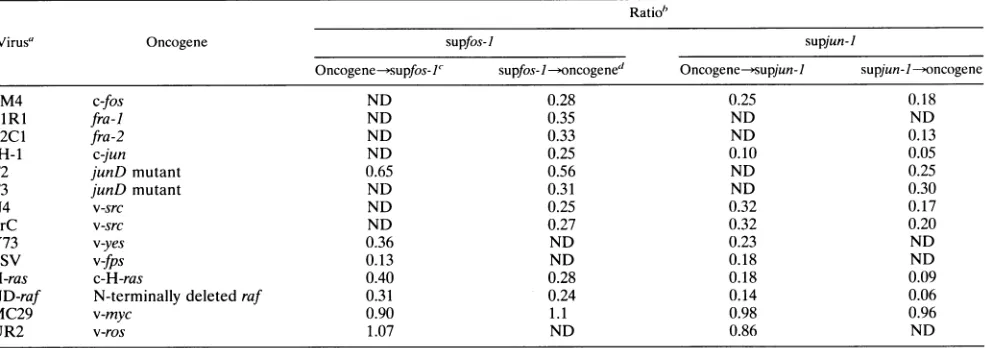
TABLE 1. Suppression of colony formation by supfos-J andsupjun-I viruses Ratio"
Virus' Oncogene supfos-I supjun-I
Oncogene-supfos-I' supfos- I-oncogened Oncogene-supjun-I supjun--/oncogene
FM4 c-fos ND 0.28 0.25 0.18
FlRl fra-] ND 0.35 ND ND
F2C1 fra-2 ND 0.33 ND 0.13
JH-1 c-jun ND 0.25 0.10 0.05
T2 junD mutant 0.65 0.56 ND 0.25
T3 junD mutant ND 0.31 ND 0.30
N4 v-src ND 0.25 0.32 0.17
PrC v-src ND 0.27 0.32 0.20
Y73 v-yes 0.36 ND 0.23 ND
FSV v-fps 0.13 ND 0.18 ND
H-ras c-H-ras 0.40 0.28 0.18 0.09
ND-raf N-terminally deleted raf 0.31 0.24 0.14 0.06
MC29 v-myc 0.90 1.1 0.98 0.96
UR2 v-ros 1.07 ND 0.86 ND
aPrC, Rous sarcoma virus Prague C; FSV,Fujinamisarcomavirus.
"The number of colonies formedbysup]un-I(A)/FM4(B)-infected CEF as shown inFig.4Dwasdivided by that formedbyDS3(A)/FM4(B)-infectedCEF(Fig.4A).
The ratio shown is the average of threeindependentexperiments.ND, notdetermined.
"Theexperimental protocolin whichtransformingvirus was infected andsupfoslwasinfected subsequently.
dTheexperimental protocol inwhichsupfosl wasinfectedfirst andtransformingvirus was infectedsubsequently.
(data notshown). The overall resultswerereproducible even when the order of introduction of the transdominant mutant andfos family oncogenewaschanged (Table 1).Forexample, whenDS3(A)-infected CEF were subsequently infected with a c-fos virus[FM4(B)],cellswereclearly transformed, assuming apolygonal cellular morphology withahigh saturation density (Fig. 3B). When CEF were initially infected with
supjun-1,
however, subsequent infection with c-fos virus did notinduce cellular transformation (Fig. 3G), and the cells exhibited a morphology similar to that of CEF infected with DS3(A) alone. These results further suggest that either of these transdominant mutants can function as ageneral inhibitor of both the Fos and Jun family proteins (AP-1).
Screening of other oncogenes whose transforming activity
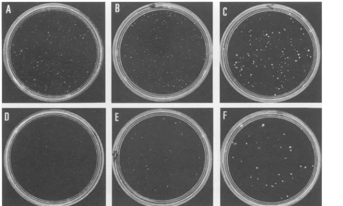
FIG. 3. Cellular morphology of doubly infected CEF. CEF were first infected with DS3(A) (A to E) orsupjun-J(A) (F to J) 4days later superinfectedwithFM4(B)(B and G), H-ras(B) (C and H),ND-raf(B)(D andI),or N4(B)(encoding v-src)(E and J) and kept under minimal essential medium containingsoft agar for5daysbeforebeing photographed.The bar corresponds to 100 ,um.
on November 9, 2019 by guest
http://jvi.asm.org/
[image:4.612.58.551.86.260.2]N1
FIG. 4. supjun-1-infected CEF are resistant to transformation by several oncogenes. CEF were first infected with DS3(A) (A to C) or
supjun-J(A)
(D to F) and 4 days later superinfected with FM4(B) (A and D), H-ras(B) (B and E), or N4(B) (C and F). At 4 days after superinfection, they were trypsinized, seeded into soft agar (60-mm-diameter plates), and propagated for 3 weeks at 38.5°C before being photographed.was suppressed by the transdominant mutants. Using these inhibitors of AP-1, we next screened CEF transformed by a
series ofnaturally isolatedorrecombinant transforming
retro-virusesto testwhether endogenousAP-1 function isessential for their transformation. Among the recombinant viruses, H-ras andND-rafwere constructed in this study and encode
the normal human Harveyc-rasgene orN-terminaltruncated
humanc-raf, respectively. Both virusestransformed CEF mod-erately, as judged from the morphological changes (Fig. 3C
andD)andtiter of focusformation[around105focus-forming units/ml forH-ras(A)orND-raf(A)].Theseresultsareingood
agreementwith those observedin murinecell lines (8, 35). As shown in Table 1 andFig. 4, supfos-1 andsupjun-1 can
efficiently suppress colony formation in soft agar caused by v-src, v-yes,v-fps, c-Ha-ras, andND-raf, though in all of these cases, suppression by
supjun-J
wasstrongerthan suppression by supfos-1. Onthe otherhand, colonies formedbyv-myc or v-roswere never suppressed by superinfectionwith either of these transdominant mutants. The reduction of anchorage-independent growth of the doubly infected CEF is in good agreementwith the reversion tothenormal cellularmorphol-ogyinmonolayercultures, someexamples of whichareshown
inFig.3(compare Fig.3CtoEwithFig.3HtoJ,respectively). In the v-src-introduced culture, however, partially refractile cells were still detectable (Fig. 3J), and their population
increased gradually when the cultures were maintained for
longer periods of time. Itis also noteworthy that CEF trans-formed by v-myc or v-ros did not revert to the normal morphologyuponsuperinfectionwith thesetwotransdominant
mutants (datanotshown).
Analysis of the AP-1 components in CEF transformed by severaloncogenes. As discussed above, wecategorized seven oncogenes into two groups by using transdominant mutants; five (v-src,v-yes, v-fps, ND-raf, and c-H-ras) require
endoge-nous AP-1 activity for their transforming activity, while two
(v-rosandv-myc)donot.Since either of thetwotransdominant mutantsused in thisstudycanfunctionas ageneralinhibitorof AP-1,wenext analyzed all known chicken AP-1 components
(c-fos,fra-2, c-jun, and
junD)
in cells transformedby several oncogenes to examine the molecular mechanisms by which AP-1 is involved in cellular transformation.Fromlogarithmically growing CEF, we prepared total cell extractsandanalyzedthe absoluteamountsofendogenous Jun andJunDbyimmunoblottingwithananti-Jun antiserum that is cross-reactive to JunD (Fig. 5A). Compared with DS3-infected oruninfected CEF, CEF transformed by v-src,v-fps, and v-rosexpressed three-tofourfoldmorec-Jun, while about twofold-greater expression was observed in v-yes-, c-Ha-ras-, and ND-raf-transformed CEF. It should be pointed out that the c-jun-transformed CEF expressed much higher levels of exogenous human c-Jun (about five times the endogenous c-Jun level). On the other hand, v-myc-transformed CEF expressedasimilar level ofc-Jun,comparabletoDS3-infected oruninfected CEF. TheJunDbandswere tooweakcompared with c-Jun bandsto allowcomparison of theexpression levels amongthese cellsquantitatively.
Foranalysis of endogenous c-Fos and Fra-2,logarithmically growingCEFweremetabolicallylabeledwith
[35S]methionine.
From cellular lysates prepared under denaturing conditions, proteins were immunoprecipitated with the Fra-2 anti-serum,whichspecifically precipitatesFra-2 (Fig.SB), and the anti-Fosantiserum,whichprecipitates allFosfamily proteins. None of thelogarithmicallygrowingCEFshowed any detect-ablec-Fosexpression (datanotshown),but basalexpressionof Fra-2wasdetectable in all cells.InCEFtransformedbyv-src,
ND-raf,
and c-H-ras, Fra-2 formeda broad band of 41 to46 kDa, compared with a sharper, thin band of 41 to 42 kDa detectedin DS3-infected CEF. Afterbacterial alkaline phos-phatase treatment of the immunoprecipitates, the protein bandsof all cells migrated as sharp 41-kDa bands, indicating thattheslowlymigratingpopulation of theproteinrepresents hyperphosphorylated forms (Fig. 5B) as we havepreviously
observed in Fra-2 immediately aftergrowth stimulation(39).
Phosphorylation of Fra-2 was most intensive in v-src-trans-formed cell and was also clear inND-raf-
or c-Ha-ras-trans-formed CEF. By comparing thedephosphorylated
form ofon November 9, 2019 by guest
http://jvi.asm.org/
e
z
e_
-*J
-I- 7 ,
x 1---,
-(4
LI- (4 44
(1) U) M) )
M W4 e0 4 co
I4
S C h4
v~ tn )Ia,~ U)t Q0 v
Xz z n X Z Q x ZZ
4W.-a6W 4ao ame 4m 4I
-BAP
.1
x,,
f,
.-
a Z46k>
40ki-C 1 2
46kor
Fr
40kp z
p* phosphorylated
Fra-2
nonphosphorylated -Ira-2
3 4
-a-2
so
*-
C-JUlFIG. 5. Expression of Jun (A and C) a
infected CEF. (A) Total cell lysate (80 V
resolved on an SDS-l0% polyacrylamide detected with anti-Jun antiserum, using the DetectionSystem. FSV, Fujinamisarcomavi triangles indicate thepositions of endogenous respectively. The arrowhead indicates thepos
CEFwerelabeled with [35S]methionine for prepared under denaturing conditions and peptide 2. Immunoprecipitates containing th activitywereincubatedwithoutorwith bacte (BAP), resolvedon anSDS-10%SDS polya
izedby fluorography afterexposureof X-ray or 12days (+BAP). (C)
supjun-J(A)-infect
DS3(A)-infected (lanes 1, 3, and 5) CEF N4(B)asshown inFig. 3JorE. After
labeling
I h, Fra-2, c-Jun, and v-Srcwere immunopr( peptide2(lanesIand2), anti-Jun (lanes 3an
5 and 6) antisera, respectively, resolved on
amide gel, and visualized by fluorography.
[image:6.612.337.520.73.269.2]+BAP
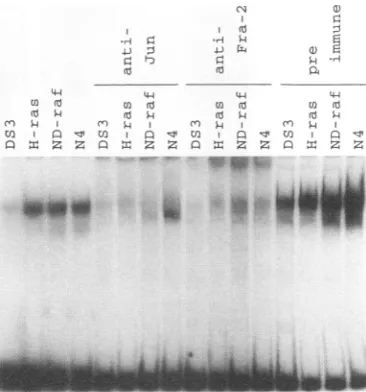
FIG. 6. Endogenous AP-l DNA-binding activity in nuclearextracts
from infected CEF. The AP-l-binding activity present in extracts
preparedfrom CEFinfected withDS3, H-ras,ND-raf,orN4(carrying
-~
>_ v-src) was determined by gel shift assays using a 32P-labeled DNA
probecontaining the AP-1-bindingsite of thecollagenase gene. Some of thenuclearextractsweretreatedwithanti-Junoranti-Fra-2peptide
1 antiserumaswell aspreimmuneserum.
Fra-2 protein, we can roughly estimate that the expression
~
* levels in CEF transformed by v-src, H-ras, and ND-raf areabout three-, two-, and twofold greater than those in DS3-infected CEF, respectively. Althoughv-myc-transformed cells
5 6 expressed lowlevelsofnonphosphorylated or
hypophosphory-lated forms of Fra-2 as in DS3-infected CEF,
v-ros-trans-formed CEF produced elevated levels of highly phosphory-lated Fra-2 asin v-src-transformed CEF (data notshown). In v-Src - theCEFtransformed byexogenouslyexpressedfra-2,theFra-2
expression levelwas increased about sevenfold(Fig. 5B).
When supjun-l-infected CEF were superinfected with N4
(Fig. 3J), the cellsweremetabolically labeled with [35S]methi-n onineand Fra-2 and Jun expressionwasanalyzed by
immuno-precipitation. The results indicated that expression levels of Fra-2 and Jun aswell asthe extent ofFra-2 hyperphosphory-lationwerenotaffectedby theexpression of supJun-1 (Fig. 5C;
compare lane 1 and 2 or lanes 3 and 4), indicating that
supJun-1 suppresses the transformation not by affecting the and Fra-2 (B and C) in componentsofendogenousAP-1.We have alsoobserved that
Lg of protein each) was
supjun-1
doesnotaffectexpression of theexogenousv-srcgenegel, and proteins were (Fig. 5C;compare lane 5 and 6).
e ECL Western Blotting We next determined the AP-1 DNA-binding activity in
irus. Theopenandclosed v-src-,ND-raf-,and c-Ha-ras-transformed CEF, usinggelshift
chickenc-JunandJunD, analysis of nuclear extracts (Fig. 6). When a DNA probe
itionof humanc-Jun.(B)
containing
theAP-1 site from the humancollagenase
genewas treated with anti-Fra-2 used, DS3-infected CEF generated a thin band of gel shiftiesameamount of radio- complex,whilegreatlyenhancedbands withthe samemobility rialalkaline phosphatase wereobserved whenCEFexpressingv-src,ND-raf,orc-Ha-ras
crylamide gel,andvisual- wereused. All of theshifted bands disappeared uponaddition
films for 32days (-BAP) of either the anti-Jun or the anti-Fra-2 antiserum but were
ed (lanes 2, 4, and 6) or insensitive to a preimmune serum. The apparently increased
were superinfected with AP-1-binding activity in the presence ofpreimmune serum is
gcwitha[dwS]methaoninefor
possibly
attributable to a carrier effect of increasedprotein
Id
4),andanti-v-Src (lanes concentration.This result indicates that theFra-2-c-Jun
com-an
SDS-10%-S
polyacryl- plex ismainly responsible for the band shifts and also for theenhancement of DNA-binding activity in these transformed cells.
A
I _
44
a) (10
co Q
Q x z
B
on November 9, 2019 by guest
http://jvi.asm.org/
[image:6.612.55.295.80.528.2]a z
44
U) l
0
IQ .0
C~
c-fos
fra-2
om<
am
c-jun
jun-D
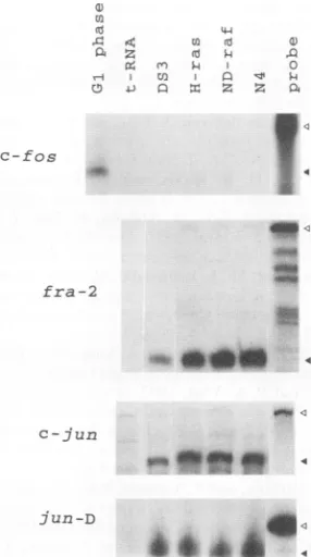
FIG. 7. Expressionlevels of mRNAs that encode AP-1components
ininfected CEF. Levelsoffos,fra-2,
c-jun,
andjunDtranscriptsin CEF infected withDS3, H-ras,ND-raf,and N4 (carrying v-src)viruseswere analyzed byRNaseprotectionassay.Open trianglesindicatepositionsof untreatedprobes;closedtriangles correspondtopositionsofprobes protected bythetranscripts. Sampleswereanalyzedina3.5%(fos,jun,
andfra-2 transcripts) or5% (forjunD) polyacrylamide gel.
Elevated levels of Fra-2 and Jun proteins result from regulation at the transcriptional level. CEF transformed by
v-src, ND-raf, and c-Ha-ras expressed elevated levels of Fra-2
and c-Jun proteins. We next tested the expression levels of c-fos, fra-2, c-jun, and junD mRNAs by RNase protection
assay.As shown inFig. 7, c-fosmRNAwasundetectable in all
of the logarithmically growing cells, while growth-stimulated CEF expressed it clearly. The fra-2 and c-jun mRNA levels
were elevated two- to fourfold in v-src, ND-raf-, or
c-Ha-ras-transformed cells. In RNase protection assays, intactness and quantity of theRNAsamplesweretestedbyethidium bromide staining of the gelorbyNorthern blot analysis using aprobe
for a housekeeping gene encoding GAPDH. This result
sug-gest that the enhancement of Fra-2 and c-Jun expression in these cells is mainly regulated at the transcriptional level. Although junD mRNA expression in v-src-transformed cells
wasslightly higher, the junDmRNA levelsweresimilaramong
other cells.
DISCUSSION
Although supFos-1 was initially designed as a specific
sup-pressorofJunfamily proteins by forminganonfunctional and
stable complexofsupFos-1 and Jun(28),wehave shown here
that itcanalsoefficientlysuppress the transformation induced by fos family proteins. Since heterodimer formation with endogenous c-Jun is thought to be essential for the transfor-mation by exogenously expressed Fos family proteins (38), supfos-1 would function as a competitive inhibitor of
exoge-nousFosinthiscase.Likesupfos-l,supjun-J suppressedeither
fos orjun transformation, and it is noteworthy that
supjun-]
was a morepotent suppressorof eitherfosorjun transforma-tion(Table 1).This strongersuppression mightbe attributable to the direct blocking of transcription by the AP-1 DNA-binding activity of supJun-1. On the other hand, non-DNA-binding heterodimers formed by supFos-1 are passivelyre-moved from the transcriptional system. supJun-1 forms homodimers and heterodimers with c-Jun, as shown in vitro (Fig. 1), and stable heterodimericcomplexeswith Fra-2were
detected in CEF (our
unpublished
result). We would expect onlyaverylimitedtransactivationactivity
inallof these homo-or heterodimers, because the in vitrotranscriptional
analysis
indicated that a similar truncated Jun showed onlymarginal
transcriptional activity even in combination withfull-length
Fos (1). supJun-1 shares some properties with A9 (20) and Tam-67 (6),whichwere previously reportedastransdominant mutantsofJunthat lacktransactivation domains. Both of the mutants were shown to have inhibitory effects on AP-1-mediated transcription in transient expression systems. Our model of the inhibition mechanism favors the
quenching
mechanism proposed byBrown et al. (6).We found that cellular transformation induced by such oncogenesassrc,
frs,
yes, ras,andraf
wassuppressed byboth of the transdominantmutants.Therefore,we decided toanalyze the amounts of all known chicken AP-1 components(c-Fos,
Fra-2, c-Jun, andJunD) in transformed cells inducedbyeach of 13oncogenes.Theexpression
level ofeitherFra-2orc-Jun inthesetransformed CEFwasabouttwo to four timesthatof the untransformed CEF, but we think that the elevation of either protein alone is not sufficient to cause the cellular transformation; the amounts ofexogenously expressed Fra-2 andc-Jun that induce cellular transformationareabout sevenandfive times,
respectively,
that of theendogenous protein
in infected CEF. Elevation of both proteins, however, would contribute synergisticallyto the enhancement ofAP-l DNA-binding activity (Fig. 6) andpossibly
also to the induction of cellular transformation. Although we did not detect elevatedexpression
from a reporterplasmid containing
asingle
AP-1-binding
site in these transformedCEF,
three- to fivefold elevation was detected when a reporter with three tandem repeats of an AP-1 site was used instead (our unpublished results).Cellular transformation induced by v-ros or v-myc was not
affectedbyeitherof the transdominantsuppressors,
suggesting
that endogenous AP-1 function is not essential for their transforming
activity.
It isinteresting
that the nuclearonco-gene v-mycand the gene
encoding
thetransmembranetyrosine
kinasev-Rosshow cleardifferencesintheir induction levels of AP-1 components. v-myc induced no change in theendoge-nous AP-1 components, indicatingthat the v-myc transforma-tion pathway is independent of AP-1. v-ros-transformed cells haveelevatedlevelsofc-Jun,
although
these enhancementsarenotessential for cellular transformation. Sincev-ros encodesa
transmembrane
tyrosine
kinase and is ahomolog
of the sevenlessgene,itmightbeexpectedtoinfluence therasandraf
signalling pathways. Therefore,
we think that there is another pathway that alone is sufficient for theros transformation.Although in thisstudywe did not address the
phosphoryla-tion status of the c-Jun
protein
invivo,
several reports have indicated that phosphorylation of amino-terminal serines 63 and 73 of c-Junaccompanies
the transformation of murine cells inducedby
v-src, activated c-Ha-ras, and activatedraf-1,
and itwasalso shown that
phosphorylation
isimportant
forratfibroblasttransformation induced
by
high-level
expressions
of both c-jun and activated c-Ha-ras(2, 3,
34).
Two lines of evidence,however,
indicatethatthesefindings
arenotdirectly
W"
400
"
fo
e<
604A 4
on November 9, 2019 by guest
http://jvi.asm.org/
[image:7.612.117.260.79.335.2]applicable to the chicken cell system. First, the N-terminal sites are apparently constitutively phosphorylated even in resting CEF and are only modestly affected by mitogenic stimulation. Second, a c-Jun mutant in which the N-terminal phosphoryla-tion site is changed from a serine to an alanine residue retains the full activity to transform CEF as a single gene (24).
Hyperphosphorylation of Fra-2 proteins, involving a mobil-ity change, was detected in v-src-, c-Ha-ras-, and
ND-raf-transformed CEF. We previously reported similar phosphory-lation of Fra-2 immediately after growth stimulation (39). In CEF, both nonphosphorylated (40-kDa) and phosphorylated (41- to 46-kDa) Fra-2 can be coimmunoprecipitated with c-Jun, and the resultant heterodimers seem to have equal specific DNA-binding activity. The 40-kDa Fra-2 protein syn-thesized in reticulocyte lysates also retains efficient dimer-forming activity with c-Jun and binding activityto AP-1 sites. Although we wouldexpectthatphosphorylation contributesto the enhancement of transactivation activity, the effect of phosphorylation on transcriptional control, as well as the protein kinases responsible for Fra-2 phosphorylation, remains tobe established.We would like to point out that these two transdominant mutants have the particular advantage that they are down-stream in the signal transduction pathways (2), and both exhibitthe blockingofcellulartransformationcausedbyawide rangeof oncogenes. Since supFos-1 and supJun-1 have onlya modest effect on normal growth, we are now introducing these genes into primary cells to look for a differentiation process that involvesendogenousAP-1 activation.
ACKNOWLEDGMENTS
Wegratefully acknowledgefruitful discussions with A. Nomoto. We thank T. Curran and P. Vogt for supplying pSP-fra-1 and pJun, respectively.WethankT.Yoshidaand H. Okuno for theconstruction and characterizationofFlRlvirus.Wealso thank JCRB forsupplying pSPT-Hras and p627. We thank Y.Suzuki,E.Suzuki,and N. Masuda for assistance inpreparationof the manuscript.
This work was supported by a research grant from the Princess Takamatsu CancerResearchFund andbyaGrant-in-AidforScientific Research from the Ministry ofEducation, Science and Culture of Japan. M. H. Sonobe is supported by a Fundacao de Amparo a
Pesquisado Estado deSaoPaulopostdoctoral fellowship. REFERENCES
1. Abate, C., D. Luk, E. Gange, R. G. Roeder, and T. Curran. 1990. Transcriptional regulation by Fos and Jun in vitro: interaction amongmultipleactivator and regulatory domains. Mol. Cell. Biol. 11:3624-3632.
2. Angel, P., and M. Karin. 1991. The role of Jun, Fos, and the AP-1 complexin cell-proliferation and transformation. Biochim. Bio-phys.Acta1072:129-157.
3. Binerty, B., S. Tod, and M. Karin. 1991. Ha-Ras augments c-Jun activity and stimulates phosphorylationofits activation domain. Nature (London)351:122-127.
4. Bohmann,D., T. J. Bos, A. Admon, T. Nishimura, P. K. Vogt, and R. Tjian. 1987. Human proto-oncogene c-Jun encodes a DNA binding proteinwith structure and functional properties of tran-scriptionalfactor AP-1.Science 238:1386-1392.
5. Bonner, T., S. B. Kerby, P.Sutrave, M. A. Gunnell, G. Mark, and U.Rapp. 1985. Structure and biological activity of human homolog of therafimiloncogene. Mol. Cell. Biol. 5:1400-1407.
6. Brown, P., H. R. Alani, L. H. Preis, E. Szabo, and M. J. Birrer. 1993.Suppressionofoncogene-induced transformation by a dele-tionmutantof c-jun.Oncogene 8:877-886.
7. Catling,A.D., J. A. Wyke, and M. C. Frame. 1993. Mitogenesis of quiescentchick fibroblasts by v-Src: dependence on events at the membraneleading to early changes in AP-1. Oncogene 8:1875-1886.
8. Chang,E.H.,M. E.Furth,E. M.Scolnick,andD.R.Lowy. 1982.
Tumorigenic transformation of mammalian cells induced by a normal human gene homologous to the oncogene of Harvey murine sarcoma virus. Nature (London) 297:479-483.
9. Cohen, D. R., and T. Curran. 1988. Fra-1: a serum-inducible, cellularimmediate-early gene that induces a Fos-related antigen. Mol. Cell. Biol. 8:2063-2069.
10. Curran, T., C. VanBeveren, N. Ling, andI. M. Verma. 1985. Viral and cellularfos proteins are complexed with a 39,000-dalton cellular protein. Mol. Cell. Biol. 5:167-172.
11. Finkel, M. P., 0. B. Biskis, and P. B. Jinkins. 1966. Virus induction ofosteosarcomas in mice. Science15:1698-1701. 12. Fujiwara, K., K. Ashida, H. Nishina, H. Iba, N. Miyajima, M.
Nishizawa, and S. Kawai. 1987. Thechicken c-fos gene: cloning andnucleotidesequence analysis. J. Virol. 61:4012-4028. 13. Granger-Schnarr, M., E. Benusiglio, M. Schnarr, and P.
Sassone-Corsi. 1992.Transformation and transactivation suppressor activ-ity ofthe c-Jun leucine zipper fused to a bacterial repressor. Proc. Natl. Acad. Sci. USA 89:4236-4239.
14. Hartl, M., J. T. Hutchins, and P. K. Vogt. 1991. The chicken junD geneand its product. Oncogene 6:1623-1631.
15. Hartl, M., and P. K.Vogt. 1992. Arearranged JunD transforms chicken embryofibroblasts. Cell Growth Differ. 3:909-918. 16. Hirai,S.-I., R.-P. Rysek, F. Mechta, R. Bravo, and M. Yaniv. 1981.
Characterization ofjun D: a new member of the jun proto-oncogenefamily. EMBO J. 8:1433-1439.
17. Iba,H., Y. Nishina, and T. Yoshida.1988.Transforming potential and growth stimulatingactivity of the v-fos and c-fos genes carried by avianretrovirusvector. Oncogene Res. 2:121-133.
18. Kameda, T., A. Akahori, M. H. Sonobe, T. Suzuki, T. Endo, and H. Iba. 1993. JunD mutantswithspontaneouslyacquired transform-ing potential have enhancedtransactivating activity in combina-tion with Fra-2. Proc. Natl. Acad. Sci. USA90:9369-9373. 19. Ledwith, B. J., S. Mana, A. R. Kraynak, W. W.Nichols, and M.0.
Bradley.1990.Antisense-fos RNAcausespartial reversion of the transformed phenotypesinduced by the c-Ha-ras oncogene. Mol. Cell. Biol. 10:1545-1555.
20. Lloyd, A., N. Yancheva, and B. Wasylyk. 1991. Transforming suppressor activity of a Jun transcriptional factor lacking its activation domain.Nature (London)352:635-638.
21. Maki, Y., T. J. Bos, C. Davis, M. Starbuck, and P. K. Vogt. 1987. Avian sarcoma virus 17carriesthejun oncogene. Proc. Natl. Acad. Sci. USA84:2848-2852.
22. Maniatis, T., E. F. Fritsch, and J. Sambrook. 1989. Molecular cloning: a laboratory manual. Cold Spring Harbor Laboratory, Cold SpringHarbor, N.Y.
23. Matsui, M., M. Tokuhara, Y. Konuma, N. Nomura, and R. Ishizaki. 1990. Isolation of human fos-related genes and their expression during monocyte-macrophage differentiation. Onco-gene5:249-255.
24. Metrivier, C., F. Piu, C. M. Pfarr, M. Yaniv, L.Loiseau, and M. Castellazzi. 1993. Invitro transformingcapacities of mouse c-jun: junDchimeric genes. Oncogene 8:2311-2315.
25. Nakabeppu, Y., K.Ryder, and D. Nathans. 1988. DNAbinding activities of three murine Jun proteins: stimulation by Fos. Cell 55:907-915.
26. Nishimura, T., and P. K. Vogt. 1988. The avian cellular homolog of theoncogenejun. Oncogene 3:659-663.
27. Nishina, H., H. Sato, T. Suzuki, M. Sato, and H. Iba. 1990. Isolation and characterization offra-2, anadditional member of thefosgenefamily. Proc. Natl. Acad. Sci. USA 87:3619-3623. 28. Okuno, H., T. Suzuki, Y.Hashimoto, T. Curran, and H. Iba. 1991.
Inhibition ofjun transformation by a mutated fos gene: design of ananti-oncogene. Oncogene6:1491-1497.
29. Panabieres, F., M. Piechaczyk, B. Rainer, C. Dani, P. Fort, S. Riaad, L.Marty, J. L. Imbach, P. Jeanteur, and J. M. Blanchard. 1984. Complete nucleotide sequence of the messenger RNA coding for chicken muscleglyceraldehyde-3-phosphate dehydro-genase. Biochem.Biophys. Res. Commun. 118:767-773. 30. Powers, S., T. Kataoka, 0.Fasano, M. Goldfarb, J. Broach, and
M.Wigler. 1984. Genesin S. Cerevisiae encoding proteins with domains homologous to themammalian ras proteins. Cell 36:607-612.
31. Riabowol, K. T., J. Vosatoka, E. G. Ziff, N. J. Lamb, and J. R.
on November 9, 2019 by guest
http://jvi.asm.org/
Feramisco. 1988. Microinjection of fos-specific antibodies blocks DNAsynthesis in fibroblast cells. Mol. Cell. Biol. 8:1670-1676. 32. Ryder, K., A. Lanahan, E.Perez-Albuerne,andD.Nathans.1989.
Jun-D:athirdmember ofthe Jungenefamily. Proc.Natl. Acad.
Sci.USA 86:1500-1503.
33. Ryder, K., and D. Nathuns. 1988. Induction ofprotooncogene
c-jun by serum growth factors. Proc. Natl. Acad. Sci. USA 85:8464-8467.
34. Smeal, T., B. Binetruy,D.Mercola, M.Birrer, and M. Karin.1991. Oncogenicandtranscriptional cooperation with Ha-Ras requires phophorylation of c-Jun onserines 63 and 73. Nature(London) 354:494-496.
35. Stanton, V. P., Jr., D. W. Nichols, A. P. Laudano, and G. M. Cooper. 1989. Definition ofthe humanraf amino-terminal
regu-latory region bydeletion mutagenesis. Mol. Cell. Biol. 9:639-647. 36. Suzuki, T., Y. Hashimoto, H. Okuno, H. Sato, H. Nishina, and H. Iba. 1991. High-level expression of human c-jun gene cause
cellular transformation of chicken embryo fibroblasts. Jpn. J. CancerRes. 82:58-94.
37. Suzuki, T., H. Okuno, T. Yoshida, T. Endo, H. Nishina,andH. lba.
1991. Difference in transcriptional regulatory function between c-Fos and Fra2.Nucleic Acids Res. 19:5537-5542.
38. Wick,M., F. C. Lucibello, and R. Muller. 1992. Inhibition of Fos-and Ras-induced transformation by mutant Fos proteins with structuralalterations infunctionallydifferentdomains.Oncogene 7:859-867.
39. Yoshida, T., H. Sato, and H. Iba. 1991. Transcription offra-2 mRNA and phosphorylation ofFra-2protein are stimulatedby serum.Biochem.Biophys.Res.Commun. 174:934-939.
40. Yoshida,T., Y. Shindo, K. Ohta, and H. lba. 1989. Identification of
a small region of the v-fos gene product that is sufficient for transforming potential and growth stimulating activity.Oncogene Res. 5:79-89.
41. Yoshida, T., T. Suzuki, H. Sato, H. Nishina, and H. Iba. 1993. Analysis offra-2 gene expression. NucleicAcids Res. 21:2715-2721.
42. Zerial, M., R. Toschi, R. P. Ryseck, M. Schuermann, R. Muller, andR. Bravo. 1989. The product ofanovelgrowth factoractivated gene, fosB, interacts with Jun proteins enhancing their DNA
binding activity. EMBO J. 8:805-813.