Copyright ©D 1994,American Society for Microbiology
Identification of
Human
T-Cell Lymphotropic Virus
Type
I
21-Base-Pair
Repeat-Specific and Glial Cell-Specific
DNA-Protein Complexes
MARIBETHTILLMANN, RENEEWESSNER, ANDBRIANWIGDAHL*
Department ofMicrobiology and Immunology, College ofMedicine, The Pennsylvania State University, Hershey,Pennsylvania 17033
Received 7 October 1993/Accepted 20 April 1994
The human T-cell lymphotropic virustype I (HTLV-I)-encoded protein, Tax, iscapableof trans-activating HTLV-Itranscriptionbyinteracting with specificsequencesin theHTLV-I longterminalrepeat (LTR) which compriseaninducible enhancer containing three imperfect tandemrepeatsofa21-bp sequence.Thereis no
evidencethatpurified TaxcanbindtoDNAintheabsenceof cellularfactors, suggesting that Taxmostlikely regulatestranscriptionvia interaction with cellular factors. Since HTLV-I isadocumentedagentofadult T-cell leukemia and tropical spastic paraparesis, disorders of the immune and nervous systems, respectively, characterization of cellular factors of lymphoid and neuroglial origin which interact with the 21-bp repeat
elementsisessentialtounderstanding ofthe mechanismsinvolved in basal and Tax-mediated transcription in cells of immune and nervoussystemorigin. Utilizing electrophoretic mobilityshift (EMS) analyses,wehave
detected both21-bprepeat-specificandglialcell-specificDNA-proteincomplexes.Several 21-bprepeat-specific DNA-protein complexesweredetected when nuclearextracts derived from cells of lymphoid (Jurkat, SupTl, and H9), neuronal (IMR-32 and SK-N-MC), and glial (U-373 MG, Hs683, and U-118) origin wereused in
reactions with eachofthe three21-bp repeatelements. In addition,aglialcell-specific DNA-protein complex wasdetected whennuclearextractsderived from U-373 MG, Hs683, and U-118 glial cell linesreactedwiththe promoter-distal and central 21-bp repeat elements. Furthermore, EMS analyses performed with nuclear extracts derived from lymphocytic and glial cell origin and a223-bp fragment oftheHTLV-I long terminal
repeatencompassingthe three21-bprepeatelements (designatedTax-responsiveelements 1 and2,TRE-1/-2) have also resulted in the detection of glial cell type-specific DNA-protein complexes. Competition EMS analyseswitholigonucleotides containingtranscription factor bindingsitesequencesindicate theinvolvement
ofacyclicAMPresponseelementbinding proteininthe formation ofDNA-protein complexes whichformwith all three21-bp repeatelements and theglial cell-specific DNA-protein complexaswellasthe involvement of
Splor an Spl-related factorintheformation ofthe21-bp repeatIII-specific DNA-protein complexes. Human T-cell lymphotropic virus type I (HTLV-I) is a
documented etiologic agent of both hematologic and
neuro-logic disorders. Specifically, HTLV-I has been determinedto
beanetiologicagentofboth adult T-cell leukemia andaslowly
progressive neurologicdisorder, tropical spastic paraparesis (3, 18, 40, 46, 50, 51). However, theviral and hostfactorsinvolved in determining the ultimate outcome of HTLV-I infection (malignancyversusneurologic dysfunction) remain tobe elu-cidated. While the interaction of HTLV-I with cells of lym-phoid origin has beenexamined extensively, relativelylittleis knownconcerningtheinteraction of HTLV-I with cells resid-inginthecentral nervous system.
Any aspect of the retroviral life cycle, including entry,
reversetranscription, integration, transcription,andassembly, may impact on the oncogenic and/or neuropathogenic pro-cesses associated with virus infection. Furthermore, these
processes maybe highly dependenton interactionsoccurring
between retrovirus- and hostcell-specific components. Subse-quent to viral entry, the outcome of HTLV-I infection in a
given celltypewithin either the immuneornervous system is critically dependent on cellular factors that interact with retroviralsequencesinvolved intranscriptional regulation.The integrated retroviralgenome is flanked at both endsby
non-coding long terminal repeat (LTR) sequences composed of
*Corresponding author. Fax: (717) 531-5580. Electronic mail ad-dress:[email protected].
threeregions, U3, R, and US (Fig. 1).The LTRs areintegral
componentsof the viralregulatorysystemcontaining informa-tionessential to the regulationofretroviral integration, tran-scription,and replication.
Evidence demonstrating that the LTR sequences of some
retroviruses play a role in tissue and cell type specificity and
may also be involved in determining the course of disease
associated with retroviral infection has been presented (7, 8, 31, 41).Use of the murine retrovirussystemhassuggestedthat the in vitro hostrangeof selected murine leukemia viruses is determined bythe retroviral LTR, specifically the U3 region (7, 8, 31, 41).Forseveral murineretroviruses,minorvariations in theproviralgenome,particularlythe LTRorenvelopegene,
result in alterations in the cellular tropism of the retrovirus and,inturn,in itspathogenicity (7, 31, 37, 41).Inaddition,the promoters of several retroviruses, including Moloney and Friend murine leukemia virusesand humanimmunodeficiency virus, have been implicated in cell type-specific expression (7-9, 31, 41).Forexample,whentransgenicmicewere
gener-atedbyutilizingtheLTRs fromeither centralnervous system-derived or T-cell-derived human immunodeficiency virus strains, expression ofthe reportergenewas detected only in
mice transgenicfor thecentral nervous system-derived LTRs
(9). Furthermore, in support of the idea thatthe LTR has a
role intissuetype-orcelltype-specific expressionofHTLV-I, the generation of transgenic mice bearing a transgene
com-posedof theHTLV-I LTR isolatedfromapatientwithtropical
4597
on November 9, 2019 by guest
http://jvi.asm.org/
A.
|LTR|
GAG
||
POL
||
ENV
I ~~U3
a,-IfA,
.300 .20§ 100 +1
m
TAT^AA
IJJ3* sJLTRI
B.
21
bp Tax-responsive
elements
I, II,
III
I
AIAGGC
C[rGACGCTCrCCC('?
+225 .404 IITIAGCGCICdTGACG
YGTC'YCCCrHI
CjACGC.GT[rGACGWCAAFCCC(T
21 bpTax-responsiveelements
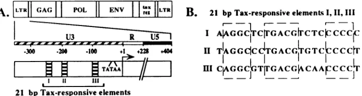
FIG. 1. (A)StructureoftheHTLV-ILTRinthecontextof theviralgenome.The21-bprepeatelementsarelocated within the U3regionof the LTRatpositions -251to-231, -203to-183, and -103to -83relativetothetranscriptionalstartsite.(B) Sequence comparisonof the three21-bp repeatelements.Theregionsofstrict conservation amongthe21-bprepeatelementsareindicatedbythe broken boxesrepresenting (fromlefttoright) domains A, B,and C.
spastic paraparesis linkedto areporter gene demonstrated that LTR-directed expression of the reporter gene occurred pri-marily in cells of the central nervous system (20).
With respect to HTLV-I, virus-specific transcription is de-pendent on cis-acting enhancer sequences comprising three 21-bp imperfectly repeated elements located within the U3
regionof the HTLV-I LTR atpositions -251 to -231, -203 to -183, and -103 to -83relativetothe transcriptionalstart site(Fig. 1) (38, 43). Immediately following virus infection,low but necessary levels of HTLV-I transcriptionwhich are
criti-cally dependent on cellulartranscriptionfactors areattained. Basal levels of transcription are greatly enhanced by the virus-encoded regulatory protein, Tax, which mediates trans activation of the HTLV-I LTR through the 21-bp repeat elements (14, 42, 44, 46, 47). As with basal transcription,
Tax-mediated transcriptional trans activation of the HTLV-I LTR requires the participation of cellular intermediaries (16,
19, 38, 46,48,53). Mutagenesis studies have shown that at least two21-bp repeat elementsarenecessaryforefficient
transcrip-tionalactivation(14,39,45,46).Anumber ofproteins,of both cellular and viral origin, which interact either directly or indirectly with sequences in the HTLV-I LTR have been identified (see references 33 and46forreviews).However,the proteins in these studieswere identified by utilizing cell lines derived from sources other than the nervous system. Because the interaction of host cellular factors with the 21-bp repeat elements and possibly other regions of the HTLV-I LTR is critical for basal andTax-mediated expression, further identi-fication and characterization of the cellular factors ofimmune systemandnervoussystemorigins which interact with the LTR regulatory unit is essential to understanding of the molecular mechanisms involved in HTLV-I LTR-directed transcription in these cellpopulations.
While each of the 21-bp repeatelements has three strictly conserved domains, termed A, B, and C (Fig. 1) (46), these domainscomprise only 13 ofthe 21 base pairs. Studies have suggested, utilizing the DNase I protection assay, that there is differential binding of cellular factors to each of the three 21-bp elements (36). Differential binding of cellular factors to the enhancer elements, in turn, may play a role in basal and Tax-mediatedHTLV-ILTR-directed transcription. Therefore, characterization of the DNA-protein complexes formed be-tweencellularfactors and each of the 21-bp repeat elements is warranted. In addition, identification of tissue type- or cell
type-specific factors which interact with these elements may
provide important information concerning the regulation of HTLV-I gene expression in cells of immune versus nervous system origin. In an effort to characterize the interaction of cellularfactors with each of the three 21-bp repeat elements,
wehaveutilized double-stranded oligonucleotides homologous to each of the 21-bp repeat elements as well as a native fragment of the HTLV-I LTR containing the three 21-bp
repeatelements(designatedTax-responsive elements 1 and2, TRE-1/-2) in reactions with nuclear extracts derived from selected cell lines oflymphocytic, neuronal, and glial originsin
electrophoretic mobility shift (EMS) analyses. Both 21-bp repeat-specific DNA-protein complexes and a glialcell
type-specific DNA-protein complex were detected, whose forma-tion mayinvolveSplor anSpl-relatedfactor andacyclicAMP response element (CRE) binding protein (CREB)-activating
transcription factor (ATF) family member(s), respectively.
These studies also indicate that the three 21-bp repeat ele-ments are not functionally equivalent with respect to DNA-protein complex formation.
MATERMILSAND METHODS
Cells. Human T-cell
lymphocytic
cell lines utilized in these studies include Jurkat(ATCCTIB152), SupTl (provided byF.Gonzalez-Scarano,Departments ofNeurology and Microbiol-ogy, University of Pennsylvania, Philadelphia, Pa.), and H9
(Thomas Folk, Retrovirus Disease Branch, Centers for Dis-ease Control and Prevention, Atlanta, Ga.). All T-cell lines werecultured andmaintained at 37°C with 5%CO2in RPMI 1640 medium. Human neuronal cell lines utilized include IMR-32 (ATCC CCL 127) andSK-N-MC (ATCC HTB 10).
Neuronal cell lineswere cultured and maintained at 37°C in Eagle minimal essential medium buffered with
N-2-hydroxy-ethylpiperazine-N'-2-ethanesulfonic acid (HEPES). Human glial cell lines utilized in these studies include U-373 MG
(ATCC HTB 17), Hs683 (ATCC HTB 138), and U-118
(ATCCHTB 15).Glial cell lines were cultured and maintained at 37°C in Eagle minimal essential medium. All medium formulations were supplemented with 10% heat-inactivated fetal calf serum, penicillin (100 U/ml), streptomycin (100
,ug/ml), 2mM L-glutamine, and 0.075%NaHCO3.
Nuclear extractpreparation.Nuclear extracts of all cell lines were prepared as described previously (11), with minor mod-ifications.Briefly, cultured cells were collected and nuclei were isolated with hypotonic buffer (10 mM HEPES [pH 7.9, 4°C], 1.5 mM MgC12, 10 mM KCl, 0.2 mM phenylmethylsulfonyl
fluoride, 0.5 mM dithiothreitol) in a Dounce homogenizer
(type B) at4°C. Nuclear proteins were extracted at 4°C with high-salt buffer (20 mM HEPES [pH 7.9,4°C], 25% glycerol,
1.5 mM MgCl2, 1 M KCl, 0.2 mM EDTA, 0.2 mM
phenyl-methylsulfonyl fluoride, 0.5 mM dithiothreitol) and dialyzed
against50volumes ofdialysis buffer (20 mM HEPES [pH 7.9,
4°C], 20% glycerol, 100 mM KCl, 0.2 mM EDTA, 0.2 mM
on November 9, 2019 by guest
http://jvi.asm.org/
[image:2.612.138.497.81.177.2]phenylmethylsulfonyl fluoride, 0.5 mM dithiothreitol) for 5 h at 4°C. Precipitated proteins and debris were removed by
centrifugation at 21,000 x g for 30 min at
4°C.
Protein quantitation of nuclear extracts was performed by the Bio-Rad protein assay (based on the Bradford dye-binding procedure [5]). Extracts were separated into aliquots, frozen in liquid nitrogen, and stored at -80°Cat a concentration of 3 mg/ml. Oligonucleotide synthesis and radiolabeling. Complemen-tary single-stranded oligonucleotides homologous to each of the three HTLV-I 21-bp repeat elements were synthesized(Macromolecular Core Facility, The Pennsylvania State Uni-versity College of Medicine) and annealed by heating for 10 min at 90°C and cooling to room temperature. For EMS analyses examining the role of flanking DNA in DNA-protein complexformation, double-stranded oligonucleotides contain-ingsequences homologous to the 21-bp repeat elements with native flanking sequences, with restriction endonuclease site-flanking DNA sequences, or without site-flanking DNA sequences were synthesized. The sequences of the full-length, double-stranded oligonucleotides were as follows: the three 21-bp repeat elements flanked by native HTLV-I LTR sequences,
5'-AGACTAAGGCTCTGACGTCTCCCCCCAGAGG-3'
(I FS), 5'-CAGGCTAGGCCCTGACGTGTCCCCCTGAA
GA-3' (II FS), and5'-GCCCTCAGGCGTTGACGACAACC
CCTCACCT-3' (III FS); the three 21-bp repeat elements alone with no flanking DNA sequences, 5'-AAGGCTCTGA
CGTCTCCCCCC-3' (I), 5'-TAGGCCCTGACGTGTCCCC
CT-3' (II), and 5'-CAGGCGTTGACGACAACCCCT-3'
(III);
and the three 21-bp repeat elements flanked by irrelevant DNAsequences in the form of restriction endonuclease sites BamHI and HindIII, 5'-GATCCAAGGCTCTGACGTCTC
CCCCCAAGCT-3' (I RS), 5'-GATCCTAGGCCCTGACGT
GTCCCCCTAAGCT-3' (II RS), and 5'-GATCCCAGGCG
TTGACGACAACCCCTAAGCT-3'
(III RS) (each 21-bpre-peat is flanked by the restriction endonuclease sequences for
BamHI [5' end of the 21-bp repeat] andHindIl [3' end of the 21-bp repeat]). For mutational analyses of 21-bp repeat III, a synthetic oligonucleotide (21-bp repeat IIIAII, 5'-GCCCT
CAGGCGTTGACGTGTCCCCCTCACCT-3')
in which fournucleotides between conserved domains B and C were changed to the corresponding nucleotides in 21-bp repeat II was synthesized. The 21-bp repeat I corresponds to nucleotides -251 to -231 (promoter distal), 21-bp repeat II corresponds to nucleotides -203 to -183 (central), and 21-bp repeat III corresponds to nucleotides -103 to -83 (promoter proximal) with respect to the transcriptional start site (Fig. 1) (38, 43).
Transcription factor binding site oligonucleotides used for competition EMS analyses include (i) a CRE oligonucleotide,
5'-GATTGGCTGACGTCAGAGAGCT-3',
and (ii) an Splbinding siteoligonucleotide, 5'-GATCGATCGGGGCGGGG CGATC-3' (Stratagene). The oligonucleotides with 5' exten-sions were labeled with
[a.-32P]dCTP
and the Klenow fragment of DNA polymerase I. The blunt-ended double-stranded oli-gonucleotides were end labeled with[y-32P]ATP
and T4polynucleotide kinase.
Generation of the 223-bp fragment of the HTLV-I LTR encompassing the three 21-bp repeat elements (TRE-1/-2).
PCR amplification wasutilized to generate a DNA fragment, TRE-1/-2, from the HTLV-I LTR, extending from the ApaI
restriction endonuclease site at position -268 to the NdeI
restriction endonuclease site at position -46 relative to the transcriptional start site. The TRE-1/-2 fragment contains the three 21-bp repeat elements and intervening sequences in the context oftheir nativepositions within the HTLV-I LTR and comprisessequences previously referred to as the Tax-respon-siveelements TRE-1 and TRE-2. The two PCR primers were
homologous to sequence -268 to -249 (5' primer) and to sequence -65 to -46 (3' primer). After PCR amplification, the resulting 223-bp fragment was end labeled with
[-y-32P]ATP
and T4 polynucleotide kinase.EMS analyses. Standard EMS analyses were performed essentially as described by Gamer and Revzin (17). Approxi-mately 50,000 cpm of radiolabeled double-stranded oligonu-cleotide (0.1 to 1.0 ng) was used in a reaction with 12 jig of protein and 2 jig of
poly(dI-dC)
in a total reaction volume of 15RI
for 30min
at30°C.
All components were simultaneously mixed by brief centrifugation. For preincubation EMS analy-ses, the nuclear extracts were incubated with the 2,ug
ofpoly(dI-dC)
for 10min
at room temperature prior toincuba-tion with radiolabeled probe DNA. For competiincuba-tion EMS analyses, unlabeled competitor oligonucleotides were simulta-neously incubated with nuclear extract,
poly(dI-dC),
and ra-diolabeled probe DNA for 30min
at30°C
and subjected to electrophoresis. Following incubation, 2RI
of loading buffer (50% glycerol, 0.1 M EDTA [pH8.0],
0.1% bromphenyl blue, 0.1% xylene cyanol) was added and each reaction mixture was subjected to electrophoresis in a 4% high-ionic-strength native polyacrylamide gel (prerun for 1.5 h at 100 V) at 30 mA. The polyacrylamide gels were dried under a vacuum at80°C
for 1.5 h prior to autoradiography.RESULTS
Detection of 21-bp repeat-specific and cell type-specific DNA-protein complexes. Because of the incomplete conserva-tion among the three 21-bp repeat elements, EMS analyses were performed to detect DNA-protein complexes which formed between each of the three 21-bp elements and nuclear factors derived from selected cell lines. Since only 13 of the 21 nucleotides of each 21-bp repeat element are strictly con-served, it is possible that 21-bp repeat-specific DNA-protein complexes form when each of the three 21-bp repeat elements individually react with cellular factors. Standard EMS analyses were performed with nuclear extracts derived from cells of lymphocytic (Jurkat, SupTl, and H9), neuronal (IMR-32 and SK-N-MC), and glial (U-373 MG, Hs683, and
U-118)
origins and each of the three 21-bp repeat elements present in the U3 region of theHTLV-I
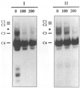
LTR. Four low-mobility DNA-protein complexes, designatedCl, C2, C3, and C4, were detected when Jurkat-,SupTl-,H9-, IMR-32-, and SK-N-MC-derived nuclear extracts were used in individual reactions with radiolabeled 21-bp repeat I (Fig. 2). In addition, several high-mobility DNA-protein complexes were also detected (Fig. 2, bracket). DNA-protein complexes with similar electrophoretic mobili-ties were detected in EMS analyses performed with nuclear extracts derived from the glial cell lines U-373 MG (Fig. 2), Hs683 (Fig. 2), and U-118 (data not shown).Four low-mobility and several high-mobility DNA-protein complexes were also detected when nuclear extracts derived from Jurkat, SupTl, H9, IMR-32, and SK-N-MC cell lines reacted with radiolabeled 21-bp repeat II (Fig. 2). The four low-mobility DNA-protein complexes had electrophoretic mo-bilities similar to those of the DNA-protein complexes, Cl to C4, detected in EMS analyses utilizing 21-bp repeat
I.
DNA-protein complexes Cl to C4 were also detected when U-373 MG (Fig. 2), Hs683 (Fig. 2), andU-118
(data not shown) nuclear extracts were incubated with 21-bp repeatII.
However, the Cl and C2 DNA-protein complexes detected when either 21-bp repeat I or II was used as a probe in EMS analyses with glial cell-derived nuclear extracts were routinely less abundant. Furthermore, a glial cell type-specific DNA-protein complex with an electrophoretic mobility between those of C2andC3on November 9, 2019 by guest
http://jvi.asm.org/
Jurkat H9 IMR-32 SK-N-MC U-373MG Hs683
_ _ _. 0- _ _ _ = _ _ _ _ _
-Ci=- _ ...
C2-Ul
-C3-WP
U2
-C4-
la
FIG. 2. EMS analyseswithnuclear extractsderived from lympho-cytic, neuronal,andglialcell lines in reaction with each21-bprepeat
element. Standard EMSreaction mixtures containing 12 ,ugof lym-phocytic (Jurkat or H9), neuronal (IMR-32 or SK-N-MC), or glial (U-373MGorHs683)cell-derived nuclearextracts,2 p.gof poly(dI-dC), and50,000cpmof radiolabeled 21-bp repeat I, II,or III were incubatedat30°Cfor 30min. High-mobility DNA-protein complexes
areindicatedbythebracket.
wasdetected when nuclearextractsderived from theglialcell lines U-373MG (Fig. 2),Hs683 (Fig. 2),and U-1 18(datanot
shown) reacted with 21-bp repeat I or II; however, the abundance of theDNA-protein complexwasfargreater when
21-bprepeatIIwasutilizedasthe radiolabeledprobeDNA.In
addition, several high-mobility DNA-protein complexes, dis-tinct from those detected in assays with radiolabeled 21-bp repeat I,were formed with radiolabeled 21-bp repeat II and
each nuclearextractwas examined (Fig. 2, bracket).
Collectively, these data demonstrate the formation of both 21-bprepeatI-andIT-specificDNA-protein complexes,aswell
as aglial cell type-specific DNA-protein complex. Since the
electrophoretic mobilities of each of the four DNA-protein complexesweresimilarfor each nuclearextractexamined, it is likelythat theproteinconstituents involved in the formation of each of the DNA-protein complexes, Cl to C4, are similar regardless of nuclear extract origin. However, subtle
differ-encesinthe relative abundanceandelectrophoretic mobilityof
eachDNA-protein complexdetectedwith the series of nuclear
extractsexamined suggestthatalthough theproteins involved in formation of each DNA-protein complex are likely to be similar for the nuclearextractsexamined, theymaynot
neces-sarilybe identical.
Unlike the patternof four low-mobility DNA-protein
com-plexes detected with 21-bp repeats I and TI, at least six DNA-protein complexeswere detectedwhen nuclearextracts
reacted with radiolabeled 21-bp repeat III. The four
low-mobility DNA-protein complexes, Cl to C4, detected when nuclear extracts reacted with 21-bp repeat elements I and II
were also detected when Jurkat-, SupTl-, H9-, IMR-32-,
SK-N-MC-, U-373 MG-, Hs683-, and U-118-derived nuclear extractsreacted with21-bprepeatIII(Fig. 2; datanotshown). InadditiontoCl toC4,twouniqueDNA-protein complexes, designated Ul and U2, were detected when each nuclear
extract reacted with 21-bp repeat III (Fig. 2 and 3). The Ul DNA-protein complex (Fig. 2 and 3) had an electrophoretic
mobility between those of C2and C3. Although the
electro-Jurkat
C2=
C3-
Of
M
U-373MGY
-lI
ci_ C2
-L1L2 C3
-S
S
-UlI
[image:4.612.362.513.77.247.2]-U2
FIG. 3. Preincubation oflymphocyticandglialcellnuclearextracts
with nonspecific DNA. Preincubation of EMS reaction mixtures containing12 ,ugof Jurkat-orU-373MG-derivednuclearextractand 2 ,ugofpoly(dI-dC)atroomtemperature for10 minwasfollowedby incubationat30°Cfor30 min with 50,000cpmofradiolabeled21-bp repeat I, II, or III. The positions of the abrogated C4 complexes detected in EMS reactionswithnopreincubationareindicatedbythe arrows.FreeradiolabeledDNAprobeisnotshown.
phoretic mobility of Ul was similar to that of the glial cell-specific DNA-protein complex,competitionEMSanalyses
indicated that the Ul and glial cell-specific DNA-protein complexes compriseddistinct protein components(seeFig.
8).
The U2 DNA-protein complex was masked by the highly
abundant C4DNA-protein complexbutwasreadilydetectable when formation of the C4 DNA-protein complexwas abro-gated by preincubation of nuclear extracts with nonspecific
DNA(Fig.3). Formation of the U2DNA-protein complexwas not, however, dependent on abrogation of the C4 DNA-protein complex, since the U2 complex could also be detected under standard EMS reaction conditions by extending the
electrophoreticseparation time(data notshown).
Asdescribed above for DNA-protein complexes Cl toC4,
each of the unique DNA-protein complexes, Ul and U2, exhibited similar electrophoretic mobilities regardless of nu-clear extract origin. Furthermore, the high-mobility DNA-protein complexes detected varied in number and relative abundancecontingentonwhich HTLV-I 21-bp repeat element was utilized as probe DNA. These data indicate that, in additionto21-bp repeatI-and
TI-specific
andglialcell-specificDNA-protein complexes, there are 21-bp repeat 111-specific
DNA-protein complexes formed when the 21-bp repeat III elementreactswith nuclear extracts derived from selected cell lines. The DNA-protein complexes discussed were routinely detectedbyutilizing multiple nuclear extract preparations and nuclear extract preparation protocols. In addition, there was no variation in the number or nature of low-mobility DNA-protein complexes formed when poly(dA-dT) or salmon sperm DNA was added as nonspecific DNA in the standard EMS reaction (data not shown).
SpecificityofDNA-protein complex formation. Several lines ofinvestigation wereexamined to determine the specificity of the DNA-protein complexes detected. First, standard EMS analyses were performed, in which increasing nuclear extract protein concentrations were utilized to ensure thatUl and U2 DNA-protein complex formation was detected only with 21-bp repeat III and that glial cell-specific DNA-protein complex
on November 9, 2019 by guest
http://jvi.asm.org/
[image:4.612.62.295.80.262.2]1 11 III pren: 3 6 9 12 15 3 6 9 1215 3 6 9 12 15
cia
C2
FIG. 4. Effect of protein concentration on DNA-protein complex formation. Standard EMS analyses in which 2 ,ug of poly(dI-dC), 50,000cpmofradiolabeled21-bp repeat I, II, orIII,andeither 3, 6, 9, 12, or 15 ,ug of Jurkat (A) or U-373 MG (B) nuclear extract were incubatedat30°C for 30 min. High-mobility DNA-protein complexes are indicatedby the bracket.
formation was detected only in assays with glial cell-derived nuclear extracts. As shown in Fig. 4, when as much as 15 ,ug of Jurkat-derived nuclear extract protein was utilized in standard EMS reactions along with radiolabeled 21-bp repeat I or II, formation of theUl and U2 DNA-protein complexes was not observed. In fact, amounts of protein up to and including 48 ,ug ofJurkat-derived nuclear extract did not result in formation of
Uland U2 inreactions with radiolabeled 21-bp repeat element I or II (data not shown). Furthermore, formation of the glial
cell-specific DNA-protein complex was not detected by EMS analyses utilizing amounts of protein (Fig. 4) up to and
including 48 ,ug of Jurkat-derived nuclear extract (data not
shown). Thesedata demonstrate that formation of theUl,U2, andglial cell-specific DNA-protein complexes was not due to differences in protein availability between EMS reactions or between nuclear extract preparations derived from the indi-catedcell lines.
In a parallel line of experimentation, increasing levels of radiolabeled probe DNA were utilized in standard EMS reactions to ensure that the inability to detect theUl and U2
DNA-proteincomplexes in assays with 21-bp repeats I and II or theglial cell-specific DNA-protein complex in assays with Jurkat-derived nuclearextracts was notdueto an inadequate
amount of radiolabeled probe DNA. Results from these studies indicated that detection of the21-bprepeatIII-specific complexorglial cell-specific DNA-protein complexwasnot a result of limited radiolabeledprobelevels in the EMS reaction
(datanot shown). Inaddition,formationof the21-bp
repeat-specificandglialcell-specific DNA-protein complexeswas not
dependent on the EMS reaction incubation time,since EMS reactions with extendedincubationtimes(uptoandincluding
60 min)didnotresultin changes in thenumberor natureof DNA-protein complexes detected (data notshown).
Cumula-tively, these results suggest that detection of the21-bprepeat
III-specificandglialcell-specific DNA-protein complexeswas in fact due to specific interactions occurring between 21-bp
repeat III and cellular factors or 21-bp repeats I and II and
glialcell-derived nuclearfactors, respectively.
Second, EMS analyses in which the nuclearextracts were
preincubated with nonspecific DNA,
poly(dI-dC), prior
to incubation witheach radiolabeled 21-bprepeat elementwereperformed. As shown in Fig. 3, when Jurkat- and U-373
0 100 200
cI
-C:2
-C3
-C4
-'p
cl
-C2
-*
C3
-C4
-11
0 100 200
.U
_
FIG. 5. Cognatecompetition EMS analyses with glial cell-derived nuclear extracts and 21-bp repeat element I orII.Competition EMS reaction mixtures containing 12 ,ug of U-373 MG-derived nuclear extract, 2jig of poly(dI-dC),50,000 cpm of radiolabeled 21-bp repeat IorII, andeither 100- or 200-fold molar excess of unlabeled 21 bp repeatII (as indicated above the autoradiographs) were incubated at 30°C for 30 min. The glial cell-specific DNA-protein complex is indicated byanasterisk.
MG-derived nuclear extracts werepreincubated with nonspe-cificDNApriortoincubation with radiolabeled 21-bp repeatI, formation of the C4 DNA-protein complex was abrogated. Similar results were also observed when radiolabeled 21-bp repeatII orIII wasutilized as probe DNA(Fig. 3). However, theCltoC3,Ul,U2, andglialcelltype-specificDNA-protein complexes observed in the standard EMS analyses were de-tected when the nuclear extracts were preincubated with
nonspecific DNA, indicating that these are specific
DNA-protein interactions occurringbetweencellular factors and the 21-bp repeat elements. In addition, and as previously
dis-cussed, detectionof the U2DNA-proteincomplexwasevident when formation of the C4 DNA-protein complex was abro-gated. Comparable resultswere also observed when nuclear extracts derived fromSupTl, H9, U937, IMR-32, SK-N-MC,
Hs683, and U-118 cell lines were utilized in similar experimen-tation (data not shown). These data suggested that the C4 DNA-protein complex is nonspecific innature.
A third line of investigation aimed at determining the
specificity ofDNA-protein complex formation included
com-petitionEMS analysesinwhich cognate DNA sequence was utilized as competitor DNA. As shown in Fig. 5, utilizing
U-373 MG-derived nuclear extract and radiolabeled 21-bp
repeatI or II, competitionwith unlabeled cognate sequence resulted in inhibition of Cl, C2, C3, and glial cell-specific DNA-protein complexformation.Incontrast,formationof the C4 DNA-protein complex was essentially unaffected by the presence of unlabeled competitor DNA. Similar resultswere obtained from EMSanalyses addressing the specificityofUl and U2DNA-protein complexformation
(data
notshown).
IncompetitionEMS
analyses
withJurkat-derivednuclearextract andradiolabeled21-bprepeatIII,formationof theCl, C2, C3,
Ul, and U2 DNA-protein complexes wasinhibited,
whereas formation of C4wasunaffectedby
the presence of unlabeled cognate competitor DNA. These data further support theprobability of the
nonspecific
nature of the C4DNA-protein
complex and demonstrate that formation ofCl, C2,
C3,
and the glialcell-specific
DNA-protein complexes
was due tospecific interactions between cellular factors and the
21-bp
repeatelements.
Dependence ofDNA-protein complexformationon
flanking
on November 9, 2019 by guest
http://jvi.asm.org/
[image:5.612.58.297.75.232.2] [image:5.612.368.499.78.223.2]Jurkat
Ci
-C2
-C3 -C4
--Ul
-U2
U-373MG
-.:I
[image:6.612.151.469.81.263.2]._U
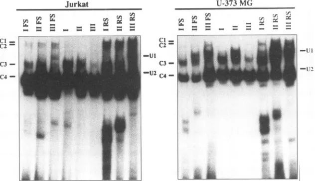
FIG. 6. EMSanalyses withlymphocyticandglialcell nuclearextractsand21-bprepeatswithorwithoutflankingDNA.Standard EMSanalyses
in which 12 ,ugofJurkat- orU-373MG-derived nuclearextract,2 ,ug ofpoly(dI-dC),and50,000cpmofradiolabeled21-bprepeatelement native flankingsequences(lanesIFS, IIFS,and IIIFS),noflankingsequences(lanesI,II, andIII),or5'HindlIl and 3' BamHIrestriction endonuclease site sequences(lanesIRS,II RS,and IIIRS)wereincubatedat30°C for30 min. Freeradiolabeled DNAprobeisnotshown.
DNA. Since the affinity of DNA-binding proteins for their cognate recognition sites is often affected bythe presence of
flanking DNA sequences, we have examined the role of flanking DNA in the formation of theDNA-proteincomplexes
detected by the three sets of 21-bp repeat double-stranded
oligonucleotidesin EMSanalyses (seeMaterials and Methods for the sequences). The first set
(I
FS, II FS, and III FS) contains sequences corresponding toeach of the three 21-bprepeatelements flankedbynative HTLV-ILTRsequences, the second set (I, II, and III) corresponds to the 21-bp repeat elements alone with no additional DNA sequences, and the third set (I RS, II RS, and III RS) contains sequences
corresponding to each of the three 21-bp repeat elements flankedbythe restrictionendonuclease sequences for BamHI
(5' end of the21-bp repeat) and HindIll (3' end of the 21-bp repeat). All data presented to this pointwere generated by utilizing 21-bp repeat elements with native HTLV-I LTR flankingDNAsequences.Consistent with the resultspresented
in Fig. 2, four low-mobility DNA-protein complexes were detected whenJurkat and U-373 MG nuclearextractsreacted with each 21-bp repeat element flanked by native HTLV-I
LTR sequences (lanes I FS, II FS, and III FS inFig. 6). In contrast,incubation of nuclearextractswith the21-bp repeat elements in the absence offlankingDNA(lanes I, II,andIIIin Fig. 6) diminished the number of DNA-protein complexes
detected,specificallyCl,C2, and Ul.However, DNA-protein
complex formation was restored by 21-bp repeat elements boundedbyirrelevant DNAsequences inthe form of restric-tionendonuclease sitesBamHIandHindlIl (lanes I RS, II RS, and III RS in Fig. 6). In each case, there was little or no homology between the restriction endonuclease sequences and the native flanking HTLV-I LTR sequences. Therefore, the
ability todetectsimilar DNA-protein complexes regardless of the sequence of the flanking DNA suggests that the require-mentfor flanking DNA in DNA-protein complex formation is not sequencedependent.
Formation of cell type-dependent DNA-protein complexes withthe TRE-1/-2 element. Having demonstrated the forma-tion ofat least one glial cell type-specific DNA-protein
com-plexin EMS analyses withHTLV-I 21-bp repeat element I or II and nuclear extracts prepared from U-373 MG (Fig. 2),
Hs683 (Fig. 2),orU-118(datanotshown) cells,weproceeded
to determine whether glial cell type-specific DNA-protein
complexes would form when the 21-bp repeat elementswere contained within thecontextof the HTLV-ILTR. Tothisend,
a DNA probe, the TRE-1/-2 element, corresponding to the sequences contained between the ApaI and NdeI restriction
endonuclease sites within the HTLV-I LTR U3 region
(posi-tions -268 to -46) was generated by PCR technology as described in Materials and Methods. The resultant 223-bp
PCR product contains the three 21-bp repeat elements and intervening sequences in thecontext oftheir native positions
within the HTLV-I LTR. When the radiolabeled TRE-1/-2
element was utilized in EMS analyses along with nuclear extractsderived fromH9, SupTl,or U-118cells, several glial
cell type-dependent DNA-protein complexes were detected
(Fig. 7, asterisks). These data demonstrate that glial cell
type-specific DNA-protein complexes could be detected not
onlywith an isolated 21-bp repeat element but also with the intact TRE-1/-2 element.
Specificity of TRE-1/-2 DNA-protein complex formation. Thespecificity of TRE-1/-2DNA-protein complex formation wasdeterminedby severallines ofexperimentation. First, the nuclear extractswere preincubated with poly(dI-dC) prior to reaction with radiolabeled TRE-1/-2 probe DNA. Under these conditions, thespecificDNA-protein complexes were detected
(Fig. 7, thin arrows), while formation of the nonspecific
DNA-protein complexeswasabrogated (Fig. 7, thick arrows). When increasing concentrations of protein or radiolabeled probeDNA wereused in EMS analyses along withlymphocytic orglialcell-derived nuclear extract, the number and nature of DNA-protein complexes detected were unchanged (data not
shown).Furthermore, formation of the glial cell-specific DNA-protein complexes was not detected in the titration EMS analyses with lymphocytic nuclear extracts, indicating that formation of these complexes was due to specific interactions between glial cell-derived factors and the TRE-1/-2 element and was not a result of differences in the abundance or availability of protein constituents. Competition EMS analyses were also performed with unlabeled cognate TRE-1/-2 as competitor DNA (Fig. 7). As expected, formation of the nonspecific DNA-protein complexes was unaffected by the
:., :rl
W r, .r.
.2
iff. - - = =O.. = = - = = - - =
cn K rA rA mcc
K Li. - Cd cc
M." = = - = = - = =
on November 9, 2019 by guest
http://jvi.asm.org/
U-,8 j TRE.l/-2
., 1SO300
WrI.6 1
[image:7.612.54.555.529.680.2]-***~~~~~~~~~~~~~~~~~~~~~~~~~~~~~~~1
FIG. 7. EMS analyses ofthe TRE-1/-2 element and nuclear ex-tractsderived from lymphocytic and glial cell-derived nuclearextracts.
Standard EMS reaction mixturescontaining 3 ,ugof H9-, SupTl-,or
U-118-derivednuclearextract, 2p,gofpoly(dI-dC),and 50,000cpmof radiolabeled TRE-1/-2 element which contains the three21-bp repeat
elements in the context of native HTLV-I LTR sequences were
incubated at30°C for 30 min. Standard EMS reactionsperformed in theabsence of preincubation with competitor DNAsare indicated by dashes. Also shown are cognate EMS analyses in which 150- and 300-fold excesses of unlabeled TRE-1/-2 were used. Thin arrows
indicate specific DNA-protein complex formation, thick arrows
indi-cate nonspecific DNA-protein complex formation, and asterisks
indi-cate glial cell-specific DNA-protein complex formation. ATL, adult T-cellleukemia.
presence of cognate competitorDNA,while formation of the specific DNA-protein complexes, including the glial cell-spe-cific DNA-proteincomplexes, wasabrogated.
Characterization of the protein components involved in DNA-protein complex formation with the HTLV-I 21-bp re-peat elements. Tobegin characterization ofthe protein com-ponentsinvolved in DNA-protein complex formation with the
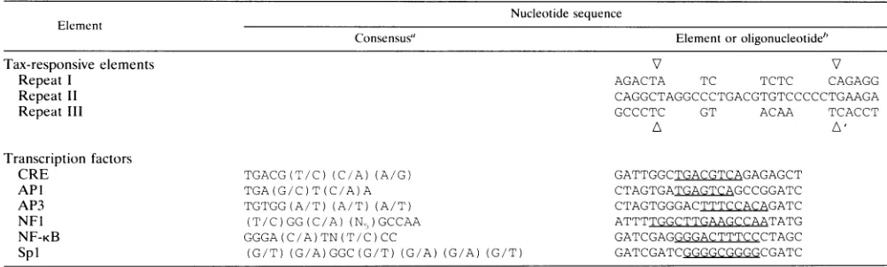
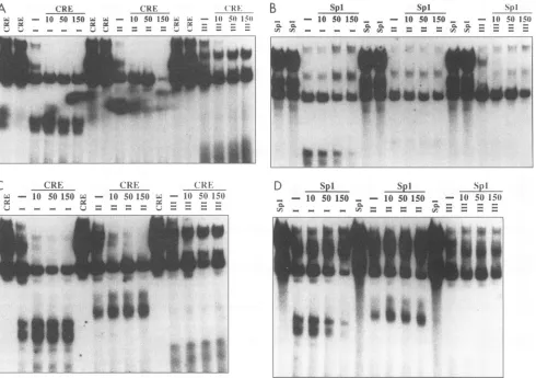
21-bp repeat elements, competition EMS analyses were per-formed with oligonucleotides containing the binding sites for one of several previously characterized transcription factors, including AP1,AP3, CREB, NF-KB, NF1, and Spl (Table 1) (12). Several ofthetranscriptionfactorbindingsite consensus sequences exhibitconsiderable homologytoselected regions of the HTLV-I LTR (Table 1) and were therefore considered likely candidates to be involved in DNA-protein complex formation with the HTLV-I 21-bp repeat elements. In addi-tion, previous studiesby other investigators (46, 48, 53) have already established that several known transcription factors interact with the21-bprepeatelements in either the absence or presence of Tax inreactions with nuclear extracts derived from lymphocytes and other cell types ofnonneuroglial origin. As shown in Fig. 8, competition EMS analyses were performed with individual radiolabeled 21-bp repeat elements, nuclear extractsderived from Jurkat or U-373 MG cells, and a 10-,50-, or 150-fold molar excess ofunlabeled competitor oligonucle-otide. In addition to each21-bp repeatelement being usedas radiolabeled probe DNA, radiolabeled transcription factor binding siteoligonucleotideswerealsoutilizedasprobe DNAs in order todetermine the electrophoreticmobilities of DNA-protein complexescorrespondingtoendogenoustranscription factors present in the Jurkat or U-373 MG nuclear extracts when they reacted with the cognate binding sites contained within the transcription factorbinding site oligonucleotides.
As shown in Fig. 8A, in competition EMS analyses with Jurkat-derived nuclear extracts and radiolabeled 21-bp repeat I element, formation of the C1-C3 DNA-protein complexes was efficiently inhibited by the presence of unlabeled CRE oligonucleotide as competitor DNA. This evidence indicates that a CREB-ATF family member(s), along with individual 21-bp repeat elements, may play a role in the formation of these DNA-protein complexes. Previous studies by other in-vestigators in whichmultimerized 21-bprepeat elementswere utilized astarget DNA(34,35, 48, 53)have alsosuggestedthat CREB-ATF family members are involved in specific DNA-protein complex formation. In contrast, formation of the C4 and high-mobility DNA-protein complexes was unaffected by CRE oligonucleotide competitor DNA (Fig. 8A). Similar resultswerealsoobtainedbyutilizing 21-bprepeat II element
TABLE 1. Nucleotidesequencesof the 21-bpTax-responsive elements and ofselectedtranscription factor bindingsiteoligonucleotides
Nucleotide sequence Element
Consensus" Elementoroligonucleotide"
Tax-responsive elements V V
Repeat I AGACTA TC TCTC CAGAGG
Repeat II CAGGCTAGGCCCTGACGTGTCCCCCTGAAGA
Repeat III GCCCTC GT ACAA TCACCT
Transcription factors
CRE TGACG(T/C)(C/A)(A/G) GATTGGCTGACGTCAGAGAGCT
API TGA(G/C)T(C/A)A CTAGTGATGAGTCAGCCGGATC
AP3 TGTGG(A/T)(A/T)(A/T) CTAGTGGGACTTTCCACAGATC
NFl (T/C)GG(C/A)(N5)GCCAA ATTTTGGCTTGAAGCCAATATG
NF-KB GGGA(C/A)TN(T/C)CC GATCGAGGGGACTTTCCCTAGC
Spi (G/T)(G/A)GGC(G/T)(G/A)(G/A)(G/T) GATCGATCGGGGCGGGGCGATC
"Coreconsensussequence for selectedtranscriptionfactorbindingsites(12).Alternative nucleotidesare indicatedasfollows:N,anynucleotide;(N,/N2),either
nucleotideN,ornucleotideN,;or(NX),aseries of nucleotides wherexrepresentsthe number of nucleotides.
'V, markers delineating the 5' and 3' boundaries of the21-bp Tax-responsiveelements. Nucleotidesoutside markers represent native HTLV-I LTRflanking
sequences. Thenucleotidedifferencesamongthe three21-bpTax-responsiveelementsareindicated,while thenucleotides conserved among the three21-bprepeats
are represented once. Nucleotide sequences of double-stranded oligonucleotides contain a minimalbinding site for thecorresponding transcription factor. The
underlinednucleotidesrepresent thetranscriptionfactorbindingsites.
SupTI
TRE-.J-2
* 15030
..:-H9
sk
.1
g ,sj TREIf-2
_
.
ILI -150 300
Sm,,
%..b
.I
on November 9, 2019 by guest
http://jvi.asm.org/
A CRE - CRE (CR
E-W^ ;w
- 10 50 150 XX9
- 10 50 150CL Xi6
-W5I 1Rci Lc ______
~~~
A<i.
, e £B
spI
__ - 10 so 150 _ _
-'a 'ai Ci cL
CA V: _ _ _ _ (A CA =
SPi
10 50 150. -
-_ _
=:cr
a*i:.. .:
C (RE
("RE
(CR
w - 10 50 150 e - 10 50 150 ; - 10 50 151)
_. P P-__ _Q __
-_._. .__ ... ... ___.Am__..._..._",.. ...;,_.A. ...I.
D
SpI- -
o1050
150 ^bit
- - 10 50 150
c_
-3. * | s ,.
FIG. 8. Competition EMS analyseswith unlabeled CREorSpl bindingsiteoligonucleotidesand Jurkat-orU-373 MG-derivednuclear extracts. Competition EMS reaction mixturescontaining 12,ugof Jurkat-derived(AandC)orU-373 MG-derived (Band D)nuclearextracts,2 jigof poly(dI-dC),50,000cpmof radiolabeled21-bprepeatI,II,orIII,anda 10-, 50-,or 150-fold molarexcessof unlabeled CRE(AandB)orSpl (C and D) binding site oligonucleotide competitor DNAwere incubated at 30°C for 30 min. Shown for comparison are EMS analyses of radiolabeled CREor Spl bindingsite oligonucleotide in reactions with 12 or9 ,g, respectively, of nuclear extract.Standard EMS reactions
performedwithoutpreincubationorcompetitorDNAsareindicatedbydashes. Free radiolabeled DNAprobeisnotshown.
asradiolabeled probeDNA,in thatformation of the Cl toC3 DNA-protein complexes was efficiently inhibited with CRE
oligonucleotide as the competitor DNA (Fig. 8A), while formation of the C4 and high-mobility DNA-protein
com-plexes was unaffected by the presence of competitor DNA.
With radiolabeled CRE oligonucleotide as the probe DNA,
DNA-protein complexes which exhibited electrophoretic
mo-bilities similar to those ofDNA-protein complexes Cl toC3 with 21-bp repeat elements I and II were detected. This
comparativeinformationprovidesadditional evidence
suggest-ingthatamember(s)of theCREB-ATFfamilymayparticipate
information oftheseDNA-protein complexes, sincethe CRE oligonucleotide andthe 21-bp repeat oligonucleotides are of
similarsizes.
Incontrast,when radiolabeled 21-bprepeatIIIwasutilized asprobeDNA insimilarcompetitionEMS analyses, unlabeled
CREoligonucleotide inhibited only the CltoC3 DNA-protein complexes efficiently (Fig. 8A). Formation of the Ul and U2 21-bp repeat III-specific DNA-protein complexes was
unaf-fected by CRE oligonucleotide competitor DNA, as was
formation of thehigh-mobility DNA-protein complexes. These resultsdemonstrate thatthe CltoC3 DNA-proteincomplexes detected when 21-bp repeats I, II, and III react with
Jurkat-derived nuclear extracts are possibly composed of protein components that recognizethe CREconsensus sequence and may include members ofthe CREB-ATFfamily of
transcrip-tion factors. Furthermore, formationof the Ul andU221-bp repeatIII-specific DNA-protein complexesdoesnotappearto
be dependenton a CREB-ATFfamily member(s).
Competition EMS analyses with nuclear extracts derived from the glialcell line, U-373 MG(Fig. 8B), demonstrated a
similar transcription factorbinding site oligonucleotide
com-petition profile. When glial cell-derived nuclear extracts
re-acted with radiolabeled 21-bprepeats I,II, andIII in
combi-nation with CRE oligonucleotide as the competitor DNA,
formation of the Cl to C3 DNA-protein complexes was
efficiently inhibited (Fig. 8B). Additionally, formation of the low-mobility, glial cell-specific DNA-proteincomplex detected in standard EMS reactions with radiolabeled 21-bprepeats I andIIwasinhibitedby CREoligonucleotide competitorDNA.
However, in contrast to the complete abrogation of Cl, C2, and C3 DNA-protein complex formation observed in EMS analyses with 10-, 50-, and 150-fold molar excesses of CRE
oligonucleotide competitor DNA, comparable levels of CRE competitor resulted in incomplete inhibition of glial cell-specificDNA-protein complex formation.
spi 10 50 150
S1pl_
5 _ I5Wso 506= =
AlrIE
b.:
IF
on November 9, 2019 by guest
http://jvi.asm.org/
[image:8.612.71.561.77.422.2]In competition EMS analyses with 21-bprepeat elements I,
II, and III, unlabeled Spl binding site oligonucleotide as competitorDNA, andJurkat-derivednuclear extract(Fig. 8C), the Cl to C4 DNA-protein complexes as well as the high-mobility DNA-protein complexes were unaffected by Spl binding site competitor DNA, suggesting that Spl is not a component ofthese DNA-protein complexes. However,when radiolabeled 21-bp repeat III was utilized as probe DNA, formation of the Ul and U2 21-bp repeat 111-specific DNA-protein complexeswas efficiently inhibited by the presence of Spl binding site competitor oligonucleotide, indicating that Spl or an Spl-related factor may playa role in the formation of these DNA-protein complexes. Similar results were ob-tained with U-373 MG-derived nuclear extract (Fig. 8D); however, competition EMS analyses with U-373 MG-derived nuclear extract, 21-bp repeat III, and competitor Spl binding site DNA resulted in only minimal competition of the Ul DNA-protein complex. This result contrasted with those ob-tained with Jurkat-derived nuclear extracts(Fig. 8C), in which Spl binding site competitorDNA efficiently abrogated forma-tion of the Ul DNA-protein complex. These results indicate that while Spl or an Spl-related factormay play a significant
rolein formation of theUl and U2DNA-protein complexesin assayswith21-bp repeat III and lymphocyticnuclear extracts, the role of Spl or related factors in DNA-protein complex formation of Ul and U2 with 21-bp repeat III and glial
cell-derived nuclear extracts is less apparent. These data further substantiate the existence of cell type-specific differ-encesin DNA-protein complex formation betweeneach of the three 21-bprepeatelements and nuclear extractsderivedfrom selected cell lines.
Insimilarexperimentation performedwith Jurkat-orU-373 MG-derived nuclearextracts and API,AP3, NFI, and NF-KB binding site oligonucleotides as competitor DNAs (data not shown), DNA-protein complex formation was essentially un-affected, indicating that these transcription factors are not involved in the formation ofany of the low- orhigh-mobility DNA-protein complexes.
Role of nonconserved nucleotides in the formation of21-bp repeat III-specific and glial cell-specific DNA-protein com-plexes. The 21-bp enhancer element located within the HTLV-ILTR isimperfectly repeated at threelocations within the U3 region. Among the 21-bp repeat elements, there are
three strictly conserved domains separated by two
noncon-servedregions(Fig. 1).Toinvestigatethe role ofnonconserved nucleotides in the formation of the 21-bp repeat
III-specific
and glial cell-specific DNA-protein complexes, the 21-bp
re-peat III element was mutated such that the four nucleotides between conserved domains B and C
(ACAA;
Fig. 1)
werechanged to the corresponding sequence of
21-bp
repeat II (TGTC, designated 21-bp repeatIIIAII;
see Materials and Methods for the entire sequence). Standard EMSanalyses
were performed with nuclear extracts derived from Jurkat or
U-373 MG cells and radiolabeled
21-bp
repeat I, II, III, orIIIAII. As shown in
Fig.
9, when21-bp
repeat IIIAII wasutilized as probe DNA, the
Ul,
21-bp
repeatIII-specific
complexwas notdetected,whereas the Cl toC4
DNA-protein
complexeswere readily detectable. In
addition,
in EMS anal-yseswith U-373 MG-derived nuclear extracts(Fig. 9),
muta-tion of the sequences between domains B and C in
21-bp
repeat IIIAII resulted in the formation of aDNA-protein
complexwithan
electrophoretic
mobility
similartothat ofthe glial cell-specificDNA-protein
complex
detectedby 21-bp
repeatelements Iand IIand
glial
cell-derivednuclearextracts.Toensure that the new
DNA-protein complex
detectedby
21-bp repeat IIIAII andglial cell-derivednuclearextracts was
Jurkat
cl
-cl- idIs
C4
-U-373 NIG(
ll
FIG. 9. EMS analysesof21-bp repeat IIIAII and nuclear extracts derived from lymphocytic and glial cells. Standard EMS reaction
mixtures containing 12
[.g
of Jurkat- or U-373 MG-derived nuclearextract, 2 VLg ofpoly(dI-dC), and 50,000cpm of radiolabeled 21-bp
repeat I, II, III, orIIIAIIwere incubatedat30°Cfor 30 min.
indeed the
glial
cell-specific DNA-protein complex
and notU
1, competition
EMSanalyses
comparable
tothosepreviously
discussed wereperformed.
Competition
EMSanalyses
with21-bp
repeatIIIAII,
U-373 MG-derived nuclear extract, andCRE or
Spl
binding
sitecompetitoroligonucleotide
(Fig. 10)
demonstrated
competition
profiles
similar to those observed with21-bp
repeat I orII,
U-373 MG-derived nuclearextract, and CREorSpl
binding
sitecompetitor
oligonucleotide.
That(:RE SPi
C31
-Ci
-(4
'iih
*.
FIG. 10.
Competition
EMSanalyses
with unlabeledoligonucleo-tides
containing
thebinding
site for CREB(CRE)
orSpl
and U-373 MG-derived nuclear extracts.Competition
EMS reaction mixturescontaining 12
p.g
of U-373 MG-derived nuclear extracts, 2p.g
ofpoly(dI-dC),50,000cpmofradiolabeled
21-bp
repeatIIIorIIIAII,anda 10-,50-,or150-foldmolarexcess
(marked
abovelanes)ofunlabeledCRE or
Spl
binding
siteoligonucleotide competitor
DNA wereincubated at
30°C
for 30 min. A standard EMS reactionperformed
without
preincubation
orcompetitor DNAsisindicatedby
the dash.Free radiolabeled DNA
probe
is not shown. *,glial
cell-specific
DNA-protein
complex.
on November 9, 2019 by guest
http://jvi.asm.org/
[image:9.612.351.521.81.252.2] [image:9.612.375.511.425.628.2]is,
formation of the newDNA-protein complex
wasinhibitedby
the presence of CREcompetitor
oligonucleotide
butnotby the presence ofSpl binding
sitecompetitor oligonucleotide.
Consequently,
these data indicate that the newDNA-protein
complex
detected in EMSanalyses
with21-bp
repeat IIIAIIand
glial
cell-derived nuclear extracts is verysimilar,
if notidentical,
to theglial cell-specific DNA-protein complex
pre-viously
detected with21-bp
repeatIorIIandglial
cell-derivednuclearextracts.
Furthermore,
these dataindicate that the four nonconserved nucleotides located betweendomainsBand C in21-bp
repeat IIaresufficient,
when in thecontextof domainsB and
C,
for formation of theglial cell-specific DNA-protein
complex.
Collectively,
these resultsdemonstrate that the fournonconservednucleotides located between domainsBandC in
21-bp
repeat III areintegrally
involved in the formation ofboth
21-bp
repeat-specific
andglial
celltype-specific
DNA-protein complexes.
DISCUSSION
A
variety
ofcellularproteins
hasbeen showntointeract withthe HTLV-I
LTR,
specifically
with the21-bp Tax-responsive
elements
(33, 46, 48, 53).
Since the interaction of cellular factors with each of the21-bp
repeatsiscritical tobothbasal and Tax-mediated HTLV-I LTR-directedexpression,
identifi-cation and characterization of theseDNA-protein
interactionsare criticalto
determining
theprecise
componentsinvolved in HTLV-I LTR-directedexpression
in immune and nervous system cell types.Furthermore,
theanalysis
of celltype-dependent
interactions will contribute todissecting
thecom-plex pathway leading
toHTLV-Icelltype-specific expression,
an event which may
directly impact
on theregulation
of(i)
productive
viralreplication; (ii) establishment, maintenance,
and reactivation of latent viral
infection;
and(iii)
viralpatho-genicity
withintargeted
cellpopulations.
Although
considerable effort has been exerted toidentify
and characterize the cellular factors of
lymphocytic origin
which interact with the HTLV-I
regulatory
unit, neuroglial
cell-derived
proteins
which interact with HTLV-I LTR se-quences have notbeenreported.
HTLV-I has been shown in vitrotoinfectcells ofnonlymphoid origin, including
anumber of cell typesofnervoussystemorigin (1, 13, 22-24, 28, 32, 49,
52). Furthermore,
PCRamplification
studies havedemon-strated the presence of HTLV-I DNA sequences in several
regions
of the brain andspinal
cord of HTLV-I-infectedtropical spastic paraparesis patients (4,
25,27),
contrastingwith results obtained for control
subjects
and adult T-cell leukemiapatients
(4, 27)
inwhichHTLV-Isequenceswerenot detected in theseregions.
Thepossibility
thatHTLV-I infectsand
replicates
in cellsofneuroglial origin warrantsinvestiga-tion of the
transcriptional regulation
of the HTLV-I LTR in these cell types.Results from our studies indicate that similar as well as
unique DNA-protein
complexesareformedbycellular factorsof
lymphocytic,
neuronal, and glial origins and each of the three21-bp
repeat elements. All of theCl,
C2, and C3DNA-protein
complexes detected from nuclear extracts inreactions with individual 21-bp repeat elements exhibited similar
electrophoretic
mobilities, suggestingthatatleast some of theDNA-protein
complexesformed between nuclearfac-tors and each ofthe 21-bp repeat elements arecomposed of similar but not
necessarily
identical proteins. In contrast, at leasttwooftheDNA-protein complexes detected,Ul
and U2,were
21-bp
repeatIIIspecific,
indicatingthat each of the21-bprepeatelements isuniquewith respecttoitsabilitytointeract with selectcellular factors.
In addition to the 21-bp repeat-specific nature of certain DNA-protein complexes detected, at least one DNA-protein complex which was detected in EMS analyses with 21-bp
repeats Iand II andglial cell-derived nuclear extracts was glial cell typespecific. Formation of glial cell-specificDNA-protein complexes was also detected by a fragment of the HTLV-I LTRcontaining the three21-bp repeat elements in thecontext of native HTLV-I LTR sequences(TRE-1/-2), demonstrating thatglial cell-specific DNA-protein complexes form with not onlyan isolated21-bp repeat element but also with the intact TRE-1/-2 element. Site-directed mutagenesis studies are under waytodetermine thespecific sequences within TRE-1/-2 which areinvolved in glial cell-specific DNA-proteincomplex forma-tion,as arestudiestoidentify the protein components of these complexes. Several possibilities exist for glial cell-derived proteins to play a role in glial cell-specific HTLV-I LTR-directed expression via interaction with the 21-bp repeat elements. For example, formation of cell type-specific DNA-protein complexes with the 21-bp repeat elements may influ-ence basal HTLV-I LTR-directed transcription, thereby
en-hancingorundercertain circumstancessuppressingthe initial phase of viralreplication.Furthermore, the preferential inter-action of cell type-specific factors with the 21-bp repeat elements may affect the overall level of Tax-mediated trans activation within select target cellpopulations.
Studies aimed at examining the role of flanking DNA in
DNA-protein complexformation demonstrated that the 21-bp repeat alone was sufficient for formation of the glial
cell-specific DNA-protein complex.Formation of the21-bp
repeat-specific complexes,in contrast,wasdependentonthe presence offlanking DNA; however,thisrequirementwas notsequence
specific.Mutational analysesof21-bprepeatIII indicated that thefour nonconserved nucleotides between domains B and C
(Fig. 1) were necessary for formation of both 21-bp repeat
III-specific and glial cell-specific DNA-protein complexes.
Replacementof these nucleotides in 21-bp repeat III (ACAA) with those of 21-bp repeat II (TGTC) abrogated Ul
DNA-protein complexformation but reconstitutedglialcell-specific complex formation, indicating that TGTC in the context of domains B and C is sufficient for glial cell-specific
DNA-protein complexformation.
Further evidence supporting the unique nature of each individual 21-bp repeat element and the presence of glial
cell-specific complexformationwasobtained through compe-tition EMS analyses with a series of unlabeled transcription factor binding site oligonucleotides. Initial studies were di-rectedatdeterminingtherole of these factors in DNA-protein
complexformation with the21-bp repeat elements, since each
21-bprepeatcontainsstrictlyconserved core sequences (Fig. 1) that exhibit homology to the CREB, AP1, NF-KB, and Spl
bindingsiteconsensussequences.On thebasis of these studies, amember(s)of the CREB-ATF family of cellular transcription factorswasdeterminedtobecritical for formation of Cl, C2, and C3, as well as for formation of the glial cell-specific
complexdetected in assaysusing 21-bp repeat I or II and glial cell-derived nuclearextracts. Ahigher level of CRE competi-torwas required to interfere with glial cell-specific complex formation than to completely abrogate formation of Cl, C2, andC3, indicatingthatthe CREB-ATF-related factor involved inglialcell-specific complex formation is not identical to that
participatinginformation ofCl, C2, and C3.
TheCREB-ATFfamily consists of a number of structurally relatedtranscription factors which bind to similar DNA ele-ments; however, individual members show distinct transcrip-tional effector functions(6, 21).Additionally, cell type-specific isoforms of CREB-ATF family members exist (30), supporting
on November 9, 2019 by guest
http://jvi.asm.org/
the hypothesis that a glial cell-specific CREB-ATF-related
factor(s)
is involved in glial cell-specific complex formation.Thisproposalmayaccountfor theobserved differencesinthe CREcompetition profilesnoted for Cl, C2, C3 and the glial
cell-specific
DNA-protein complex. Alternatively, anotherCREB-related factor, the CRE modulator (CREM) protein, may participate either alone or in conjunction with CREB-ATF factors in formation of the glial cell-specific complex.
Like
CREB,
several CREM isoforms which are capable ofinteracting
with CREB-ATF factors and bindingto the CREmotif, thereby enhancingorblocking transcriptional activation mediated by CREB factors, are produced (10, 15, 29).
Fur-thermore,
certain CREM isoforms exhibit tissue type-specificexpression
and are developmentally regulated (10, 15, 29).Since one ofthe
primary
sites of CREM expression includes the central nervous system (10, 15, 29), current research is focusedondeterminingthe role ofCREM isoformsin forma-tion of theglial cell-specific
DNA-protein complexaswell asthe
Cl, C2,
and C3 complexes.In addition to the involvement of a CREB-ATF family
member(s)
inDNA-protein complexformationwiththe21-bprepeatelements, competitionEMSanalysesindicatedthat the
transcription
factorSplor anSpl-likefactorparticipatesinUland U2 formation. However, a significant difference was observedinthe abilities ofSpl bindingsiteoligonucleotide to inhibit formation ofUl andU2 in competition EMS analyses
performed
with Jurkat- and U-373 MG-derived nuclearex-tracts. Thepresence ofSpl
binding
site oligonucleotidecom-pletely abrogated formation of Ul and U2 in analyses with Jurkat-derived nuclear extracts, whereas similar assay
condi-tionsresultedin
incomplete
inhibition with U-373 MG-derived nuclearextracts.DespitethepresenceofSplinmostcelltypes,tissue-specific
expression of Spl as well as tissue-specificenhancement of
Spl-mediated
gene expression have beenreported
previously
(2, 26). Cell-and/or tissue-specific expres-sion ofSpl
or an Spl-related factor may account for the observeddifferences inSpl
binding
siteoligonucleotide
com-petition profiles
in assays with 21-bp repeat III and nuclearextracts of
lymphocytic
orglial origin. Alternatively,
the ob-servedUIcomplex
detected inassayswith U-373 MG-derived nuclearextractscould betwo ormorecomigrating
complexes,only
one ofwhich isdependentonSpl for formation.In summary,we presentevidence that21-bprepeat-specific
and
glial cell-specific DNA-protein complexes
can formbe-tweentheHTLV-I
21-bp Tax-responsive
elements and cellular factors of immuneandnervoussystemorigins.
Current studiesare directed at
identifying
andcharacterizing
the factors involvedin formation of theglial cell-specific
complex,assess-ing
the functional role of the21-bp
repeatsincells oflympho-cytic
andglial
cellorigins,
anddefining
the functional roleofneuroglial cell-specific protein
factors in celltype-specific
expression.
ACKNOWLEDGMENTS
Wegratefully acknowledgethe technical assistanceofWangDian.
This researchwassupported bya grantawardedto B.W.from the National Cancer Institute(PublicHealthServicegrant CA54559).
REFERENCES
1. Akagi, T., Y. Hoshida,T. Yoshino, N.Teramoto, E. Kondo, K.
Hayashi, and K. Takahashi. 1992. Infectivity of human T-cell leukemia virus type Itohumannervous tissue cellsin vitro. Acta
Neuropathol.84:147-152.
2. Bessereau, J., D. Mendelzon, C. LePoupon, M. Fiszman, J. Changeux,andJ.Piette. 1993.Muscle-specific expression of the acetylcholine receptoralpha-subunit gene requiresboth positive
and negative interactions between myogenic factors,SpI andGBF factors. EMBO J.12:443-449.
3. Bhagavati, S., G. Ehrlich, R. W. Kula, S. Kwok, J. Sninsky, V.
Udani, and B. J. Poiesz. 1988. Detection of human T-cell lympho-ma/leukemia virus type I DNA and antigen in spinal fluid and blood of patients with chronic progressive myelopathy. N. Engl. J. Med. 318:1141-1147.
4. Bhigjee,A.I., C. A. Wiley, W. Wachsman, T. Amenomori, D.Pirie,
P.L. A. Bill, andI. Windsor. 1991. HTLV-I-associated myelopa-thy: clinicopathologic correlation with localization of provirus to the spinal cord. Neurology 41:1990-1992.
5. Bradford, M. M. 1976. A rapid and sensitive method for the quantitation of microgram quantities of protein utilizing the principle of protein-dye binding. Anal. Biochem. 72:248-254. 6. Brindle,P.K., and M. R. Montminy. 1992. The CREB family of
transcription activators. Curr. Opin. Genet. Dev. 2:199-204.
7. Celander, D.,and W.A. Haseltine. 1984.Tissue-specific transcrip-tion preference as adeterminant of cell tropism and leukaemo-genic potential ofmurineretroviruses. Nature (London)
312:159-162.
8. Chatis,P. A., C.A. Holland, J. W. Hartley, W. P. Rowe, and N. Hopkins. 1983. Role for the 3' end of the genome in determining disease specificity of Friend and Moloney murine leukemia
vi-ruses. Proc. Natl.Acad. Sci. USA 80:4408-4411.
9. Corboy, J. R., J.M. Buzy,M. C.Zink, and J. E. Clements. 1992. Expression directed from HIV long terminal repeats in the central
nervoussystem of transgenic mice. Science 258:1804-1808.
10. Delmas, V., B. M. Laoide, D. Masquilier, R. P. de Groot, N. S. Foulkes,and P.Sassone-Corsi. 1992. Alternative usage of initia-tion codons in mRNA encoding the cAMP-responsive-element modulator generates regulators with opposite functions. Proc. Natl.Acad. Sci. USA 89:4226-4230.
11. Dignam, J. D.,R.M.Lebovitz, and R. G. Roeder. 1983. Accurate transcription initiationbyRNApolymerase II inasoluble extract from isolated mammalian nuclei. Nucleic Acids Res. 11:1475-1489.
12. Faisst, S.,and S.Meyer. 1992.Compilationof vertebrate-encoded transcriptionfactors. Nucleic Acids Res. 20:3-26.
13. Fanauchi, M.,T. Saida,K. Saida,M.Makajima, S. Matsuda, E.
Nishiguchi,and H.Nishitani. 1989.Cytopathic effects ofHTLV-I infection in human neural celllines,p.333-336.In G. C.Roman, J.-C. Vernant, and M. Osame (ed.), HTLV-I in the nervous system.Alan R. Liss, Inc., New York.
14. Felber, B. K., H. Paskalis, C. Kleinman-Ewing, F. Wong-Staal,
and G. N. Pavlakis. 1985. The pX protein of HTLV-I is a
transcriptional activator of its long terminal repeats. Science 229:675-679.
15. Foulkes, N. S., E. Borelli, and P. Sassone-Corsi. 1991. CREM gene:useof alternativeDNA-bindingdomainsgeneratesmultiple antagonists of cAMP-induced transcription. Cell 64:739-749. 16. Fujisawa, J.-I., M. Toita, T.Yoshimura, and M. Yoshida. 1991.
The indirect association of human T-cell leukemia virus tax
protein with DNA results in transcriptional activation. J. Virol. 65:4525-4528.
17. Garner,M.M.,and A. Revzin.1981.Agelelectrophoresismethod forquantifyingthe bindingofDNA regions: applicationto
com-ponents of the Escherichia coli lactose operonregulatorysystem. Nucleic AcidsRes.9:3t)47-3060.
18. Gessain, A., J. C. Vernant, L. Maurs, F. Barin, 0. Gout, A.
Celander,andG. de The. 1985.Antibodies tohuman T-lympho-tropic virus type I in patients with tropical spastic paraparesis. Lancetii:407-410.
19. Giam, C.-Z., and Y.-L. Xu. 1989. HTLV-I tax gene product activates transcriptionviapre-existingcellular factors and cAMP responsiveelement.J. Biol. Chem. 264:15236-15241.
20. Gonzalez-Dunia, D., G. Grimber, P.Briand, M. Brahic, and S. Ozden.1992.Tissueexpressionpattern directed intransgenicmice by theLTRofanHTLV-Iprovirusisolated froma caseoftropical spasticparaparesis.Virology187:705-71t).
21. Hai, T.-Y.,F.Liu,W.Coukos,andM. Green. 1989.Transcription factor ATF cDNAclones: an extcnsive familyof leucine zipper proteins able to selectively form DNA binding heterodimers. GenesDev.3:2083-2090.