‘DOES BLADDER OUTLET OBSTRUCTION PREDISPOSE
‘DOES BLADDER OUTLET OBSTRUCTION PREDISPOSE
TO ABDOMINAL STRAINING IN MEN?’
A dissertation submitted to The Dr. M.G.R. Medical University,
Tamilnadu, in partial fulfillment of the requirements for M.Ch.
CERTIFICATE
This is to certify that this dissertation entitled ‘DOES BLADDER OUTLET OBSTRUCTION PREDISPOSE TO ABDOMINAL STRAINING IN MEN?’
is bonafide work done by Dr.Sameer Grover in partial fulfillment of the rules and regulation for M.Ch. Br. IV (Genitourinary Surgery) examination of the Tamil Nadu Dr. M.G.R. Medical University, Chennai, to be held in August 2009.
Dr. Nitin Kekre M.S, DNB (Urology) Professor & Head,
Dept. of Urology,
ACKNOWLEDGEMENTS
I wish to express my deep gratitude to Dr. Nitin Kekre, M.S. DNB (Urology)
Professor & Head, Dept. of Urology, Christian Medical College & Hospital,
Vellore for his guidance and constant encouragement throughout the course
of this study.
I also thank Mr.Prassana Samuel Department of Biostatistics, for his
comprehensive statistical analysis.
I am thankful to all Urology department staff for their kind co-operation in
doing necessary tests in the treatment room.
I express my deep gratitude and sincere thanks to all the patients who
actively participated in this study and helped me to complete this study.
CONTENTS
Page no
1. Introduction 1
2. Aim of the study 3
3. Review of literature
• Pathophysiology of Obstruction 4
• Natural History of BPH 8
• LUTS and BPH 11
• Urodynamic Evaluation 13
• Straining During voiding and in BOO 14
4. Patients and methods 28
5. Results 36
6. Discussion 44
7. Conclusions 54
8. Limitations 55
9. Bibliography 56
10. Annexure 66
1 INTRODUCTION
Abdominal straining is assumed to be a symptom of bladder outlet
obstruction( BOO). It is included in the various symptom scores which are
used for the assessment of patients presenting with lower urinary tract
symptoms (LUTS) suggestive of BOO.1-4 It is presumed that straining
augments urinary flow. However there is a sparse literature linking
abdominal straining and outlet obstruction. Furthermore there is little
evidence that straining is specifically a feature of BOO and the effect it has
on the flow of young asymptomatic men or symptomatic men. It has been
shown that upto 25% of men strain habitually though no large longitudinal
population based studies have been done5. Uroflowmetry and postvoid
residual urine volume (PVR) are useful screening tests in the evaluation of
men with LUTS but neither can make a definitive diagnosis of BOO. Most
men with BOO have diminished flow rates, 6 and 90% of neurologically
normal men with a maximum flow rate (Qmax) of
decreased flow are not obstructed. Decreased uroflow can result from
impaired detrusor contractility or obstruction. Without the synchronous
measurement of detrusor pressure (Pdet), uroflow is unable to distinguish
between these 2 entities.8-10. Similarly, a normal uroflow does not exclude
outlet obstruction. Urodynamics with pressure flow studies remain the gold
standard for diagnosing BOO and other voiding and storage abnormalities
responsible for LUTS and voiding dysfunction. We wished to determine if
there is any correlation between the symptoms of straining to objective
3
AIM OF THE STUDY
To evaluate the relationship between bladder outlet obstruction and
abdominal straining in men > 45 yrs of age.
4
Review of Literature
Pathophysiology of BOO: The partially obstructed urethra, detrusor muscle
and the central nervous system function interact to produce lower urinary tact
symptoms (LUTS). These were historically referred to as ‘prostatism’ . There
are several mechanisms by which Benign prostatic hyperplasia (BPH) may
cause obstruction such as a prominent median lobe acting as a ball valve, a
dynamic obstruction related to the contractile properties of prostatic smooth
muscle, a static obstruction resulting from an enlarged prostate enveloping
the prostatic urethra, or a restricted surgical capsule. Each of these
mechanisms is clinically feasible and components of each are likely to be
present in most instances. The result is a raised intravesical pressure and a
reduction in flow which leads to the gradual development of secondary
changes in the muscle itself.
6
cellular and molecular alterations in bladder wall, which result from
obstruction of the urethra impair its function and add to the symptomatology
of BPH11. Hypertrophy of the detrusor muscle in early phases of outflow
obstruction allows a compensatory increase in detrusor pressure in order to
maintain flow in the presence of increased outflow resistance. With persistent
obstruction however decreased compliance in the bladder wall and impaired
emptying occur owing to the deposition of increasing amounts of
extracellular matrix (ECM)12. Acute urinary retention may occur during the
process and may be related to bladder failure, as well as to sudden increase in
outflow obstruction. The alteration in ECM is probably the predominant
pathophysiological feature in long term obstruction. Studies from the rabbit
model of obstruction have shown that significant smooth muscle hyperplasia
is induced when the load is increased and that this is associated with down
regulation of myosin light chain (MLC) Expression. This effect contributes to
the decreased smooth muscle contractility and moreover results in
Figure 1:The pathophysiology of BOO*(Adapted from Wein: Campbell-Walsh Urology, 8th ed. Physiology and Pharmacology of the Bladder and Urethra, p1303)
8
Natural History of Benign Prostatic Enlargement:
The natural history of a disease refers to the progression of the untreated
development of more severe symptoms, bladder dysfunction manifested by
incomplete emptying or detrusor instability, more severe bladder outlet
obstruction, acute urinary retention (AUR), recurrent UTI, urosepsis, chronic
renal insufficiency, bladder stones, incontinence, and hematuria. The natural
history of BPH is incompletely understood because of the absence of a
uniform definition of the disease and the lack of rigorous studies. Insights
into the natural history of benign prostatic enlargement can be gleaned from
the longitudinal follow-up of the Olmstead County Study of Urinary
Symptoms and Health Status.14A relatively small subset of men between the
ages of 40 and 79 were randomly selected from the Olmstead County
community and underwent transrectal ultrasonography at baseline
9
and 6 years later. A mixed-effects regression model showed that prostate
volume increased by about 1.6% per year on average.
Men with larger prostates at baseline experienced the greatest increase in
prostatic volume. Jacobsen and colleagues27 reported
on LUTS progression in the Olmstead County Study over an interval of 42
moderate to severe (8-35). There was much movement across symptom
categories during the follow-up interval. At 42 months, 22% of men with
mild symptoms crossed over to moderate to severe symptoms. A regression
model showed that the average symptom score change over time was 0.18
symptom units per year. The AUA symptom score increased during this
interval of time in all age categories. The greatest mean symptom score
progression was observed in the 60- to 69-yearold
age group. The Medical Therapy of Prostatic Symptoms (MTOPS) study
represents the longest placebo-controlled trial to date of men
10
with BPH.15 It is important to note that prostate volume was not an inclusion
criterion for enrollment. Thus, the placebo arm provides
insights into the natural history of men with moderate to severe LUTS and
decreased peak urinary flow rates, which imply some level of bladder outlet
obstruction. The objective of the MTOPS study was to examine the impact of
medical therapies on BPH progression. In this study, BPH progression was
AUR, chronic renal insufficiency or socially unacceptable incontinence, or
recurrent UTI or urosepsis. The final analysis of the MTOPS study was
recently conducted with a mean follow up of 4.5 years. The only clinically
relevant progression rates were
observed for symptom progression and AUR. The overall progression rate
(events/100 patient-years) was 4.5 in the placebo group. The MTOPS study
demonstrated that the development of AUR is quite common in men with
clinical BPH. This is consistent
11
with the Olmstead County Study of Urinary Symptoms. and Health Status,
which reported a cumulative incidence rate for AUR of
6.8 Per thousand person-years. With a multivariate analysis, age at baseline,
symptom severity, and peak flow rate independently predicted risk of AUR.
Prostate volume was not evaluable as a predictive factor as only a small
subset of men underwent prostate volume determination at baseline. Based
on information from the Olmstead County Study, a 60-year-old man with
moderate to severe symptoms has a 13.7% chance of developing AUR by age
effectiveness of the 5ARIs dutasteride 16,17 and finasteride18 provide insights
into the risk of AUR in men with LUTS, bladder outlet obstruction, and an
enlarged prostate. In men with prostates over 58 cm3, the risk of AUR in the
finasteride study placebo group over the 4-year period was 22%.
12
LUTS and BPH: Benign prostatic hyperplasia (BPH) is a progressive
disease that is commonly associated with bothersome
lower urinary tract symptoms (LUTS) such as urinary frequency, urgency,
nocturia, decreased and intermittent force of stream and the sensation of
incomplete bladder emptying. The term ‘BPH’ actually refers to a
histological condition, namely the presence of stromal-glandular hyperplasia
within the prostate gland19. The condition becomes clinically relevant if and
when it is associated with bothersome LUTS; however, the relationship
between BPH and LUTS is complex, because not all men with histological
BPH will develop significant LUTS, while other men who do not have
histological BPH will develop LUTS. Benign prostatic enlargement (BPE) is
complex relationship between age related changes in the prostate, not all men
with histological BPH will develop BPE; in addition, not all men with LUTS
will have concomitant BPE, and not all men with BPE will have bothersome
13
LUTS. The final component of this complex relationship is bladder outlet
obstruction (BOO). This results from a pressure gradient at the bladder neck ⁄
prostatic urethra and may lead to compression of the urethra, compromised
urinary flow and deterioration of the upper urinary tract with renal failure.
Yet again, not all men with BPH⁄ BPE and LUTS will have BOO, and there
are causes of BOO other than BPH⁄ BPE (e.g. primary bladder neck sclerosis
or a
urethral stricture). The causes of LUTS are multifactorial, although
BPE secondary to BPH is a major contributing factor. The prevalence of
LUTS in Europe varies with age, ranging from 14% for men in their fourth
decade of life to > 40% for men in their sixth decade21. Although bothersome
LUTS are commonly the
only determinant for a BPH diagnosis in clinical practice, simple
LUTS because of BPH. The European Association of Urology (EAU)
guidelines recommend a series of initial evaluations for men with LUTS
suggestive of bladder obstruction;
14
these include taking a clinical history, using a validated questionnaire to
assess symptoms, conducting a physical examination, creatinine
measurement, urinalysis, flow rates, postvoid residual (PVR) volume and
serum prostate-specific antigen (PSA) measurement (particularly when a
diagnosis of
prostatic carcinoma would affect the decision about which therapeutic option
to use)22. The initial evaluations recommended by the American Urological
Association (AUA) are a clinical history, use of a validated questionnaire to
assess symptoms, a physical examination, urinalysis and serum PSA
measurement 23
It has been demonstrated that there is a high correlation between diagnoses
using medical history, serum PSA, digital rectal examination (DRE) and
International Prostate Symptom Score (IPSS) and those based on a full
15
Urodynamics in Diagnosis: Urodynamic studies are the most
definitive tests available to determine the etiology of voiding dysfunction and
lower urinary tract symptoms. The urodynamic study can be divided into 2
parts, the filling and storage phase (cystometrogram) and the voiding phase
(voiding pressure flow
study). The voiding phase allows one to definitively make a diagnosis of
obstruction, as detrusor pressure and urinary flow rate can be measured and
outlet resistance calculated. However the filling and storage phase measured
by the cystometrogram (CMG) can provide useful information in the patient
in whom obstruction is suspected, for example, detrusor overactivity, or
involuntary contractions, may be present (with or without obstruction) and
may account for symptoms. Sensation and capacity also can be determined.
Another urodynamic parameter is impaired compliance. Normally the
bladder should hold increasing volumes of urine at low pressures indicating a
pressure). Impaired
16
compliance may result from several conditions including neurogenic voiding
dysfunction, radiation cystitis, tuberculosis,
and chronic bladder outlet obstruction. In the case of obstruction,
compliance appears to deteriorate as a result of high intravesical
pressure generated by bladder muscular activity opposed by inappropriately
high outlet resistance. Prolonged high-storage pressures are known to be
detrimental to renal function.25 The simultaneous measurement of detrusor
pressure and urinary flow
rate during voluntary voiding is one of the best ways currently available to
access 2 critical parameters of bladder and outlet function: detrusor
contractility (normal vs. impaired) and outlet resistance (obstructed vs.
unobstructed). In general, pressure-flow
studies identify 3 fundamental voiding states:
1) Low detrusor pressure and high flow rate (unobstructed)
2) High detrusor pressure and low flow rate (obstructed)
contractility).
17
It is important to understand that pressure-flow studies do not always allow
for an absolute classification into one distinct category. Borderline cases exist
as well as cases in which there is a combination of impaired contractility and
obstruction.
Measures of Outlet Resistance and Obstruction: Attempts to
mathematically define urethral resistance date back to the early 1960s.26 Early
equations calculating urethral resistance, such as
R = Pves/Q (where R = resistance, Pves = vesical pressure, and Q = flow
rate), followed standard hydrodynamic formulae calculating outlet resistance.
Unfortunately, these concepts failed to consider that the urethra has an active
and distensible nature and is not a rigid tube. They also failed to consider the
importance of bladder volume. Rigid tube hydrodynamics were abandoned in
favor of more dynamic ways to analyze micturition. In 1972, Griffiths
introduced Bladder Output Relation (BOR), which depicted the interrelation
between bladder pressure and uroflow at a given volume.27,28 According to
the BOR, for any given bladder there is
A specific bladder output relation and the higher the bladder pressure, the
lower the flow and vice versa. The BOR essentially measures the function of
the bladder independent of the function of the urethra. Griffiths further
defined a method to evaluate urethral resistance independent of bladder
function: the urethral resistance relation (URR).29According to this relation,
as bladder pressure rises; the flow rate will be zero until the intrinsic bladder
pressure
equals the intrinsic urethral pressure. At this point flow will start
and the flow rate will rise rapidly with further increases in the intrinsic
bladder pressure. If pairs of simultaneously measured values of detrusor
pressure and flow rate are plotted against one another throughout the course
of a micturition event, a curve is obtained that shows the resistance to flow
independent of detrusor function, representing the urethral resistance relation.
A change in one of these relations during micturition would not affect the
curve representing the other relation but would result in the point of
intersection to move along that curve. In 1979, Abrams and
19
males.30 The researchers collected pressure flow data on 117 males older than
age 55 years, who were evaluated for possible prostatic obstruction. By
comparing pressure-flow data
between these patients and plotting the Qmax on the X axis and the
detrusor pressure (Pdet) at maximum flow (Pdet @ Qmax) rate on the Y axis,
they created 3 zones representing obstructed, unobstructed, and equivocal
micturition. The zone boundaries were created by a combination of empiric
observations and theoretical
considerations. Conceptually, the Abrams-Griffiths nomogram does not
permit a diagnosis of impaired detrusor contractility with or without
coexisting BOO. The passive urethral resistance relation
(PURR) developed by Schafer 31,32 in 1983 constitutes a simplified
model of Griffith’s URR. The PURR curve describes the relationship
between pressure and flow during the period of lowest urethral resistance
(i.e., during complete relaxation), and therefore defines the lowest urethral
resistance during a single voiding event.
20
tube is considered. Outlet function is characterized by 2 simple parameters:
the minimum opening pressure, reflecting
collapsibility of the tube, and the cross-sectional area of the flow-rate
controlling zone, reflecting extensibility.33 Therefore, the PURR curve is a
method of assessing the presence or absence of BOO independent of inherent
detrusor strength. The PURR was the first attempt to quantify relevant
features of the voiding cycle describing the interplay of detrusor capability
and bladder outlet resistance. Schafer subsequently modified the PURR by
using a straight line instead of a parabolic curve.34 Schafer divided this linear
PURR (LinPURR) curve into 7 zones labeled 0 to VI corresponding to
increasing grades of obstruction: grades 0 and 1, no obstruction; grade 2,
equivocal or mild obstruction; grades 3 to 6, increasing severity of
obstruction. The boundary between grades 2 and 3 corresponds to the
boundary between equivocal and obstructed in the Abrams-Griffiths
nomogram. The linear PURR
21
also allowed for the assessment of detrusor contractility independent of
Finally in 1989, Griffiths and associates developed a single urethral
resistance parameter, URA.35 Using data from a mixed group of patients, they
determined that obstruction is represented by URA values greater than 29 cm
H2O. In the past 10 years, work has been done to simplify the diagnosis of
BOO in men and to create a standardized method for diagnosis, based on the
work of different authors described above. In 1997, the International
Continence Society (ICS) introduced the provisional ICS nomogram, which
is recommended for the diagnosis of obstruction in older men with LUTS
suggestive of benign prostatic
obstruction (BPO).36 This was based on extensive studies and concepts
developed by Griffith, Abrams and Schafer. Lim and Abrams showed that
patients were identically classified by the Abrams-Griffiths and Schafer
nomograms and there was only a 6% discrepancy between these and the
URA nomogram described by
22
Griffiths.38 They also described the Abrams-Griffiths number derived from
the slope of the dividing obstructed and equivocal
obstructed (II) and slightly obstructed (III) on the Schafer nomogram. The
Abrams-Griffiths number was later renamed the bladder outlet obstruction
index (BOOI) and is represented by the equation: BOOI = Pdet @ Qmax – 2
Qmax.
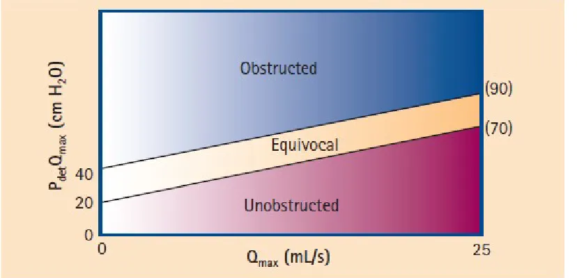
Figure 2: The ICS Nomogram. The patients are divided into 3 classes:
unobstructed, obstructed and equivovocal based on the bladder outlet
obstruction index (BOOI). From Abrams 35 ICS
[image:25.612.56.470.242.442.2]23
International continence society; Pdet detrusor pressure; Qmax maximum
flow rate.
[image:26.612.58.469.201.397.2]
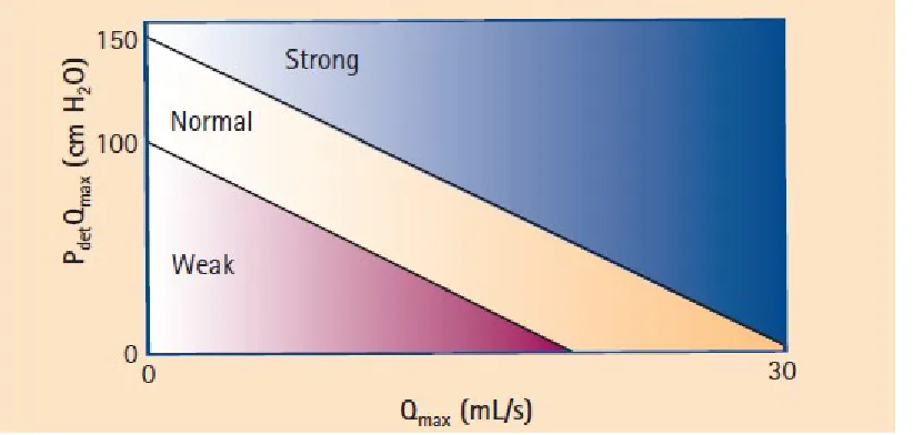
Figure 3: Bladder contractility nomogram. The patients are divided 3 classes;
strong, normal and weak contractility based on Bladder Contractility Index
(BCI). From Abrams 35 Pdet: detrusor pressure; Qmax Maximum flow rate
Using this ICS nomogram, men can be divided into obstructed, equivocal,
and unobstructed according to their BOOI:
BOOI > 40 = obstructed;
BOOI 20-40 = equivocal; and
24
BOOI< 20 = unobstructed (Figure 2).
For purposes of standardization, this nomogram is recommended for use in
older men with LUTS suggestive of BPO.
Significance of straining: Jensen et al36 studied the frequency of abdominal
straining during cystometry and pressure flow studies in men more than 50
yrs who presented with LUTS. Approximately 25-35% of men in this study
either strained before the start of detrusor contraction, during the rise in
detrusor pressure or during flow. Reynards et al37 studied the prevalence of
abdominal straining during free flow in patients with symptomatic BPH and
it showed that approximately 25% of men strained during voiding. Garraway
et al38 in a study of the prevalence of symptoms of prostatic dysfunction in
men aged 40-79 years in Scotland , found that only 0.4% of men said they
strained to void and 11% said they occasionally strained to void. A survey of
urinary symptoms in an unselected community based group of men in united
25
states39reported straining in 15% of men aged over 50 years. Two thirds of
these subjects reported this symptom to be bothersome. In the study by
Reynolds et al straining some or most of the time was reported by 13% of
men and straining occasionally by 44%. A higher prevalence of symptoms is
to be expected in men who specifically present with voiding problems.
Straining to void is an even more common symptom in men undergoing
TURP, prevalence rates of 30-40% being reported in two studies from United
States.40, 41 There are few reports relating the symptom of straining to void
with objective evidence of straining during voiding. Jensen et al noted that
half of the patients who claimed not to strain during voiding did actually
show objective evidence of straining during micturition. Only a few studies
have explored the relationship between abdominal straining and BOO. Susset
et al commented that during voiding patients with obstruction usually
required added pressure provided by straining. However the number in this
study was small and no formal statistical
26
comparison of the prevalence of straining in the obstructed and the non
obstructed groups. Further more the symptoms of straining were still present
in 11 of 15 men following TURP in study by Jensen et al. Relief of
obstruction therefore does not seem to remove the need to strain. Mefflan et
al 42 and Christmas et al43 studied the effect of abdominal straining on urinary
flow rates in young men. In the former study, three men aged between 30 and
40 years performed multiple flow tests with and without abdominal straining.
Straining caused a marked increase in flow rate. In a group of normal young
men and women Christmas et al found that abdominal straining increased
flow raters in men but not in women. Claridge et al44 studied the effect of
straining to void during free flows in a group of men with prostatic or bladder
neck obstruction defined on basis of high voididng detrusor pressure and on
cystoscopic findings. He found that in 29 of 31 patients abdominal straining
had no effect on the flow rate. Infact in 10 men the flow rate fell with
straining. He concluded that the fall in
27
flow rate indicated a rise in outflow resistance, probably as a result of
external pressure on the intra abdominal part of the urethra. The flow rates
remained unchanged or fell in most of the patients who Reynolds et al
studied.
28
Patients & Methods:
Research Questions:
1. Is there an association between BOO and abdominal straining?
2. What is the incidence of straining during voiding in normal men?
3. Is there an Objective co-relation between the symptom of straining and
abdominal pressure?
Inclusion Criteria for Cases:
Men more than 45 years of age with urodynamic proven obstruction as
defined by the ICS criteria.
Inclusion criteria for controls: Normal Men more than 45 years who had
no lower urinary tract symptoms
Exclusion Criteria for Cases:
1. Presence of Co morbidities like diabetes, hypertension, Ischaemic heart
disease
29
2. Previous surgery of urethra, prostate or bladder.
3. Previous pelvic procedures likely to cause bladder denervation like
Hysterectomy and Abdominoperineal excision of rectum.
4. Neurological diseases likely to influence the lower urinary tract 5.
Overactive bladder
6. Bladder calculus.
7. Medications like anticholinergics, diuretics, antidepressants and
antipsychotics.
8. Urinary tract infection
Calculation of Sample size: The sample size was calculated from a
retrospective study comparing abdominal straining in those with BOO to
30
Methodology: The study was conducted in urology outpatient clinic of
Christian medical college, Vellore. This was prospective case control
study.
CASES: Men of age more than 45 years with LUTS attending the urology
outpatient clinic were recruited for the study. They were asked to answer
an international prostatic symptom score (IPSS) questionnaire (annexure
1). All the demographic and clinical data were recorded in the proforma
(annexure 2). Evaluation of patients began with detailed history and
examination done by the investigator. Estimation of serum creatinine, and
urine microscopy was done for all. Urine culture and sensitivity was done
for those who were subjected to urodynamics. Those who have positive
culture with LUTS were excluded from the study. All subjects did a
representative uroflowmetry. Post void residual urine was measured using
abdominal ultrasound using prolate ellipsoid formula; Volume (V) in ml =
31
Those with urinary free flow rate less than 10ml/sec underwent another
free flow with abdominal pressure monitoring and urodynamic evaluation
after written informed consent. The pressure-flow studies were done using
medical measurement systems (MMS) UD 2000 equipment. Single dose
of prophylactic antibiotic was given. Pretest residue was measured prior to
urodynamic evaluation by placing two 6Fr infant feeding tubes. One of
these tubes was used for filling as well as for intravesical pressure
measurement. During cystometry in sitting posture, bladder was filled
with physiological saline at 37ºC at a filling rate of 50ml/min. First
sensation of bladder filling (ml), maximum cystometric capacity (ml),
detrusor overactivity (presence or absence), incontinence (presence or
absence), and compliance (cmH2O) were assessed during filling phase.
Maximum urinary flow (Qmax, ml/sec), Maximum intravesical pressure
on voiding (cmH2O), Voided volume (ml), events like abdominal
32
phase. Abdominal pressure was recorded by using perforated rectal
balloon catheter. Detrusor pressure was calculated by subtracting
intra-abdominal pressure from intravesical pressure. Detrusor pressure at
maximum urinary flow rate (Pdet at Qmax, cm H2O) was measured to
evaluate detrusor contractility. Methods, definitions and units were
appropriate to the standards recommended by the international continence
society.
Controls:
Men more than 45 years without any LUTS were recruited as controls
after written informed consent. All did a representative uroflowmetry.
Those who had flow rates more than 25 ml/sec were included. These
underwent another uroflowmetry with simultaneous measurement of
abdominal pressure. This was measured using urodynamic perforated
33
Definitions:
1) Urinary incontinence was defined if the complaint of any involuntary
leakage of urine was present in which stress urinary incontinence (the
complaint of involuntary leakage on effort on exertion, or on sneezing or
coughing), or urge urinary incontinence (the complaint of involuntary
leakage accompanied by or immediately preceded by urgency) were
noted.
2) Normosensitive bladder - Volume at first sensation of 150 -200ml
3) Delayed first sensation – Appreciation of first sensation of filling at
volume ≥250ml or greater than 50% of maximal cystometric capacity.
4) Detrusor overactivity – Involuntary phasic increase in detrusor pressure
that was difficult to control or could not be controlled by patient resulting
in incontinence or voiding.
5) Normal compliance – Filling detrusor pressure of 5-20cmH2O in the
absence of simultaneous detrusor contraction at maximum cystometric
6) Normal maximum cystometric capacity – Volume 350 to 600ml, at
which there was bladder contraction that resulted in voiding or patient
discomfort.
7) Normal urinary flow rate – Catheterized urine flow rate of more than
12ml/sec.
8) Normal Pdet at Qmax - > 10cm H2O or <40cmH2O during voiding
with catheterized flow rate if more than 12ml/sec.
9) Normal post void residue – 50ml
10) Hypocontractile detrusor – Pdet at Qmax less than 10cmH2O or flat
trace during voiding with or without abdominal straining.
11) Bladder outflow obstruction – Pdet at Qmax more than 40cmH2O with
catheterized urine flow rate less than 12ml/sec. Abrams Griffith Number
calculated as Pdet-2Qmax more than 40
35
Analysis
There were three groups for analysis
Group 1: Those with poor flow and proven BOO
Group 3: Normal men without LUTS
All statistical analyses were performed using Statistical Package for the
social Sciences (SPSS 11.0) for windows. Categorical data was presented
using frequencies and percentage. Continuous data was described using
mean ± standard deviation or median and range. Associations between
categorical variables were assessed using chi-square test with yates’
correction or fisher’s exact test. Continuous variables were compared
using student‘t’ tests and Mann-Whitney tests were used for non-normal
36
Results
Patients included in final analysis: Total of 100 males were included in
the study . Of these 60 were those who presented with LUTS and 40 who
were asymptomatic. These were subdivided for analysis into three groups
as above. The mean age of cases was 58 years and that of the controls was
53 years. Neither had any co morbidities.
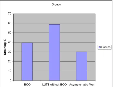
Prevalence of Straining: Of those who presented with LUTS and had
BOO on urodynamics (Group 1) approximately 40% strained on
urodynamic studies as per the criteria defined for straining. In those who
had LUTS but were not obstructed on urodynamics(group B) 58%
strained. 30% of asymptomatic men also strained on free flow done with
abdominal pressure monitoring by a rectal balloon. Comparison between
37
Groups
0 10 20 30 40 50 60 70
BOO LUTS without BOO Asymptomatic Men
S
tr
ai
n
in
g
%
Groups
Figure 4: Comparison of objective evidence of straining on
urodynamic/ free flow with abdominal pressure monitoring between
the three groups
[image:40.612.78.460.96.390.2]38
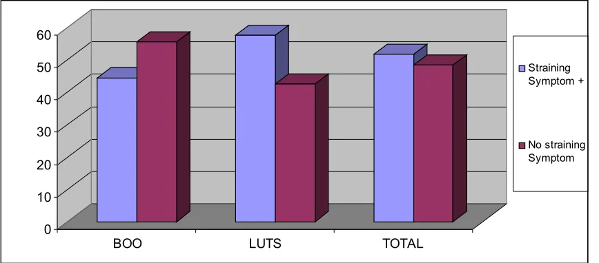
Straining as a symptom: 50% of those who presented with LUTS
complained of straining to void. There was no statistical difference between
the symptom of straining in those who had BOO and those who didn’t. (P
value 0.12). 52% of those who had no symptom of straining on the IPSS but
had other LUTS had obstruction on urodynamics. Straining symptom
therefore was a poor indicator of BOO.
0 10 20 30 40 50 60
BOO LUTS TOTAL
Straining Symptom +
[image:41.612.77.511.363.554.2]No straining Symptom
Figure 5: Comparison of the symptom of straining between those who had
39
Straining in Asymptomatic Men: 30% of asymptomatic men strained during
free flow. Most of them commented that it was habitual and representative of their
normal voiding pattern.
0% 10% 20% 30% 40% 50% 60% 70%
Straining + No straining
Objective Evidence of Straining on Free Flow in Asymptomatic Men
Asymptomatic Men
Figure 6: Straining in controls on free flow with rectal pressure
[image:42.612.95.498.283.491.2]40
Effect of Straining on the free flow rates: To ascertain the effect of
straining on free flow rates a comparative analysis was done between those
who strained and those who didn’t in each of the groups. There was no
statistically significant difference in mean, median, minimum or maximum
flow rates in either group. Among cases the mean flow rate was 6 ml/sec and
6.2 ml/sec in those who strained and those who didn’t respectively.
Comparison of peak flow rates
0 2 4 6 8 10 12 Qmax Mean
Minimun Maximum Qmax Mean
Minimum Maximum
Cases Straining No straining
[image:43.612.62.485.324.610.2]F lo w R at e m l/ se c
Figure: Comparison of flow rates in those with LUTS
41
difference in the mean, minimum or maximum flow rates among those who
strained and those who didn’t. (p value 0.311). An example of straining on
free flow is shown in figure 7.
Comparison of peak flow rates in controls
0 5 10 15 20 25 30 35 Qmax Mean
Minimun Maximum Qmax Mean
Minimum Maximum
Controls Straining No straining
[image:44.612.80.460.168.475.2]Q m ax m l/ se c
Figure 6: Comparison of flow rates in asymptomatic men
42
Figure 7: straining pattern on free flow with abdominal pressure
monitoring in an asymptomatic male.
43
IPSS Scores: The mean IPPS score among the cases was 18. Apart from the
symptom of straining other symptoms were analyzed in the cases. Among
those who had BOO none of the storage or other voiding LUTS were
statistically significant for BOO. Among those who had LUTS but no BOO
on urodynamics none of the other symptoms had any statistically significant
44
Discussion
The term ‘‘lower urinary tract symptoms’’ (LUTS) is an umbrella term
that was introduced originally in 1994 to dissociate urinary symptoms in
the male patients from any implied specific site of origin of symptoms,
such as the prostate19. It is now recognized that LUTS is a global term that
encompasses all urinary symptoms, including storage, voiding, and
postmicturition symptoms. This terminology links well with the
classification proposed by Wein20, which suggested that disorders of
micturition would be more elegantly characterized
as ‘‘failure to store’’ or ‘‘failure to empty.’’ In this context, it is important
to acknowledge the fact that it has been known that symptoms do not
relate to the underlying pathophysiology in many patients; indeed the
phrase ‘‘the bladder is an unreliable witness’’ was coined to acknowledge
this45.Historically, voiding symptoms have been related to obstruction of
the so-called symptoms of ‘‘prostatism.’’ However, it is well recognized
that voiding symptoms poorly correlate with underlying
pathophysiology46. Similar symptoms can also be produced by any other
form of obstruction, such as a urethral stricture or, conversely, by poor
function of the lower urinary tract in circumstances in which there is
impaired detrusor contractility. This has led to the recognition that,
although LUTS may commonly be related to bladder outlet obstruction
(BOO) as a result of benign prostatic obstruction (BPO), which is often
associated with benign prostatic enlargement resulting from the
histological condition of benign prostatic hyperplasia (BPH), this is not
invariably the case. Failure to empty can be related either to an outlet
obstruction or to detrusor underactivity of the bladder, or to a combination
of both. Postmicturition symptoms, such as post void dribbling, occur in
both sexes, but most often in men, in whom these symptoms are highly
common, very troublesome, and cause significant interference with quality
of life. Storage symptoms are currently largely encompassed by the term
overactive bladder (OAB) syndrome, which is defined as urgency,
be correlated with underlying detrusor overactivity. These symptoms tend
to be more bothersome than voiding symptoms, especially if they are
associated with incontinence48.The most recent international
population-based survey, the EPIC study49, was conducted in five countries using the
2002 ICS definitions for LUTS38. This survey assessed prevalence of
OAB, urinary incontinence, and LUTS in more than 19 000 men and
women. The data showed that there is a higher prevalence of storage
(51.3%) versus voiding symptoms (25.7%) in men and all LUTS,
including OAB, in addition to histologic BPH increase in prevalence as
men age. The EPIC study demonstrated that the majority of men with
voiding symptoms did not experience these in isolation but had either
storage and/or postmicturition
symptoms as well. Specifically, among men with LUTS, 9% experienced
both storage and voiding symptoms, whereas an additional 9%
experienced storage, voiding, and postmicturition symptoms. It has also
been shown that there is a far better correlation between storage LUTS and
urodynamics than with Voiding LUTS. In our study however there was no
voiding LUTS in the IPPS with the urodynamic findings. The EpiLUTS
study, a cross-sectional, population representative Internet survey
conducted in the United States, the United Kingdom, and Sweden to assess
prevalence of LUTS, also found that both males and females who reported
having voiding
symptoms were more likely to experience either storage or postmicturition
symptoms, or both50. Data from the US study showed that 10.7% of the
male population 40 yr and older had voiding symptoms, 10.1%
experienced both voiding and storage symptoms, and 24.2% experienced
voiding, storage, and postmicturition symptoms. In our study also most of
the men who complained of voiding LUTS also had storage and post
micturition symptoms.
Uroflowmetry remains a useful screening test in the evaluation of LUTS.
It assesses the combination of detrusor force and outflow opening and,
thus, gives an indirect indication of these aspects of bladder function. Flow
rates must be interpreted together with the voided volume sine low
volumes may give inaccurate flow-rate measurements. The most important
additional information is gained by looking at the voiding time and the
flow pattern. It is mandatory to have more than one flow-rate
measurement, as they can be variable (depending on voided volume,
diurnal variation). The voided volume should be>150ml. For patients with
decreased flow rates who are suspected of BPO, urodynamic studies have
shown that BOO was present in 88% of those with a Qmax <10 ml/s, in
57% of those with a Qmax of 10–14 ml/s, and in only 33% of those with a
Qmax >15 ml/s 51. Thus, a decreased flow rate implies a high likelihood of
BOO due to BPO. Following this study by Abrams et al, a Qmax cut-off
of 15 ml/s has been widely accepted as signifying BPO requiring
treatment. To further increase the probability of detecting BOO we
included those with flow rates of less than 10ml/sec as cases. Also a flow
rate before the urodynamics was done to reduce the effect of intravesical
lines on the maximum flow. Straining during voiding is one of the
questions in the IPSS. There has been sparse literature on the correlation
between the symptom and the objective documentation on urodynamics.
In the previous studies by Jensen et al36 and Reynard et al37 25-35% of men
our study in which 44% of men with urodynamic proven BOO strained.
The incidence of straining was more in those with LUTS without
obstruction (60%; 10/17) on pressure flow studies but it was not
statistically significant. This could be due to the predominance of
hypocontractile detrusors in this group. In this study the symptom of
straining was present in overall 50% of patients with 44% of those with
BOO complaining of straining. Reynard et al had reported straining some
or most of the time in 13% and occasionally in 44% in those with BOO. A
higher prevalence of symptoms is expected in men who specifically
present with voiding problems. Indeed, straining to void is an even more
common symptom in men undergoing transurethral resection of prostate
(TURP) with prevalence rate of 35-40% being reported by Fowler and
Bruskewitz et al.40
There have been few reports relating the symptom of straining to void
with objective evidence of straining during voiding. Jensen et al noted that
half of patients who claim not to strain did actually show objective
evidence of straining during micturition. Thirteen of 61(21%) patients in
of straining and objective evidence of its presence. In our study twenty
seven (27/60) men with LUTS complained of straining to void. Only 12 of
these (44%) had objective evidence of straining on pressure flow studies.
Conversely of the 33 who did not complain of straining 19 actually
strained. In the control group of normal men without any LUTS 30%
strained. These observations suggest that a history of straining or not
during urination may be unreliable and there is a poor correlation between
the symptom of straining and its objective evidence. There was no
consistent pattern in our study as to the timing of straining in the voiding
curve. However in those without LUTS terminal straining was
predominant though not statistically significant. This could be more due to
habit of trying to expel the last few drops of urine. All of these men when
retrospectively inquired also complained of straining to defecate
suggesting a habitual pattern.
Few studies have looked at the relationship between abdominal straining
and BOO. Jensen et al did not find any difference in detrusor pressures in
men with and without straining. In our study there was no statistically
without BOO or in normal men. 44% of those with BOO and 30% of
normal men strained during voiding. There was a poor correlation between
straining to void and the presence of bladder outlet obstruction.
Straining to void in patients with BOO is believed to be an initiating cause
of inguinal hernia. Most of the surgeons believe that relief of obstruction if
possible be carried out prior to hernia repair. However as we have shown
that the symptom of straining is unreliable and upto 50% of those who
claim not to strain in fact do strain to void on objective assessment.
Further more the straining pressures observed in patients who show the
presence of straining are, in general, low compared to those observed with
coughing. These findings in combination with the observation that
straining does not cease after the relief of obstruction (Jensen et al)
suggest that there is no support for the argument that evaluation to rule out
BOO should be performed prior to inguinal hernia repair.
There was no difference in the mean or maximum flow rates between
those who strained and those who didn’t in all the 3 Groups. This is
consistent with the study by Claridge et al who found that in 29 of
study the rate in fact fell in 10 men. This was only seen in 4 men in our
study who initially did not strain during free flow with abdominal pressure
monitoring but later strained during urodynamics and was not significant.
Our current understanding of the physiology of the urethra is that the
urodynamic behavior of the bladder outlet is determined by the principles
governing flow through distensible tubes. There is a flow controlling
Zone, which in prostatic obstruction is located in the prostatic urethra,
where it is under the influence of abdominal pressure. A rise in abdominal
pressure probably results in an increase in outlet resistance thereby
decreasing the flow. Another way to assess the effect of straining on flow
rate could be by asking patients to whistle during voiding and then
54
Conclusions:
From this study it can be concluded that:
1) The relationship between the symptom of straining and the objective
evidence of its presence is poor
2) Straining has a poor correlation with BOO and is not a sensitive
measure of BOO
55
Limitations:
1) Small sample size
2) The effect straining has on the flow rate can be best assessed by having
consecutive flows with subjects being asked to strain at different times
during voiding. This can be incorporated in future studies.
3) The lack of associations between some of the parameters evaluated in
this study might be due to the relatively small sample size and the
consequent low power of the study. Studies with larger sample sizes can
56
BIBLOGRAPHY
1. Boyarsky S. Jones G, Paulson DF, Prout GR. New look at bladder neck
obstruction by the food and Drug Administration regulators Am Ass
Genitourinary Surg 1977; 68: 29-32
2. Madson PO, Iversen P. A point system for selecting operative
candidates. In Hinman F ed. Benign Prostatic Hypertrophy. New York:
Springer- Verlag 1983: 763-765
3. Barry MJ. Fowler FJ, O’Leary MP et al. The American Urological
Association Symptom Index for Benign Prostatic Hyperplasia. J Urol
1992: 148:1549-57
4. Hald T, Nordling J, Anderson JT, Bilde T, Meyhoff HH, Walter S. A
patient weighted symptom score system in evaluation of uncomplicated
benign prostatic hyperplasia. Scand J Urol Nephrol ( Suppl) 1991; 138;
59-62
5. Madsen PO, Iverson P. A point system for selecting operative
Springer Verlag, 1983:763-765
6. Blaivas JG. Multichannel urodynamic studies in men with benign
prostatic hyperplasia: indications and interpretation. Urol Clin North Am.
1990; 17:543-552.
7. Abrams P, Bruskewitz R, De La Rosette J, et al. The diagnosis of
bladder outlet obstruction: urodynamics. In: Cockett ATK, Khoury S, Aso
Y,et al, eds. Proceedings, the 3rd International Consultation on BPH.
World Health Organization;
1995:299-367.
8. Chancellor MB, Blaivas JG, Kaplan SA, Axelrod S. Bladder outlet
obstruction versus impaired detrusor contractility: the role of uroflow. J
Urol. 1991; 145:810-812.
9. Gerstenberg TC, Andersen JT, Klarskov P, et al.High flow infravesical
obstruction in men: symptomatology, urodynamics and the results of
surgery. J Urol. 1982; 127:943-945.
10. George N, Slade N. Hesitance and poor stream in men without bladder
11. Isaacs J, Coffey DS. Etiology and disease process of benign prostatic
hyperplasia. Prostate.1987; 2(suppl):33-50.
12. Elliot SJ, Zorn BH, McLeod DG, Moul JW, Nyberg L, Striker LJ,
Striker GE. Prostate Cancer Prostatic Dis. 2003; 6(2):138-42.
13. Levin RM, Haugaard N, O'Connor L, Buttyan R, Das A, Dixon JS,
Gosling JA. Neurourol Urodyn. 2000; 19(5):609-29
14. Rhodes T, Girman CJ, Jacobsen SJ, et al. Longitudinal prostate growth
rates during 5 years in randomly selected community men 40-79 years old.
J Urol. 1999; 161:1174-1179
15. Jacobsen SJ, Girman CJ, Guess HA, et al. Natural history of
prostatism: longitudinal changes in voiding symptoms in community
dwelling men. J Urol. 1996; 155:595-600.
16. Roehrborn CG, Marks LS, Fenter T, et al. Efficacy and safety of
dutasteride in the fouryear treatment of men with benign prostatic
hyperplasia. Urology. 2004; 63:709-715.
17. Roehrborn CG, Boyle P, Nickel JC. PSA is a significant
predictor of objective parameters in men at risk for BPH progression. J
18. McConnell JD, Bruskewitz R, Walsh P, et al. The effect of finasteride
on the risk of acute urinary retention and the need for surgical treatment
among men with benign prostatic hyperplasia.
Finasteride Long-Term Efficacy and Safety Study Group. N Engl J Med.
1998; 338:557-563.
19. Abrams P. New words for old: lower urinary tract symptoms
for ‘‘prostatism’’. BMJ 1994; 308:929–30.
20. Wein AJ. Pathophysiology and classification of voiding dysfunction.
In: Wein AJ, Kavoussi LR, Novick AC, Partin AW, Peters CA, editors.
Campbell-Walsh urology. ed. 9. Philadelphia: Saunders/Elsevier; 2007. p.
1973–85.
21. Milsom I, Abrams P, Cardozo L, et al. How widespread are the
symptoms of an overactive bladder and how are they managed? A
population-based prevalence study. BJU Int 2001; 87:760–6.
22. Madersbachera S, Alivizatosb G, Nordlingc J, Sanzd CS, Embertone
M, Jean J.M.C.H. de la Rosettef. EAU 2004 Guidelines on
Assessment,Therapy and Follow-Up of Men with Lower Urinary Tract
European Urology 46 (2004) 547–554
23. Kaplan SA. Update on the American urological association guidelines
for the treatment of benign prostatic hyperplasia. Rev Urol. 2006; 8 Suppl
4:S10-7.
24. Jensen, K M., Jmgensen, J. B. and Mogensen, P.: Urodynamics in
prostatism w. Search for prognostic patterns as evaluated by linear
discriminant analysis. Scand. J. Urol. Nephrol, suppl., 114: 84, 1988.
25. McGuire EM, Woodside JR, Borden TA. Prognostic value of
urodynamic testing in myelodysplasic children. J Urol. 1981;
126:205-209.
26. Gleason DM, Lattimer J. The pressure-flow study: a method for
measuring bladder neck resistance. J Urol. 1962; 87:844-852.
27. Griffiths DJ. The mechanics of the urethra and of micturition. Br J
Urol. 1973; 45:497-507.
28. Griffiths DJ. Basics of pressure-flow studies. World J Urol. 1995;
13:30-33.
29. Schafer W. The contribution of the bladder outlet to the relation
Boyarsky, S, eds. Benign Prostatic Hypertrophy.
New York, NY: Springer Verlag; 1983: 470-496.
30. Schafer W. Urethral resistance? Urodynamic concepts of physiological
and pathological bladder outlet function during voiding.Neurourol
Urodyn. 1985; 4:161-201.
31. Schafer W. Urodynamics of micturition. CurrOpin Urol. 1992;
2:252-256.
32. Schafer W. Principles and clinical application of advanced urodynamic
analysis of voiding function.Urol Clin North Am. 1990; 17:553-566.
33. Griffiths DJ, van Mastrigt R, Bosch R.Quantification of urethral
resistance and bladder function during voiding, with special reference to
the effects of prostate size reduction in urethral obstruction due to benign
prostatic hyperplasia. Neurourol Urodyn.1989; 8:17-27.
34. Lim CS, Abrams P. The Abrams-Griffiths nomogram. World J Urol.
1995; 13:34-39.
35. Abrams P. Bladder outlet obstruction index, bladder contractility index
and bladder voiding efficiency: three simple indices to define bladder
36. Jensen KM, Bruskewitz RC, Iverson P, Madson PO. Abdominal
straining in benign prostatic hyperplasia. J Urol 1983; 129; 44-7
37. Reynard JM, Peters T.J, Lamond E, Abrams P. The significance of
abdominal straining in men with lower urinary tract symptoms. BJU
1995; 75: 148-153
38. Garraway WM, Collins GN, Lee RJ. High prevalence of benign
prostatic hypertrophy in the community. Lancet 1991; 338; 469-71
39. Chute CG, Panse LA, Girman CJ et al. The prevalence of prostatism; a
population based survey of urinary symptoms. J Urol 1993; 150: 85-9
40. Fowler FJ, Wennberg JE, Timothy RP, Barry MJ, Mulley AG, Hanley
D. Symptom status and quality of life following prostatectomy. JAMA
1988; 259:3018-22
41. Bruskewitz RC, Larsen EH, Madsen PO, Dorflinger T. 3 year follow
up of urinary symptoms after transurethral resection of prostate. J Urol
1986; 136; 613-615
42. Meffan PJ, Nacey JN, Delahunt B. Effect of abdominal straining on
urinary flow rate in normal males. BJU 1991; 67:134-139
RT. Does abdominal straining influence the flow rate? Proceedings of the
International Continence Society, 18th Annual Meeting. 1988: 36-7
44. Claridge M. Analysis of obstructed micturition. Ann Roy Coll Surg
1966; 39: 30-53
45. Turner-Warwick R, Whiteside CG, Worth PH, Milroy EJ, Bates CP.
An urodynamic view of the clinical problems associated with bladder neck
dysfunction and its treatment by endoscopic incision and trans-trigonal
posterior prostatectomy. Br J Urol 1973; 45:44–59.
46. De la Rosette JJ, Witjes WP, Schafer W, et al. Relationships
between lower urinary tract symptoms and bladder outlet obstruction:
results from the ICS-‘‘BPH’’ study. Neurourol Urodyn 1998; 17:99–108.
47. Abrams P, Cardozo L, Fall M, et al. The standardization of
terminology in lower urinary tract function: report from the
standardization sub-committee of the International Continence Society.
Urology 2003; 61:37–49.
48. Scarpa RM. Lower urinary tract symptoms: what are the implications
for the patients? Eur Urol 2001;40:12–20.
urinary incontinence, overactive bladder, and other lower urinary tract
symptoms in five countries: results of the EPIC study. Eur Urol 2006;
50:1306–15.
50. Coyne K, Sexton C, Kopp Z, et al. The prevalence, bother, and overlap
of LUTS in the US, UK, and Sweden: EpiLUTS. Eur Urol Suppl 2008;
7:238.
51. Abrams P, Bruskewitz R, De La Rosette J, et al.The diagnosis of
bladder outlet obstruction:urodynamics. In: Cockett ATK, Khoury S, Aso
Y,et al, eds. Proceedings, the 3rd International Consultation on BPH.
World Health Organization; 1995:299-367.
66
Annexure: Worksheet for Controls. N=40,1 Present; 2 Absent
Age H.No free flow ml/sec voided volume ml PVR flow with pressure straining +/- initial at peak
flow continuous intermittent
48 675133B 30 350 10 28 2
51 404437D 32 400 20 30 1 1
46 103507D 28 460 20 30 2
50 410568D 25 520 30 26 2
Srimantha 56 272972D 30 450 20 33 2
47 401340D 26 500 20 27 2
Shantana 55 413305D 28 440 25 27 1
Mohd.Alam 48 567462C 25 400 15 25 1 1
59 381381D 25 400 20 26 2
68 382343D 24 380 20 22 1 1
57 383228D 25 600 30 26 1 1
Janardhan 55 386103D 24 450 22 22 2
57 388192D 25 360 20 24 2
49 375628D 27 700 30 24 2
52 386318D 25 300 10 26 2
49 951812B 25 360 20 23 2
Sarvanan 55 893974C 26 550 20 25 1
Armugam 51 396548D 25 400 10 25 1
52 396548D 29 570 20 28 2
Amal Soni 54 389564D 33 680 20 30 2
Mohd Narul 66 395738D 26 440 20 25 2
Santosh 66 396399D 28 360 10 30 2
55 176675D 24 340 20 27 2
55 401128D 25 410 30 24 1 1
Nirul haque 48 080571D 26 300 20 24 2
49 135497D 25 380 10 20 1 1
46 771286 28 550 40 25 2
54 455759C 24 600 10 25 1 1
52 278952D 29 320 37 27 2
49 392635D 25 300 46 24 1
46 369550D 25 290 18 25 1
Jothy singh 67 558220A 25 300 16 20 2
55 397575D 30 480 28 25 2
48 405447D 26 380 22 22 2
49 407173D 28 600 38 25 2
50 225054D 26 300 22 22 2
Sampath 52 780564B 25 360 26 28 2
49 411211D 25 300 10 27 2
47 417001D 26 350 24 20 2
initial at peak flow continous intermittent terminal
1
1 1
1 1
1 1
1
1
1
IPSS SCORE /
35 straining symptom intensity /5 flow Qmax ml/sec voided volume ml PVR Qmax with pressure
563756 22 1 3 7 200 60 7
365961d 24 1 2 7 250 40 6
959924c 30 2 6 180 30 7
366688
D 18 2 5 240 100 6
368870
B 22 2 8 300 30 9
092845
D 20 1 2 8 350 20 6
375429d 23 1 4 5 158 4
376159
D 18 2 9 250 30 11
371729
D 20 1 4 8 200 60 9
055778
D 14 2 9 170 150 10
388265
D 20 1 1 4 200 40 5
386486
D 23 2 8 340 20 10
393868
D 12 1 2 8 560 10 7
016226
C 20 1 2 3 200 40 4
200691
D 26 2 4 380 60 3
264925
D 21 1 1 8 260 20 6
259532d 15 2 6 180 80 8
239814
D 23 2 3 210 20 4
370191
C 22 2 8 350 10 8
271645
D 24 1 2 4 230 40 4
266784d 22 1 3 5 180 50 6
703435
A 18 1 2 5 300 30 4
217568d 16 1 3 4 210 40 3
240894
D 15 2 6 300 20 5
325725
D 17 2 9 400 20 10
388265
D 22 1 2 4 200 10 3
333196
D 12 1 3 8 350 30 9
187549
D 25 2 3 210 20 4
301835
D 20 1 4 4 170 30 5
939026
B 18 1 1 8 350 16 7
299978
D 26 2 6 300 20 6
263976
B 23 1 2 8 400 60 7
295835
D 17 2 6 320 150 8
298854
D 22 1 3 5 220 110 4
283849
D 20 1 2 4 160 120 5
234133
D 22 2 9 500 30 10
068450
1 1 43 7 29 1
1 1 71 7 63 1
2 85 7 71 2
1 1 158 4 150 2
2 108 10 88 2
2 53 7 39 2
1 1 15 2 11 1
2 67 13 39 2
1 1 15 6 3 1
1 1 30 9 12 1
2 74 4 66 2
1 1 97 9 79 1
1 1 42 9 24 1
2 102 3 99 2
2 129 5 61 2
1 1 73 10 53 2
1 1 80 8 64 2
1 1 194 2 190 1
1 1 56 12 32 2
2 40 5 30 2
2 85 8 69 2
2 91 3 85 2
1 1 47 3 41 1
1 1 100 5 90 1
2 44 10 24 2
2 80 3 73 2
1 1 56 9 38 1
1 1 132 3 126 1
2 66 3 60 2
2 80 9 68 2
1 1 110 5 100 1
2 40 10 20 1
1 1 26 10 6 1
2 120 5 110 2
2 76 5 66 2
2 110 10 90 2
2 60 6 48 2
1 1 30 8 14
1 1 183 7 169 1
2 100 5 90 2
2 102 4 94 2
2 68 5 58 2
2 77 5 67 2
1 1 104 8 96 1
2 83 6 71 2
1 1 110 5 100 1
2 129 7 115 2
2 60 10 40 2
2 60 12 46 2
2 95 8 79 2
1 1 83 4 75 1
2 60 3 53 2
1 1 102 2 98 1
2 138 8 122 2
2 88 3 82 2
1 1 91 7 77 1
1 1 78 8 62 1 1
2 88 5 78 2
1 1 90 6 78 1 1
Straining
+/- initial at peak flow continous intermittent terminal compliance Instability
1 1 1 1 2 2 2 1 2 1 1 2
1 1 poor
2 1 1 2 2 2 2 1 1 2 1 1 2 2 2 1 2 1 1 2 2 1 1 2 2 1 1 1 1 2 1 1
Worksheet for Cases N=60 1: Present