“IMMUNOHISTOCHEMICAL EXPRESSION OF CK-19
IN THYROID NODULES AND ITS CORRELATION
WITH HISTOPATHOLOGY”
Dissertation submitted in
Partial fulfillment of the regulations required for the award of M.D. DEGREE
In
PATHOLOGY – BRANCH III
THE TAMILNADU
DR. M.G.R. MEDICAL UNIVERSITY CHENNAI
DECLARATION
I hereby declare that the dissertation entitled
“
IMMUNOHISTOCHEMICAL EXPRESSION OF CK-19 IN THYROIDNODULES AND ITS CORRELATION WITH HISTOPATHOLOGY” is a bonafide research work done by me in the Department of Pathology,
Coimbatore Medical College during the period from July 2014 to July
2015 under the guidance and supervision of Dr. A .Dhanalakshmi M.D., Associate Professor Department of Pathology, Coimbatore Medical
College.
This dissertation is submitted to The Tamilnadu Dr.MGR Medical
University, Chennai towards the partial fulfilment of the requirement for
the award of M.D., Degree (Branch III) in Pathology. I have not
submitted this dissertation on any previous occasion to any University for
the award of any Degree.
Place: Coimbatore
Date: Dr.P. Suriyaprabha
CERTIFICATE
This is to certify that the dissertation entitled
“Immunohistochemical expression of CK-19 in thyroid nodules and its
correlation with histopathology” is a record of bonafide work done by
Dr. P. Suriyaprabha in the Department of Pathology, Coimbatore Medical College, Coimbatore under the guidance and supervision of
Dr. A. Dhanalakshmi M.D., Associate Professor, Department of Pathology, Coimbatore Medical College and submitted in partial
fulfilment of the requirements for the award of M.D. Degree (Branch III)
in Pathology by The Tamilnadu Dr. MGR Medical University, Chennai.
Guide Head of the Department
Dr. A. Dhanalakshmi, M.D., Dr. C.Lalitha, M.D.,
Associate Professor, Professor,
Department of Pathology, Department of Pathology, Coimbatore medical college, Coimbatore medical college, Coimbatore. Coimbatore.
Dr. A.EDWIN JOE.M.D
The Dean,
ACKNOWLEDGEMENT
To begin with, I thank the almighty GOD for his blessings and
guidance in all my activities.
I wish to express my sincere thanks to the honourable Dean,
Dr. A.Edwin Joe, M.D., Coimbatore Medical College and Hospital,
Coimbatore, for permitting me to conduct this study in this hospital.
I extend my gratefulness and thanks to Prof Dr. C. Lalitha, M.D.,
Professor and Head, Department of Pathology for her able guidance and
support and also for providing all facilities to carry out this study.
It‟s a great pleasure to express my humble gratitude to my guide
Dr. A. Dhanalakshmi, M.D., Associate Professor, Department of
Pathology for her innovative suggestions, constant encouragement and
guidance during this endurable work.
I thank Professor Dr. A. Arjunan, M.D., all the Associate
Professors , all Assistant Professors and Tutors of Pathology department,
Coimbatore medical college for their constant support and valuable
opinions.
I wish to thank all my colleagues for their timely help and
encouragement.
I would like to thank the department of General Surgery and
department of Surgical Oncology for their constant support.
It would not be complete without mention of my husband,
Dr. S. Arulananthan, B.D.S., for his encouraging words, extensive help
and constant support throughout this project.
I express my gratitude to my lovable child A. Shriram, my dear
brother P. Parthiban Pradeep, B.E., my respectable parents, other family
members and my friends for their tireless support, encouragement,
prayers and source of strength all through this endeavour.
Finally, I am obliged to all the patients without whom this study
would not have been possible and I dedicate this study to them.
CONTENTS
SI.NO. PARTICULARS PAGE NO.
1. INTRODUCTION 1-3
2. AIM & OBJECTIVES 4
3. REVIEW OF LITERATURE 5-52
4. MATERIALS AND METHODS 53-62
5. OBSERVATION AND RESULTS 63-80
6. DISCUSSION 81-88
7. SUMMARY AND CONCLUSION 89-91
8. BIBLIOGRAPHY
9. ANNEXURES
ANNEXURE I – PROFORMA & CONSENT FORM
ANNEXURE II – MASTER CHART
LIST OF TABLES
SI.NO TITLE PAGE
NO
1 Age distribution of thyroid nodules 64
2 Sex distribution of thyroid nodules 66
3 Distribution of different thyroid neoplasms 67
4 Association of age with histopathological diagnosis 69
5 Association of sex with histopathological diagnosis 71
6 Variants of different thyroid carcinomas 73
7 Incidence of Papillary Carcinoma variants 75
8 Intensity of staining of cytokeratin19 in thyroid nodules 76
9 Intensity of staining of cytokeratin19 in well
differentiated thyroid carcinoma
78
10 Statistical analysis data of cytokeratin19 staining in
thyroid nodules
LIST OF CHARTS
SI.NO TITLE PAGE NO
1 Age distribution of thyroid nodules 65
2 Sex distribution of thyroid nodules 66
3 Distribution of various thyroid neoplasms 68
4 Association of age with histopathological diagnosis 70
5 Association of sex with histopathological diagnosis 72
6 Proportion of variants of different thyroid carcinomas 74
7 Percentage of variants of papillary carcinoma 75
8 Intensity of staining of cytokeratin19 in thyroid nodules 77
9 Intensity of staining of cytokeratin19 in papillary
carcinoma of thyroid
LIST OF COLOUR PLATES
S.NO COLOUR PLATES
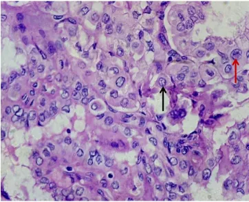
1 Papillary carcinoma of thyroid- H& E (10X)
2 Papillary carcinoma of thyroid showing nuclear grooves and
nuclear pseudoinclusion – H & E (40X)
3 Diffuse 3+ cytoplasmic positivity of cytokeratin19 in papillary
carcinoma (10X)
4 2+ positivity of Cytokeratin19 in papillary carcinoma (10X)
5 Follicular variant of papillary carcinoma – H & E (10X)
6 Follicular variant of papillary carcinoma showing 2+ positivity
with cytokeratin19 (10X)
7 Follicular variant of papillary carcinoma showing 1+ positivity
with cytokeratin19 (10X)
8 Follicular carcinoma of thyroid with capsular invasion- H & E
(10X)
9 Follicular carcinoma thyroid showing vascular invasion- H & E
(10X)
10 Follicular carcinoma of thyroid showing focal 1+ positivity with
cytokeratin19 (10X)
11 Follicular carcinoma showing negative staining with
cytokeratin19 (10X)
12 Follicular adenoma – H & E (10X)
13 Follicular adenoma showing focal 1+ positivity with
cytokeratin19 (10X)
14 Follicular adenoma showing negative staining with cytokeratin19
(10X)
15 Metastatic papillary carcinoma deposits in lymph node – H & E
(10X)
16 Diffuse 3+ positivity of cytokeratin19 in metastatic papillary
1
INTRODUCTION
Thyroid neoplasms constitute the most commonly occurring
endocrine tumors worldwide. Thyroid nodules commonly occur between
30-60 years of age. About 4% to 8% of adult women and 1% to 2% of
adult men present with thyroid nodules that can be identified by physical
examination. With the advent of ultrasonography, the detection rate has
increased to 30%. Majority of the thyroid nodules are benign with
malignant nodules comprising only 10%. Thyroid tumors can arise either
from the epithelial cells lining the follicles or from parafollicular C cells.
Malignant tumors of thyroid are prevalent worldwide. A survey
conducted by WHO during 2010 revealed that around 44,670 new cases
had appeared of which 1690 deaths occurred due to thyroid malignancy.
Papillary carcinoma is the most common malignant tumor constituting
80-85% of all the thyroid carcinomas and that too the classic type,
followed by follicular carcinoma comprising 10- 15%.But the mortality
rate is only 6.5%. According to the surveillance and epidemiology, the 10
year survival rates for malignant thyroid tumors are
Papillary carcinoma – 98% Follicular carcinoma – 92% Medullary carcinoma – 80%
2
Early diagnosis of thyroid tumors and appropriate management
will prolong the survival rate of patients. However distinguishing various
thyroid lesions by hematoxylin and eosin sections alone is really
challenging to pathologist.
This is well stated by Baloch and Livolsi as, “Thyroid follicular
lesions are the bane of the Pathologist” in their article.
As many thyroid tumors have overlapping morphological features,
exact diagnosis is very essential for surgical and post- operative
management of patients. Especially papillary carcinoma and its follicular
variant which mimics follicular carcinoma can be treated by simple
thyroidectomy, if diagnosed early. Differentiation of follicular adenoma
and follicular carcinoma depends on capsular and vascular invasion.
When it is inconclusive, false diagnosis of benignity may lead to
extensive vascular dissemination and dismal prognosis. An increasing
number of immunohistochemical markers are used in the differential
diagnosis of both benign and malignant thyroid lesions. They are
cytokeratin19, CD56, HBME-1, Galectin-3, Ret oncoprotein, p17,
CITED1, PAX8 and EGFR (epidermal growth factor receptor).
Cytokeratin19, a low molecular weight protein of 40kDa belonging
to keratin family is an intermediate filament involved in protein binding
3
diagnosis of thyroid tumors. Many studies have reported it as sensitive
marker in differentiating benign from malignant thyroid tumors.
The purpose of this study is to analyse the usefulness of
Cytokeratin19 in differentiating thyroid nodules by grading the intensity
of staining in cytoplasm of cells and to correlate it with the
4
AIM OF THE STUDY
To study the immunohistochemical expression of Cytokeratin19 in
various types of thyroid nodules and its correlation with histopathology.
OBJECTIVES:
1. To study the expression of Cytokeratin19 in different thyroid
lesions.
2. To study the value of cytokeratin19 in differentiating benign
from malignant thyroid nodules.
3. To study the correlation of cytokeratin19 expression in thyroid
5
REVIEW OF LITERATURE
Thyroid gland is the major endocrine gland that controls the
metabolic functions of the human body. THOMAS WHARTON, an
English physician and anatomist from London, in 1656 named this
endocrine gland as THYROID GLAND, as it resembled the SHIELD,
used in Ancient Greece.
The occurrence of palpable thyroid nodules in adults is about 5%.
The main goal in clinical medicine is to identify malignant lesions in a
cost effective manner. In iodine deficient areas thyroid nodules are more
frequent with higher occurrence in women and also with increasing age.
The palpability of nodules depends on its location in the thyroid gland
and anatomy of patient’s neck. These nodules can be detected by thyroid
ultrasound, CT scan and pathological studies. Nowadays, image guided
biopsies or FNAC is more helpful. But FNAC is not confirmatory test as
follicular adenoma and follicular carcinoma cannot be differentiated by
this test.
Papillary carcinoma which is identified by its characteristic nuclear
features like nuclear grooves , intranuclear cytoplasmic inclusions are
also present in atypical adenoma ,Hyalinising trabecular adenoma,
6
Hence, histopathological study of the tissue sections are essential.
But certain tumors of thyroid have overlapping features and often
pose a diagnostic difficulty especially in the tumors having follicular
pattern such as follicular adenoma, follicular carcinoma and follicular
variant of papillary carcinoma.
As the prognosis and management are different, differentiating
these lesions is essential. As follicular patterned lesions are identified
based on cytological criteria like nuclear grooving, nuclear overlapping
and intranuclear pseudoinclusions , interobserver variations are common.
This leads to inappropriate nomenclature or diagnosis.
Thyroid follicular lesions are either capsulated or unencapsulated
but with follicular architecture. The four important lesions that should be
differentiated are
Hyperplastic colloid nodule
Follicular adenoma
Follicular carcinoma
Follicular variant of papillary carcinoma
In an attempt to overcome this diagnostic difficulty many
immunohistochemical markers are evaluated in distinguishing papillary
7
CK19, HBME-1, galectin-3, CD 56, Leu-7 (CD 57), CITED-1,
fibronectin-1, CD 15, PAX8, CD 44 and platelet derived growth factor.
CD 56 expression is lost in papillary carcinoma but is expressed in benign
and other malignant lesions of thyroid including normal thyroid.
EMBRYOLOGY:
Thyroid gland starts developing around 2nd or 3rd week of gestation
and completes by 11th week and becomes functional at third month.1
Thyroid gland develops from median endodermal thyroid diverticulum,
which arises from foramen caecum, present between tuberculum impar
and copula linguae in the base of the tongue. From foramen caecum,
thyroglossal duct develops and descends down behind the hyoid bone to
the neck. It lies in front of the trachea and bifurcates to form two lobes of
thyroid gland.
The thyroglossal duct then obliterates. Sometimes the lower end of
thyroglossal duct may persist forming pyramidal lobe or Lalou ette’s
pyramid. Parafollicular or ‘C’ cells are derived from caudal pharyngeal
complex or ultimobranchial body derived from fourth and fifth
pharyngeal pouches.2 Solid cell nests, having collections of stratified
epithelial cells with mucin production focally and cyst formation are the
8
On 9th week follicular cells are present as cords and plates. On 10th
week follicular lumina appears and is small , by 12thweek colloid
secretion begins and at 14th week well formed follicles lined by cuboidal
cells, containing colloid within the lumen are present .
HISTOLOGY:
Thyroid gland is covered by a fibrous capsule and the septa
arising from it divides the gland into lobules. Each lobule is composed of
many follicles of approximately 200µm in diameter. They are lined by
follicular cells with central lumen containing colloid. The interstitium
contains parafollicular C cells, lymphatics and blood vessels. C cells are
called as clear cells or light cells. They are polyhedral with eccentrically
placed oval nuclei.
The follicular cells vary in their shape depending upon their
function. The normal cells are cuboidal, whereas the inactive or resting
cells are flat to squamous and become columnar when hyperactive.3
ANATOMY:
The normal thyroid gland weighs about 25 grams in adults and is
9
left joined by isthmus. Each lobe measures about 5cm x 2.5 cm x 2.5 cm.
Isthmus measures 1.2 cm x 1.2 cm. Each lobe extends from the middle of
thyroid cartilage to fourth or fifth tracheal ring. The isthmus extends
between second to fourth tracheal rings.
Thyroid gland has both true and false capsule. True capsule arises
from connective tissue of gland and false capsule from pre-tracheal
fascia. Suspensory ligament of Berry connects the thyroid gland to
cricoid cartilage, posteriorly.
It has rich blood supply from superior thyroid artery, inferior
thyroid artery, thyroidea ima artery in 3%of individuals, branches of
tracheal and oesophageal arteries. Thyroid gland is drained by superior,
middle and inferior thyroid veins.
Lymphatic drainage is to the upper and lower deep cervical nodes,
pre-tracheal and para-tracheal nodes. Nerve supply is from the middle
cervical ganglion, mainly and small contributions from the superior and
inferior cervical ganglia.
PHYSIOLOGY:
The thyroid gland plays important role in regulation of basal
metabolic rate, calcium metabolism, somatic and psychic growth by the
10
potent than T4 (prohormone of T3). Thyroperoxidase is the primary
enzyme of thyroid hormone synthesis.
These hormones are regulated by negative feedback mechanism of
hypothalamic-pituitary-thyroid axis. Thyrotrophin releasing hormone
(TRH ) from hypothalamus, enters anterior pituitary gland and stimulates
secretion of thyroid stimulating hormone ( TSH ) which in turn acts on
the thyroid gland, to produce and release T3 and T4. They bind to thyroid
binding globulin (TBG) in plasma. Unbound forms are the active one in
tissues.
THYROID TUMORS - AN OVER VIEW:
Thyroid tumors are the most common endocrine tumors. The
estimated age standardized annual incidence is 1.0 to 2.9 cases per 1, 00,
000 men and 3.4 to 9.1 cases per 1, 00, 000 women according to
GLOBACON 2008. Thyroid tumors are more common in developed
countries. The incidence of thyroid tumors has increased in past two
decades, predominantly papillary carcinoma of thyroid.5The liberal
criteria for diagnosis of papillary carcinoma of thyroid and detection of
small tumors by imaging techniques and environmental factors led to
11
RISK FACTORS FOR THYROID CARCINOMA :
Irradiation to head and neck
Age : <20 or >45 years
Bilaterality
Female gender
Iodine deficiency (follicular cancer)
Positive Family history for thyroid carcinoma or MEN 2 syndrome
FEATURES INDICATING MALIGNANCY:
Extrathyroidal extension.
Fixation of nodules to adjacent soft tissues and structures.
Paralysis of vocal cord
Involvement of lymph nodes.
Nodule size > 4 cm.
Progressively enlarging neck mass.
GENERAL CHARACTERISTICS OF PRIMARY THYROID CANCERS:
1. Most common histologic type is papillary carcinoma.7
2. Females are most commonly affected than men. 8
3. Young patients have well differentiated tumors whereas in older
patients less differentiated tumors are common.
4. Young females below 40 years have slightly better prognosis
12
5. Size of the primary tumor with staging is essential and is an
important factor that determines the prognosis.
CHARACTERISTIC FEATURES OF PRIMARY THYROID CARCINOMAS IN CHILDREN:
1. Most common is papillary carcinoma of thyroid. National
Cancer Institute in 2013 in their statistical analysis also found
that papillary carcinoma is common comprising 70-80%.10
2. Radiation exposure plays important role, example : Hiroshima
Nagasaki bomb explosion.11
3. 60 – 80% of them present with lymph node metastasis and
recurrence is more common in these patients.12
4. Thyroid carcinomas in children, though aggressive has slightly
good prognosis having mortality rate 2.6 % only.
FAMILIAL THYROID TUMORS:
25% of tumors occur in familial form, not only medullary
carcinoma, but also familial non - medullary thyroid carcinoma can
occur.
Familial non-medullary thyroid carcinoma syndrome is diagnosed
if three or more first degree relatives have non - medullary thyroid
13 Examples are:
1. Familial papillary thyroid carcinoma with or without oxyphilia
chromosome locus 19p13.2 (TCO).
2. Familial papillary thyroid carcinoma with renal papillary
neoplasia chromosome 1q21 (FPTC/PRN).
3. Familial non- medullary thyroid carcinoma type –I chromosome
2q21 (NMTC-1).
4. Familial multinodular goiter syndrome chromosome 14q31
(MNG – 1).
SYNDROMES ASSOCIATED WITH THYROID TUMORS:
S.
NO SYNDROME
GENE
INVOLVED INCIDENCE THYROID TUMORS
1. Familial adenomatous polyposis
APC (5q21) 2-12% Papillary carcinoma cribriform - morular variant often
2. PTEN- hamartoma tumour (Cowden syndrome )
PTEN (10q23.2)
> 10% Follicular carcinoma, papillary carcinoma occasionally, benign follicular nodules 3. Carney complex PRKAR1α
(17q22-24)
15% Follicular carcinoma, papillary carcinoma, benign follicular nodules
4. Werner syndrome WRN (8p11-12)
14
WHO CLASSIFICATION (2004) OF PRIMARY THYROID TUMORS
Tumors of thyroid follicular or metaplastic epithelium:
1. Follicular adenoma (includes Hürthle cell adenoma)
2. Papillary carcinoma
3. Follicular carcinoma (includes Hürthle cell carcinoma)
4. Mucinous carcinoma
5. Mucoepidermoid carcinoma
6. Sclerosing mucoepidermoid carcinoma with eosinophilia
7. Poorly differentiated thyroid carcinoma
8. Anaplastic / Undifferentiated carcinoma (including squamous cell
carcinoma and carcinosarcoma)
Tumors showing C-cell differentiation
1. Medullary carcinoma
Tumors showing both follicular and C-cell differentiation
1. Collision tumor: follicular/papillary and medullary carcinomas
15
Tumors showing thymic or related branchial pouch differentiation
1. Ectopic thymoma
2. Carcinoma showing thymus-like element (CASTLE)
3. Spindle epithelial tumor with thymus-like differentiation
(SETTLE)
Tumors of lymphoid cells
1. Malignant lymphoma
2. Extramedullary plasmacytoma
Mesenchymal and other tumors
1. Benign and malignant mesenchymal tumors such as solitary
fibrous tumor, peripheral nerve sheath tumor, smooth muscle
tumor, and angiosarcoma
2. Paraganglioma
3. Teratoma
16
Tumor, Node, Metastasis (TNM) Staging of Tumors of the
Thyroid:
TUMOR (T):
TX - Primary tumor cannot be assessed
T0 - No evidence of primary tumor
T1 - Tumor ≤ 2cm in greatest dimension and limited to the thyroid
T2 - Tumor >2 cm but <4 cm and limited to the thyroid
T3 - Tumor >4 cm in greatest dimension and limited to
the thyroid or tumor with minimal extrathyroid
extension (e.g., extension to perithyroid soft tissues
or sternothyroid muscle)
T4a - Tumor of any size extending beyond thyroid capsule
and invades subcutaneous tissue, larynx, trachea,
esophagus or recurrent laryngeal nerve
T4b - Tumor invades prevertebral fascia / encases carotid
artery/ mediastinal vessels.
All Anaplastic carcinomas are considered T4
17
T4a - Intrathyroidal anaplastic carcinoma - any size
T4b - Extrathyroidal anaplastic carcinoma - any size
REGIONAL LYMPH NODES (N) :
NX - Regional lymph nodes cannot be assessed
N0 - No regional lymph node metastasis
N1a - Metastasis to Level VI (pretracheal, paratracheal, and
prelaryngeal/ Delphian lymph nodes)
N1b - Metastasis to unilateral, bilateral or contralateral
cervical or Superior mediastinal nodes.
DISTANT METASTASIS (M) :
MX - Distant metastasis cannot be assessed
M0 - No distant metastasis
M1 - Distant metastasis present
STAGE GROUPING
Separate stage groupings are recommended for papillary (or)
18 Papillary or Follicular (<45 years) :
Stage I Any T Any N M0
Stage II Any T Any N M1
Papillary or Follicular (45 years and older):
Stage I T1 N0 M0
Stage II T2 N0 M0
Stage III T3 N0 M0
Stage III T1 N1a M0
Stage III T2 N1a M0
Stage III T3 N1a M0
Stage IVA T4a N0 M0
Stage IVA T4a N1a M0
Stage IV A T1 N1b M0
Stage IVA T2 N1b M0
Stage IVA T3 N1b M0
Stage IVA T4a N1b M0
Stage IVB T4b Any N M0
19 Medullary Carcinoma :
Stage I T1 N0 M0
Stage II T2 N0 M0
Stage III T3 N0 M0
Stage III T2 N1a M0
Stage III T2 N1a M0
Stage III T3 N1a M0
Stage IVA T4a N0 M0
Stage IVA T4a N1a M0
Stage IVA T1 N1b M0
Stage IVA T2 N1b M0
Stage IVA T3 N1b M0
Stage IVA T4a N1b M0
Stage IVB T4a N1b M0
Stage IVC Any T Any N M1
Anaplastic Carcinoma :
All anaplastic carcinomas are considered Stage IV
Stage IVA T4a Any N M0
Stage IVB T4b Any N M0
20
ROLE OF IMMUNOHISTOCHEMISTRY IN THYROID LESIONS:
As there is morphological overlap between many thyroid tumors
with follicular pattern as seen in follicular adenoma, follicular carcinoma
and follicular variant of papillary carcinoma and the nuclear features
characteristic of papillary carcinoma like nuclear grooves and inclusions
are also seen in multinodular goiter with papillary hyperplasia and
Hyalinising trabecular adenoma , immunohistochemistry is helpful in
differentiating the tumors. A panel of immunomarkers that are useful
includes Cytokeratin 19, HBME-1, galectin -3, CD 56, PAX 8 and Ret –
oncoprotein.
CYTOKERATIN 19:
Cytokeratin 19 belongs to the keratin family. It is a 40 kDa protein
that is encoded by KRT 19 gene in human being. It is an intermediate
filament involved in protein binding, organization of myofibres and
maintains structural integrity of epithelial cells. This acidic protein
arranged in pair of heterotypic keratin chains unlike its related family
members is not paired with basic cytokeratin in epithelial cells. They are
clustered in region of chromosome 17q12 - q21.
Cytokeratin 19 is widely applied as diagnostic marker of
21
found cytokeratin19 as a sensitive marker in diagnosis of papillary
carcinoma and its variants.15It is also expressed in defined zone of basal
keratinocytes, sweat gland, mammary gland ductal and secretory cells ,
GIT, ectocervix epithelium and urothelium.
HBME-1:
HBME-1 (Hector Battifora mesothelial ) is a monoclonal antibody
which act against the antigen present on mesothelial cell membrane. It is
named after Dr. Hector Battifora, who introduced this marker.The target
epitope is located in microvilli. It is expressed in thyroid papillary and
follicular carcinoma but is not expressed in nodular goiter or in nodular
hyperplasia.16
GALECTIN-3:
Galectin-3 is a 31kDa protein belonging to the lectin family and is
encoded by the gene LGALS3 located in chromosome 14 in the locus
q21-q22. It binds to beta-galactosides and play important role in
regulation of cell to cell or cell to matrix interaction, repair of cell
damage and cell migration. Galectin-3 also plays an important role in
neoplastic transformation and inflammation. It aids in distinguishing
papillary carcinoma and follicular variant of papillary carcinoma from
22 CD 56:
CD56, a neural adhesion molecule plays an important role in
regulating migrating capabilities of neoplastic cells. Loss of CD 56
expression leads to increase in metastatic potential of tumor cells and
leads to poor prognosis. CD 56 is normally expressed by normal thyroid
follicular epithelial cells. Low expression of CD 56 is useful in diagnosis
of papillary carcinoma of thyroid.18
PAX 8:
PAX 8, a transcription factor is essential for the development of
thyroid follicular cells and also expresses thyroid specific genes. It is
expressed in papillary carcinoma, follicular neoplasms, medullary
carcinoma and poorly differentiated carcinoma. It is also expressed in B
cell lymphomas, renal cell carcinoma and normal B lymphocytes.
RET ONCOPROTEIN:
Ret gene play an important role in the production of tyrosine
kinase, a transmembrane receptor. Ret gene is located in chromosome
10q. It is not expressed in normal thyroid follicular cells, but gene
rearrangement commonly occurs in papillary carcinoma and hence useful
in the diagnosis of papillary carcinoma, which is proved by the study
23 BENIGN THYROID NODULES:
It is classified as hyperplastic nodules and benign epithelial
neoplasm. Hyperplastic nodules includes dyshormonogenetic goiter
and nodular hyperplasia. Benign epithelial tumors are follicular
adenoma and hyalinising trabecular adenoma.
DYSHORMONOGENETIC GOITER:
It occurs due to defect in hormone synthesis due to peroxidase
deficiency, deiodinase deficiency, defective iodide transport, defective
coupling, decreased thyroglobulin synthesis and loss of function of
pendrin gene ( ion channel for transport of iodine).20 Thyroid gland is
grossly enlarged and multinodular. Microscopically it may show
microfollicular, solid, papillary and insular pattern. Marked nuclear
atypia of cells inbetween hyperplastic nodules and increased mitoses are
seen. Follicular carcinoma and papillary microcarcinoma are incidental
findings.21T4 replacement therapy can produce thyroid tumors in these
24 NODULAR HYPERPLASIA:
It is also called as multinodular goiter or adenomatous goiter or
adenomatous hyperplasia. It exists in two forms, endemic and sporadic
goiter. Endemic goiter occurs in geographical areas with low iodine
content in soil and water leading to defective thyroid hormone synthesis
which stimulates TSH release and causes diffuse or nodular colloid
goiter.
Sporadic goiter is due to dietary deficiency of iodine or increased
excretion of iodine by kidney or defective hormone synthesis by
antibodies. In both, thyroid gland is enlarged and has multiple nodules
surrounded by complete or incomplete capsule. Few dilated follicles have
conglomerate of small follicles at one pole which are active called as
Sanderson polsters.
Some follicles are cystically dilated with papillary hyperplasia and
the papillae face towards the center of the cyst which may mimic
papillary carcinoma24. Nuclear atypia is seen in cells within nodules due
to previous irradiation incontrast to dyshormonogenetic goiter where it is
seen inbetween hyperplastic follicles. It is also different from adenoma
which is solitary and completely encapsulated. Chromosomal
abnormalities are rare and may have TSHR mutations, extra copy of
25 FOLLICULAR ADENOMA:
Follicular adenoma is a solitary benign encapsulated tumor and the
patients are in euthyroid state. In radioactive iodine scan, usually
follicular adenoma is cold, at times, warm and rarely hot. Hot nodules
indicate benign lesion.
PLUMMER ADENOMA :
Follicular adenoma with hyperthyroidism is called as toxic
adenoma or PLUMMER ADENOMA.23Plummer adenomas have
activating mutation of TSHR or GNAS1. Intra luminal calcium oxalate
crystals present in thyroid follicles, with hyperfunctioning nodule outside
is a sign of hypofunction.Mitosis is rare. Secondary degenerative changes
like hemorrhage, cystic degeneration and fibrosis are common.
Some follicular adenoma have papillary structures reported as
papillary adenoma in past which gave confusion with papillary carcinoma
is now termed as follicular adenoma with papillary architecture.24
Follicular adenoma has four different patterns:
1) Normofollicular / simple type
2) Microfollicular / fetal type
26 4) Trabecular /embryonal / solid type
Rarely, papillary pattern can occur.
DIFFERENTIAL DIAGNOSIS OF LARGER FOLLICLES:
1) Hyperplastic nodule
2) Follicular variant of papillary carcinoma
DIFFERENTIAL DIAGNOSIS OF SOLID / TRABECULAR / NESTED PATTERN:
1) Medullary carcinoma
2) Poorly differentiated carcinoma
But they are mostly invasive. Calcification, edema and bone
formation are more common.
IMMUNOHISTOCHEMISTRY OF FOLLICULAR ADENOMA:
1) Low molecular weight keratin - cytoplasmic positivity
2) TTF 1- nuclear positivity.
27 MOLECULAR GENETICS:
No molecular test effectively distinguish follicular adenoma from
follicular carcinoma because both has similar chromosomal abnormalities
like activating RAS stimulation, PAX 8 / PPARγ rearrangement.26
Other Chromosomal abnormalities specific for follicular adenoma -
are translocations involving Chromosomes 19q13 having break point at
ZNF 331 gene locus and chromosome 2p21 - break point at (THADA)
thyroid adenoma associated gene locus.27Sporadic follicular adenoma has
rarely alteration of PI3K / PTEN/ AKT pathway.
VARIANTS OF FOLLICULAR ADENOMA:
1) Hurthle cell adenoma.
2) Hyalinizing trabecular adenoma.
3) Atypical adenoma – has irregular cytoarchitecture but, lacks
capsular and vascular invasion.28
4) Adenoma with bizzare nuclei- cells occur in clusters and have huge
hyperchromatic nuclei. Other malignant features are absent.
5) Clear cell type
6) Adenolipoma
28
8) Spindle cell adenoma - resembles meningioma somehow.
9) Black adenoma - minocycline induced.29
TREATMENT:
Lobectomy
Levothyroxine to suppress the nodule and
I131 for toxic adenoma.
HYALINIZING TRABECULAR ADENOMA:
It was first identified by Langhans and the term given by
Carney.30
MICROSCOPY:
Tumor cells are arranged in trabecular pattern. Cytoplasm shows
prominent hyaline material due to intermediate filament accumulation
which is also present in extracellular matrix. Hyalinised collagen and
basement membrane material are present.
Cytoplasmic yellow body - pale yellow inclusion bodies situated
near nucleus with refractile quality is present.31 Psammoma bodies,
29 IMMUNOHISTOCHEMISTRY:
Thyroglobulin and TTF -1 is strongly positive, galectin -3in half of
cases and NSE and neurotensin – only focally positive.
Pathogenetic link between hyalinising trabecular adenoma and papillary carcinoma:
Both Papillary carcinoma and hyalinising trabecular adenoma
have nuclear grooves, nuclear pseudoinclusions and psammoma
bodies.
Both express epithelial type keratins.
Hyalinising trabecular adenoma can have foci of papillary
carcinoma.
Papillary carcinoma with hyalinising trabecular adenoma like
pattern with cervical node metastasis is seen.
Both have RET/PTC rearrangement.
Because of the overlapping features it is now termed as
30
HURTHLE CELL OR ONCOCYTIC TUMORS:
HURTHLE CELL ADENOMA:
It is common in female adults .Tumors are solid, tan, encapsulated
and has rich vascularity. The tumor cells have follicular, papillary,
trabecular or solid pattern and has inspissated colloid having concentric
laminations. Cells have deeply eosinophilic, granular cytoplasm. Nuclei
sometimes exhibit pleomorphism and may have prominent nucleoli. Few
bizarre forms are also seen. But they do not indicate malignancy. Most of
them are benign and show reactivity for thyroglobulin. Benign tumors are
called as Hurthle cell adenoma.
ATYPICAL HURTHLE CELL ADENOMA:
It is also called as Hurthle cell tumors of uncertain malignant
potential (HCT-UMP) .They have solid or trabecular pattern of growth
with increased nuclearcytoplasmic ratio. There is no capsular or vascular
invasion and does not metastastize to other sites.
HURTHLE CELL CARCINOMA:
They are aggressive tumors and have solid pattern of growth,
increased mitoses and have capsular and/or vascular invasion. They
metastatise to bone and lungs. These tumors exhibit aneuploidy and
31
Hurthle cell tumors commonly undergo acute infarction following
fine needle aspiration. Hurthle cell tumors >4 cm have poor prognosis.
MALIGNANT THYROID TUMORS:
PAPILLARY CARCINOMA:
It is the most common primary thyroid carcinoma and affects any
age with mean age of 40 years having female preponderance. In children,
90% of thyroid malignancy is constituted by papillary carcinoma.
Irradiation to head and neck causes papillary carcinoma in 5 -10 % of
cases and can arise in patients with Hashimoto’s thyroiditis.
Papillary carcinoma in thyroid gland alone is 67%, Thyroid and
cervical nodes - 13%, Lymph nodes alone -20%.33
GROSS APPEARANCE:
Size varies from microscopic to larger nodule and most of the
tumor nodules are < 1 cm. Grossly it appears as infiltrating nodule
with ill - defined border and is grey white to tan and granular.
Encapsulated variant has thick capsule and constitutes < 10%.34
32 MICROSCOPIC FEATURES:
Papillary carcinoma consist of numerous branching papillae having
central fibrovascular core and lined by stratified cuboidal cells having
characteristic nuclear features like Ground glass appearance / orphan
annie nuclei / optically clear with nuclear overlapping, nuclear
pseudoinclusions (round acidophilic vacuoles due to cytoplasmic
invagination into nucleus ) and nuclear grooves along the long axis of
nucleus due to infolding of redundant nuclear membrane and nuclear
microfilament.59
Psammoma bodies are seen in papillary stalk or between tumor
cells or in fibrous stroma. Psammoma bodies are concentric lamellated
basophilic structures occurring as a result of calcification of individual
necrotic tumor cells and should be distinguished from inspissated
secretions in Hurthle cell tumor.
VARIANTS OF PAPILLARY CARCINOMA:
1) PAPILLARY MICROCARCINOMA:
It is usually ≤1 cm in diameter, formerly called as occult sclerosing carcinoma or non - encapsulated sclerosing tumor. It is most
common in males.36 RET / PTC rearrangements and BRAF mutations
33 2) ENCAPSULATED VARIANT:
Tumor nodule is completely encapsuled.
D /D: Hyperplastic nodule with central cystic degeneration which
appears hot on thyroid scan and their papillae face towards the centre of cystic cavity and has pale vacuolated colloid. Immunohistochemistry
shows negativity with high molecular weight keratin.
3) FOLLICULAR VARIANT:
Tumor cells are arranged in follicles and is invasive. Psammoma
bodies, colloid with scalloped margins, abortive papillae and distinctive
nuclear features are present. Follicular variant of papillary carcinoma has
many types like solid variant, macro follicular variant, diffuse /
multinodular variant and encapsulated variant called as LINDSAY
TUMOR.37
4) DIFFUSE SCLEROSING VARIANT:
It involves one or both lobes of thyroid gland. Dense sclerosis,
solid foci, psammoma body, squamous metaplasia and lymphocytic
infiltration are present.38 Lymph node and brain metastasis are common.
They exhibit both RET/PTC1 and RET/ PTC3 rearrangements.
34
5) ONCOCYTIC / OXYPHILIC VARIANT:
Tumor cells have abundant granular eosinophilic cytoplasm with
papillary or follicular pattern with nuclear features of papillary
carcinoma.39 It has good prognosis.
6) TALL CELL AND COLUMNAR CELL CARCINOMA:
Tall cell variant has single layer of tall cells whose height is equal to three times the breadth.40 It has papillary structures, nuclear pseudoinclusions and lymphocytic infiltration of stroma. It is more
aggressive and affects older age group. Extrathyroidal extension is
common.
Columnar cell carcinoma has stratified layer of columnar cells, with subnuclear vacuolation and papillary carcinoma nuclear features are
present. It has high proliferative index and has poor prognosis.
7) CRIBRIFORM MORULAR VARIANT:
It has cribriform growth pattern with morular formation.
Ultrastructurally - accumulation of microfilaments made of biotin leads to nuclear clearing and is different from papillary carcinoma with strong
35
8) PAPILLARY CARCINOMA WITH EXUBERANT NODULAR FASCITIS LIKE STROMA:
It has prominent stromal reaction giving, fibroadenoma like
appearance. It resembles nodular fasciitis and fibromatosis.42
MOLECULAR GENETICS:
Mitogen activated protein kinase pathway which regulates cell
proliferation differentiation and survival plays major role in causation of
papillary carcinoma. MAPK - pathway activation leads to RET/PTC
rearrangements, TRK rearrangements, BRAF mutation and RAS
mutation in follicular cells of thyroid. These mutations are mutually
exclusive.
REARRANGEMENTS:
Gene rearrangements constitute 20 - 40% of papillary carcinoma.
RET oncogene present in chromosome 10q11.2 is a transmembrane
tyrosine kinase receptor , the point mutations of which causes medullary
carcinoma and rearrangements causes papillary carcinoma. RET
rearrangements occurs in intron 11 by intrachromosomal inversions
involving long arm of chromosome 10, interchromosomal translocations.
RET fuses with 12 different genes leading to 17 different chimeric
36
RET/PTC1 (RET fusion with CCDC6 a.k.a H4 or D10S170),
RET/PTC3 (RET fusion with ncoa4 a.k.a, RFG, ELE1 or ARA 70), these
two occur by intrachromosomal rearrangements and are more common.
RET/PTC2 (RET fusion with PRKAR-1A, gene which is inactivated in
patients with Carney complex) occurs in one third of the cases. Other
mutations are rare.
RET/PTC is common in children and young adults and those
exposed to radiation.43 These rearrangements occur in classical papillary
carcinoma or microcarcinoma. They represent low stage with little
proliferative capability and less likely to undergo dedifferentiation. These
features are more commonly seen in RET/PTC1 rearrangement.
RET/PTC3 may behave aggressively. These rearrangements can be
detected by reverse transcriptase polymerase chain reaction or by
fluorescent in situ hybridization.
BRAF:
BRAF activating mutations are the most common genetic alteration
constituting 30-70% of papillary carcinoma.It belongs to RAF family and
is a serine threonine kinase of MAPK pathway. Most common molecular
alteration is thymidine to adenine transversion in nucleotide 1799 of exon
15 which leads to valine to glutamate substitution in residue 600 of
37
BRAF mutations are more specific for papillary carcinoma of
thyroid and is present in papillary carcinoma arising in struma ovarii.
BRAF mutation is characteristically present in tumors with papillary
architecture. It is uncommon in follicular variant of papillary carcinoma.
Other rare mutations of BRAF are K601E mutation, paracentric inversion
of chromosome 7 and small deletions near codon 600.
BRAF mutations have been attributed to male sex, older age group,
extrathyroidal extension, metastasis to lymph nodes and distant sites,
recurrence, high tumor stage at initial presentation and reduced survival.
BRAF mutation are reduces gene expression required for enzymes
production in thyroid hormone synthesis and it makes the tumor
refractory to treatment radioactive iodine.
RAS:
RAS mutations are seen in follicular patterned thyroid tumors like
follicular adenoma, follicular carcinoma, follicular variant of papillary
carcinoma.44
NTRK1:
NTRK1 gene encodes a transmembrane tyrosine kinase receptor
and it binds with nerve growth factor. NTRK1 rearrangements constitute
38
recombination at NTRK locus in chromosome 1q22 producing chimeric
oncogene leading to spontaneous activation of NTRK1 tyrosine kinase.
METASTASIS:
Cervical lymph nodes are commonly involved with cystic
degeneration. It is commonly seen in young patients .Blood borne
metastasis is less frequent and involves lungs, bone, soft tissue, central
nervous system, breast and pancreas. In lung the metastatic foci appear as
miliary micronodules and this can be identified with I131 scintiscan.
Occasionally tumor spreads to nearby parathyroid glands.
PROGNOSIS:
• Good prognostic factors:
• Children and adults < 40 years45
• Females and
• Encapsulated variant.46
• Bad prognostic factors:
• Age is > 40 years
• Extra thyroid extension
39 • Multicentric tumor
• Tumors with distant metastasis
• Poorly differentiated and anaplastic carcinoma 47and
• Tumors with aneuploidy and BRAF mutations.48
IMMUNOHISTOCHEMISTRY:
Cytokeratin 19, high molecular weight keratin demonstrated by
34βE12, thyroglobulin and TTF -1 are strongly positive.49 Other markers are TTF-2, PAX8, S 100, HBME-1, galectin 3, CD 15, CD 57, EMA,
CEA, antichymotrypsin, insulin like growth factor, HER2/neu ,
c-Met/hepatocyte growth factor receptor and vimentin.
FOLLICULAR CARCINOMA:
Follicular carcinoma, a rare neoplasm of elderly females
predominantly constitutes 10 – 18 % of all the primary thyroid tumors. It
is identified by the capsular or vascular invasion or invasion of adjacent
thyroid.50
It arises commonly in patients with endemic goiter and iodine
deficiency. Rarely, it arises from follicular adenoma. Irradiation and
40 GROSS EXAMINATION:
Follicular carcinomas is solid tan to light brown fleshy, sometimes
glistening with, areas of hemorrhage and cystic degeneration .Minimally
invasive variant are encapsulated. Size varies from 1cm to 10 cm.
MICROSCOPIC FEATURES:
Follicular carcinoma has thick fibrous capsule in which the tumor
cells are arranged in closely packed follicles, trabecular pattern or solid
sheets. The tumor cells are cuboidal to low columnar and have round
nuclei with inconspicuous nucleoli sometimes exhibiting, nuclear
pleomorphism. Mitosis is uncommon. Vascular invasion and capsular
invasion are the diagnostic features and it differentiates follicular
adenoma from follicular carcinoma.
VARIANTS (based on invasion ):
1) Minimally invasive follicular carcinoma.51
2) Widely invasive follicular carcinoma.52
MINIMALLY INVASIVE FOLLICULAR CARCINOMA:
It is encapsulated variant resembling follicular adenoma of
embryonal or fetal type. Invasion into vessels of venous caliber within or
41
wall of vessels lined by endothelium or protrudes into lumen. CD31,
Ulex europaeus, Factor –VIII related antigen and Fli –I endothelial cells
marker are useful. Capsular invasion should be present and
pseudoinvasion has to be ruled out. Pseudoinvasion is due to herniation
of tumor tissue due to breech in capsule made by surgeon on fresh
specimen.
TERMS USED IN FOLLICULAR NEOPLASM:
Follicular carcinoma – tumors with definite capsular invasion
Follicular tumor of uncertain malignant potential – has
questionable capsular invasion but does not have nuclear features
of papillary carcinoma.
Well differentiated tumors of uncertain malignant potential – has
questionable nuclear changes (? Papillary carcinoma type)
WIDELY INVASIVE FOLLICULAR CARCINOMA:
Tumors that are encapsulated and having four or more blood vessel
invasion or those having wide spread infiltration into blood vessels and /
or adjacent thyroid tissue are termed as widely invasive follicular
carcinoma. Metastasis to sternum, shoulder girdle, skull and iliac bone is
common. Those tumors that look alike normal thyroid tissue is called as
42
has affinity for radioiodine.53 < 5% of minimally invasive type of tumors
have metastasis.
MOLECULAR GENETICS:
1. Castro and Colleagues postulated that chromosomal gains and
aneuploidy leads to microfollicular/ solid / trabecular pattern. Diploidy
and near diploidy forms normofollicular pattern.
2. Loss of heterozygosity, (LOH) of 20% per chromosomal arm produce
follicular carcinoma and only 5% loss occurs in follicular adenoma
and codon 12 and 13 of K-RAS occurs in follicular carcinoma.54
3. PAX 8/ PPAR gamma rearrangement due to t (2:3) (q13; p25) is
common in females, young age, highly cellular and invasive tumors.55
4. PI3K / PTEN /AKT pathway activation is common in COWDEN SYNDROME, CARNEYS COMPLEX I and WERNER SYNDROME .56 5. TSHR gene mutations are rare and occurs in hyperfunctioning
follicular carcinoma
6. VEGFR1 genes are also involved.
POORLY DIFFERENTIATED CARCINOMA:
Tumors falling in between well differentiated and anaplastic type
are the poorly differentiated carcinoma .INSULAR CARCINOMA is the
43
carcinoma can also progress to poorly differentiated carcinoma. It is
common in old age about 60 years. Recurrence, extrathyroidal extension
and metastasis to lymph nodes (14 – 48 %) and distant sites (12-44%) are
common. Mortality rate is increased to 50%. TP53 mutation and β catenin mutations are common.58Insular carcinoma analogous to “Langhans’ wuchernde struma “ is common in South America and Europe.
Gross and Microscopic Appearance:
The tumor is solid grey white, partly encapsulated or invasive with
areas of hemorrhage and necrosis. Microscopically the tumor cells are
arranged in insular pattern with retraction artifact or as diffuse sheets.
Coagulative necrosis giving peritheliomatous appearance is common.59
Tumors cells are small with vesicular or hyperchromatic nuclei and
vascular invasion is commonly seen. Tumor cells are positive for TTF-1
and PAX8 and have increased Ki-67 index.
UNDIFFERENTIATED / ANAPLASTIC CARCINOMA:
Anaplastic carcinoma constitutes 2-5% of primary thyroid cancers.
Women of >70 years are commonly affected. They are aggressive tumors
growing rapidly in a shorter period with increased incidence of recurrence
44
Tumor cells produce granulocyte colony stimulating factor causing
marked increase in leucocytes. They are resistant to chemotherapy.
Younger patients with tumors < 4 cm can be operated with radical
surgery along with adjuvant chemoradiation.
They have mutation in TP53 gene (70%), β-catenin mutation (65%), RAS mutation (30%) mutations in BRAF and RET/ PTC, PTEN,
APC, PIK3CA and APC.
GROSS AND MICROSCOPIC FEATURES:
Grossly tumor completely replaces entire thyroid gland and
invades surrounding soft tissue. Microscopically cells are either of
squamoid type or sarcomatoid type – having spindle cells and giant cells.
Epithelial looking cells are arranged in sheets and large polygonal with
highly pleomorphic nuclei and many giant cells and bizarre forms are
seen. Spindle cell components are alike to undifferentiated pleomorphic
sarcoma, with extensive necrosis and hemorrhage.
VARIANTS:
Angiomatoid variant
Osteoclastic variant
Rhabdoid variant
45
Paucicellular variant
Carcinosarcoma
Adenosquamous carcinoma
Squamous cell carcinoma
Immunohistochemistry:
Cytokeratin positivity is variable (47-90%) depending on
proportion of carcinoma component antigen and PAX8 (76%).61
MEDULLARY CARCINOMA:
Medullary carcinoma is a malignancy with parafollicular C cell
differentiation. It may be sporadic or part of familial or multiple
endocrine neoplasia 2A or 2B.62 Sporadic form is common in 44- 50
years, and around 10-30 years in MEN syndrome. In sporadic form
bilateral tumors are 0-32%, 40-50% of nodal metastasis and 12% of
distant metastasis with intermediate prognosis. Hereditary medullary
carcinomas have autosomal dominantly acquired RET proto-oncogene
mutation and 90% are bilateral tumors.
Familial and MEN-2A associated medullary carcinoma have
indolent course with mutation in exon 10, 11, 13, 14 or 15. MEN 2B
incur mutation at exon 16 (ATG ACG; methionine to threonine) and
46
parathyroid tumors and cutaneous lesions; MEN 2B Pheochromocytoma,
neuromas of mucosa and intestine and Marfanoid features. Lymph node
metastasis is 10 – 30 % in hereditary form with metastasis to distant sites
rarely except MEN 2B (38%).
Grossly the tumor is small firm, greywhite to tan or reddish brown
.Tumor is most commonly present in middle third of lateral lobe because
of increased C cell number there. They may have capsule. Larger tumors
have necrosis and hemorrhage. Microscopically tumor cells are arranged
in nest, sheet, trabecular, tubular, pseudopapillary, cribriform or
microglandular pattern. Cells are round to polygonal cells having
amphophilic cytoplasm with round nuclei having stippled chromatin.
Nuclear pleomorphism and mitoses are infrequent. Increased vascularity
is a striking feature.80-85% of cases have amorphous eosinophilic
material called as amyloid.63
VARIANTS:
Glandular / follicular type
Oncocytic / oxyphilic type
Pseudopapillary type
Clear cell type, small cell type , pigmented variant
Spindle cell type ,Giant cell variant,
47
Pseudoangiosarcomatous like , carcinoid like
Hyalinising trabecular adenoma like
Immunohistochemistry:
Tumors show strong positivity with cytokeratin,
pan-neuroendocrine markers, TTF1, calcitonin, CEA- (80 -100%).
Amyloid can be stained with congo-red and under polarized light
gives apple green birefringence.
PROGNOSTIC FACTORS:
GOOD prognosis:
Female sex
Medullary carcinoma in MEN 2A64
Medullary microcarcinoma (<1 cm) and
Small tumors.
BAD prognosis:
Above 45 years of age
MEN 2B associated medullary carcinoma Small cell type
Calcitonin poor tumors65 and
48 MUCOEPIDERMOID CARCINOMA:
Primary mucoepidermoid carcinoma is a low grade malignant
neoplasm which is rare.67 Females are commonly affected around 10-83
years of age . 20% of the individuals presented with thyroid mass having
extrathyroidal extension. Lymph node metastasis occur commonly but
distant metastasis is rare.
Harach stated “Mucoepidermoid carcinoma of thyroid arises from
ultimobranchial body”.68 Some other study insisted as tumor of
thyroglossal duct origin.
Histologically the tumor is not circumscribed and has cellular
islands present in a sclerotic background.some of the cells contain
intracytoplasmic mucin. Few of the cells are squamoid. Comedo type of
necrosis, nuclear pleomorphism and psammoma bodies are present.
Rarely glands that are lined by ciliated columnar epithelium is seen.
Mucoepidermoid carcinoma can occur along with papillary
carcinoma.two cases of mucoepidermoid carcinoma associated with
follicular carcinoma (Hurthle cell variant) are also on record.69
IMMUNOHISTOCHEMISTRY:
49
SCLEROSING MUCOEPIDERMOID CARCINOMA WITH
EOSINOPHILIA:
It is a rare tumor of low grade malignant behavior and occurs in a
background of Hashimoto thyroiditis.70 It arises from metaplastic
squamous epithelium. It is common in adults with mean age of 55 years
and with female preponderance. It is an aggressive tumor.71
Tumor is composed of nests and anastomising cords of cells in a
dense sclerotic stroma which is infiltrated with eosinophils and
lymphocytes. Tumor is infiltrative and it extends to perithyroidal tissue.
Cells are polygonal with mild to moderate nuclear pleomorphism with
prominent nucleoli. Some foci show squamoid nests and mucin pools.
Perineural invasion and blood vessel obliteration are common. Lymph
node metastasis resembles Hodgkin lymphoma.72
IMMUNOHISTOCHEMISTRY:
CYTOKERATIN AND TTF-1 are positive in these tumors.
MUCINOUS CARCINOMA:
Primary mucinous carcinoma is very rare in thyroid .Only seven
cases have been reported in literature.73 these tumors metastatize rapidly
with mean survival of about 6 months to 4 years. It is similar to colloid
carcinoma occurring in other sites.
50
TUMORS SHOWING DIFFERENTIATION OF BOTH FOLLICULAR AND C-CELL :
COLLISION TUMORS:
Collision tumors are composed of two recognizable types of
carcinoma of thyroid. They are
1. Medulllary carcinoma and follicular carcinoma74,75
2. Papillary carcinoma and medullary carcinoma76
They occur contiguously and are more aggressive.
MIXED MEDULLARY CARCINOMA AND FOLLICULAR CELL CARCINOMA:
It is also called as Follicular-parafollicular carcinoma or differentiated carcinoma of intermediate type.77This rare tumor arises from stem cells hence showing dual component. They are not capsulated.
They have features of medullary carcinoma along with follicles. Other
patterns like nests, cribriform, trabecular and solid pattern are also seen.
Amyloid is present in few cases. These cells show neurosecretory
granules, cells having intermediate features, follicular cells and
indifferent cells ultrastructurally.
Immunohistochemistry :
51
TUMORS OF HEMATOLYMPHOID CELLS:
MALIGNANT LYMPHOMA:
Primary thyroid lymphomas, constitutes 2.5 to 3% of extranodal
lymphomas and comprises 4 to 5 % of thyroid malignancies. It commonly
occurs in elderly females. Lymphomas, commonly arises from
lymphocytic thyroiditis or Hashimoto thyroiditis. Thyroid lymphomas
form non -circumscribed rubbery or soft mass. Cut surface bulges out and
is fleshy homogeneous and light tan coloured. Size of the tumor may vary
from 1 to 14 cm.
Non Hodgkin lymphomas are common in thyroid than
Hodgkin lymphoma. Diffuse large B-cell lymphoma (constitute 70%) and
extranodal marginal zone lymphoma of MALT type occur most
commonly.78 Follicular lymphoma and Burkitt lymphoma are very rare.79
Some cases of intravascular large B cell lymphoma and T cell lymphoma
that expressing γδ receptors of T cells are reported.
METASTATIC MALIGNANT TUMORS IN THYROID:
As thyroid has rich blood supply and lymphatics invasion or
metastasis to thyroid is common. Tumors like lung adenocarcinoma,
colorectal carcinoma, renal cell carcinoma, malignant melanoma, breast
52
psammoma bodies can be seen in metastatic deposits giving
misconception with papillary carcinoma.81
Metastatic neuroendocrine carcinoma from intra-abdominal site
and bronchus present as solitary or multiple nodules within the thyroid
gland giving misinterpretation as medullary carcinoma. The metastatic
carcinomatous deposits usually present as multiple nodules with
increased vascularity and hemorrhage. Immunohistochemistry with
53
MATERIALS AND METHODS
Study Design:
Prospective study
Study Period:
From July 2014 – July 2015
Study Place:
Coimbatore Medical College and Hospital, Coimbatore.
Sample Size:
A total number of 30 cases.
From case records brief clinical data were collected, which included age,
sex, clinical diagnosis and surgical procedure .
The following inclusion and exclusion criteria were adopted.
Inclusion Criteria :
1. All thyroidectomy specimens (hemithryoidectomy, subtotal and near
total thyroidectomy and total thyroidectomy) done for solitary
nodule or multiple neoplastic nodules.
2. Patients in all age groups
54 Exclusion criteria:
Multinodular goiter
Toxic goiter
Methods:
Among the total thyroidectomy specimens that were received in
the department of Pathology in our hospital during the study period, 30
cases were taken into study as per inclusion criteria and were evaluated
further .
All those 30 thyroidectomy specimens (one with lymph node
metastasis) selected were then fixed in 10% formalin, embedded in
paraffin and stained with hematoxylin and eosin.
HEMATOXYLIN AND EOSIN STAINING METHOD:
REAGENTS USED:
1. Hematoxylin solution- Erhlich’s hematoxylin
2. Eosin Y 1% solution
3. Acid alcohol 1% solution
PROCEDURE:
55
2. Place the sections in Isopropyl alcohol for 15 minutes.
3. Wash in running tap water.
4. Stain in Erhlich’s hematoxylin for 10 to 15 minutes.
5. Differentiation is done with 1% acid alcohol two to three dips.
6. Blueing is carried out for 10 minutes.
7. Counterstain with eosin 1% solution 3 to 4 dips.
8. Running tap water wash.
9. Air dry
10.Mount with DPX
After hematoxylin and eosin staining, all slides were reviewed by
pathologist and categorized as following
1. Follicular adenoma
2. Minimally invasive Follicular carcinoma.
3. Widely invasive follicular carcinoma
4. Papillary carcinoma
5. Follicular variant of papillary carcinoma
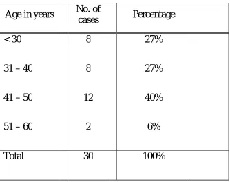
The age group varied between 19 to 60 years.
Follicular adenoma:
Follicular tumors that are completely encapsulated having

![TABLE 6: VARIANTS OF DIFFERENT THYROID CARCINOMAS [N=30]](https://thumb-us.123doks.com/thumbv2/123dok_us/288488.61567/90.595.128.503.145.552/table-variants-different-thyroid-carcinomas-n.webp)