Int. J. Electrochem. Sci., 5 (2010) 815 - 827
International Journal of
ELECTROCHEMICAL
SCIENCE
www.electrochemsci.org
Computational Studies on Effects of Efavirenz as an Anticancer
Drug on DNA: Application in Drug Design
S. Riahi a,b*, S. Eynollahi b and M.R. Ganjali b,c ,P. Norouzi b,c
a Institute of Petroleum Engineering, Faculty of Engineering, University of Tehran, Tehran, Iran b
Center of Excellence in Electrochemistry, Faculty of Chemistry, University of Tehran, P. O. Box 14155-6455, Tehran, Iran
c
Endocrinology & Metabolism Research Center, Tehran University of Medical Sciences, Tehran, Iran
*
E-mail: [email protected]
Received: 20 May 2010 / Accepted: 12 June 2010 / Published: 20 June 2010
This research is designed to further understand the physicochemical interaction between the novel drug EFZ, and its biological receptor DNA. The ultimate goal is to design drugs that interact more with DNA. Understanding the physicochemical properties of the drug as well as the mechanism by which it interacts with DNA should ultimately enable the rational design of novel anti-cancer or anti-viral drugs. Molecular modeling on the complex formed between EFZ and DNA presented this complex to be fully capable of participating in the formation of a stable intercalation site. Furthermore, the molecular geometries of EFZ and DNA bases (Adenine, Guanine, Cytosine and Thymine) were optimized with the aid of the B3LYP/6-31G* method. The properties of the isolated intercalator and its stacking interactions with adenine···thymine (AT) and guanine···cytosine (GC) nucleic acid base pairs were studied with the DFTB method, an approximate version of the DFT method that was extended to cover the London dispersion energy. The B3LYP/6-31G* stabilization energies of the intercalator···base pair complexes were found to be -5.55 kcal/mol and -7.52 kcal/mol for AT···EFZ and GC···EFZ, respectively. It was concluded that dispersion energy and the electrostatic interaction contributed to stability of the intercalator·DNA base pair complexes. Results from comparison of the DFTB method and HF method conclude close results and support each other.
Keywords: DNA, Intercalator, EFZ, DFTB, Drug Design, Stacking Interaction
1. INTRODUCTION
Combination of anti-HIV agents has long been an indispensable tool in fighting the AIDS epidemic. Combination of drugs from different classes has proven to be beneficial in terms of sustained efficacy and long-term safety, provided there are no significant negative pharmacokinetic drug-drug interactions [3].
In recent years the DFT method was applied in different branches of chemistry [4-38]. This paper presents the recently introduced approximate DFT method, DFTB technique (density functional tight-binding), empirical London dispersion energy term, which is accurate and reliable for computational studies [39-40], and calculations performed using the DFTB technique for H-bonded and stacked DNA base pairs [41-42]. Furthermore, this computationally very efficient procedure can yet be used in quantum mechanical (QM) and QM/molecular mechanical (MM) MD simulations very conveniently and accurately [43-44].
The quantum mechanical description of interactions between EFZ and DNA base pairs (Watson-Crick base pairing) employing the DFTB method are reported in this paper. To achieve this goal EFZ and DNA base pairs were simulated and; atomic charges, geometrical values (bond lengths, bond angles and dihedral angles), dipole moment, polarizability, and energies of the frontier molecular orbitals (HOMO and LUMO) were obtained. According to a literature survey, this is the first paper that studies EFZ and DNA base pair intercalations using the DFT method.
The aim of this work was to study the geometries, electronic EFZstructures and its molecular complexes with nucleobases by DFTB methods. This study will shed more light on the nature of intercalations between a drug and DNA dominantly from the viewpoint of: charge transfer, dispersion and electrostatic forces. Hence, the study can help design new intercalators (drugs) to interact more with DNA.
2. COMPUTATIONAL METHODS
Calculations on the isolated molecules and molecular complexes were performed within GAUSSIAN 98 package [45].
Each species was initially optimized with PM3 method and, then the optimized structures were again optimized with density functional theory using the 6-31G* basis set. Full geometry optimizations and frequency calculations were performed and each species was found to be a minimum by having no negative values in the frequency calculation. The calculations gave internal energies at 0 K. In order to obtain gas phase free energies at 298.15 K, it is necessary to calculate the zero-point energies and thermal corrections together with entropies to convert the internal energies to Gibbs energies at 298.15 K [46-47].
Frequency calculations on these structures verified that they were true minima and provided the necessary thermal corrections to calculate H (Enthalpy) and G (Gibbs free energy). Finally, full optimizations and frequency calculations for each species were performed with the DFT/6-31G* [48-49].
guess structures are considered based on PM3 semi-empirical calculations followed by full geometry search based on Newton–Rapson procedure as implemented in GAMESS quantum chemistry code [50]. The most stable geometries were achieved when the intercalator (EFZ) and base pairs (AT and GC) were situated in coplanar planes in such a way that the major system axes were parallel. There is special definition for the molecular geometries of DNA base pairs. In all cases, the QM-optimized geometries of the base pairs and the intercalators were used for the QM calculation. Thus, when the idealized geometries were utilized, the interacting molecules were overlaid by their B3LYP/6-31G* optimized geometries, based on the least-squares fitting method. In the case of the empirical potential calculations, either the subsystem geometries were relaxed by the empirical potential or the QM-optimized geometries were saved. This difference had an insignificant effect on the calculated energies.
The other one-electron properties (dipole moment, polarizability, energies of the frontier molecular orbital) were also determined at the B3LYP/6-31G* level. For the charged species, the dipole moment was derived with respect to their mass center, because for the non-neutral molecules the calculated dipole moment depended on the origin of the coordinate system.
The stabilization energies of the selected complexes were determined with the help of the DFT calculations and calculated with a recently introduced method, based on the combination of the approximate tight-binding DFTB with the empirical dispersion energy. The DFT methods are known to be inherently very deficient for stacking interactions, as they basically ignore the dispersion attraction [51-53]. As a consequence, their enlargement by an empirical dispersion term currently appears to be a very reasonable way to improve the major deficiency of the DFT method for the evaluation of the molecular complexes. It should also be mentioned that the interaction energies were obtained as the difference between the complex energy and the combined energies of the molecules in isolation [54].
Processes in DNA environment depend on a delicate balance between stacking interactions, hydrogen bonding and hydration effects [55] Hydration free energies could be calculated by implicit models like solvent reaction field [56] and Langevin dipoles [57] methods or by explicit models in conjunction with free-energy calculations and molecular dynamics simulations [58] Due to complexity of these calculations, hydration effects will be evaluated in future studies.
3. RESULTS AND DISCUSSION
3.1. EFZ characteristics
a)
b)
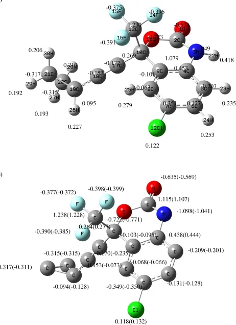
Figure 1. (a) The optimized structure and the atom charges of EFZ. (b) The optimized structure and the atom charges of EFZ after the complex formation with GC and AT (Parentheses include the changes after the complex formation with AT)
-0.377(-0.372)
-0.103(-0.095)
-0.068(-0.066) -0.390(-0.385)
-0.722(-0.771) -0.398(-0.399)
-0.635(-0.569)
-1.098(-1.041)
0.438(0.444)
-0.209(-0.201)
-0.349(-0.350) -0.131(-0.128)
0.118(0.132) -0.170(-0.235) -0.153(-0.073)
-0.094(-0.128) -0.315(-0.315) -0.317(-0.311)
1.115(1.107)
0.264(0.277) 1.238(1.228)
-0.375
-0.531
0.279 0.235
0.418 -1.049
-0.391
-0.351 -0.396
0.253 0.122
-0.172 -0.153
-0.095 0.210
0.206
0.192
0.193
0.227 -0.315
-0.101
-0.203 0.453
1.079 -0.723
0.269 1.230
-0.127 -0.067
[image:4.596.56.518.82.718.2]
Table 1. Significant computed geometrical parameters for EFZ before and after complex formation
EFZ does not have a planar structure. It should also be mentioned that the atom charge distribution in the EFZ is delocalized. The C2 exhibited the highest positive charges which was the cause of two bonding to the oxygen atoms with high electronegativity. The most negative charge is O11, because it contacted to two carbons are electropositive. The presence of electronegative elements in EFZ facilitated its interaction with the DNA molecule through hydrogen bonding with the GC and AT hydrogen. In addition, there are two kinds of interactions between EFZ and DNA; electrostatic interactions and dispersion interactions, being discussed in the next paragraphs.
Bond
lengths EFZ
EFZ- GC
EFZ- AT
Bond
Angles EFZ
EFZ - GC
EFZ - AT
Bond
Dihedrals EFZ
EFZ - GC
EFZ - AT
R(1,2) 1.20 1.22 1.21 A(1,2,3) 124.21 124.15 124.81 D(1,2,3,4) -175.92 -174.46 -176.30
R(2,3) 1.36 1.34 1.35 A(1,2,11) 120.67 119.22 119.55 D(1,2,3,22) 1.57 3.19 1.55
R(2,11) 1.36 1.36 1.36 A(2,3,4) 124.88 124.08 124.62 D(11,2,3,4) 2.43 3.87 2.04
R(3,4) 1.40 1.34 1.41 A(2,3,22) 115.30 115.86 115.62 D(11,2,3,22) 179.93 -178.46 179.99
R(3,22) 0.99 1.12 0.92 A(4,3,22) 119.76 119.77 119.72 D(1,2,11,10) -164.96 -167.20 -164.12
R(4,5) 1.39 1.39 1.38 A(3,4,5) 120.28 119.81 120.13 D(2,3,4,5) 170.20 169.26 170.14
R(5,6) 1.38 1.38 1.38 A(4,5,6) 120.00 119.84 120.00 D(2,3,4,9) -9.04 -9.62 -9.05
R(5,23) 1.07 1.07 1.07 A(4,5,23) 119.93 119.61 120.04 D(22,3,4,5) -7.19 -8.30 -7.73
R(6,24) 1.07 1.07 1.07 A(11,10,13) 104.18 103.91 103.53 D(3,4,5,6) -179.30 -179.20 -179.34
R(10,11) 1.44 1.44 1.45 A(11,10,17) 107.65 107.66 107.84 D(3,4,5,23) 0.33 0.07 0.30
R(10,17) 1.45 1.45 1.45 A(2,11,10) 125.90 125.54 125.73 D(23,5,6,24) 0.47 0.70 0.49
R(17,18) 1.19 1.19 1.19 (18,19,20) 119.76 119.90 120.71 D(17,10,11,2) -150.04 -148.05 -150.82
R(18,19) 1.43 1.44 1.44 A(19,20,26) 116.57 25.66 25.73 D(11,10,13,14) -60.23 -60.12 -60.41
R(19,20) 1.52 1.52 1.52 A(19,20,27) 117.01 117.03 116.44 D(11,10,13,15) 60.15 60.18 59.35
R(20,28) 1.07 1.07 1.07 A(19,20,28) 117.37 117.40 117.45 D(17,10,13,14) -175.31 -175.09 -175.61
Table 2. Dipole moment [D], polarizibility [B3], HOMO and LUMO energies (in eV) of the drug, the bases and the base pairs
Compound HOMO LUMO Dipole moment Polarizability
AT -8.64 3.01 1.28 213.2
GC -7.35 2.74 2.51 223.4
EFZ -9.18 -2.16 4.68 177.4
A -8.83 3.12 2.49 101.2
T -9.53 2.94 3.88 89.1
G -8.45 3.52 2.76 109.2
[image:5.596.153.457.619.725.2]
Table 2 depicts the one-electron properties (dipole moment and polarizability) and the energies of the frontier molecular orbital (HOMO and LUMO) of EFZ using the DFTB computational method. The dipole moment, which is the first derivative of the energy, with respect to an applied electric field as a measure of asymmetry in the molecular charge distribution. The high values of the dipole moment and the polarizability present that the electrostatic and the dispersion contribution will play a key role in the interaction with the nucleobases.
3.2. Base pairs characteristics
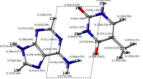
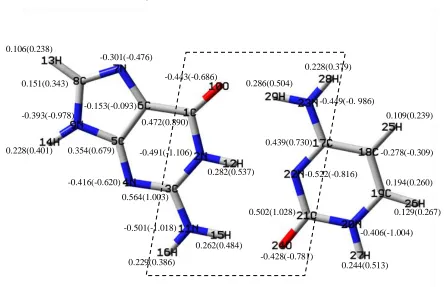
[image:6.596.38.510.306.566.2]The optimized structures of the adenine...thymine (AT) and guanine...cytosine (GC) based pairs in the Watson-Crick structures are visualized in Figures 2 and 3, respectively. Tables 3 and 4 show the significant computed geometrical parameters, using the DFTB method before and after the complex formation.
Figure 2. Optimized structure and charge of AT base pair & EFZ···AT before and after the complex formation (Parentheses include the changes after the complex formation)
In addition, Table 2 presents the one-electron properties (dipole moment and polarizability) and the energies of the frontier molecular orbital (HOMO and LUMO) of the bases and the base pairs. From Table 2, it is clear that all the bases and base pairs are very poor electron acceptors (all LUMO energies are positive in contrast to the LUMO energy of EFZ which is negative).
The bases and the base pairs are apparently good electron donors and among the isolated bases the best one is guanine. This is in accordance with experimental and theoretical studies showing, that ultimate carcinogens primarily react with DNA at the N7 atom of guanine [59-60]. The electron donor
0.358(0.649) -0.337(-0.549)
0.162(0.298)
-0.512 (-0.815) 0.128(0.254)
0.115(0.246)
-0.104(-0.040) 0.237(0.405)
-0.476(-0.995) -0.390(-0.973)
0.170(0.364)
-0.335(-0.515)
0.492(0.794)
0.237(0.394)
0.260(0.474) 0.491(1.080)
0.304(0.550)
-0.337(-0.191)
-0.225(-0.427) 0.246(0.480)
-0.364(-0.638)
-0.410(-0.662) 0.220(0.226)
0.472(0.846) -0.457(-1.073)
-0.418(-1.013)
0.097(0.157)
0.129(0.184)
[image:7.596.69.513.138.425.2]
ability of all bases is further magnified by base pairing. For example, the HOMO energy of guanine (-8.45 eV) increases by 1.1 eV upon pairing by cytosine. Furthermore, the high polarizability and dipole moment values of AT and GC (but more than those of EFZ) reveal that the electrostatic and dispersion contribution influence considerably the interaction with the intercalator.
Figure 3. Optimized structure and charge of GC base pair & EFZ···GC, before and after the complex formation (Parentheses include the changes after the complex formation)
Table 3. Significant computed geometrical parameters for AT and EFZ before and after the complex formation
Bond
lengths AT
AT-EFZ Bond Angles AT AT-EFZ
Bond
Dihedrals AT AT-EFZ
R(3,12) 1.08 1.07 A(2,3,4) 128.11 126.38 D(6,1,2,26) 179.98 -179.51
R(16,24) 1.21 1.23 A(2,3,12) 114.81 115.82 D(2,1,6,7) -179.99 179.98
R(16,17) 1.37 1.36 A(4,3,12) 117.07 117.09 D(10,13,23,19) -0.19 -0.69
R(17,21) 1.37 1.38 A(3,2,26) 117.13 115.19 D(2,26,18,16) -175.78 -169.35
R(17,25) 1.00 0.10 A(3,2,1) 119.69 120.69 D(2,1,10,13) 0.03 -0.02
R(21,27) 1.08 1.07 A(17,16,18) 113.20 114.60 D(26,2,3,12) 0.01 -0.43
R(2,3) 1.34 1.34 A(18,16,24) 124.41 122.47 D(18,16,17,21) 0.00 -0.44
R(3,4) 1.33 1.38 A(16,17,21) 123.63 122.98 D(18,16,17,25) -178.00 178.80
R(16,18) 1.38 1.25 A(16,17,25) 115.23 117.33 D(24,16,17,21) 180.00 179.28
R(18,26) 1.04 1.39 A(21,17,25) 127.13 129.67 D(24,16,17,25) 0.00 -1.47
R(2,26) 1.82 1.89 A(16,18,26) 115.89 116.56 D(24,16,18,26) 0.00 0.28
0.472(0.890)
-0.491(-1.106)
0.229(0.386)
0.262(0.484) 0.228(0.401)
0.106(0.238)
0.282(0.537)
-0.501(-1.018)
-0.443(-0.686)
-0.393(-0.978) 0.151(0.343)
-0.301(-0.476)
-0.153(-0.093)
0.354(0.679)
-0.416(-0.620)
0.564(1.003)
-0.428(-0.781) 0.502(1.028)
-0.522(-0.816) -0.449(-0. 986) 0.228(0.379)
0.129(0.267)
-0.406(-1.004) 0.194(0.260) -0.278(-0.309) 0.439(0.730)
0.109(0.239)
[image:7.596.47.551.534.720.2][image:8.596.44.540.164.395.2]
From the previous papers we can understand that the DFT method is more accurate. Moreover, the results concluded from the comparison of the DFTB method and the HF method indicates that these methods show close results and support each other.
Table 4. Significant computed geometrical parameters for GC and EFZ before and after the complex formation
Bond
lengths GC GC-EFZ Bond Angles GC GC-EFZ
Bond
Dihedrals GC GC-EFZ
R(1,2) 1.41 1.39 A(2,1,10) 119.62 119.21 D(6,1,2,3) 0.00 0.73
R(1,10) 1.23 1.24 A(6,1,10) 128.91 128.97 D(6,1,2,12) 180.0 179.71
R(2,3) 1.37 1.37 A(1,2,3) 125.48 125.41 D(10,1,2,3) -180.0 -178.95
R(2,12) 1.03 1.01 A(1,2,12) 115.25 115.31 D(10,1,2,12) 0.00 0.06
R(10,29) 1.76 1.83 A(3,2,12) 118.44 119.26 D(2,1,10,29) 0.00 8.27
R(11,15) 1.02 1.00 A(2,3,11) 116.85 117.06 D(6,1,10,29) 180.0 171.32
R(11,16) 1.00 0.99 A(1,10,29) 127.16 129.24 D(1,2,3,4) 180.0 163.4
R(12,22) 1.90 1.96 A(3,11,15) 123.14 122.51 D(1,2,3,11) -180.0 -179.21
R(17,22) 1.33 1.33 A(3,11,16) 116.76 117.28 D(12,2,3,11) 0.00 0.28
R(17,23) 1.33 1.32 A(15,11,16) 120.32 120.21 D(2,3,11,15) -1.00 1.22
R(20,21) 1.41 1.37 A(21,20,27) 115.77 117.51 D(2,3,11,16) -180.0 -176.71
R(20,27) 1.00 1.00 A(20,21,22) 117.03 118.56 D(4,3,11,15) -179.92 -179.00
R(21,22) 1.35 1.34 A(20,21,24) 118.36 128.98 D(4,3,11,16) 0.06 3.01
R(21,24) 1.23 1.25 A(22,21,24) 124.67 122.39 D(27,20,21,22) -180.0 -179.87
R(23,29) 1.03 1.00 A(17,22,21) 121.64 121.31 D(27,20,21,24) 0.00 -0.09
3.3. Complex characteristics
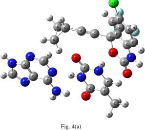
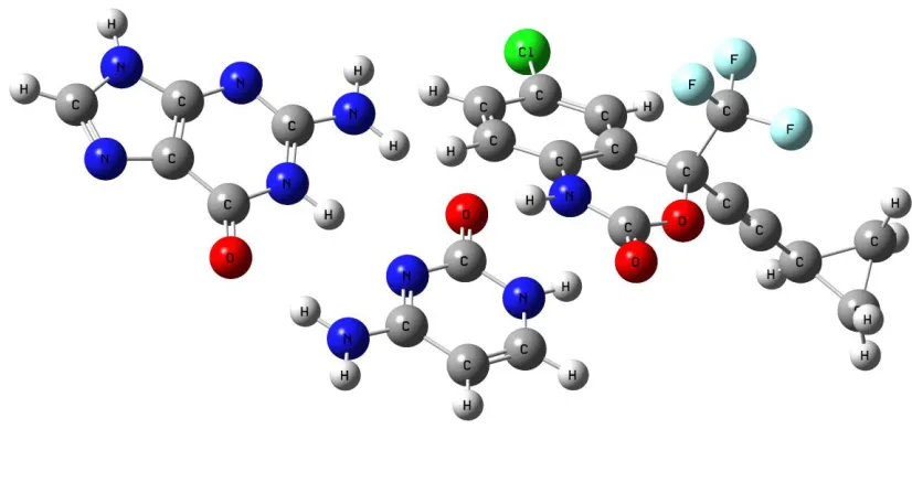
The EFZ···AT and EFZ···GC optimized geometries are summarized in Figures 4a and 4b, respectively.
[image:8.596.153.450.493.760.2]
Fig. 4(b)
Figure 4. (a,b) Optimized structures of EFZ ···AT and EFZ ···GC, respectively
The atom charge differences of EFZ, GC and AT before and after complex formation are presented in Figures 1(1a and 1b), 2 and 3 respectively. For instance, the O1 charge differences are -0.531 to -0.635, bond length (1, 2) shifted from 1.20Å to 1.22Å and the N3 charge moves from -1.049 to -1.098 bond length (3, 22) shifted from 0.99Å to 1.12Å. These changes indicated that the oxygen receives a part of its charge from the hydrogen atoms in GC. Therefore, the weak hydrogen bonding was formed between EFZ and GC.
The study of the atom charges in GC and EFZ···GC exhibits that the part, shown with dash marks (the only part which is going to be discussed afterwards), displays the highest changes, because of the EFZ and GC interactions. Similar changes have also been obtained in AT. Since the EFZ heteroatoms interact with the GC hydrogen in the zone, the charge changes are not important for the other heteroatom of the GC or AT bases pairs. This observation was proved by the increase in the GC hydrogen charges (i.e. H12 atom charge shifted from 0.282 to 0.537), revealing that a weaker hydrogen bonding had been formed between these atoms and the heteroatoms in the drug, and the decrease in their bond length (i.e. the R bond length (12, 22) shifted from 1.90Å to 1.96Å and bond length (10, 29) shifted from 1.76Å to 1.83Å).
[image:9.596.80.493.119.338.2]
in the above mentioned area. To avoid repetition, the results attained for AT are only listed in Table 1 and Figure 4, which are in agreement with those of GC.
In general, a way for information collection regarding the electrons distribution is by computing the polarizability. This property depends on the second derivative of the energy relating to an electric field. Table 2 delineates the high EFZ, GC and AT polarizability values, supporting the fact that the dispersion energy is always important. Another way is dipole moment of the base pairs and the studied intercalator which is presented in Table 2. The significant polarizability and dipole moment values proved the existence of the dispersion and electrostatic interactions between DNA and EFZ. The polarizability and the dipole moment of the intercalator have the same effects on the interaction with DNA. Hence, a drug should be designed with high polarizability and dipole moment to increase the interactions between DNA and the drugs.
[image:10.596.145.403.332.582.2]Furthermore the intercalation reaction between EFZ and different double base pairs of DNA (A–T/A–T, A–T/T–A, A–T/G–C, A–T/C–G, C– G/G–C, C–G/C–G) were also studied by the PM3 method. Figure 5 is a sample related to this study. The double base pairs of DNA were built by the nucleic acid database of Hyperchem and their 3D geometry was optimized with PM3 method [61-62].
Figure 5. Optimized structures of EFZ with different DNA double base pairs
4. CONCLUSIONS
1. Since LUMO energy of EFZ is negative, it is a good electron acceptor, but AT and GC base pairs have positive LUMO energies and are good electron donors.
homogeneous charge distribution with no centers of high charge accumulation. This, accounts for the low contribution of the electrostatic forces in the binding involving these molecules. Both intercalators and bases have high polarizability allowing a leading role for the dispersion forces.
3. Effects of vertical distance between the stacked molecules on the interaction energy were investigated. We found that, of the binding forces acting on these interactions, charge transfer and dispersion play an important role in our investigated systems.
4. In designing a drug changes in the structure and addition of specific groups should be in order to increase values of the main parameters such as polarizability, dipole moment and interaction energy. With high values of these factors it can be concluded that the drug design is suitable.
ACKNOWLEDGEMENTS
We gratefully acknowledge the support of this work by the Institute of Petroleum Engineering, Tehran University Research Councils.
References
1. [Online] available: http://en.wikipedia.org/wiki/Efavirenz
2. M. Gotte, G. Maier, A. M. Onori, L. Cellai, M. A. Wainberg and H. Heumann, J. Biol. Chem., 274 (1999) 11159
3. J. Y. Feng, J. K. Ly, F. Myrick, D. Goodman, K. L White, E. S. Svarovskaia, K. Borroto-Esoda and M. D. Miller, Retrovirology, 6 (2009) 44
4. L. A. Birchall, N. P. Bailey and G. R. Blackledge, Br. J. Clin. Pract., 45 (1991) 208. 5. U. Bren, M. Hodoscek and J. Koller, J. Chem. Inf. Model., 45 (2005) 1546.
6. M. R. Ganjali, P. Norouzi, F. Faridbod, S. Riahi, J. Ravanshad, J. Tashkhourian, M. Salavati- Niasari and M. Javaheri, IEEE Sens. J., 7 (2007) 544.
7. M. R. Ganjali, P. Norouzi, F. Sadat Mirnaghi, S. Riahi and F. Faridbod, IEEE Sens. J., 7 (2007) 1138.
8. L. Nathanson, Cancer Treat. Rev., 11 (1984) 289.
9. D. Faulds, J. A. Balfour, P. Chrisp and H. D. Langtry, Drugs, 41 (1991) 400.
10. D. S. Goodin, B. G. Arnason, P. K. Coyle, E. M. Frohman and D. W. Paty, Neurology, 61 (2003) 1332.
11. L. S. Lerman, J. Mol. Biol., 3 (1961) 18.
12. S. Wang, T. Peng and C. F. Yang, J. Biophys. Chem., 104 (2003) 239.
13. K. Reszka, P. Kolodziejczyk, J. A. Hartley, W. D. Wilson and J. W. Lown, In Anthracycline and Anthracenedionebased Anticancer Agents, Elsevier Press, Amsterdam, (1998), pp. 401-405.
14. S. Riahi, M. R. Ganjali, R. Dinarvand, S. Karamdoust, K. Bagherzadeh and P. Norouzi Riahi, Chem. Biol. & Drug Design, 71 (2008) 474.
15. S. Riahi, P. Norouzi, A. B. Moghaddam, M. R. Ganjali, G. R. Karimipour and H. Sharghi, Chem. Phys. 337 (2007) 33.
16. S. Riahi, A. B. Moghaddam, M. R. Ganjali and P. Norouzi, J. Mol. Struct. (THEOCHEM), 814 (2007) 131.
17. S. Riahi, M. R. Ganjali, A. B. Moghaddam, P. Norouzi and S. S. Hosseiny Davarani, Spectrochim Acta, Part A, 70 (2008) 94
20. S. Riahi, A. Beheshti, M. R. Ganjali and P. Norouzi, Spectrochim. Acta Part A, 74 (2009) 1077. 21. M. R. Ganjali, A. Alipour, S. Riahi, B. Larijani and P. Norouzi, Int. J. Electrochem. Sci., 4 (2009)
1262.
22. S. Riahi, M. R. Ganjali, A. B. Moghaddam, P. Norouzi and M. Niasari, J. Mol. Struct. (THEOCHEM), 774 (2006) 107.
23. S. Riahi, M. R. Ganjali, A. B. Moghaddam, P. Norouzi and M. Latifi, J. Mol. Struct. (THEOCHEM), 807 (2007) 137.
24. S. Riahi, M. R. Ganjali, A. B. Moghaddam and P. Norouzi, J. Theor. Comput. Chem., 6 (2007) 331.
25. S. Riahi, M. R. Ganjali, A. B. Moghaddam and P. Norouzi, J. Theor. Comput. Chem., 6 (2007) 255.
26. S. Riahi, F. Jalali Farahani, M. R. Ganjali, A. B. Moghaddam and P. Norouzi, J. Mol. Struct. (THEOCHEM), 850 (2008) 48.
27. S. Riahi, M. R. Ganjali, A. B. Moghaddam, P. Norouzi, J. Mol. Model., 14 (2008) 325.
28. S. Riahi, F. Edris-Tabrizi, M. Javanbakht, M. R. Ganjali and P. Norouzi, J. Mol. Model., 15 (2009) 829.
29. S. Riahi, M. R. Ganjali and P. Norouzi, J. Theor. Comput. Chem., 7 (2008) 317.
30. S. Riahi, M. R. Ganjali, A. B. Moghaddam and P. Norouzi, Spectrochim. Acta A, 71 (2008) 1390. 31. S. Riahi, P. Pourhossein, M. R. Ganjali and P. Norouzi, Petrol. Sci. Technol, 28 (2010) 68. 32. F. Faridbod, M. R. Ganjali, B. Larijani, P. Norouzi, S. Riahi and F. Sadat Mirnaghi, Sensors, 7
(2007) 3119.
33. S. Riahi, A. B. Moghaddam, M. R. Ganjali and P. Norouzi, J. Mol. Struct. (THEOCHEM), 896 (2009) 63.
34. S. Riahi, A. B. Moghaddam, M. R. Ganjali and P. Norouzi, Int. J. Electrochem. Sci., 4 (2009) 122. 35. S. Riahi, S. Eynollahi and M. R. Ganjali, Int. J. Electrochem. Sci., 4 (2009) 1407.
36. S. Riahi, S. Eynollahi and M. R. Ganjali, Int. J. Electrochem. Sci., 4 (2009) 1128. 37. S. Riahi, M. R. Ganjali and M. Bagheri, Mater. Sci. Eng. C., 29 (2009) 1808. 38. S. Riahi, S. Eynollahi, M. R. Ganjali, Int. J. Electrochem. Sci., 4 (2009) 551.
39. S. Riahi, M. R. Ganjali, M. Hariri, S. Abdolahzadeh and P. Norouzi, Spectrochim. Acta Part A, 74 (2009) 253.
40. J. Dolenc, U. Borstnik, M. Hodoscek, J. Koller and D. Janezic, J. Mol. Struct. (TEOCHEM), 718 (2005) 77.
41. M. Elstner, P. Hobza, T. Frauenheim, S. Suhai and E. Kaxiras, J. Chem. Phys., 114 (2001) 5149. 42. T. M. EL-Gogary, The role of charge transfer complex formation on the overall structure activity
relationships of DNA radioprotectants and radiosensitizers. Ph.D. Thesis, Mansoura University, Egypt/De Montfort University, Leicester, UK (1998).
43. T. M. El-Gogary and G. Koehler, J. Mol. Struct. (THEOCHEM), 808 (2007) 97. 44. J. B. Chaires, Curr. Opin. Struct., 8 (1998) 314.
45. M. J. Frisch, G. W. Trucks, H. B. Schlegel and et al., Gaussian Inc. Pittsburgh, PA (1998) 46. J. J. P. Stewart, J. Comp. Chem., 10 (1989) 210.
47. J. J. P. Stewart, J. Comp. Chem., 10 (1989) 221.
48. W. Yang and Q. Wu, Phys. Rev. Lett., 89, (2002) 143002/1. 49. R. G., Parr and W. Yang, Annu. Rev. Phys. Chem., 46 (1995) 701.
50. M. W. Schmidt, K. K. Baldridge, J. A. Boatz, S. T. Elbert, M. S. Gordon, J. H. Jensen, S. Koseki, N. Matsunaga, K. A. Nguyen, S. J. Su, T. L. Windus, M. Dupuis, J. A. Montgomery, J. Comput. Chem., 14 (1993) 1347.
51. F. B. Duijneveldt, J. G. C. M. Duijneveldt-van de Rijdt and J. H. Lenthe, Chem. Rev., 94 (1994) 1873.
53. H. Y. Zhou, E. Tajkhorshid, T. Frauenheim, S. Suhai and M. Elstner, Chem. Phys., 277 (2002) 91.
54. P. Hobza and R. Zahradnik, Intermolecular Complexes, Elsevier, Amsterdam (1988) 55. U. Bren, V. Martinek and J. Florian, J. Phys. Chem. B, 110 (2006) 10557.
56. S. Mierts, E. Scrocco and J. Tomasi, Chem. Phys., 55 (1981) 117. 57. J. Florian and A. Warshel, J. Phys. Chem. B, 101 (1997) 5583.
58. U. Bren, V. Martinek and J. Florian, J. Phys. Chem. B, 110 (2006) 12782.
59. U. Bren, M. Zupan, F. P. Guengerich and J. Mavri, J. Org. Chem., 71 (2006) 4078. 60. U. Bren, F. P. Guengerich and J. Mavri, Chem. Res. Toxicol., 20 (2007) 1134.
61. M. J. Frisch, J. E. Del Bene, J. S. Binkley and H. F. Schaefer, J. Chem. Phys., 84 (1986) 2279. 62. D. W. Schwenke and D. G. Truhlar, J. Chem. Phys., 82 (1985) 2418.
![Table 2. Dipole moment [D], polarizibility [B3], HOMO and LUMO energies (in eV) of the drug, the bases and the base pairs](https://thumb-us.123doks.com/thumbv2/123dok_us/1942358.154272/5.596.46.564.90.374/table-dipole-moment-polarizibility-homo-lumo-energies-bases.webp)