THE EFFECTS OF VANILLOID-LIKE
AGENTS ON
PLATELET AGGREGATION
Safa Yousef Almaghrabi, MBBS
School of Human Life Sciences
Submitted in fulfilment of the requirements for the degree of
Master of Biomedical Science (Research)
DECLARATION
I hereby declare that this thesis entitled The Effects of Vanilloid-Like Agents on Platelet Aggregation contains no material which has been accepted for a degree or
diploma by the University or any other institution, except by way of background
information and duly acknowledged in the thesis, and to the my knowledge and
belief no material previously published or written by another person except where
due reference is made in the text of thesis, nor does the thesis contain any material
that infringes copyright.
Date: 24th Oct 2012 Signed:
AUTHORITY OF ACCESS
This thesis may be made available for loan and limited copying and communication
in accordance with the Copyright Act 1968.
Date: 24th Oct 2012 Signed:
STATEMENT OF ETHICAL CONDUCT
The research associated with this thesis abides by the international and Australian
codes on human and animal experimentation, the guidelines by the Australian
Government’s Office of Gene Technology Regulator and the rulings of the Safety,
Ethics and Institutional Biosafety Committees of the University.
Date: 24th Oct 2012 Signed:
ACKNOWLEDGEMENTS
First of all, I would like to thank the Government of Saudi Arabia (King Abdulaziz
University) for the scholarship and sponsorship.
I would also like to sincerely acknowledge my supervisors, Dr. Murray Adams,
A/Prof. Dominic Geraghty, and Dr. Kiran Ahuja for their guidance, tolerance and
being there whenever needed.
I will not forget Merrilyn Johnson for her great help in the haematology laboratory
and special thanks to the volunteers who donated with their precious blood and time.
At last but not the least, I would like to thank my husband, Abdul, for his
understanding, love and full support throughout these years. Many thanks as well to
Table of Contents
DECLARATION i ACKNOWLEDGEMENTS ii
Table of Contents iii
List of Figures vi
List of Tables viii
List of Abbreviations ix
Abstract xii Chapter 1 LITERATURE REVIEW 1
1.1 Introduction 2
1.2 Haemostasis 3
1.2.1 Platelets 3
1.2.1.1 Platelet Structure and Function 3
1.2.1.2 Platelet Activation 5
1.2.1.3 Platelet Receptors 9
1.2.2 Blood Coagulation 14
1.2.2.1 Tissue Factor Pathway 14
1.2.2.2 Natural Inhibitors of Blood Coagulation 16
1.2.3 Fibrinolysis 17
1.3 Cannabinoid Receptors 18
1.3.1 Endogenous Cannabinoids (Endocannabinoids) 19
1.4 Transient Receptor Potential Vanilloid Channels 20
1.4.1 TRPV1 Structure 21
1.4.2 TRPV1 Function 22
1.4.4 Biochemical Pharmacology of TRPV1 27
1.5 Vanilloids 28
1.5.1 Endogenous Vanilloids 29
1.5.2 Plant Derived Vanilloids 29
1.5.2.1 Plant-Derived Vanilloid Targets and Actions 31
1.5.2.2 Clinical Applications of Plant-Derived Vanilloids 32
1.5.2.3 Plant-Derived Vanilloid Toxicity 38
1.6 Project Aims 39
Chapter 2 Materials and Methods 40
2.1 Ethics 41
2.2 Materials 41
2.2.1 Preparation of Platelet Aggregation Agonists 42
2.2.2 Preparation of Vanilloids 42
2.3 Methods 42
2.3.1 Platelet Aggregation 42
2.3.2 Lactate Dehydrogenase (LDH) Cytotoxicity Assay 45
2.3.3 Measurement of Alpha Granule Release 45
2.3.4 Statistical Analysis 46
Chapter 3 Results 47
3.1 Effect of Vanilloid-Like Agents on ADP-Induced Aggregation 48
3.2 Effect of Vanilloid-Like Agents on Collagen-Induced Aggregation 53
3.3 Effect of Vanilloid-Like Agents on Arachidonic Acid-Induced Aggregation 57
3.4 Effect of SB-452533 on ADP-induced Platelet Aggregation 65
3.5 Cytotoxicity Assay 65
Chapter 4 Discussion 69
BIBLIOGRAPHY 79 APPENDICES 109
Appendix 1: Manual of PF4 ELISA Kit 109
Appendix 2: Manual of β-TG ELISA Kit 116
List of Figures
Figure 1-1 ADP and Platelet Activation ... 13
Figure 1-2 Schematic Diagram of the Coagulation Cascade ... 15
Figure 1-3 Chemical Structures of Endogenous Cannabinoids ... 20
Figure 1-4 Regions and Amino Acids involved in TRPV1 Function ... 21
Figure 1-5 Chemical Structures of the Capsaicinoids ... 31
Figure 3-1 Effect of CAP (A), DHC (B), NADA (C) and OLDA (D) on ADP-induced Aggregation ... 49
Figure 3-2 Effect of Plant-Derived Vanilloids on ADP-induced Platelet Aggregation ... 51
Figure 3-3 Effect of Endovanilloids on ADP-induced Platelet Aggregation ... 52
Figure 3-4 Effect of NADA (A) and OLDA (B) on Collagen-induced Aggregation 53 Figure 3-5 Effects of Plant-Derived Vanilloids on Collagen-induced Platelet Aggregation ... 55
Figure 3-6 Effects of Endovanilloids on Collagen-induced Platelet Aggregation ... 56
Figure 3-7 Effect of Vanilloids on the Lag-Time of Collagen-induced Platelet Aggregation ... 57
Figure 3-8 Effect of Capsaicin (A), DHC (B) and NADA (C) on Arachidonic Acid-induced Aggregation ... 58
Figure 3-9 Effects of Plant-Derived Vanilloids on Arachidonic Acid-induced Platelet Aggregation ... 60
Figure 3-10 Effects of Endovanilloids on Arachidonic Acid-induced Platelet Aggregation ... 61
Figure 3-12 Effects of SB-452533 on Capsaicin- and OLDA-mediated inhibition of
Platelet Aggregation induced by ADP ... 66
List of Tables
Table 1-1 Contents of Platelet Granules ... 4
Table 1-2 Platelet Alpha Granule Contents and Functions ... 6
Table 1-3 Major Sub Endothelial Matrix Constituents that support Platelet Adhesion
... 7
Table 1-4 Platelet Receptors for Adhesive Protein ... 10
Table 1-5 Platelet Integrins ... 10
Table 3-1 Effect of Vanilloids on ADP, Collagen, Arachidonic acid-induced Platelet
Aggregation. ... 63
List of Abbreviations
AA APC ADP AEA 2-AG ATP AUC CAP CGRP CB COX DHC DRG ELISA FVII FAAH GPCRs GP 2-HPETE 5-HT 5-iodo-RTX IP3 JAMs Arachidonic acidActivated protein C
Adenosine diphosphate
N-arachidonoyl-ethanol-amide
2-Arachidonylglycerol
Adenosine triphosphate
Area under curve
Capsaicin
Calcitonin gene-related peptide
Cannabinoid receptors
Cyclooxygenase
Dihydrocapsaicin
Dorsal root ganglia
Enzyme-linked Immunosorbent Assay
Factor VII
Fatty acid amide hydrolase
G protein-coupled receptors
Glycoprotein
12-Hydro-peroxyeicosatetraenooic
5-Hydroxytryptamine
5-Iodo-resiniferatoxin
1,4,5- Inositol triphosphate
LDH LDL Max MAGL NADA OLDA OCS PIP2 PLC PLG PAI-1 PDGF PECAM-1 PF4 PRP PPP PAR PKC SD SEM β-TG THC TAFI TxA2 TF Lactate dehydrogenase Low-density lipoprotein Maximum aggregation Monoacylglycerol lipase N-arachidonoyl-dopamine N-oleoyldopamine
Open canalicular system
Phosphatidylinositol-4,5-bisphonate
Phospholipase C
Plasminogen
Plasminogen activator inhibitor-1
Platelet derived growth factor
Platelet endothelial cell adhesion molecule 1
Platelet factor 4
Platelet rich plasma
Platelet poor plasma
Protease-activated receptor
Protein kinase C
Standard deviation
Standard error of the mean
β-thromboglobulin
Delta-9-tetrahydrocannabinol
Thrombin-activatable fibrinogen inhibitor
Thromboxane A2
TFPI
tPA
TRPV1
TM
uPA
vWF
Tissue factor pathway inhibitor
Tissue plasminogen activator
Transient receptor potential vanilloids
Transmembrane
Urokinase
Abstract
Capsaicin, the ‘hot’ principle found in chilli, and other vanilloids exert their effects
on neuronal cells through activation of transient receptor potential vanilloid 1
(TRPV1). TRPV1 is widely distributed in neuronal and non-neuronal cells. It has
been proposed that consumption of vanilloid-like agents, including capsaicinoids,
inhibits platelet aggregation and may protect against the development of
cardiovascular disease. The aim of this study was to investigate the effects of a range
of vanilloid-like agents on in vitro platelet aggregation.
Venous blood was collected from healthy subjects who avoided antiplatelet
medications and dietary chilli for at least 10 and 2 days, respectively. Collagen (4
and 8 g/mL), ADP (10 and 5 μM) and arachidonic acid (AA) (300 and 400 mg/mL)
-induced platelet aggregation was determined using platelet rich plasma (PRP;
250x109/L) in the absence and presence of the capsaicinoids [capsaicin and
dihydrocapsaicin (DHC)] and the endocannabinoid/endovanilloid agents
[N-oleoyldopamine (OLDA) and N-arachidonoyl-dopamine e (NADA)]. %Maximum
aggregation (%Max), % area under curve (%AUC) and slope of platelet aggregation
were determined. Platelet lactate dehydrogenase (LDH), which is released rapidly
after cell membrane damage, was investigated to determine the direct toxic effects of
these agents on platelets. Platelet factor 4 (PF4) and β-thromboglobulin (β-TG)
release were examined to determine the effects of vanilloids on alpha granule
release. Finally, the effects of TRPV1 antagonist (SB-452533) on capsaicin- and
ADP-induced (5 μM) platelet aggregation was inhibited in a concentration-dependent
manner by capsaicin (%Max, meanSEM; 0 vs 100 μM, 83.80.9% vs 45.22.4%,
n=6, p0.001); OLDA (0 vs 100 μM, 71.68.2% vs 9.41.4%, n=4, p0.001); and
NADA (0 vs 100 μM, 71.55.9% vs 38.21.4%, n=4, p0.008). Similar results were
observed using 10 μM ADP. OLDA and NADA, but not capsaicin and DHC,
inhibited platelet aggregation induced by 4g/mL collagen: OLDA (Max%, 0 vs 100
μM, 89.31.4% vs 45.512.5%, p<0.001); and NADA (0 vs 100 μM, 87.70.8% vs
28.58.2%, p<0.001). AA-induced (300 mg/mL) aggregation was inhibited in a
concentration-dependent manner by capsaicin (Max%, 0 vs 100 μM, 89.60.9% vs
110.8%, p<0.001); DHC (0 vs 100 μM, 88.32.1% vs 18.76.9%, p<0.001); and
NADA (0 vs 100 μM, 841.8% vs 21.94.7%, p<0.001). Similar results were
observed using 400mg/mL AA. The inhibition of platelet aggregation by all agents
was not due to direct toxic effects as LDH release from platelets was unaffected by
any of the vanilloids. SB-452533 did not inhibit the effects of OLDA (SB-45253;
Max 0 vs 10μM, 55.92.1% vs 58.41.37%) and capsaicin (SB-45253; Max 0 vs
10μM, 65.150.44% vs 65.551%) on platelet aggregation, suggesting that
inhibition of ADP-induced aggregation is not TRPV1 mediated. ADP-stimulated PF4
release from platelets was impaired by capsaicin, DHC and OLDA whereas NADA
enhanced ADP-stimulated PF4 release. Furthermore, OLDA and capsaicin impaired
the release of β-TG from ADP-stimulated platelets.
The present study using human platelet shows that capsaicin, DHC, OLDA and
NADA inhibit in vitro aggregation. The inhibitory effects of vanilloids are not
inhibit platelet aggregation by interfering with granule release, although further
1.1 Introduction
Platelets play an essential role in cardiovascular diseases both in pathogenesis of
atherosclerosis and in the development of acute thrombotic events (Harker et al.,
1976, Zucker, 1980). Their importance in coronary disease and in acute coronary
syndromes is indirectly confirmed by the benefit of antiplatelet agents in treating
these disorders. Several research groups have studied natural compounds as potential
antiplatelet agents. One of the exciting discoveries from these studies was that the
active ingredient of the hot chilli pepper, capsaicin, inhibits in vitro platelet
aggregation (Adams et al., 2009, Hogaboam and Wallace, 1991, Raghavendra and
Naidu, 2009).
The mechanism(s) by which capsaicin inhibits platelet aggregation is still poorly
understood. To date only few studies have been conducted on human, rabbit and dog
platelets to determine the effect of capsaicin and DHC on platelet aggregation and
the mechanism(s) of its action(s) and those limited studies have produced conflicting
data (Adams et al., 2009, Mittelstadt et al., 2012, Hogaboam and Wallace, 1991,
Harper et al., 2009, Raghavendra and Naidu, 2009). The effects of both endogenous
and plant-derived vanilloid-like agents on in vitro human platelet aggregation and the
potential mechanism(s) of action were therefore systematically investigated in this
study. The aims were to: (1) investigate the effects of exogenous and endogenous
vanilloids on in vitro platelet aggregation, (2) determine whether vanilloid-like
agents are toxic to platelets, (3) determine effect of vanilloid-like agents on platelet
alpha and dense granule release, and (4) investigate the potential involvement of
1.2 Haemostasis
The haemostatic response to blood vessel damage includes a series of interactions
between platelets, the subendothelial matrix and coagulation proteins. Under normal
conditions, endothelial cells prevent tissue factor (TF) and the subendothelial matrix
from being exposed directly to platelets and coagulation proteins. The contact
between platelets and the subendothelial matrix induces platelet activation to
facilitate plug formation. The haemostatic plug works as a catalytic surface for
stimulation and recruitment of coagulation proteins, as well as localization and
amplification of the coagulation system (Bombeli and Spahn, 2004, Hoffman, 2003,
Hoffman and Monroe, 2001, Lawson and Murphy, 2004, Walsh, 2004). The
haemostatic system is controlled by many anticoagulant proteins and inhibitors, as
well as the fibrinolytic system. When working in balance, these systems ensure that
the formed thrombus stops bleeding and that revascularization occurs afterward to
maintain blood flow (Spahn and Rossaint, 2005).
1.2.1 Platelets
Platelets are small anuclear cytoplasmic fragments of megakaryocytes that are
produced in the bone marrow (Avraham, 1993). Their major function is to maintain
normal hemostasis and wound repair, but they also participate in many other
pathophysiological processes such as atherosclerosis. Platelets possess many
receptors that regulate platelet function (Ruggeri, 2002, De Botton et al., 2002).
1.2.1.1 Platelet Structure and Function
Platelets are small irregularly shaped cell fragments, with dimensions of
heterogeneity in size and structure. The circulating number of platelets in the
peripheral blood ranges from 150-400 109/L (George, 2000). They play a major
role in the physiological and pathological processes of hemostasis; wound healing,
inflammation, host defense, and tumor metastasis (Jurk and Kehrel, 2005, Hoak,
1988). The main components of platelets are alpha () granules, dense granules, the
dense tubular system, open canalicular system (OCS), cytoplasmic membrane,
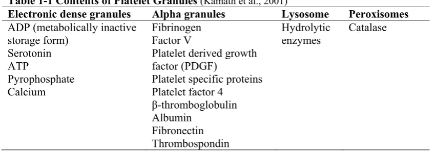
mitochondria, peroxisomes, lysosomes and cytoskeleton (Table 1-1) (Kamath et al.,
[image:19.595.106.531.329.478.2]2001, Fukami and Salganicoff, 1977).
Table 1-1 Contents of Platelet Granules (Kamath et al., 2001)
Electronic dense granules Alpha granules Lysosome Peroxisomes
ADP (metabolically inactive storage form)
Serotonin ATP
Pyrophosphate Calcium
Fibrinogen Factor V
Platelet derived growth factor (PDGF)
Platelet specific proteins Platelet factor 4
β-thromboglobulin Albumin
Fibronectin Thrombospondin
Hydrolytic enzymes
Catalase
Platelets are full of secretory granules that are crucial for their normal function
(Coppinger et al., 2004). Dense granules store small molecules including serotonin,
calcium ions and nucleotides, particularly adenosine triphosphate (ATP) and
adenosine diphosphate (ADP). These constituents induce localized vasoconstriction
and cause activation of other platelets, and any defect in their release may affect
normal haemostasis (Ren et al., 2008). Platelets also contain other structures such as
peroxisomes, that store catalase, and lysosomes that contain hydrolytic enzymes such
as hexosaminidase and cathepsins, that may have a role in further platelet activation
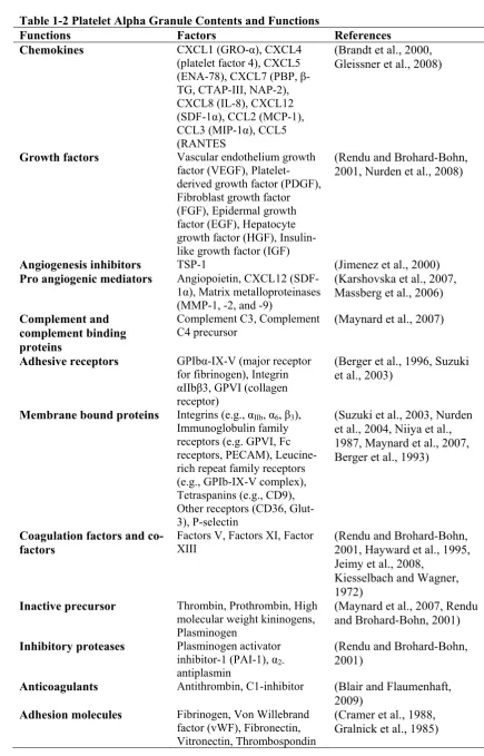
The most abundant platelet secretory granules are -granules, accounting for ~10%
of platelet volume, which is ten-fold more than dense granules. Furthermore, the
total membrane surface area α-granules in a platelet is 14m, which is eight-fold
more than dense granules and roughly equal to that of the open canalicular system.
There are approximately 50-80 -granules per platelet and their size ranges from
200-500nm (Frojmovic and Milton, 1982). Moreover, the role of -granules in
atherosclerosis, angiogenesis, host defense, inflammation, wound healing,
malignancy, and antimicrobial have been well described (Table 1-2) (Blair and
Flaumenhaft, 2009).
1.2.1.2 Platelet Activation
The process of platelet activation can be classified into three overlapping phases:
initiation, extension and perpetuation (Hoffman et al., 2008). Although several
agonists initiate platelet activation in vitro, in vivo activation is normally initiated by
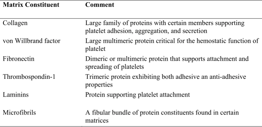
thrombin and collagen. Platelets can adhere to subendothelial matrix proteins such as
collagen once the endothelium has been disrupted, with these proteins having an
important role in platelet attachment and the amplification reaction (Table 1-3). The
disrupted vessel wall captures and then activates platelets via exposed collagen
attached to von Willbrand factor multimers, building a monolayer that aids thrombin
generation and the subsequent aggregation of more platelets (Falati et al., 2003,
Gross et al., 2005, Del Conde et al., 2005). Essential to these events are platelet
surface receptors that assist the vWF-dependent binding of platelets, glycoprotein
Ib/IX/V (GPIb-IX-V) and, to a lesser extent, integrin αIIbβ3 and subsequent
Table 1-2 Platelet Alpha Granule Contents and Functions
Functions Factors References Chemokines CXCL1 (GRO-α), CXCL4
(platelet factor 4), CXCL5 (ENA-78), CXCL7 (PBP, β -TG, CTAP-III, NAP-2), CXCL8 (IL-8), CXCL12 (SDF-1α), CCL2 (MCP-1), CCL3 (MIP-1α), CCL5 (RANTES
(Brandt et al., 2000, Gleissner et al., 2008)
Growth factors Vascular endothelium growth factor (VEGF), Platelet-derived growth factor (PDGF), Fibroblast growth factor (FGF), Epidermal growth factor (EGF), Hepatocyte growth factor (HGF), Insulin-like growth factor (IGF)
(Rendu and Brohard-Bohn, 2001, Nurden et al., 2008)
Angiogenesis inhibitors TSP-1 (Jimenez et al., 2000)
Pro angiogenic mediators Angiopoietin, CXCL12 (SDF-1α), Matrix metalloproteinases (MMP-1, -2, and -9)
(Karshovska et al., 2007, Massberg et al., 2006)
Complement and complement binding proteins
Complement C3, Complement
C4 precursor (Maynard et al., 2007)
Adhesive receptors GPIbα-IX-V (major receptor for fibrinogen), Integrin
αIIbβ3, GPVI (collagen receptor)
(Berger et al., 1996, Suzuki et al., 2003)
Membrane bound proteins Integrins (e.g., αIIb, α6, β3), Immunoglobulin family receptors (e.g. GPVI, Fc receptors, PECAM), Leucine-rich repeat family receptors (e.g., GPIb-IX-V complex), Tetraspanins (e.g., CD9), Other receptors (CD36, Glut-3), P-selectin
(Suzuki et al., 2003, Nurden et al., 2004, Niiya et al., 1987, Maynard et al., 2007, Berger et al., 1993)
Coagulation factors and co-factors
Factors V, Factors XI, Factor
XIII (Rendu and Brohard-Bohn, 2001, Hayward et al., 1995, Jeimy et al., 2008,
Kiesselbach and Wagner, 1972)
Inactive precursor Thrombin, Prothrombin, High molecular weight kininogens, Plasminogen
(Maynard et al., 2007, Rendu and Brohard-Bohn, 2001)
Inhibitory proteases Plasminogen activator inhibitor-1 (PAI-1), α 2-antiplasmin
(Rendu and Brohard-Bohn, 2001)
Anticoagulants Antithrombin, C1-inhibitor (Blair and Flaumenhaft, 2009)
Adhesion molecules Fibrinogen, Von Willebrand factor (vWF), Fibronectin, Vitronectin, Thrombospondin
Table 1-3 Major Sub Endothelial Matrix Constituents that support Platelet Adhesion
(Hoffman et al., 2005b)
Matrix Constituent Comment
Collagen Large family of proteins with certain members supporting platelet adhesion, aggregation, and secretion
von Willbrand factor Large multimeric protein critical for the hemostatic function of platelet
Fibronectin Dimeric or multimeric protein that supports attachment and spreading of platelets
Thrombospondin-1 Trimeric protein exhibiting both adhesive an anti-adhesive properties
Laminins Protein supporting platelet attachment
Microfibrils A fibular bundle of protein constituents found in certain matrices
further platelets to extend on the endothelium and form a nidus for subsequent
platelet-platelet interactions (Massberg et al., 2003).
At the site of vessel injury, collagen receptors help the capture of fast-moving
platelets, resulting in activation of platelets and reorganization of the cytoskeleton.
Platelets flatten out and attach more closely to the exposed vessel wall. vWF
enhances this event by increasing the binding site affinity of collagen for platelets
(Massberg et al., 2003, Kato et al., 2003, Poole et al., 1997, Nieswandt et al., 2001,
Nieuwenhuis et al., 1985, Sixma et al., 1997). In inflammatory and thrombotic
diseases, platelet activation is initiated by thrombin through G protein-coupled
receptors (GPCRs) of the protease-activated receptor (PAR) family. The initiation
step is sufficient to form a platelet plug but not to prevent bleeding (Hoffman et al.,
The second stage of platelet aggregation occurs when more platelets are recruited,
activated and aggregate on top of the collagen-bound monolayer (Hoffman et al.,
2008). The secretion of agonists, such as thromboxane A2 (TxA2), thrombin and
ADP, recruit further platelets to the site of injury and activate phospholipase C
(PLC). The PLC isoform, PLCγ2, hydrolyzes phosphatidylinositol-4,5-bisphonate
(PIP2) to form 1,4,5- inositol triphosphate (IP3) and diacylglycerol. IP3 raises the
cytosolic Ca2+ concentration by opening Ca2+ channels in the platelet-dense tubular
system. This leads to Ca2+ influx through the plasma membrane of the platelet
(Nesbitt et al., 2003, Kulkarni et al., 2004). The most important cohesive interaction
that maintains the adhesion between the platelets is the binding of vWF or fibrinogen
to αIIbβ3. Locally secreted or circulating catecholamines result in vasoconstriction
and enhance platelet activation by increasing the effects of other platelet agonists.
The majority of platelet agonists exert their action to extend the platelet plug through
GPCRs. The characteristics of GPCRs make them especially well-suited for this
function. Human platelets have almost 10 forms of Gα, which fall into the G12α, Giα,
Gsα and Gqα groups (Offermanns, 2006).
The last stage of perpetuation occurs when the platelet plug is stabilized to prevent
premature disaggregation (Hoffman et al., 2008). The interactions between platelets
can be indirect, for example polyvalent adhesive proteins bind to activate αIIbβ3 on
other platelets, or direct, where one cell adhesion molecule links to adjacent platelet
in trans. Theoretically, both mechanisms support an additional adhesive force and
another source of intracellular signaling (Brass et al., 2005). The binding between
αIIbβ3 and fibrin, fibrinogen or vWF provides the main cohesive strength that
molecules aside from integrins support the adhesion and intracellular signaling such
as, platelet endothelial cell adhesion molecule 1 (PECAM-1; CD31) (Newman and
Newman, 2003), junctional adhesion molecules (JAMs) (Muller, 2003, Bazzoni,
2003) and signaling lymphocytic activation molecule (SLAM; CD150) (Krause et
al., 2000, Martin et al., 2001, Nanda et al., 2005). Platelet activation is thus a
dynamic process where effector pathways and many receptors are controlling at each
phase of platelet plugs (Hoffman et al., 2008).
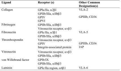
1.2.1.3 Platelet Receptors
Several nomenclature systems have been used to classify platelet membrane proteins
(Table 1-4) (Hoffman et al., 2008). The majority of platelet adhesive proteins belong
to the integrin family, a broadly distributed group of heterodimeric cell surface
molecules (having two subunits, and ). Eight -subunits are known that display
high sequence homology, ranging from 35% to 45% at the first amino acid sequence
level and a common structural organization. The -subunits are also similar but show
less extensive sequence identity (Hynes, 1992, Fitzgerald et al., 1987). Every
-subunit joins in a noncovalent complex with an --subunit to make an efficient
adhesive protein receptor. A solitary -subunit can combine with many -subunits.
The 1 and 3 (2 is reported to be present at low levels) are the major -subunits
expressed on platelet, as well five -subunits (Table 1-5) (Philippeaux et al., 1996).
The IIb3 integrin (GPIIb/IIIa) is restricted predominantly to platelets and
megakaryocytes and plays a major role in platelet aggregation and several other
platelet reactions (Grossi et al., 1988, Honn et al., 1992, Boukerche et al., 1989).
GPIb-V-IX however, is not a member of the integrin family but is involved in
Table 1-4 Platelet Receptors for Adhesive Protein (Hoffman et al., 2005b)
Ligand Receptor (s) Other Common
Designation(s)
Collagen GPIa/IIa,
GPIIb/IIIa, IIb3 GPIV
GPVI
VLA-2
GPIIIb, CD36
Fibrinogen GPIIb/IIIa, IIb3
Vitronectin receptor, v3 Fibronectin GPIc/IIa, 51
GPIIb/IIIa, IIb3
VLA-5
Thrombospondin Vitronectin receptor, v3 GPIV
Integrin-associated protein
GPIIIb, CD36 IAP
Vitronectin Vitronectin receptor, v3 GPIIb/IIIa, IIb3
von Willebrand factor GPIb/IX
GPIIb/IIIa, IIb3
Laminin GPIc/IIa region, 61 VLA-6
Table 1-5 Platelet Integrins (Hoffman et al., 2005b)
Integrin Major Ligand (s) 21 (GPIa/IIa, VLA-2) Collagen
51 (GPIc/IIa, VLA-5) Fibronectin
IIb3 (GPIIb/IIIa) Fibrinogen, fibronectin, vitronectin, von Willebrand factor, CD40L
v3 (vitronectin receptor) Fibrinogen, fibronectin, vitronectin, von Willebrand factor, thrombospondin, osteopontin
[image:25.595.107.533.451.599.2]haemostasis can be traced directly to its function as the vWF receptor as well as a
binding site for thrombin (Moroi et al., 1982, Okumura and Jamieson, 1976).
Therefore, by adhesion with matrix proteins, vWF mediates directly reversible and
rapid platelet adhesion that supports the rolling of platelets along the surface of the
disrupted endothelium (Sadler, 2002). The most likely receptors responsible for the
firm platelet-adhesive bound that enduringly stops the rolling of the platelet are
integrins. A significant consequence of occupancy of GPIb-V-IX by vWF is the
stimulation of intracellular signaling events that lead to the activation of IIb3 and
platelet aggregation (Berndt et al., 2001, Savage et al., 1992).
Collagen in the subendothelial matrix is an important initiator of platelet responses.
It is both a substrate for platelet adhesion and a potent platelet agonist. Platelets have
four types of collagen receptors. Two bind directly to collagen (21 and GPVI),
and two bind to collagen through vWF (IIb3 and GPIb-IX-V). GPVI acts as the
primary collagen receptor and is responsible for platelet secretion and aggregation
induced by collagen. In addition, 21 works as an anchor for platelets to adhere to
exposed collagen after endothelial injury (Hoffman et al., 2005a, Hoffman et al.,
2008, Clemetson et al., 1999).
ADP is another platelet agonist that causes shape change of platelets from smooth
discoid shape to speculated spheres, liberation of granule contents and release of
thromboxane A2, ultimately causing platelet aggregation (Jin et al., 1998). ADP is
released actively from the platelet dense granules in response to the physiological
platelet agonists, which are thrombin, TXA2 and collagen, and amplifies its own
(Shankar et al., 2006, Maffrand et al., 1988, Born, 1985). Damaged vessel walls
(endothelial cells) and erythrocytes also release ADP passively, which induces
aggregation of platelets through integrin IIb3 activation and subsequent fibrinogen
binding (Mills, 1996). ADP mediates platelet aggregation via binding to two
G-protein-coupled receptors subtype, P2Y1 and P2Y12 (Figure 1-1) (Jantzen et al.,
1999, Daniel et al., 1998). Activation of P2Y1 is sufficient to cause platelet shape
change, while co activation of both P2Y1 and P2Y12 is required to induce platelet
aggregation (Paul et al., 1999, Jin et al., 1998, Jin and Kunapuli, 1998, Savi et al.,
1998).
The P2Y1 receptor is coupled to the heterotrimeric protein Gq. P2Y1 activation leads
to activation of phospholipase C, production of diacylglycerol and inositol phosphate
(IP). Moreover, calcium mobilization from cytosolic stores in response to IP
formation results in activation of protein kinase C (PKC) and phosphorylation of
myosin light chain, signaling events that play a major role in agonist-induced platelet
shape change (Daniel et al., 1998, Daniel and Adelstein, 1976, MacKenzie et al.,
1996). ATP weakly antagonizes these effects of ADP at the P2Y1 receptor, in
comparison to strong P2Y1 receptor-specific adenine nucleotide analog antagonists,
A3P5PS, A3P5P, and A2P5P. These three antagonists are required in high concentrations (20-fold molar excess) to block functional responses to 10mM ADP, while a 200-fold molar excess of ATP is required to completely inhibit the P2Y1 receptor (Boyer et al., 1996, Eckly et al., 2001). The absence of P2Y1 in mouse models of thrombosis has been shown to increase survival after the administration of
The secon inhibition al., 2001) phenotype Moreover impairs th concentrat Figu Plate secon plate two G text. Mech surfa incre
nd ADP rec
of cAMP f
). Lack of
e (Hollopete
, deletion o
he response
tions (Foste
ure 1-1 ADP
elet activation ndary agonist
let dense gran G protein–cou Drugs such hanisms that p ace of endothe ease the conce
ceptor, P2Y
formation by
P2Y12 in
er et al., 200
of either P2
e of platele
er et al., 200
P and Platele
by potent ago ts such as thr nules. Platelet upled receptor h as, Ticlop
place a limit o elial cells, wh
ntration of cA
Y12, is cou
y adenylyl
n humans p
01, Cattane
2Y1 or P2Y
ets to TxA2
01, Fabre et
et Activation
onists such as romboxane A2
t responses to rs, P2Y1 and idine and C on unwarrant hich hydrolyze AMP and cGM
upled to Gi
cyclase (Ho
produces a
o and Gach
Y12 in mic
2, ADP and
al., 1999, L
n
s thrombin or 2 (TxA2) and o ADP require P2Y12, whos Clopidogrel b ed platelet ac es ADP to AM MP within plat
2 and its a
ollopeter et
relatively
het, 1999, N
ce prolongs
d thrombin
Leon et al.,
collagen caus d the secretio e the coordina se actions are block activati tivation inclu MP, and PGI2 telets (Woulfe
activation re
al., 2001, Z
mild hem
Nurden et al
bleeding t
n, especially
1999).
ses the release on of ADP fr ate activation described in tion of P2Y ude CD39 on 2 and NO, wh e et al., 2001b)
esults in
Zhang et
morrhagic
., 1995).
time and
y at low
1.2.2 Blood Coagulation
The blood coagulation system contributes to the stabilization of the primary platelet
plug. Natural endogenous anticoagulants regulate blood coagulation to ensure that,
under normal conditions, haemostasis remains balanced. Any disturbance of this
balance between coagulation and anticoagulation due to acquired or genetic factors
may cause bleeding or thrombosis (Dahlback, 2000).
1.2.2.1 Tissue Factor Pathway
Blood coagulation is initiated by the exposure of TF, which is present in the cells that
encircle the vascular bed as well as expressed by cells circulating in the blood such
as leukocytes. TF binds to factor VII (FVII) and the activated form of FVII (FVIIa).
The TF-FVIIa complex triggers the conversion of FIX and FX to FIXa and FXa
respectively (Kirchhofer and Nemerson, 1996, Mann et al., 1998). FIXa and FXa
remain attached to the TF-bearing cell or diffuses into the blood to adhere to
activated platelets (Hoffman et al., 1996). Platelet activation is combined with the
disclosure of negatively charged phospholipids which have high affinity to bind and
assemble enzyme-cofactor complex and coagulation factors that are essential for
effective propagation of the system (Zwaal et al., 1998).
The prothrombinase complex, which is a phospholipid-bound complex composed of
FV and FXa, activates prothrombin to thrombin. Thrombin is a key enzyme in the
coagulation system due to its multiple roles including platelet activation, fibrinogen
to fibrin conversion and feedback amplification of coagulation (Figure 1-2).
Thrombin activates FV, FVIII and FXI as feedback amplification. Another
of factor IXa that can lead to activation of factor X. vWF is an important adhesive
protein for the creation of the primary platelet plug and FVIII circulates bounding to
this factor. FVIII become activated and dissociated from vWF and make a complex
with FIXa on the platelet surface, which eventually leads to FX activation (Gailani
and Broze, 1991). Following fibrin clot formation, thrombin generation reaches its
peak (Mann et al., 1998), which is important for the activation of FXIII and
thrombin-activatable fibrinolysis inhibitor (TAFI) as well as for further fibrin
generation (Davie, 1995).
Thrombin generation is considered to be more important than fibrin deposition due to
the fact that thrombin plays an important role in the activation of platelets, which is a
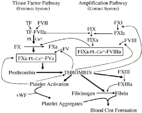
[image:30.595.194.442.398.596.2]crucial reaction for haemostasis response to vascular injury (Dahlback, 2000).
Figure 1-2 Schematic Diagram of the Coagulation Cascade
1.2.2.2 Natural Inhibitors of Blood Coagulation
The coagulation system is controlled by three natural anticoagulants, the protein C
anticoagulant system, tissue factor pathway inhibitor (TFPI) and antithrombin
(Hoffman et al., 2008). Protein C is activated by thrombin and its cofactor
thrombomodulin, to form activated protein C (APC). Thrombin alone is inefficient in
activating protein C without its cofactor thrombomodulin; this complex increases the
rate of protein C activation by 1000-fold. The protein C system inhibits the
coagulation by controlling the activities of FVa and VIIIa, cofactors of the
prothrombinase and tenase complexes, respectively (Dahlback and Villoutreix, 2005,
Esmon, 2003, Nicolaes and Dahlback, 2002). Protein S and FV stimulate the activity
of APC. Protein S is enough to inhibit activated factor V, while for regulation of
activated factor VIII needs a synergistic APC cofactor function of both factor V and
protein S (Nicolaes and Dahlback, 2002).
TFPI is the major inhibitor of TF-VIIa complex and plays an important role in
regulating thrombin generation (Broze, 1995). TFPI inhibits activated factor VII by
two mechanisms. Firstly, TFPI binds to and inhibits activated factor X that has been
activated via TF-activated factor VII. Secondly, the complex of TFPI-activated factor
X binds to TF-activated factor VII building an inactive quaternary complex
(Hoffman et al., 2008). Heparin binds to antithrombin (AT) causing conformational
change, which makes AT more reactive to thrombin, activated factor X and activated
factor IX (Silverman et al., 2001, Lane et al., 2005, Carrell et al., 1991, Schulze et
1.2.3 Fibrinolysis
Fibrinolysis helps to balance coagulation to ensure blood flow and prevent bleeding
(Degen, 2001, Esmon et al., 1999, Hajjar, 2003, Kolev and Machovich, 2003,
Cesarman-Maus and Hajjar, 2005). Tissue plasminogen activator (tPA) and
urokinase (uPA) are the major endogenous activators of fibrinolysis (Kasai et al.,
1985, Pennica et al., 1983). tPA and uPA convert the circulating plasma zymogen
[plasminogen, (PLG)] to plasmin, which is the main fibrinolytic protease (Holvoet et
al., 1985). Fibrin enhances plasmin generation by binding PLG and tPA on its
surface to protect it against the inhibitor, 2-antiplasmin. The affinity between tPA
and PLG is reduced in the absence of fibrin but increased in its presence. tPA has
much higher affinity for fibrin than uPA, but uPA is an effective PLG activator in the
presence and the absence of fibrin (Gurewich et al., 1984, Lijnen et al., 1986).
2-Antiplasmin is the major plasmin inhibitor, which immediately neutralizes
plasmin in the flowing blood or in the vicinity of a platelet-rich thrombus (Holmes et
al., 1987). In addition, plasmin activity is inhibited by 2-macroglobulin, but to a
lesser extent compared to 2-antiplasmin (Aoki et al., 1978). Plasminogen activator
inhibitor-1 (PAI-1) is the most important and rapidly acting inhibitor of tPA and uPA
(Ny et al., 1986, Samad et al., 1996). Activated thrombin-activatable fibrinogen
inhibitor (TAFI) is a potent attenuator of fibrinolysis (Eaton et al., 1991, Nesheim,
2003). There are two kinds of fibrinolytic receptors, activation and clearance
receptors. These receptors include IIb3, integrin M2, -enolase, annexin 2,
Heymann nephritis antigen and amphoterin which are expressed on platelets,
leukocytes, monocytoid cells, endothelial cells, renal epithelial cells and
1.3 Cannabinoid Receptors
The endocannabinoid system is composed of the cannabinoid receptors (CB),
endogenous cannabinoids (endocannabinoids) and enzymes that degrade and
synthesis endocannabinoids (Howlett et al., 2002, Freund et al., 2003, Mackie,
2006). Endocannabinoid effects are mostly mediated by two G protein-coupled
receptors (GPCRS), CB1 and CB2, even though other receptors might be involved
such as TRPV1 (Howlett et al., 2002). CB1 receptors are mainly present in several
brain regions and peripheral nerves and mediate retrograde synaptic inhibition of
endocannabinoids (Herkenham et al., 1991, Matsuda et al., 1993, Marsicano and
Lutz, 1999). CB2 receptors have a limited distribution, being present in some
immune cells, some neurons and inflammatory cells (Galiegue et al., 1995, Van
Sickle et al., 2005, Ofek et al., 2006).
In addition, CB1 and CB2 receptors are present in platelets but their role in platelet
activation is unclear. Delta-9-tetrahydrocannabinol (THC) is a main compound of
marijuana (natural cannabis) and it activates platelets by increasing P selectine and
glycoprotein IIb-IIIa expression and not through activation of CB1 or CB2 (Deusch
et al., 2004). Furthermore, it increases 2-arachidonylglycerol (2-AG) levels, resulting
in activation of platelets through the AA pathway (Nakahata, 2008). A further study
has shown that anandamide activates rabbit platelets through the same pathway
(Braud et al., 2000). Both studies showed that it is not CB1 or CB2 mediated. On the
other hand, it has been reported that anandamide (arachidonoylethanlamide, AnNH)
activates human platelets through a mechanism independent of the AA pathway
(Maccarrone et al., 1999). Later, the same group showed that 2-AG mediated
CB1 and CB2 are similar to other GPCRs, depending on pharmacological influences,
such as functional selectivity (Breivogel and Childers, 2000, Prather et al., 2000,
Mukhopadhyay and Howlett, 2005, Urban et al., 2007), partial agonism (Straiker and
Mackie, 2006) and inverse agonism (Vasquez and Lewis, 1999, Kenakin, 2001,
Kenakin, 2007), which play a significant role in regulating the cellular response to
specific cannabinoid receptor ligands (Mackie, 2008).
1.3.1 Endogenous Cannabinoids (Endocannabinoids)
Endocannabinoids include ‘endovanilloids’ mentioned later, such as the AA
derivatives and eicosanoid family members, 2-arachidonylglycerol (2-AG),
N-arachidonoyl-dopamine (NADA), anandamide (N-arachidonoyl-ethanol-amide,
AEA), O-arachidonoyl-ethanolamine (virodhamine) and 2-arachidonylglyceryl-ether
(noladin ether) (Figure 1-3) (Blankman et al., 2007). In comparison to other chemical
signals in the brain, endocannabinoids are not produced and stored in the neural cells
but synthesized on demand from their precursors and then released (Simon and
Cravatt, 2006, Alexander and Kendall, 2007, Basavarajappa, 2007, Okamoto et al.,
2007).
Anandamide shares with capsaicin the ability to stimulate TRPV1. The main
endocannabinoids, anandamide and 2-AG, are produced in response to an increasing
concentration of intracellular calcium by diacylglycerol lipase, and are degraded and
hydrolyzed by two enzymes, fatty acid amide hydrolase (FAAH) and
monoacylglycerol lipase (MAGL) (Blankman et al., 2007, Bisogno, 2008). 2-AG
enzymes, (Nakahata CB1 rece antagonism 2010). Figu Anan (nola
1.4 Tran
The transi subtypes,level to T
TRPV1 is
it is the o
peppers) a
which in
a, 2008). Mo
eptors but
m have no
ure 1-3 Chem
ndamide (AEA adin) (Tomida
nsient Rec
ient recepto TRPV1-6. TRPV1 ares the only re
only membe
and its poten
turn con
oreover, 2-A
through a
effect on
mical Struct
A), 2-arachido a et al., 2004)
ceptor Po
or potential v
The protei
TRPV3 (4
eceptor that
er activated
nt relative, r
nvert AA
AG induces
activation o
2-AG- indu
tures of End
onoyl glycero
otential Va
vanilloid (T
ins with m
43%), TRPV
t truly deser
d by capsai
resiniferatox
into prosta
s platelet ag
of the CO
uced platele
dogenous Ca
l (2-AG), and
anilloid C
TRPV) chan
most similar
V2 (46%) a
rves the van
icin (the ac
xin (RTX) (
aglandins a
ggregation n OX pathway et aggregat annabinoids d 2-arachidony
Channels
nnel family homology and TRPV4 nilloid recep ctive constit (Roberts et and thromnot via activ
y. CB1 an
tion (Keow
s
yl-glyceryl et
is compose
at the am
4 (43%). H
eptor name,
ituent of ‘h
al., 2004).
mboxanes
vation of
nd CB2
wn et al.,
her
ed of six
ino acid
However,
because
1.4.1 TR
TRPV1, o
transmemb
between t
stimulated
capsaicin
(OLDA) a
Julius, 20
leads to bu
for capsai
plasma m
identical r that capsa Figu Resid indic indic amin (PKC resid indic
RPV1 Stru
or the capsa
brane (TM
the fifth and
d by a wid
and RTX)
and NADA]
001, Zhong
urning and
icin, which
membrane, a
responses w
icin can cro
ure 1-4 Regio
dues reported cates a TRP d cated region in no (N)-termini
C) or CaM k
dues indicated cated by arrow
ucture
aicin recept
M) domains
d sixth TM
de variety
), endogen
], in additio
and Wang
painful sen
is lipophil
as patch cl
when added
oss the lipid
ons and Am
to be involve domain. Phos n the carboxy i. A indicates
kinase II (Ca
by arrows. P
ws (Tominaga
tor, is a no
s and a sh
M domains (
of agents,
nous vanillo
on to heat (>
g, 2008). A
nsations (Sz
lic in natur
lamp studi
to either si
d bilayer to m
mino Acids in
ed in vanilloid sphatidylinosit yl (C) terminu
the first anky
aMKII) phosp
Protons act on and Tominag n-selective hort, pore-(Figure 1-4 including oid-like lip
> 43 0C) an
Activation o
allasi and B
e, appear to
es have sh
ide of a pat
mediate its
nvolved in T
d binding are tol (4,5)-bisph s. Calmodulin yrin repeat. P phorylate ove n the two Glu
a, 2005) cation chan forming hy 4) (Caterina plant-deriv pids (e.g.
nd acid (pH
f TRPV1 o
Blumberg, 1
o be presen
hown that c
tch, consist
effects (Cat
TRPV1 Func
shown in gre
hosphate (PIP
n (CaM) binds
Protein kinases rlapping Ser u (E) in the ex
nnel contai
ydrophobic
a et al., 199
ved vanilloi
[N-oleoyldo
H < 7) (Cate
on sensory
1999). Bind
nt on both
capsaicin p
tent with th
terina et al.,
ction
ey. TRP in a b P2) binds to
ds to both C- a s A (PKA) or
(S) or Thr
xtracellular lo
ining six
c stretch
97). It is
ids (e.g. opamine erina and neurons ding sites sides of produces he notion , 1997). box the and r C (T)
1.4.2 TRPV1 Function
There are several amino acids and amino acid sequences of the TRPV1 protein that
have defined functions, such as mediating capsaicin action, heat activation,
phosphorylation and modulation by lipids, multimerization, proton action,
permeability and desensitization. TRPV1 is therefore fundamental in peripheral
nociception. Understanding the sequences and amino acids of TRPV1 could lead to
the development of anti-nociceptive or anti-inflammatory agents (Tominaga and
Tominaga, 2005).
1.4.2.1 Capsaicin Action
Capsaicin is structurally related to endogenous TRPV1 agonists, such as
N-arachidonyl dopamine (NADA), 12-hydro-peroxyeicosatetraenooic (12-HPETE) and
anandamide (Huang et al., 2002, Hwang et al., 2000, Zygmunt et al., 1999).
Capsaicin and its congeners, for example RTX, are lipophilic, which allows them to
pass through the cell membrane and act on binding sites on the intracellular domain
of TRPV1, providing a possible explanation for the lag time between capsaicin
intake and pungent sensation (Jung et al., 1999). TRPV1 has a similar structure to
voltage-gated K+ channels, involving the six-TM topology. As specified by the
contemporary helix-packing models of the voltage-gated K+ channels, the first,
second and third TM domains are placed on the lipid-facing side of the tetrameric
channel complex. In contrast, the fifth and sixth TM domains are placed nearer to the
pore-forming channel core. Presuming TRPV1 is similar to helix packing, the
capsaicin lipophilic moiety might bind to the second and third TM domains on the
channel-lipid interface. The vanilloid moiety might interact with residues around
domains (Kuzhikandathil et al., 2001, Tominaga and Tominaga, 2005, Welch et al.,
2000).
1.4.2.2 Heat Activation
TRPV1 heat-evoked currents exhibit similar characteristics to those of
capsaicin-evoked currents. However, the TRPV1 responses to heat and capsaicin are different,
although the mechanisms overlap. Several TRP family ion channels (TRP
(melastatin) 8 (TRPM8), TRP subfamily A member 1 (TRPA1), TRPV1, TRPV2,
TRPV3, TRPV4) are thermosensitive, suggesting that the domains of temperature
sensor are present in these protein channels (Jordt et al., 2003, Patapoutian et al.,
2003, Tominaga and Caterina, 2004). TRPV1 displays the property of
voltage-dependent gating, when it is activated by changes in temperature and depolarization,
and results in graded shifts in its voltage-dependent activation curve (Gunthorpe et
al., 2000).
1.4.2.3 Phosphorylation
TRPV1 is phosphorylated by kinases including protein kinase C (PKC), protein
kinase A (PKA) and Ca2+|Ca2+M-dependent kinase II (CaMKII). Inflammatory
mediators such as prostaglandins activate PKA-dependent pathway, which affects
heat- or capsaicin- mediated effects on sensory nerve by acting on TRPV1 (Bhave et
al., 2002, Mohapatra and Nau, 2003). In addition, it has been reported that PKA
phosphorylates the Ser 116 and Thr 370 in the amino terminus, which leads to
TRPV1 desensitization. Ser 116 phosphorylation by PKA suppresses TRPV1
TRPV1 receptor PKC-dependent phosphorylation results from activation of
Gq-coupled receptors via many inflammatory mediators including prostaglandins, ATP,
bradykinin and tryptase or trypsin (Dai et al., 2004, Moriyama et al., 2003,
Moriyama et al., 2005, Sugiura et al., 2002, Tominaga et al., 2001). PKC-dependent
phosphorylation of TRPV1 decreases the temperature threshold for TRPV1
activation plus potentiates proton- or capsaicin-evoked responses, such that normally
non-painful temperatures (normal range) are able to stimulate TRPV1 and lead to
painful sensation (Numazaki et al., 2002). CaMKII regulates the activity of TRPV1
upon TRPV1 phosphorylation at Thr704 and Ser502 by controlling capsaicin
binding. Therefore, TRPV1 phosphorylation via three different kinases appears to
regulate the activity of the channel via the dynamic stability between
dephosphorylation and phosphorylation processes (Jung et al., 2004).
1.4.2.4 Modulation by Lipids
Lipids derived from cell membranes are also known to regulate some ion channels,
including TRPV1 (Ahern, 2003, Hwang et al., 2000, Zygmunt et al., 1999).
Phosphatidylinositol-4,5-bisphonate (PIP2) seems to be associated with TRPV1,
leading to ionic channel inhibition, while PIP2-mediated inhibition appears to be
discharged upon stimulation of PLC via metabotropic receptors, resulting in
hydrolysis of PIP2 to inositol (1,4,5) trisphosphate and diacylglycerol. PIP2 deletion
from TRPV1 upon hydrolysis by PLC or experimental sequestration results in
1.4.2.5 Multimerization
TRPV1, like many other TRP channels, has a carboxyl terminus consisting of a TRP
domain near to the sixth TM and a long amino terminus consisting of three
ankyrin-repeat domains (Sedgwick and Smerdon, 1999). The ankyrin ankyrin-repeat is composed of a
~33-residue motif labeled after ankyrin cytoskeletal protein, calmodulin (CaM) is a
one protein that bind to the first ankyrin repeat domain of TRPV1 (Rosenbaum et al.,
2004, Sedgwick and Smerdon, 1999). TRPV1 forms multimers with a homotetramer
as the predominant form and heterooligomerises with TRPV3, which is another
heat-sensitive TRP channel (Smith et al., 2002).
1.4.2.6 Proton Action
TRPV1 function is affected by acidification of the extracellular media in two ways.
First, extracellular protons lower the threshold for TRPV1 channel activation, which
increases the strength of capsaicin or heat as a TRPV1 agonist. Second, extracellular
protons can act as an agonist by increasing the probability of opening the TRPV1
channel at room temperature with further acidification (to pH <6.0) (Tominaga et al.,
1998). Mutational analysis has demonstrated that TRPV1 Glu 600, situated in
extracellular putative domain, works as the main regulatory site for proton
potentiation of TRPV1 activity, while Glu 648 is associated with direct
proton-evoked TRPV1 activation (Jordt et al., 2000).
1.4.2.7 Permeability
The TRPV1 shows remarkable preference for bivalent cations, however the cation
permeability region in TRPV1 is not well defined (Welch et al., 2000, Caterina et al.,
(Bevan and Szolcsanyi, 1990). Extracellular Ca2+ is an important factor in
desensitization caused in presence of capsaicin (Holzer, 1991), therefore, it was
found that elimination of extracellular Ca2+ in that it has decreased desensitization to
capsaicin (Liu and Simon, 1996, Garcia-Hirschfeld et al., 1995). Moreover, any
change in TRPV1 permeability to Ca2+ leads to loss of Ca2+-dependent
desensitization in the presence of extracellular Ca2+ (Mohapatra et al., 2003).
1.4.2.8 Desensitization
Capsaicin has paradoxical effects, in that it has algesic and analgesic action. These
may relate to the ability of capsaicin to desensitize nocieptive terminals after
prolonged exposure (Szallasi and Blumberg, 1999, Caterina et al., 1997).
Desensitization to capsaicin is a complicated process with different kinetic
components. These are a “rapid” component that depends on Ca2+ influx through
TRPV1 and a “slow” component, which does not (Docherty et al., 1996, Koplas et
al., 1997, Liu and Simon, 1996, Piper et al., 1999).
1.4.3 TRPV1 Distribution
TRPV1 is widely distributed throughout the peripheral nervous system, central
nervous system and non-neuronal tissue. TRPV1 is highly expressed in more than
50% of human dorsal root ganglia (DRG) (Sanchez et al., 2001). It is more prevalent
in small to medium sized neurons and TRPV1-like immunoreactivity has been
demonstrated in thinly myelinated and unmyelinated fibers, conforming to the
original hypothesis that TRPV1 was expressed mainly in nociceptors (Caterina and
Julius, 2001). TRPV1 is also present in the spinal cord (mainly in sensory efferent
hypothalamus (Sanchez et al., 2001), and non-neuronal tissues and cells such as
bladder urothelium and smooth muscle (Birder et al., 2001), macrophages (Chen et
al., 2003), liver hepatocytes (Reilly et al., 2003), pancreatic -cells, endothelial cells,
lymphocytes (Lai et al., 1998), epithelial cells lining human airways, keratinocytes
(Southall et al., 2003) and polymorphonuclear granulocytes (Heiner et al., 2003).
Furthermore, lung (Kollarik and Undem, 2004), dental pulp (Renton et al., 2003),
urinary bladder (Yiangou et al., 2001b), gastrointestinal tract (GIT) (Ward et al.,
2003) and prostate (Van der Aa et al., 2003), have fibres with TRPV1-like
immunoreactivity, consistent with capsaicin-sensitive pathway distribution (Szallasi
and Blumberg, 1999). Fluorescent labels have identified TRPV1 in more precise
subcellular areas on the Golgi complex, cell membrane and smooth endoplasmic
reticulum (Veronesi and Oortgiesen, 2006).
1.4.4 Biochemical Pharmacology of TRPV1
Expression of TRPV1 in DRG and its ability to mediate pain responses to vanilloids
suggest that the expression of TRPV1 might be different in acute and chronic pain
models in rat (Ji et al., 2002, Sanchez et al., 2001). It has been observed that the
number of TRPV1-like immunoreactive fibres increases in the colon of patients with
irritable bowel syndrome and active inflammatory bowel disease, and in rectal
biopsy from patients with fecal urgency and rectal hypersensitivity (Chan et al.,
2003, Yiangou et al., 2001a). Moreover, the number of TRPV1 receptors in DRG
increased after DRG avulsion injury (e.g. central axotomy) (Smith et al., 2002). This
information suggest that upregulated TRPV1 expression might be related to certain
types of pathophysiologies that lead to pain, providing support for the therapeutic
of TRPV1 by neurons that do not express TRPV1 normally has been associated to
the occurrence of inflammatory hyperalgesia and neuropathic pain (Hudson et al.,
2001, Rashid et al., 2003).
Activation of TRPV1 in neurons and non-neuronal tissues results in rapid increases
in intracellular Ca+2 levels. Cloned TRPV1 does not discriminate between
monovalent cations, but exhibits significant preference for those that are divalent
(sequence of permeability: Ca+2 >Mg+2 >Na+ K+ Cs+). The relative permeability
of TRPV1 is very high to Ca+2 (PCa/PNa = 9.60; PMg/PNa = 4.99), which exceeds what
has been observed for most non-selective cation channels (Mayer and Westbrook,
1987, Seguela et al., 1993). In cultured sensory neurons, the continuous exposure to
vanilloid leads to desensitization according to electrophysiological analyses of
vanilloid-evoked response. However, in the absence of extracellular calcium,
vanilloid–evoked responses showed little or no desensitization during continuous
capsaicin application. Indeed, the response to capsaicin is depended on ambient
calcium levels (Holzer, 1991, Liu and Simon, 1994).
1.5 Vanilloids
Capsaicinoids are the hot compounds in placental tissues of Capsicum fruits. They
are responsible for capsicum’s pungency by eliciting a sensation of burning pain by
selectively activating sensory neurons that convey the noxious stimuli to the central
nervous system (Caterina, Schumacher et al. 1997; Aza-Gonzalez, Nunez-Palenius et
1.5.1 Endogenous Vanilloids
TRPV1 is activated by endogenous AA derivatives that include the
endocannabinoids, OLDA, NADA, N-acylethanolamines [N-oleoylethanolamine,
anandamide, N-linoleoylethanolamine]; plus lipoxygenase products [e.g., leukotriene
B4, 12-(S)- and 15-(S)-hydroperoxyeicosatetraenoic]. These substances have
different affinities to TRPV1 and are increasingly recognized as an important group
of signaling molecules affecting tissue injury, pain and inflammation (Zhong and
Wang 2008). The most potent and selective endogenous TRPV1 agonist is OLDA,
which is 30 times more potent than capsaicin, and 50 times more potent at TRPV1
than at CB1 receptors. Furthermore, OLDA is metabolized (inactivated) slowly,
suggesting it is a stable compound that may stay for hours in biomembranes and
activate the receptors for longer periods. Moreover, it may function as a central or
peripheral mediator of TRPV1 activation.
NADA was the first endovanilloid identified. It has nanomolar potency to TRPV1
and CB1, and is more potent than capsaicin but less so than OLDA. NADA is found
in brain regions with high TRPV1 expression (i.e., DRG, hippocampus, striatum and
cerebellum). The TRPV1 antagonists, capsazepeine and iodoresiniferatoxin, block
the action of NADA (Chu et al., 2003, Zhong and Wang, 2008, Hu et al., 2009).
1.5.2 Plant Derived Vanilloids
There are many capsaicinoid analogs found naturally (Kozukue et al., 2005,
Thompson et al., 2005a, Thompson et al., 2005b). The six most abundant analogs are
DHC, capsaicin, homocapsaicin, homodihydrocapsaicin, nordihydrocapsaicin, and
Mueller-Seitz et al., 2008). It has been well described that capsaicinoids are
important agonists of TRPV1 (Caterina et al., 1999, Zygmunt et al., 1999, Bhave et
al., 2002), and they may produce their effects through both receptor-dependent and
receptor-independent pathways (Ziglioli et al., 2009).
Each analog binds to and activates TRPV1, but with different potency depends upon
alkyl chain structure (Hayes et al., 1984, Walpole et al., 1993a, Walpole et al.,
1993c, Walpole et al., 1993b) and a 3-methxy-4-hydroxybenzylamine (vanilloid)
ring. The most pungent and potent analogs are capsaicin > nonivamide > DHC,
followed by the other analogs (Reilly and Yost, 2006).
Capsaicinoids are formed by condensing a branched fatty acid (from 9 to 11 carbon
atoms) produced from either leucine or valine, to a molecule of vanillyllamine
(Curry et al., 1999, Thiele et al., 2008) and are stable in nonpolar and polar solvents
(Tanaka et al., 2009). It has been shown that capsaicinoids exert numerous beneficial
physiological and pharmacological effects (Luo et al., 2011). However, capsaicinoids
are also toxic to numerous cells through TRPV1-dependent and independent
pathways with toxicity dependent on the route of administration (Glinsukon et al.,
1980, Lee et al., 2000, Maccarrone et al., 2000, Macho et al., 2000, Surh, 2002,
Reilly et al., 2003, Agopyan et al., 2004, Reilly and Yost, 2005, Johansen et al.,
Figu
2010
1.5.2.1 P
It is well
however,
et al., 201
activating nervous sy the sensor This respo the nerve 2008). TRPV1 ca
Ca2+ entry
increases
ure 1-5 Che
0)
Plant-Deri
l known th
the exact m
1). In the n
TRPV1 in
ystem. Acti
ry neuron m
onse is follo
(Wang and
an be detect
y, or Ca2+
platelet cy emical Struc
ived Vani
hat capsaic mechanism(s nervous systn sensory ne
vated TRPV
membrane r
owed by a
Woolf, 200
ted in plate
release fro
ytosolic Na
ctures of th
illoid Tar
inoids exer
s) for this i
tem, capsai
eurons that
V1 allows s
resulting in
long lasting
05, Gerner e
lets using W
om platelet
a+. Those r
he Capsaici
rgets and
rt their act
interaction i
cinoids act
t convey no
sodium and
depolarizat
g refractory
et al., 2008,
Western blo
t intracellul
reactions w
noids (Aza-G
Actions
tions by st
is still poor
as natural i
oxious sensa
calcium ion
tion and no
y period and
Kissin, 200
otting, and c
lar stores, were inhibit Gonzalez et timulating rly understo irritants, sel
ation to the
ns to move
ociceptive r
d desensitiz
08, Knotkov
capsaicin st
and capsai
ted by the al., TRPV1, ood (Luo lectively e central through esponse. zation of
va et al.,
imulates
cin also
antagonists, 5-iodo-resiniferatoxin (5-iodo-RTX) and AMG 9810 in a concentration
dependent manner. It was also reported that TRPV1 contributed to the activation of
platelets by ADP and thrombin which presumably is due to formation of
endovanilloids in response to the agonists (Harper et al., 2009).
1.5.2.2 Clinical Applications of Plant-Derived Vanilloids
Capsaicinoids have a number of therapeutic properties but have to date been limited
by high toxicity and low selectivity (Luo et al., 2011). Examples include,
antineoplastic action (Macho et al., 2003, Yang et al., 2010), pain relief (Wong and
Gavva, 2009, Knotkova et al., 2008), anti-obesity, antinflammatory and antioxidant
properties (Ramirez-Romero et al., 2000, Rosa et al., 2002, Sancho et al., 2002, Joo
et al., 2010).
1.5.2.2.1 Analgesia
Capsaicin is the most studied capsaicinoid for pain relief. It has been found that
administration of capsaicin locally or orally leads to reduced rheumatoid arthritis
pain, inflammatory heat and noxious chemical hyperalgesia (Fraenkel et al., 2004).
Furthermore, capsaicin is the main component in Adlea, which is a long-acting
analgesic ointment used to treat osteoarthritis and post-surgical pain (Remadevi and
Szallisi, 2008). Capsaicin is also added to many popular over-the counter creams as a
pain reliever, at a concentration of 0.075% or less (Knotkova et al., 2008). Generally,
the efficacy of capsaicin-containing creams to treat chronic pain is poor to moderate
(Luo et al., 2011). Capsaicin is thought to exert pain relief by an initial irritation and
refractory state i.e. desensitization (Gerner et al., 2008, Kissin, 2008). That effect
makes capsaicinoids unique compare to other natural irritants.
1.5.2.2.2 Cancer Treatment
The potential of capsaicin and DHC in treating cancer has been widely investigated
in both in vitro and in vivo studies (Surh, 2002). In animal experiments, capsaicin
reduced the size of MDAMB231 breast cancer masses in mice after oral
administration by 50%, and effectively suppressed the growth of breast
pre-neoplastic lesions by up to 80%. In addition, systemic administration of capsaicin
decreased MDAMB231 breast cancer tumors size by 80% (Thoennissen et al., 2010).
Furthermore, capsaicin was able to arrest migration of the cultured breast cancer cell
and destroy cultured prostate cancer cells, and DHC was reported to enhance the
autophagy in cultured human HCT116 colon cancer cells (Oh et al., 2008,
Thoennissen et al., 2010, Yang et al., 2010). Moreover, in clinical studies capsaicin
was reported to suppress the development of leukemic cells expressing wild-type p53
(Ito et al., 2004).
It is well known that proliferation of cells plays the main role in carcinogenesis and
is an important marker for cancer prevention. The chemopreventive and anticancer
effects of capsaicinoids are closely linked to their potential to inhibit cell
proliferation and migration, and to induce apoptosis. Moreover, capsaicin and DHC
have been found to suppress the development of different malignant cell lines by
inducing apoptosis, cycle arrest, autophagy, and/or by the suppression of metabolic
cellular activation (Zhang et al., 2003, Choi et al., 2010b, Choi et al., 2010a, Ghosh
DHC might suppress an isoform of cytochrome P450, which is an enzyme associated
with metabolic activation and detoxification of variant low-molecular-weight
carcinogens (Singh et al., 2001). Interestingly, capsaicin selectively induces
apoptosis or inhibits the growth of malignant or immortalized cell lines, while at the
same dosage does not affect normal cell lines (Kim and Moon, 2004). The
underlying mechanisms of this phenomenon are still poorly understood (Luo et al.,
2011).
In contrast, chilli extracts or capsaicin may instead act as a tumor promoter or
co-carcinogen (Surh and Lee, 1996). Epidemiological studies have found that people
who consumed chilli peppers in large quantities are at higher risk of stomach cancer
than non-consumers. Moreover, capsaicin metabolites (such as the reactive phenoxy
radicals) may affect DNA and trigger malignant transformation and mutagenicity
(Baez et al., 2010). Thus, capsaicin has both chemopreventive and carcinogenic
characteristics, which make it a ‘double edged sword’ (Luo et al., 2011).
1.5.2.2.3 Weight Reduction
It is well known that energy expenditure and thermogenesis play a major role in
obesity regulation, and that chilli peppers are able to increase energy expenditure to
produce a sensation of heat when eaten. Therefore, capsaicinoids are considered as a
potential natural substance for obesity management (Cui and Himms-Hagen, 1992,
Leung, 2008, Joo et al., 2010). In clinical studies and animal experiments it has been
found that capsaicinoids suppress obesity by decreasing body fat accumulation (Shin