Development of real-time PCR and hybridization methods for
detection and identification of thermophilic
Campylobacter
spp. in pig faecal samples
A.N. Jensen
1, M.T. Andersen
2, A. Dalsgaard
2, D.L. Baggesen
1and E.M. Nielsen
3Departments of1Microbiological Food Safety and2Veterinary Pathobiology, Danish Institute for Food and Veterinary Research, The Royal Veterinary and Agricultural University, Copenhagen, Denmark, and3Statens Serum Institut, Copenhagen, Denmark
2004/1173: received 11 October 2004, revised 23 December 2004 and accepted 26 January 2005
A B S T R A C T
A . N . J E N S E N , M . T . A N D E R S E N , A . D A L S G A A R D , D . L . B A G G E S E N A N D E . M . N I E L S E N . 2005.
Aims:
To develop a real-time (rt) PCR for species differentiation of thermophilic
Campylobacter
and to develop a
method for assessing co-colonization of pigs by
Campylobacter
spp.
Methods and Results:
The specificity of a developed 5
¢
nuclease rt-PCR for species-specific identification of
Campylobacter jejuni
,
Campylobacter coli
,
Campylobacter lari
,
Campylobacter upsaliensis
and of a
hipO
gene nucleotide
probe for detection of
C. jejuni
by colony-blot hybridization were determined by testing a total of 75 reference
strains of
Campylobacter
spp. and related organisms. The rt-PCR method allowed species-specific detection of
Campylobacter
spp. in naturally infected pig faecal samples after an enrichment step, whereas the hybridization
approach enhanced the specific isolation of
C. jejuni
(present in minority to
C. coli
) from pigs.
Conclusions:
The rt-PCR was specific for
Campylobacter jejuni
,
C. coli
,
C. lari
, and
C. upsaliensis
and the
colony-blot hybridization approach provided an effective tool for isolation of
C. jejuni
from pig faecal samples typically
dominated by
C. coli
.
Significance and Impact of the Study:
Species differentiation between thermophilic
Campylobacter
is difficult
by phenotypic methods and the developed rt-PCR provides an easy and fast method for such differentiation.
Detection of
C. jejuni
by colony hybridization may increase the isolation rate of this species from pig faeces.
Keywords:
Campylobacter jejuni
,
hipO
gene, hybridization, identification, pigs, real-time PCR, thermophilic
Campylobacter
.
I N T R O D U C T I O N
Campylobacter jejuni is the dominant cause of human campylobacteriosis. This dominance of C. jejuni is also found in most healthy carrier animals, for example, broiler chickens, cattle and wild-living birds and mammals (Nielsen
et al.1997; Petersen et al.2001). In contrast, most studies on conventional pigs show a dominance of Campylobacter coli often with colonization of several Campylobacter sero-types in the same pig (Munroe et al. 1983; Manser and Dalziel 1985; Weijtens et al. 1999), although an American
study found a high occurrence of C. jejuni (Young et al.
2000).Campylobacter jejunimay also coexist withC. coli, but are typically present in 10–100-fold lower numbers than
C. coli.Hence,C. jejuniwill be less likely isolated from such samples, because typically only a few colonies are identified to the species level with conventional culturing and biochemical testing techniques.
Correct differentiation of thermophilicCampylobacterspp. by phenotypic tests is difficult (Steinhauserovaet al.2001), and can be problematic if only one character distinguishes the two species. The use of hippurate hydrolysis, which is the only specific characteristic that differentiates C. jejuni
from C. coli, is problematic, because hippurate-variable or hippurate-negative strains have been reported (Tottenet al.
Correspondence to: Annette Nygaard Jensen, Bu¨lowsvej 27, DK-1790 Copenhagen V, Denmark (e-mail: [email protected]).
1987; Wainø et al. 2003). Hence, hippurate hydrolysis-negativeC. jejunistrains may be misclassified asC. coli.
Nucleic acid-based methods provide good alternatives for identification of Campylobacter and in particular, PCR was widely applied for identification and detection of Campylo-bacter spp. (e.g. Linton et al. 1997; Denis et al. 1999; Burnettet al.2002; Cloak and Fratamico 2002; Lu¨becket al.
2003; On and Jordan 2003). Development of real-time PCR (rt-PCR) assays is preferable to conventional PCR because of the increased specificity, sensitivity and possible quanti-tative approach. In addition, there is a reduced risk of cross-contamination of the closed-tube system of rt-PCR, requiring no further handling of the products opposed to gel electrophoresis of conventional PCR. Despite these advan-tages of the rt-PCR technique, its usage for a molecular identification of C. coli, C. jejuni, Campylobacter lari and
Campylobacter upsaliensis at species level has not yet been reported. Recently, rt-PCR for identification of Campylo-bacter jejuni and/or C. coli isolates (Nogva et al. 2000; Wilson et al. 2000; Best et al. 2003) and detection of
C. jejuni in naturally contaminated samples such as foods (Sails et al. 2003), poultry, milk and water (Yang et al.
2003), cattle faeces (Inglis and Kalischuk 2004) and chicken cecum/faeces (Rudi et al. 2004) have been reported. An alternative two-step rt-PCR method reported by Logan
et al.(2001) differentiated by melting peak analysis between different Campylobacterspp. but not between C. jejuni and
C. coli.
Application of rt-PCR may show the presence of specific bacteria or gene sequences, however, it is still required to obtain bacterial isolates for further elucidation of the diversity among strains by serotyping and genotyping such as restriction fragment length polymorphism.
Therefore, one of our aims in this study was to develop a rt-PCR method for species-specific identification of isolates ofC. coli,C. jejuni, C. lariandC. upsaliensis. This rt-PCR method was also examined for its ability to detectC. jejuni
among predominantC. coliin naturally infected enriched pig faecal samples for an assessment of co-colonization of
Campylobacterspecies in pigs. Another aim was to develop a method for specific detection ofC. jejunicolonies in mixed populations as an alternative to random selection of Campy-lobacterspp. used in conventional methods. For this purpose, a nonradioactive labelled colony-blot hybridization method for detection of C. jejuni colonies from pig faecal samples including the ability to obtainC. jejuniisolates was evaluated.
M A T E R I A L S A N D M E T H O D S
Bacterial strains
A total of 75 different Campylobacter spp. and related
Helicobacter,WollinellaandArcobacterreference strains from
the CCUG, ATCC or NCTC culture collections were used for testing the specificity of primers and probes for rt-PCR identification of C. jejuni,C. coli,C. lari andC. upsaliensis
and for evaluation of C. jejuni colony detection by colony-blot hybridization with a digoxigenin (DIG)-labelled probe (Table 1). In addition, 29 hippurate hydrolysis-negative but presumptive C. jejuniisolates (human and pig origin) from our inhouse collection were included (Table 1). Campylo-bacter jejuni, C. coli, C. lari, C. upsaliensis, Campylobacter concisus,Campylobacter hyointestinalis,Campylobacter showae
andCampylobacter mucosaliswere grown at 41Æ5C for 48 h in microaerobic atmosphere on calf blood agar (BA) plates (Statens Serum Institut, Denmark).Arcobacter skirrowiiand
Arcobacter cryaerophiluswere grown under similar conditions but at 30C. Arcobacter nitrofigilis was grown at 25C for 48 h in microaerobic atmosphere on BA plates containing 2% NaCl. Campylobacter curvus, Campylobacter gracilis,
Campylobacter rectus andWollinella succinogens were grown anaerobically at 37C for 48–72 h. The remaining strains were incubated at 37C for 48 h in microaerobic atmosphere on BA plates (Table 1).
Hippurate hydrolysis test
A loopful of cell material (c. 10ll) from BA plates was added to 0Æ4-ml sterile filtered 1% sodium hippurate solution and incubated at 37C for 2 h after which 0Æ2-ml 3Æ5% ninhydrin solution dissolved in acetone–butanol (1 : 1 v/v) was added. The appearance of a purple colour reaction within 10 min of incubation at 37C was denoted as a positive result.
Real-time PCR for differentiation of thermophilic Campylobacter
One loopful (c.10 ll) of colony material from pure bacterial cultures on BA plates was suspended in 200 ll of sterile destilled water and lysed in a heat block at 100C for 10 min. Lysates were centrifuged (4C) at 10 000g for 3 min to pellet cell debris and the supernatant was used for rt-PCR analysis.
Species-specific primers and probes for rt-PCR (5¢ nuclease) identification of C. jejuni, C. coli, C. lari and
C. upsaliensis were designed based on the nucleotide sequence of the glyA gene, which encodes serine hydroxy-methyltransferase, ofC. jejuniATCC 33560,C. coliATCC 33559,C. lariATCC 35221 andC. upsaliensisATCC43954
(GenBank accession numbers AF136493–AF136496)
(ATCC46916 and ATCC13218) to avoid regions with sequence homology for design of primers and probes. Primer Express Software (version 2Æ0; Applied Biosystems, Foster City, CA, USA) together with the corresponding guidelines (User’s Manual; Applied Biosystems) was used in
[image:3.592.46.545.106.574.2]the design of the primers and probes. Primers and probes were run through a BLAST search, which showed that the sequences were species specific. The oligonucleotides used were synthesized by DNA Technology (Aarhus, Denmark) (Table 2).
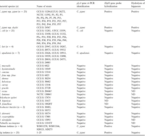
Table 1 Evaluation and comparison of real-time-PCR (glyA), DIG probe colony-blot hybridization (hipO) and hippurate hydrolysis by testing
Campylobacterssp. and related organisms
Bacterial species (n) Name of strain
glyAgene rt-PCR identification
HipOgene probe hybridization
Hydrolysis of hippurate
C. jejunissp.jejuni(n¼23) CCUG 11284,CCUG 26272, NCTC 11392, P1, P2, P3, P4, P5j, P6, P7, P8, P11, P15, P18, P19, P21, P23, P27, P33, P42, P44, P55, P57
C. jejuni Positive Positive
C. jejunissp.doylei CCUG 24567 C. jejuni Positive Positive
C. coli(n¼21) CCUG 11283, ATCC 33559, CCUG 33450, CCUG 11352, P5c, P14, P20, P24, P25, P26, P28, P30, P34, P39, P46, P48, P49, P54, P56, P59, P61
C. coli Negative Negative
C. lari(n¼4) CCUG 23947, CCUG 18267, CCUG 20575, CCUG 19512
C. lari Negative Negative
C. upsaliensis(n¼7) CCUG 23626, CCUG 14913, CCUG 19559, CCUG 33890, CCUG 20818, CCUG 24571, CCUG 24803
C. upsaliensis Negative Negative
C. mucosalis CCUG 6822 Negative Negative Negative
C. hyointestinalis CCUG 14169 Negative Negative Negative
C. concisus CCUG 13144 Negative Negative Negative
C. fetusssp.fetus CCUG 6823 Negative Negative Negative
C. showae CCUG 30254 Negative Negative Negative
C. helveticus CCUG 30682 Negative Negative Negative
C. curvus CCUG 13146 Negative Negative Negative
C. gracilis CCUG 27720 Negative Negative Negative
C. rectus CCUG 20446 Negative Negative ND
C. lanienae NCTC 13004 Negative Negative ND
Helicobacter pylori CCUG 17874 Negative Negative Negative
H. hepaticus CCUG 33637 Negative ND Negative
H. fennellieae CCUG 18820T Negative ND Negative
Arcobacter butzleri(n¼2) CCUG 30485, CCUG 10373
Negative Negative Negative
A. skirrowii CCUG 10374 Negative Negative Negative
A. cryaerophilus CCUG 17801 Negative Negative Negative
A. nitrofigilis CCUG 15893 Negative Negative ND
Wolinella succinogenes CCUG 13145T Negative Negative ND Human isolates (n¼4) WS011270, WS011166,
S20123, S20275
C. jejuni Positive Negative
Pig isolates (n¼25) 1–25 C. jejuni Positive Negative
n, number of strains.
NCTC, National Collection of Type Cultures, (Colindale, UK); CCUG, Culture Collection, University of Go¨teborg (Go¨teborg, Sweden); ATCC, American Type Culture Collection (Manassas, Va); T typestrain and PPenner schemeserostrains (CCUG reference strains) based on heat-stable (HS) antigens (Penner and Hennessey 1980).
The rt-PCR was carried out for eachCampylobacterspp. (single-plex) in a 20-ll PCR reaction volume containing TaqMan Universal Mastermix (Applied Biosystems), sterile destilled water, 600 nmol l)1 each primer, 200 nmol l)1 each probe and 2ll of template DNA prepared as described above. The rt-PCR was performed in the ABI Prism 7700 sequence detection system (Applied Biosystems) with thermocycler conditions and postPCR analysis as described by Nielsen and Andersen (2003).
HipO gene nucleotide probe for detection of C. jejunicolonies
A primer-pair termed HIP400F and HIP1134R for specific amplification of C. jejuni (735-bp sized amplicon) was previously designed on basis of the sequence of the
N-benzoylglycine amidohydrolase hippuricase (hipO) gene, which is absent from Campylobacter spp. other than C. jejuni (Hani and Chan 1995; Linton et al. 1997). These primers were used (1lmol l)1 each) to prepare aC. jejuni -specific hipO gene nucleotide probe labelled with DIG by incorporating DIG-11-dUTP using the PCR DIG Probe Synthesis Kit (Roche Diagnostics, Mannheim, Germany) according to the manufacturer’s recommendations. A 2-ll of DNA from pure cultures was prepared as described above from C. jejuni CCUG 11284, C. jejuni Penner scheme
serotype strains 2, 4, 6, 11, 23 and 35, respectively, and used as template DNA in the preparation of probes. The PCR thermocycler conditions were 94C for 5 min; 30 cycles of 1 min at 94C, 1 min at 62C and 2 min at 72C and final extension for 8 min at 72C.
The specificity of the hipO gene probe for detection of
C. jejuni by colony-blot hybridization was evaluated with
the 73 Campylobacter spp. and related organisms listed in Table 1. Cell material from single colonies of these strains were inoculated in the pattern of a grid onto two BA plates successively using a 1-ll loop, and incubated as appropri-ate for 24–48 h, followed by colony-blot hybridization according to the guidelines of the user’s manual (Roche Diagnostics). The DNA was cross-linked to the mem-branes by exposure to UV illumination for 3 min and membranes were prehybridized at 60C for 1 h before hybridization overnight at 60C with a mixture of the prepared denatured hipO gene nucleotide probes (2 ll probe solution per ml of hybridization buffer). The DIG-labelled probe-target hybrids were detected using alkaline phosphatase conjugated anti-DIG antibody and 5-bromo-4-chloro-3-indolyl phosphate (BCIP)/4-nitroblue-tetrazolium chloride (NBT) as colour substrate according to the manufacturers recommendations (Roche Diagnostics). Development of a purple colour reaction on the mem-branes was denoted as hybridization positive colonies (C. jejuni).
Detection of C. jejuniin pig faecal samples
A total of 261 rectal faecal samples from 7 to 13-week-old pigs were examined within 24 h after collection, for the presence ofC. jejuni. Four different methods were compared for the ability to detect the presence ofC. jejuniin pig faecal samples, (i) analysing enriched samples (Bolton broth without blood, prepared according to the recommendations of the Bacteriological Analytical Manual Online; Hunt
[image:4.592.222.551.86.253.2]et al. 1998) by rt-PCR, (ii) colony-blot hybridization with the hipO gene probe, either on (ii-a) enriched sample (referred to as enriched hybridization) or (ii-b) nonenriched
Table 2 Probes and primers used for
auto-mated 5¢nuclease PCR assay (glyAgene) Species (size in bp)
Primer or probe name
Primer and probe sequences 5¢ fi3¢
Reporter dye*
C. coli(80) Cc-F GTTGGAGCTTATCTTTTTGCAGACA Cc-R TGAGGAAATGGACTTGGATGCT
Cc-P TGCTACAACAAGTCCAGCAATGTGTGCA TET
C. jejuni(135) Cj-F TAATGTTCAGCCTAATTCAGGTTCTC Cj-R GAAGAACTTACTTTTGCACCATGAGT
Cj-P AATCAAAGCCGCATAAACACCTTGATTAGC FAM
C. lari(96) Cl-F CAGGCTTGGTTGTAGCAGGTG Cl-R ACCCCTTGGACCTCTTAAAGTTTT
Cl-P CATCCTAGTCCATTCCCTTATGCTCATGTT TET
C. upsaliensis
(65)
Cu-F TCGTAGCTGGTGAGCATCCTAG Cu-R GGTTTTGTGTGTGGTTGAGCTT
Cu-P CCTTTCCCTCACGCACACATCG FAM
bp, length in base pairs of the species-specific PCR products.
sample (referred to asdirect hybridization), and finally by (iii) random picking ofCampylobactercolonies (see flow chart in Fig. 1).
[image:5.592.44.364.434.711.2]Microbiological method. One gram of faecal material was suspended in 9 ml of Bolton broth, 10-fold dilution series (10)1–10)4) were prepared and 0Æ1 ml of each dilution was inoculated onto modified charcoal-cefoperazone-deoxycho-late agar pcharcoal-cefoperazone-deoxycho-lates (CCDA) [CampylobacterBlood-Free Select-ive Agar Base (Oxoid, Basingstoke, UK) with CCDA Selective Supplement (SE155E; Oxoid)] for enumeration, isolation and colony-blot hybridization. The enrichment broth dilutions (a total of 134 samples for rt-PCR screening and enriched hybridization) and CCDA plates (a total of 261 samples for enumeration and direct hybridization) were incubated for 48 h at 41Æ5C in a microaerobic atmosphere (Fig. 1).
Rt-PCR screening of enriched faecal samples. The enriched sample dilutions were analysed for the presence of
C. jejuniand C. coliby the rt-PCR assay described above. For this, template DNA was prepared by centrifugation of 1 ml of the enrichment broth at 10 000gfor 5 min. Then, the pellet was washed in 1 ml 0Æ9% NaCl and re-suspended in 50ll of modified TE buffer (10 mmol l)1 Tris–HCl, 0Æ1 mmol l)1 EDTA, pH 7Æ6) after centrifugation. Finally, the cells were lysed and analysed in the rt-PCR assay as described for pure cultures.
Although quantification is possible with PCR, this rt-PCR was used to determine if C. jejuni and C. coli were present (qualitatively) in the enriched sample dilutions. Generally, samples with cycle threshold (Ct) >30 and clear indication of probe cleavage (judged by the multicomponent analysis) were considered positive as described by Nielsen and Andersen (2003).
Colony-blot hybridization on enriched samples. In case an enriched faecal sample was detectedC. jejunipositive in rt-PCR, the sample was subjected to analysis by colony-blot hybridization to detect C. jejuni and to possibly obtain bacterial isolates by the enriched hybridization method described earlier. As agar plates with single colonies were preferable for optimal colony hybridization,C. jejuni-positive enrichment broth dilutions were further 10-fold diluted in 0Æ85% saline solution with 0Æ1% peptone before inoculation onto CCDA plates. After 48 h of incubation, the CCDA plate with most single colonies was selected and the membrane discs and plates were marked for orientation before perform-ing colony-blot hybridization as described above.
Colony-blot hybridization on nonenriched samples.
Direct hybridization was applied for detection of C. jejuni
colonies as a simpler alternative to the enriched hybridiza-tion. Thus, the CCDA plates inoculated with the dilution series of nonenriched faecal material (261 samples for enumeration of Campylobacter spp.) were subjected to
Bolton
(n = 134) CCDA plates (n = 134)
(i) Rt-PCR screening (n = 134)
(ii-a) Enriched hybridization (n = 16)
(iii) Random isolation (n = 134) C. jejuni positive
(n = 17)
C. jejuni positive (n = 10)
C. jejuni positive (n = 24) (ii-b) Direct hybridization
(n = 261) Pig faecal samples
(n = 261)
Dilution series
Selective enrichment
CCDA plates (n = 127)
Rt-PCR isolate identification
colony-blot hybridization (direct) with no prior screening of theC. jejunistatus.
Bacterial isolates. When probe-target hybrids (presump-tiveC. jejuni colonies) were detected by hybridization, cell material from the corresponding colony on the CCDA plate (reincubated for 48 h after the colony lift) was picked and subcultured by restreaking single colonies twice onto new BA plates in an attempt to obtain pure cultures.
The specific detection of C. jejuni was compared with
random isolation of Campylobacter spp. These random
isolates were obtained from five presumptiveCampyloacter
spp. colonies (based on colony and cell morphology) on the CCDA plates inoculated with nonenriched pig faecal samples (used for direct hybridization). Colonies were only obtained from those faecal samples that were subjected to both direct hybridization and rt-PCR (enriched hybridiza-tion) (n¼134) (Fig. 1). The isolates were selected to represent the different colony morphologies present on each plate. The presumptive C. jejuni isolates obtained from hybridization and the random Campylobacter spp. isolates were all subjected to the hippurate hydrolysis test and the rt-PCR assay for identification.
R E S U L T S
Evaluation of real-time PCR detection of Campylobacterspp.
A total of 23C. jejunissp.jejuni, oneC. jejunissp.doylei, 21
C. coli, sevenC. upsaliensis, and four C. lariwere correctly amplified by single-plex rt-PCR with no cross reactions between the four species (Table 1). In addition, only negative results were obtained when 10 otherCampylobacter
spp., five Arcobacter strains, one Wolinellastrain and three
Helicobacter species were tested in the rt-PCR test to evaluate the specificity of the primers and probe.
Evaluation of thehipOgene probe for detection of Campylobacter jejuni
The colony-blot hybridization of 24C. jejunistrains with the DIG-labelled hipO gene nucleotide probe yielded clearly purple probe-target hybrids after chromogenic detection (Table 1). However, the detection ofC. jejuniPenner strains 2 and 11 was sometimes less distinct. An increase of the denaturation time for the bacterial cells while blotting onto the membrane (according to user’s manual) from 5 to 15 min enhanced the signal of the Penner strains 2 and 11 (data not given). Slater and Owen (1997) showed that the hippuricase gene is highly conserved in Penner heat-stable serotypes 1, 4 and 11. In addition, 42 Campylobacter
non-jejuni and seven closely related non-Campylobacter strains yielded no target-probe hybrids, which indicated a good specificity of thehipOprobe.
Detection of C. jejuniin pig faecal samples
Four methods were compared for the ability to detect
C. jejuni in pig faecal samples: including screening of enrichment broth by real-time PCR; hipO gene probe colony-blot hybridization either on enriched or nonenriched (direct) samples; and finally by random picking of Campy-lobacter colonies. The rt-PCR on enrichment cultures revealed the presence ofC. coli in all samples andC. jejuni
in 17 of the 134 samples (Table 3). Thus, the developed of rt-PCR method for differentiation of thermophilic Campy-lobacter spp. enabled specific detection of C. coli and
C. jejuni in mixed populations of naturally infected pig faecal samples after an enrichment step.
[image:6.592.46.552.574.671.2]Comparison of rt-PCR and colony-blot hybridiza-tion. Identification of C. jejuni colonies by colony-blot hybridization showed that most, but not all 17 samples, which contained C. jejunias shown by rt-PCR, were found
Table 3 Comparison of the rt-PCR results (no. of samples*) with colony-blot hybridization and random isolation for detection ofCampylobacter jejuniin pig faecal samples
Result of rt-PCR screening (n)
Colony-blot hybridization (DIG-labelled probe-hipOgene)
Enrichment followed by
hybridization Direct hybridization Random isolation
Positive Negative ND Positive Negative ND Positive Negative
C. jejuni-positiven¼17 10 6 1 8 9 0 5 12
C. jejuni-negativen¼117 117 3 101 13 1 116
n, number of faecal samples.
to yield purple colonies by the two colony hybridization approaches (Table 3). The two different hybridization approaches gave the same result in nine (five negative, four positive) of the 16 comparable samples with neither of the methods being superior to the other. However, the CFU ratio between C. jejuni (target-probe hybrids) and total
Campylobacter ssp. seemed to be higher for the enriched samples with 635 of 1612 CFU (enriched hybridization) compared with 166 of 1274 CFU for the nonenriched samples (direct hybridization).
The direct hybridization approach was performed on 134 samples and detected target-probe hybrids (C. jejuni) in three of the 104 samples foundC. jejuni-negative by rt-PCR (hybridization was not feasible for the remaining 13 samples that were C. jejuni-negative by rt-PCR because the colony density was too high) (Table 3). However, confirmation of
C. jejuniby isolation from the presumptiveC. jejunicolonies was only performed for one of the three samples. In addition, the direct hybridization approach was applied to 127 samples as the singleC. jejunidetection method, which further indicated the ability of thehipOgene probe to detect
C. jejuni colonies in minority to Campylobacter ssp. (99 of 2335 CFU) in 13 cases.
Identification of bacterial isolates. A total of 117 isolates (representing 27 samples) were picked to confirm the identity of the presumptiveC. jejunicolonies detected by either direct or enriched hybridization. Eighty-nine strains were con-firmed to be C. jejuni by rt-PCR, whereas 17 wereC. coli, one was neitherC. colinorC. jejuni, and 10 colonies failed for unknown reasons to show growth. In 10 cases, the filter membranes showed some vaguely coloured target-probe hybrids (referred to as suspect), but the isolates obtained from these were identified asC. coliby the rt-PCR method. The presumptiveC. jejuniisolates identified as non-C. jejuni
in the rt-PCR test, were retested by colony-blot hybridization with the hipO probe. All these strains were negative by hybridization in agreement with the rt-PCR identification. Hence, the non-C. jejuni isolates obtained from the pre-sumptiveC. jejuni colonies were apparently not because of poor specificity of thehipOgene probe. Instead, the problem may have been to isolate the right colony or colonies of mixed strains, where the restreaking of colony material in an attempt to obtain pure cultures may have lead to a loss ofC. jejuni.
The isolates were also tested for their ability to hydrolyse hippurate, which is a specific characteristic for C. jejuni, normally used for phenotypic discrimination from other
Campylobacterspp. In this study, the development of colour indicating hydrolysis of hippurate either failed or was very weak in two repeated tests of 25 isolates, varied for 30 isolates and was positive for 44 isolates of 89 isolates. This occurred despite they were all identified as C. jejuni by rt-PCR test and thehipOgene probe (Table 1).
Random isolation of isolates. Finally, the specific detec-tion of C. jejuni by rt-PCR or colony hybridization was compared with isolation ofCampylobacterspp. by picking of five random colonies from each sample. Rt-PCR identifica-tion of the obtained isolates showed that six samples were
C. jejunipositives with 18 of the 560 isolates beingC. jejuni
(Table 3).Campylobacter jejuniwas also obtained from these six samples by at least one of the hybridization methods, but rt-PCR screening of the enriched faecal samples failed to detect the presence in one of these samples.
D I S C U S S I O N
The developed 5¢nuclease rt-PCR based on theglyA gene correctly identified representative reference culture strains of C. jejuni, C. coli, C. lari and C. upsaliensis in pure cultures, providing a good and easy alternative to the often inadequate microbiological phenotypic methods for differ-entiation between these species. Furthermore, the rt-PCR test also enabled the detection of C. jejuni and C. coli
present in pig faecal samples enriched for 2 d in Bolton broth. This indicates that the enrichment in Bolton broth support the growth ofC. jejunieven when present in lower numbers than C. coli. This was also indicated by an increased ratio of hybridization target-probe hybrids (C. jejuni) to totalCampylobacterCFU, when samples that were enriched were compared with nonenriched samples (direct hybridization). Nevertheless, enrichment ofC. jejuniin pig faecal samples can be difficult due to overgrowth by the more numerous C. coli and a high background flora in general (Corry et al. 1995; Madden et al.2000). This may explain the detection of C. jejuni in three nonenriched samples either by direct hybridization (three samples) or random isolation (one sample), while the enrichments of these samples were foundC. jejuninegative by rt-PCR. As
C. coli was amplified in all the enriched faecal samples, it was unlikely that the prepared DNA could have been inhibitory to the rt-PCR enzyme reaction and thereby leading to false negatives.
The rt-PCR screening enabled the detection of Campy-lobacter spp. in enriched faecal samples and the differenti-ation betweenC. coliandC. jejuni. Thus, rt-PCR can detect a possible co-colonization of Campylobacter species, which may be missed by conventional methods because of the often low numbers of C. jejuni in pigs. However, the applied boiling method for lysis of bacterial cells may have biased the detection because of the existence of Campylobacter
strains resistant to lysis by boiling that would then be undetectable by PCR (Englen and Kelly 2000). A compar-ison of rt-PCR screening results based on DNA prepared by different methods would have addressed this.
further characterization would be valuable, for example, to study diversity ofCampylobacterspp. from pigs by serotyp-ing and genotypserotyp-ing (Weijtenset al.1999).
This study showed that the developed DIG-labelled nucleotide probe based on the hippuricase (hipO) gene in combination with either enriched or direct colony-blot hybridization provided a tool for specific identification and isolation of C. jejuni present in low numbers in mixed populations of pig faecal samples. The 17 bacterial isolates not confirmed as C. jejuni (C. coli) could probably be explained by difficulties in identifying the correct colony or overgrowth from neighbouring colonies rather than a poor specificity of thehipO nucleotide probe, as rehybridization of the isolates was negative. Other reports onCampylobacter
detection by colony blot hybridization did not provide the opportunity of obtaining bacterial isolates for subsequent characterization (Taylor and Hiratsuka 1990; Ng et al.
1997). Furthermore, our PCR DIG-labelling of a specific nucleotide sequence by use of specific primers for the hippuricase gene provided a more specific and easy method compared with the cloning approach described by Taylor and Hiratsuka (1990).
When comparing the two different hybridization approa-ches, only nine samples were found to yield the same hybridization results and four of these were hybridization negative. Furthermore, with target-probe hybrids (C. jejuni) found in 10 and eight samples for enriched and direct hybridization, respectively, neither of the approaches seemed to be superior to the other. Hence, the preferable approach may depend on the expected frequency of
C. jejuni, for example, rt-PCR screening will reduce the number of hybridizations if only few samples contain
C. jejuni but instead require preparations of DNA and rt-PCR analysis. Further, pre-enrichment may be necessary to obtain colonies for hybridization in the case of a low level ofCampylobacter.
It was assumed that only the most predominant species was likely to be chosen by the conventional method with random picking of a few colonies. However, when five isolates were obtained from each of 134 pig faecal samples,
C. jejuniwas found in six cases, compared with 10 and 11 for the two hybridization methods. Isolation of five colonies with different morphology instead of only one colony as in normal procedure may have enhanced the likelihood of obtainingC. jejuniso the beneficence of specific detection by hybridization became less distinct.
The prevalence of hippurate-negative C. jejuni in this study was surprisingly high, as detection ofC. jejuniby the ability to hydrolyse hippurate only, would have misclassified 25 (28%) rt-PCR C. jejuni isolates as C. coli. Whereas, Totten et al. (1987) and Wainø et al. (2003) found that hippurate-negative C. jejuni represented 1Æ6 and 13Æ4%, respectively, of C. jejuni strains obtained from human
patients and chickens, respectively. An additional 30 (33%) strains showed a variable result in two testings and this variation occurred despite a standard method being applied. These results emphasize the need for nonphenotypic method alternatives, as the rt-PCR test described.
The hippurate-hydrolysis negativeC. jejuniwere detected by the hipO nucleotide probe, which was based on the hippuricase gene sequence. This indicates the presence of this gene and its absence is therefore not a likely explanation of the hippurate-hydrolysis negative strains, which is in agreement with the studies by Hani and Chan (1995) and Slater and Owen (1997). However, the cause of the hydrolyse activity defect has not yet been elucidated.
In conclusion, this study shows that the developed rt-PCR assays provide easy and fast differentiation of C. jejuni,
C. coli,C. lari, andC. upsaliensis. Furthermore, the colony-blot hybridization approach provided a tool for isolation of
C. jejuni from pig faecal samples with a Campylobacter
population that was dominated byC. coli.
A C K N O W L E D G E M E N T S
We thank the campylobacter group at Danish Institute for Food and Veterinary Research and the veterinary assistants at Rørrendega˚rd for sampling of the pigs. This study was partly funded by the Danish Research Centre of Organic Farming (DARCOF, II.10).
R E F E R E N C E S
Al Rashid, S.T., Dakuna, I., Louie, H., Ng, D., Vandamme, P., Johnson, W. and Chan, V.L. (2000) Identification ofCampylobacter jejuni,Camp. coli,Camp. lari,Camp. upsaliensis,Arcobacter butzleri, andA. butzleri-like species based on theglyAgene.J Clin Microbiol 38, 1488–1494.
Best, E.L., Powell, E.J., Swift, C., Grant, K.A. and Frost, J.A. (2003) Applicability of a rapid duplex real-time PCR assay for speciation of
Campylobacter jejuni and Campylobacter coli directly from culture plates.FEMS Microbiol Lett229, 237–241.
Burnett, T.A., Hornitzky, M.A., Kuhnert, P. and Djordjevic, S.P. (2002) Speciating Campylobacter jejuni and Campylobacter coli
isolates from poultry and humans using six PCR-based assays.
FEMS Microbiol Lett216, 201–209.
Cloak, O.M. and Fratamico, P.M. (2002) A multiplex polymerase chain reaction for the differentiation ofCampylobacter jejuniand Campy-lobacter colifrom a swine processing facility and charaterization of isolates by pulsed-field gel electrophoresis and antibiotic resistance profiles.J Food Prot2, 266–273.
Corry, J.E.L., Post, D.E., Colin, P. and Laisney, M.J. (1995) Culture media for the isolation of campylobacters.Int J Food Microbiol26, 43–76.
Englen, M.D. and Kelly, L.C. (2000) A rapid DNA isolation procedure for the identification ofCampylobacter jejuniby the polymerase chain reaction.Lett Appl Microbiol31, 421–426.
Hani, E.C. and Chan, V.L. (1995) Expression and characterization of
Campylobacter jejuni benzoylglycine amidohydrolase (hippuricase) gene inEscherichia coli.J Bacteriol177, 2396–2402.
Hunt, J.M., Abeyta, C. and Tran, T. (1998) Campylobacter. In
Bacteriological Analytical Manual Online, 8th edn, revison A. ed. Jackson, G.J., Merker, R.I. and Bandler, R. Available at: http:// www.cfsan.fda.gov/ebam/bam-7.html.
Inglis, G.D. and Kalischuk, L.D. (2004) Direct quantification of
Campylobacter jejuni and Campylobacter lanienaein faeces of cattle by real-time quantitative PCR. Appl Environ Microbiol 70, 2296– 2306.
Linton, D., Lawson, A.J., Owen, R.J. and Stanley, J. (1997) PCR detection, identification to species level and fingerprinting of
Campylobacter jejuni and Campylobacter coli direct for diarrheic samples.J Clin Microbiol35, 2568–2572.
Logan, J.M.J., Edwards, K.J., Saunders, N.A. and Stanley, J. (2001) Rapid identification of Campylobacter spp. by melting peak analysis of biprobes in real-time PCR. J Clin Microbiol 39, 2227–2232.
Lu¨beck, P.S., Wolffs, P., On, S.L.W., Ahrens, P., Ra˚dstro¨m, P. and Hoorfar, J. (2003) Toward an international standard for PCR-based detection of foodborne thermotolerant campylobacters: assay devel-opment and analytical validation.Appl Environ Microbiol69, 5664– 5669.
Madden, R.H., Moran, L. and Scates, P. (2000) Optimising recovery of
Campylobacter spp. from the lower porcine gastrointestinal tract.
J Microbiol Methods42, 115–119.
Manser, P.A. and Dalziel, R.W. (1985) A survey of campylobacter in animals.J Hyg95, 15–21.
Munroe, D., Prescott, J.F. and Penner, J.L. (1983)Campylobacter jejuni
andCampylobacter coliserotypes isolated from chickens, cattle, and pigs.J Clin Microbiol18, 877–881.
Ng, L.-K., Kingombe, C.I.B., Yan, W., Taylor, D.E., Hiratsuka, K., Malik, N. and Carcia, M.M. (1997) Specific detection and confir-mation of Campylobacter jejuni by DNA hybridization and PCR.
Appl Environ Microbiol63, 4558–4563.
Nielsen, E.M. and Andersen, M.T. (2003) Detection and character-ization of verocytotoxin-producingEscherichia coliby automated 5¢
nuclease PCR assay.J Clin Microbiol41, 2884–2893.
Nielsen, E.M., Engberg, J. and Madsen, M. (1997) Distribution of
Campylobacter jejuniandCamp. colifrom Danish patients, poultry, cattle and swine.FEMS Immunol Med Microbiol19, 47–56. Nogva, H.K., Bergh, A., Holck, A. and Rudi, K. (2000) Application of
the 5¢nuclease PCR assay in evaluation and development of methods for quantitative detection of Campylobacter jejuni. Appl Environ Microbiol66, 4029–4036.
On, S.L.W. and Jordan, P.J. (2003) Evaluation of 11 PCR assays for species-level identification ofCampylobacter jejuniandCampylobacter coli.J Clin Microbiol41, 330–336.
Penner, J.L. and Hennessey, J.N. (1980) Passive hemagglutination technique for serotyping Campylobacter fetus subsp. jejuni on the basis of soluble heat-stable antigens.J Clin Microbiol12, 732–737. Petersen, L., Nielsen, E.M., Engberg, J., On, S.L. and Dietz, H.H.
(2001) Comparison of genotypes and serotypes of Campylobacter jejuniisolated from Danish wild mammals and birds and from broiler flocks and humans.Appl Environ Microbiol67, 3115–3121. Rudi, K., Høidal, H.K., Katla, T., Johansen, B.K., Nordal, J. and
Jakobsen, K.S. (2004) Direct real-time PCR quantification of
Campylobacter jejuni in chicken faecal and cecal samples by integrated cell concentration and DNA purification. Appl Environ Microbiol70, 790–797.
Sails, A.D., Fox, A.J., Bolton, F.J. and Wareing, D.R.A. (2003) A real-time PCR assay for the detection ofCampylobacter jejuni in foods after enrichment culture.Appl Environ Microbiol69, 1383–1390. Slater, E.R. and Owen, R.J. (1997) Restriction fragment length
polymorphism analysis shows that the hippuricase gene of Campy-lobacter jejuniis highly conserved.Lett Appl Microbiol25, 274–278. Steinhauserova, I., Ceskova, J., Fojtikova, K. and Obrovska, I. (2001) Identification of thermophilicCampylobacterspp. by phenotypic and molecular methods.J Appl Microbiol90, 470–475.
Taylor, D.E. and Hiratsuka, K. (1990) Use of non-radioactive DNA probes for detection ofCampylobacter jejuniandCampylobacter coliin stool specimens.Mol Cell Probes4, 261–271.
Totten, P.A., Patton, C.M., Tenover, F.C., Barret, T.J., Stamm, W.E., Steierwalt, A.G., Lin, J.Y., Holmes, K.K.et al.(1987) Prevalence and characterization of hippurate-negative Campylobacter jejuni in King County, Washington.J Clin Microbiol25, 1747–1752. Wainø, M., Bang, D.D., Lund, M., Nordentoft, S., Andersen, J.S.,
Pedersen, K. and Madsen, M. (2003) Identification of campylobac-teria isolated from Danish broilers by phenotypic tests and species-specific PCR assays.J Appl Microbiol95, 649–655.
Weijtens, M.J.B.M., Reinders, R.D., Urlings, H.A.P. and Van der Plas, J. (1999)Campylobacter infections in fattening pigs; excretion pattern and genetic diversity.J Appl Microbiol86, 63–70.
Wilson, D.L., Abner, S.R., Newman, T.C., Mansfield, L.S. and Linz, J.E. (2000) Identification of ciprofloxacin-resistant Campylobacter jejuniby use of a fluorogenic PCR assay.J Clin Microbiol38, 3971– 3978.
Yang, C., Jiang, Y., Huang, K., Zhu, C. and Yin, Y. (2003) Application of real-time PCR for quantitative detection ofCampyobacter jejuniin poultry, milk and environmental water. FEMS Immunol Med Microbiol38, 265–271.