1-1-2000
Expression and partial purification of recombinant
bovine intercellular adhesion molecule-1
Anton Martin Roach
Iowa State UniversityFollow this and additional works at:
https://lib.dr.iastate.edu/rtd
This Thesis is brought to you for free and open access by the Iowa State University Capstones, Theses and Dissertations at Iowa State University Digital Repository. It has been accepted for inclusion in Retrospective Theses and Dissertations by an authorized administrator of Iowa State University Digital Repository. For more information, please [email protected].
Recommended Citation
Roach, Anton Martin, "Expression and partial purification of recombinant bovine intercellular adhesion molecule-1" (2000). Retrospective Theses and Dissertations. 17701.
by
Anton Martin Roach
A thesis submitted to the graduate faculty
in partial fulfillment of the requirements for the degree of MASTER OF SCIENCE
Major: Immunobiology
Major Professors: James A. Harp and James A. Roth
Iowa State University Ames,Iowa
Graduate College Iowa State University
This is to certify that the Master's thesis of Anton Martin Roach
has met the thesis requirements of Iowa State University
Signatures have been redacted for privacy
TABLE OF CONTENTS
ABSTRACT ... v
GENERAL INTRODUCTION ... 1
Thesis Organization ... . 1
Literature Review ........................... .. 1
Adhesion Molecules In Leukocyte Trafficking ... 3
The Selectins ... 3
The Integrins ... 6
lntercellular Adhesion Molecules ... 8
ICAM-1 ... 9
ICAM-2 ... 15
ICAM-3 ... 17
ICAM-4 ... 19
ICAM-5 ... 19
Conclusion ... 21
MATERIALS AND METHODS ... 23
Polymerase Chain Reaction (PCR) ... ...... 23
The Expression Vector ............. 25
Ligations ... . 25
Transformations ......... . 26
Small Scale Expression of Soluble Recombinant Bovine ICAM-1 ... 27
Large Scale Expression and Affinity Purification ...... 28
Inclusion Body Production, Solubilization, and Purification Using Ion Exchange Chromatography ...... 29
Ion Exchange Chromatography ... 30
Electrophoresis ... 31
Coomassie Blue Stain ... 31
Silver Stain ... 32
Western Blotting ... 32
Antibody Production ......... ...... 33
Enzyme Linked Immunosorbent Assay (ELISA) ... 34
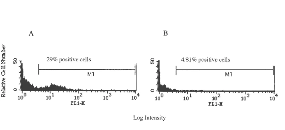
Immunofluorescence Analysis by Flow Cytometry to Detect Mononuclear Cell Binding of rbICAM-1 ... 35
Preparation of Recombinant Protein ... 35
Bovine Blood Collection ... 35
RES UL TS ... 37
PCR
..
...
...
....
.
....
...
...
...
...
...
...
...
.
..
.
.
..
...
...
.
.
...
.
...
37
Ligations/Transformations
...
.
...
.
...
....
...
...
.
...
.
....
.
.
...
. 37
Small Scale
Expression
of Soluble Recombinant Bovine ICAM-1
... 38
Large Scale Expression and Affinity Purification
...
..
...
...
...
...
.41Inclusion Body Production, Solubilization, and Purification Using Ion Exchange
Chromatography
...
...
..
...
...
...
.
...
.
...
...
....
...
.
..
....
....
.
...
.
...
.
...
.
...
41Antibody Production and ELIS
As
...
...
...
...
...
... 46
Fluorescent
Activated
Cell Sorting Analysis (F ACScan)
...
...
..
...
46
DISCUSSION ... 49
GENERAL CONCLUSIONS ... 56
RE FE REN CES ... 57
ABSTRACT
Recombinant bovine intercellular adhesion molecule-I (ICAM-1) has potential as an
anti-inflammatory agent for treating bovine mastitis and other inflammatory conditions. The
objective of this study was to produce biologically active recombinant bovine ICAM-1 in order
to investigate its use as a potential immunomodulator. Using a cDNA encoding the complete
bovine ICAM-1 open reading frame, polymerase chain reaction (PCR) was used to prepare
cDNAs encoding domains 1 and 2 (Bl), or domains 1 through 5 (A6), of bovine ICAM-1.
These were cloned into the pTrxFus expression vector down stream and in frame with the
sequence coding for the expression tag, thioredoxin. The constructs were verified by DNA
sequence analysis. The E. coli strain GI724 was transformed with the constructs and
expression of the recombinant protein was induced by tryptophan. The ICAM-1 (domains 1
and 2 or 1-5) constructs were mainly expressed as insoluble protein in inclusion bodies, with
little recombinant protein in the soluble fraction of cell lysates. The recombinant protein was
identified by Western blots using a mAb to the thioredoxin tag. The inclusion bodies were
solubilized by the addition of 6 M guanidine-HCl. Guanidine-HCl was then slowly removed
by dialysis, and the recombinant protein was partially purified by ion exchange chromatography
using DEAE Sephacel. Recombinant protein from the soluble portion of the cell lysates was
used to immunize mice in attempts to obtain a mouse mAb to recombinant bovine ICAM-1.
GENERAL INTRODUCTION
Thesis Organization
This thesis is organized in a standard format consisting of six chapters: a General
Introduction, Literature Review, Material and Methods, Results, Discussion, and Conclusions. The General Introduction consists of a review of the literature of leukocyte trafficking with
emphasis on the intercellular adhesion molecules. The remaining chapters outline and detail the
means by which the goals of this project were addressed.
Literature Review
Leukocytes play a key role in the surveillance of tissues for pathogens and tissue injury.
The nature of the inflammatory stimulus, the location of the insult, and the surface molecules
present on various leukocytes determine the subpopulation of cells that interact with the
endothelium [1]. Generally, neutrophils are the first line of defense in an inflammatory response [2, 3, 4]. Though an inflammatory response is needed to fight infection, leukocytes,
and particularly neutrophils, can cause extensive tissue damage in the host [2, 5, 6]. This can
occur in adult respiratory distress syndrome in humans, bovine respiratory disease complex in
cattle, and mastitis in both humans and animals [7, 8, 9]. Sometimes it is necessary to limit an immune response to lessen the severity of damage host cells may cause while mounting an
effective immune response.
Immune cells adhere to one another and to other cells using four families of adhesion
proteins: the selectin, cadherin, integrin, and immunoglobulin, families [10, 11, 12].
Chemoattractants including but not limited to interleukin-8 (IL-8), RANTES (Regulated Upon Activation, Normal T cell Expressed and Secreted), macrophage chemotactic protein 1 (MCP-1),
injury [I, 2]. Subsequent leukocyte extravasation into sites of infection induces a multi-step
process involving the four families of adhesion proteins and chemoattractants.
Extravasation includes the steps of leukocyte rolling, tight binding, diapedesis,
(movement of leukocytes between the endothelial cell junctions) and lastly migration to sites of
infection [4, 6, 13, 14]. Leukocytes roll along the endothelial lining of blood vessels under the
shear force of blood flow [I, 2, 3, 6]. A transient rolling or "sticking" is accomplished by
carbohydrate ligands on leukocytes binding with E-selectin and P-selectin on the surface of
endothelial cells, and L-selectin on leukocytes adhering to its carbohydrate ligand on endothelial
cells [I, 13, 15]. This transient interaction is reversible within seconds and is not sufficient to
allow white blood cells to remain bound to the vessel wall without further activation of
accessory molecules [13].
Activation of leukocytes by chemoattractants and cytokines can trigger tight adhesion of
leukocytes already tethered to the endothelial lining of blood vessels [2, 13]. This is facilitated
by the activation of the
B
2 integrins, specifically LFA-1 and Mac-I on the surface of white blood cells interacting with their ligand, intercellular adhesion molecule-I (ICAM-1) onendothelial cells [2, 13]. The LFA-1 / ICAM-1 interaction is sufficient to cause leukocyte arrest
at sites of infection even under the shear force of the blood flow. This interaction, however, is
still reversible within minutes [4, 15].
Diapedesis is dependent on glycosylated aminoglycans and the
B
2 integrins on the leukocyte plasma membrane adhering to platelet endothelial cell adhesion molecule-I(PECAM-1, CD31) at the intercellular junctions of endothelial cells [2, 3, 6, 15]. These interactions allow
the leukocyte to squeeze between the endothelial cells, thereby leaving the blood vessel and
entering into sites of injury [15, 16].
The last step in the extravasation process, leukocyte migration to specific sites of
infection within the tissue matrix, is facilitated by the chemotactic gradient generated by
Leukocyte extravasation can be regulated at the various steps in the process, thus modulating the inflammatory response. Selectivity in the process of leukocyte recruitment to sites of tissue injury comes from the diversity of molecules capable of mediating each step [1, 14].
The goal of this research was to produce a biologically active form of recombinant bovine Intercellular Adhesion Molecule-1 (rbICAM-1) in a prokaryotic expression system, and produce a monoclonal antibody (mAb) against bovine ICAM-1. The rationale behind these goals was as follows: the recombinant protein could be used as an immunomodulator in vivo, and in vitro for the detection of surface molecules present on bovine endothelial cells and leukocytes. These surface molecules would include the ligands for ICAM-1, the ~2 integrins. The rbICAM-1 could also be used to detect ICAM-1 expression in bovine tissue in vitro, and as an immunomodulator in vivo. This is the first report of the production of biologically active rbICAM-1.
Adhesion Molecules In Leukocyte Trafficking
The Selectins
The selectin family of adhesion proteins mediates the initial role in leukocyte
recruitment to sites of infection [2, 4, 13]. They are involved in the initial tethering and rolling of leukocytes along postcapillary venule endothelial cells lining the vessel walls. The adhesive proteins L-selectin (CD62L), E-selectin (CD62E), and P-selectin (CD62P) are the selectin molecules involved and are named after cells on which they were first identified [2, 3, 4, 17, 18]. L-selectin was discovered on lymphocytes, and has been found on all circulating leukocytes, except for a subpopulation of memory lymphocytes [1, 2].
E-selectin was first found on endothelial cells, but only after stimulation by
rapidly mobilized to the plasma membrane in response to mediators of inflammation such as
thrombin and histamine [l, 2], [3].
In contrast to L- and P-selectin, E-selectin is the only selectin that requires de novo synthesis [1]. All three selectins are involved in the recruitment of leukocytes, but there are
fundamental differences in their distribution, activation, and mode of expression [16].
The three members of the selectin family are all transmembrane glycoproteins that
possess a carbohydrate-recognizing domain (CRD) at their amino terminus, an epidermal
growth factor (EGF)-like domain, and two (L-selectin), six (E-selectin), or nine (P-selectin)
complement regulatory domains [15, 16]. There is also a transmembrane segment and a short
cytoplasmic domain [2, 16, 17]. The CRD at the amino terminus end of the selectins is homologus to the Ca2+ dependent (C-type) lectins [l, 16].
The level of homology among the three selectins is 60-65% at the CRD and 40-45% at
the EGF-like domain [2, 16]. There is little homology among the three in the transmembrane
and cytoplasmic domains [15, 16]. The lectin domain plays the most crucial role in ligand
binding. This domain mediates cell-cell contact through Ca2+-dependent interactions with
cell-surface carbohydrates [1, 15, 16].
All selectins recognize a sialylated carbohydrate moiety on their counterreceptors. E
and P selectin recognize carbohydrate structures that are distinct, but closely related to the
tetrasaccharide sialyl Lewisx and its isomer sialyl Lewis" [1, 3, 15, 16]. A high affinity ligand
for P-selectin, termed P-selectin glycoprotein ligand-1 (PSGL-1) has been characterized [1, 3,
12, 16]. PSGL-1 also serves as a counterreceptor for E-selectin [16]. PSGL-1 is expressed on
all blood neutrophils, monocytes, and lymphocytes [l, 16]. Another glycoprotein ligand has
been identified specific for E-selectin, E-selectin ligand-1 (ESL-1) [ 16].
The ligand for L-selectin is also related to the Lewisx,a moieties, but contains sialic acid
and sulfate as well [1, 15]. Two sulfated glycoprotein ligands have been identified for
is expressed mainly by the endothelial cells of high endothelial venules (HEVs) of lymphoid
tissue, and can be secreted into the blood stream as well [l, 2, 15, 16]. The second, termed
peripheral node addressin (PNAd, CD34), is expressed by the endothelial cells of many tissues
as well as hemapoietic stem cells [l, 2, 15, 16].
Binding of the selectins to their ligands is transient and reversible in seconds under the
shear force of blood flow (i.e., rolling) [4, 13]. Leukocytes roll along the endothelial lining of
the vasculature as part of their immunosurveillance. L-selectin on the tips of the leukocyte
microvilli transiently binds with its receptors GlyCAM-1 or CD34, and without further
activation, detach and re-enter the circulation to roll elsewhere along the vasculature [15].
L-selectin expression on leukocytes is plentiful, especially on unactivated leukocytes [15].
The importance of the carbohydrate moieties on the selectin ligands for adhesion is
made evident by the genetic disease leukocyte adhesion deficiency type II (LAD II). In 1992
Etzioni et al. identified two unrelated children with neutrophilia, recurrent bacterial infections,
and failure of their neutrophils to bind cytokine-activated endothelial cells [19]. The patients
also exhibited short stature and mental retardation [19]. Further in vitro analysis of leukocytes
from these patients revealed they failed to express the sialyl Lewisx structure and therefore are
unable to bind E-selectin and P-selectin [19]. Etzioni et al. (1992) reported this disorder as
LAD II. Consistent with the proposed role of selectins in extravasation, leukocytes from these
individuals fail to roll along cytokine activated endothelial cells of postcapillary venules.
Furthermore, these individuals suffer from recurrent bacterial infections, fail to form pus, and
exhibit short stature and mental retardation due a disorder in fucose metabolism [4, 12]. Fucose
is an important component of sialyl Lewisx.
During an inflammatory response, cytokines and chemoattractants present at sites of
infection activate leukocytes and the surrounding vascular endothelial tissue. E-selectin and P-selectin expression is upregulated as well as their ligands. This also includes a rapid up
this activation process, L-selectin is shed and receptors for the selected proinflammatory cytokines and chemoattractants on leukocytes are upregulated. This activation process along with vasodilation leads to an accumulation of leukocytes at the site of tissue injury.
The lntegrins
Another family of adhesive proteins permits leukocyte arrest and stops the transient flow of white blood cells at sites of inflammation and tissue injury. This class is termed the integrins. Integrin proteins are involved in the integration of cells with the extracellular matrix, hence the name integrin [10, 20]. The integrin superfamily consists of more than 20
structurally homologous proteins that promote cell-cell or cell-matrix interactions [3, 21]. These interactions are a basic component of cell migration and recognition, and underlie many biologic processes, including embryogensis, tissue repair, and immune and inflammatory responses [12, 21].
The integrins are composed of non-covalently linked
a
and13
chains selected from among 17a
and 813
subunits that heterodimerize to produce various receptors[lO]. Three major groups of integrins,/3i, /3
2 and the/3
7 subfamilies are important in the interactions ofleukocytes with endothelium [l, 15].
In the
!3
1 subfamily thea
4/3
1 integrin is an important adhesion molecule based on itsinteraction with vascular cell adhesion molecule-I (VCAM-1), found on activated endothelium at sites of inflammation [1, 15]. The
!3
2 integrin subfamily consists of LFA-1 (CD11a/CD18),Mac-1 (CD11b/CD18), p150,95 (CD11c/CD18), and
act/3
2 (CD11d/CD18) [15]. The thirdsubfamily,
/3
7, includes an important member, the a4/3
7 integrin. This integrin is important in1 ymphocyte-endothelium interactions [ 15].
integrin subfamily interacts with intercellular adhesion molecules (ICAMs), specifically ICAM-1, 2, and 3 [22].
LFA-1, Mac-1, and p150,95 (though less characterized) are the ~2 integrins that play a
major role in leukocyte adherence to activated endothelium via the ICAMs [23, 24]. They contain different
a
chains (CD 1 la,b,c, and d) but possess a common ~ chain, CD 18 [11, 23, 25]. The globular heads of thea
subunits of the ~2 integrins contain an "I" (inserted) domainnot seen in many integrins [2]. The I domain is a 200 amino acid sequence at the N terminus of the
a
chain [25]. The I domain contains bivalent cation-binding sites that are essential for ligand binding [2, 26]. Mg2+ and Ca2+ are the two essential cations that mediate ligand bindingand the activation state of the integrins [1, 27].
LFA-1 is found on Band T lymphocytes, monocytes and neutrophils [2, 28]. Upon activation, LFA-1 can selectively bind ICAM-1, -2, or-3 [29, 30, 31]. Mac-1 is expressed by monocytes, neutrophils, and some mast cells and natural killer cells (NK cells) [2, 18]. Mac-1 is able to bind ICAM-1, ICAM-2, fibrinogen, and the complement opsonin iC3b [18, 22, 32].
p150,95 is found on monocytes, neutrophils, natural killer cells, and platelets [18, 33]. Upon activation p150,95 is able to bind fibrinogen and the complement opsonin iC3b [18, 24]. Activated cells expressing p150,95 are able to bind stimulated endothelial cells, with the postulated ligand being ICAM-1 [24].
The ~2 integrins are mainly activated by a process termed "inside-out" signaling [30].
That is, intracellular signals that activate (produce conformational changes) first in the
cytoplasmic domains, and then in the extracellular portion of the integrin heterodimers are only triggered once an accessory molecule on the leukocyte has bound its ligand [22, 30, 34]. The best example of inside-out signaling to activate LFA-1 is found in peripheral blood
lymphocytes (PBLs).
circulating cells and clogging of the blood vessels [30]. Intracellular signals that activate LFA-1
are generated only after activation of a lymphocyte mainly by the T-cell receptor TCR/CD3
complex [30, 34, 35]. In addition to the TCR/CD3 complex, several other leukocyte receptors
can activate LFA-1 through the G proteins or protein tyrosine kinases [15, 30, 36]. The
quantity of intracellular divalent cations, specifically Mg2+, Ca2+, and Mn2+ present in the
microenvironment of the cytoplasmic tail of the integrin also plays a critical role in the
activation/deactivation process [18, 22, 30].
The importance of the ~2 integrins in mounting an effective immune response is made
evident by the genetic disease leukocyte adhesion deficiency type I (LAD I) identified in
humans [28] dogs [37] and bovine leukocyte adhesion deficiency (BLAD) in cattle [38] [39].
Humans and animals afflicted with these diseases have various genetic defects in the CD18
molecule of the ~2 integrin heterodimer [28, 39]. Leukocytes from LAD I patients are able to
roll along the blood vessel wall, but are unable to arrest at sites of infection [28], [4, 38]. This
leads to leukocytosis and the threat of death from recurrent infections [28, 40]. Humans
afflicted with the severe form of LAD I (no CD 18 expression) die at childhood [ 4, 28] and
BLAD animals rarely survive past nine months of age [38, 39, 41].
Intercellular Adhesion Molecules
Intercellular adhesion molecules (ICAMs) are Type I transmembrane glycoproteins that
belong to the immunoglobulin gene superfarnily [20, 42, 43]. Their cellular distribution, degree
of glycosylation, and molecular weights vary [20, 44, 45]. ICAMs promote adhesion of
immune cells to the extracellular matrix and aid in the interactions between antigen presenting
cells (APCs) and lymphocytes during immunological and inflammatory reactions by binding
their ligand, the
f3
2 integrins [11, 43, 46, 47]. There are currently five members in the ICAMICAM-2, and ICAM-3 [11, 20], [22, 45]. The role ofICAM-1 in leukocyte adherence and extravasation has led to it being targeted as an anti-inflammatory agent [6, 48, 49].
ICAM-1
ICAM-1 is the most widely distributed intercellular adhesion molecule and is the most
extensively studied molecule in the ICAM family [22]. At the Leukocyte Typing Workshop IV
(Vienna, Austria 1989) it was given the cluster of differentiation (CD) designation of CD54
[50]. It is expressed constitutively on the surface of endothelial cells, T and B lymphocytes,
monocytes, dendritic cells, and epithelial cells at low levels [11] [47] [50] [43]. These cells
upregulate their expression of ICAM-1 in the presence of proinflammatory cytokines including
IL-1 (interleukin-I) and tumor necrosis factor alpha (TNF a) [11, 43, 50]. Recent evidence
indicates that ICAM-1 belongs to the family of acute-phase response genes which are induced
by interleukin-6 (IL-6) [51].
The upregulation of ICAM-1 by proinflammatory cytokines occurs in a cell type
specific manner [50]. ICAM-1 surface expression on T cells is upregulated by interleukin-2
(IL-2) and to a lesser extent by gamma interferon (IFN-y) [50]. In contrast, IFN-y, TNF a, and
IL-1 are all good inducers of ICAM-1 on fibroblasts. Endothelial cells increase their surface
expression of ICAM-1 in response to TNF a, lymphotoxin, and IL-1, with a less potent response from IFN-y [50]. Increased ICAM-1 surface expression can be detected within four
hours of stimulation of the various cell types, and expression is often maintained for greater than twenty-four hours [14, 50].
The key role ofICAM-1 in the inflammatory process is to promote leukocyte arrest
under the shear force of the blood flow by binding its ligand (the ~2 integrins) found on all
leukocytes. This binding stops leukocyte rolling at sites of infection for subsequent
chimpanzee, murine, rat, canine, and the bovine [43, 52, 53, 54, 55]. Among these species there
is 55-65% homology at the DNA level [43].
ICAM-1 Structure
The human ICAM-1 gene consists of seven exons separated by six intrans [51].
Studies of ICAM-1 cDNA have led to its structural and functional analysis, including its
identification as a member of the immunoglobulin superfamily [42]. The 5' -flanking region of
the ICAM-1 gene contains binding sites for many transcription factors. These include the
glucocorticoid receptor binding sites, IFN-y- responsive elements, NF-KB (Nuclear Factor-KB)
consensus elements and AP-1 and -2 (Activator Protein) can be found in this region [51, 56].
ICAM-1 contains five extracellular immunoglobulin-like domains and varies in
molecular weight from 80-115 kilodaltons (kDa) depending on the degree of glycosylation
[42]. The five extracellular domains oflCAM-1 are each encoded by their own exon [51]. The
immunoglobulin-like domains are comprised of 453 predominantly hydrophobic amino acids,
followed by a hydrophobic transmembrane domain, and a short charged cytoplasmic tail [43].
Domains one through four contain 88, 97, 99, and 101 amino acid residues respectively, which
are typical of immunoglobulin domain size; domain five is truncated with 68 amino acid
residues [42]. The extracellular immunoglobulin-like domains contain a
B
sheet structurewhich is stabilized by disulfide bridges between highly conserved cysteine residues in domains
one, two, three, and five [42, 43].
It was originally hypothesized that ICAM-1 was a monomeric protein [42]. Further
experimental analysis proved that membrane bound ICAM-1 is indeed a homodimer with
greater affinity and avidity for its ligand, the
B
2 integrins, versus monomeric ICAM-1 [57, 58,59]. The exact mechanism leading to homodimer formation of ICAM-1 has yet to be
ICAM-1 and Ligand Binding
As mentioned previously ICAM-1 binds the
B
2 integrins LFA-1 and Mac-1. Many integrins that bind extracellular matrix proteins bind to a calcium binding RGD (Arg-Gly-Asp)sequence in their ligand [60]. Human ICAM-1 is the first identified ligand for the integrins that
does not contain a calcium binding RGD sequence [43, 60]. In addition, ICAM-1 serves as the
receptor for a major group of human rhinoviruses (at least 90% of the approximately 100
identified serotypes), the causative agent of the common cold [61, 62, 63]. It is also the ligand
for Plasmodiumfalciparum on erythrocytes, the pathogen responsible for malaria [64]. Upon activation, the
B
2 integrins undergo an extracellular conformational changeenabling them to bind to ICAM-1 (see above). Mutational analysis of the cDNA encoding
ICAM-1 has revealed that the LFA-1 binding site is located in domain one [60, 65].
Glycosylation of domain one is not essential to ICAM-1/LFA-1 binding as none of the 8
potential glycosylation sites in ICAM-1 are located in domain 1 [42]. In vitro analysis of monomeric vs. dimeric ICAM-1 binding to LFA-1 revealed that the homodimer form has a
higher degree of binding to LFA-1 [57, 58].
The binding site for Mac-1 is located in the third immunoglobulin-like domain of
ICAM-1 [46]. Mutational analysis of the cDNA encoding human ICAM-1 demonstrated that
the degree of glycosylation in domain three affected Mac-1 binding. Amino acid substitutions
that eliminated the N-linked glycosylation sites in domain three ofICAM-1 resulted in higher
affinity binding of Mac-1 compared to wild type ICAM-1 [46].
The ICAM-1 LFA-1/Mac-1 interaction is a key step in leukocyte trafficking and the
generation of an immune response. The interactions of these molecules not only causes
leukocyte arrest at sites of inflammation but aids in facilitating T-cell mediated killing, T-helper
and B-lymphocyte responses, killing by Natural Killer cells (NK), and antibody-dependent cell
to limit or abrogate the inflammatory response emphasizing the importance of the ICAM-1
LFA-1/Mac-l interaction [23, 24, 43, 66].
Domain one of human ICAM-1 serves as the major receptor for rhinoviruses [42, 63].
Mutagensis of the cDNA encoding human ICAM-1 showed that glycosylation is not required
for rhinovirus attachment [60, 63]. Truncated forms of ICAM-1 lacking domains 3,4, and 5
were able to bind rhinoviruses but to a lesser extent (10 fold decrease) than wild type ICAM-1
[60, 63]. Domains 3-5 oflCAM-1 may play a role in virus binding by enhancing the tertiary
structure of the first two domains by allowing greater accessibility to the virus receptor site in
domain 1. Rhinoviruses are able to bind recombinant monomeric and dimeric ICAM-1 in vitro,
but exhibit a higher affinity for the dimeric form [58]. Attachment of human rhinoviruses to
human ICAM-1 is species specific. Cells from other species, which express host ICAM-1, are
not able to bind human rhinovirus [63].
The primary event in the pathogenesis of malaria is the attachment of Plasmodium
falciparum-infected erythrocytes to at least three different receptors on capillary endothelium
[64, 68]. Berendt et al. (1989) identified human ICAM-1 as one of these receptors. The other
two receptors are glycoprotein IV (CD36) and thromobospondin [64]. Mutagenesis of
hICAM-1 cDNA revealed that the binding site for P. falciparium-infected erythrocytes is located in domain one of ICAM-1 [69]. Attachment of infected red blood cells to hICAM-1
seems to be species specific as infected erythrocytes do not bind murine ICAM-1 in vitro [68].
Murine ICAM-1 has 50% homology to human ICAM-1 at the amino acid level [53] and,
therefore, may not have receptors for infected erythrocytes.
Though LFA-1, human rhinovirus, and P.falciparum-infected erythrocytes are able to
bind domain one oflCAM-1, their respective binding sites are distinct with only a small degree
of overlap (60, 68, 69]. Monoclonal antibodies against ICAM-1 which block the
LFA-1/ICAM-1 interaction do not inhibit human rhinoviruses and infected erythrocytes from binding
antibodies is required to disrupt ICAM-1 binding to human rhinovirus and infected erythrocytes [68, 69].
ICAM-1 has also been implicated as a receptor for fibrinogen, sialophorin (CD43), and the matrix factor hyaluronan [43, 70, 71]. The extent these proteins interact with ICAM-1 during ICAM-1 cell mediated adhesion has not been determined.
Bovine ICAM-1
In 1997 bovine ICAM-1 was cloned and sequenced by Lee et al. Sequence comparison with ICAM-1 from other species aligns bovine ICAM-1 in the lg superfamily. The open reading frame of 1605 nucleotides encodes 535 amino acids that include a signal sequence, five extracellular lg-like domains, a hydrophobic transmembrane region, and a cytoplasmic tail [72]. There are 13 potential N-glycosylation sites in bovine ICAM-1 [72]. Unstimulated bovine mononuclear cells and neutrophils exhibit low and undetectable levels of ICAM-1 mRNA respectively [72]. Upon stimulation with inflammatory mediators these cells have an increase in total ICAM-1 mRNA [72].
At the amino acid level bovine ICAM-1 exhibits 57% homology with human ICAM-1 with the greatest degree of homology existing in domain two (70%) [72]. Like the typical integrin ligand, bovine ICAM-1 contains the calcium binding RGD sequences in its extracellular domains [72].
Therapeutic Uses of Recombinant ICAM-1 (rlCAM-1) or an ICAM-1 Monoclonal Antibody
adhesion molecules or using recombinant forms of leukocyte receptors. It is well documented
that antibodies to the
B
2 integrins can block leukocyte adhesion in vitro and in vivo [23, 31, 66].Recently the use of ICAM-1 mAbs and rICAM-1 to block leukocyte adhesion has been
investigated. Preliminary investigations into blocking the ICAM-1/LFA-1 interaction assessed
the role that this binding plays in exacerbating immune induced complications. These would
include the role of ICAM-1 in ischemia-reperfusion injury, allograft rejection, and in chronic
inflammation such as rheumatoid arthritis. The use of rICAM-1 has also been investigated in
attempts to block rhinovirus attachment in vivo [73].
Antibodies to ICAM-1 have been shown to inhibit both lymphocyte and granulocyte
adhesion processes in in vitro assays including mixed lymphocyte reactions, cytotoxic T-cell
activity, and granulocyte and lymphocyte attachment to endothelium [47, 49]. In vivo testing
has yielded promising results in treating inflammatory disorders [6, 49, 73].
The leukocyte cell adhesion cascade plays a critical role in myocardial
ischemia-reperfusion injury. Reperfusion of ischemic tissue leads to an upregulation of inflammatory
mediators and an influx of leukocytes to the coronary microvasculature [6, 74]. Neutrophils are
the predominant leukocyte implicated in myocardial ischemic-reperfusion injury. During
ischemia, inflammatory mediators are released into the coronary vasculature. Upon reperfusion
neutrophils are recruited to this area and view the ischemic tissue as foreign. This process
exerts a greater damage to the myocardium contributing to necrosis of cardiac tissue than would
occur by ischemia alone (i.e., reperfusion itself may exacerbate the injury sustained during the
ischemic period) [6, 74].
The ICAM-1/LFA interaction is a key step in the adherence of neutrophils to the
coronary vasculature. Depletion of circulatory leukocytes or reperfusion in the absence of
neutrophils markedly attenuates the degree of reperfusion injury [6, 74, 75]. In a feline model
of reperfusion injury, administration of mAbs to ICAM-1 greatly reduced myocardial infarct
seen using mAbs to ICAM-1 in reducing the severity of myocardial ischemia-reperfusion injury
[6, 75].
In human studies, administration of a mAb to ICAM-1 greatly improved allograft
survival in renal and liver transplants [6, 49]. For patients with rheumatoid arthritis, chronic
inflammation in the joints was reduced by treatment with a mAb to ICAM-1 compared to
controls [49, 76, 77].
The therapeutic use of a recombinant form of human ICAM-1 has been investigated for
treating rhinovirus infection. 'Tremacamra' a recombinant soluble form ofICAM-1 produced
by Boehringer Ingelheim Pharmaceuticals Inc. (Ridgefield, CT) was used in an experimental
rhinovirus infection model [73]. This experiment yielded promising results as subjects given
Tremacamra intranasally exhibited less symptoms of the common cold (e.g. nasal congestion,
cough, sneezing, and sore throat), yielded lower virus titers, and had lower nasal mucus weights
versus the control group [73].
ICAM-2
ICAM-2 was discovered when it became apparent that antibodies to ICAM-1 did not
block all LFA-1 dependent adhesions in vitro, thus suggesting there was a second LFA-1 ligand
[78, 79, 80]. ICAM-2 is a cell surface protein with a broad distribution, being constitutively
expressed on platelets, T and B lymphocytes, monocytes, and to a higher degree on vascular
endothelial cells [81, 82, 83]. It is not expressed on neutrophils [82].
ICAM-2 is given the CD designation of CD102 [83] and has not been as extensively
characterized as ICAM-1. Studies on cells bearing surface expression ofICAM-2 reveal that it
is not upregulated by proinflammatory cytokines or other mediators of inflammation [78, 80,
ICAM-2 Structure
The ICAM-2 gene contains five exons, two of which encode the extracellular
lg-domains [ 44]. The remaining three exons encode the signal sequence, the transmembrane
segment, and the cytoplasmic tail [44]. ICAM-2 has a molecular weight of 46 - 65 kDa
depending on the degree of glycosylation [78, 81, 82]. There are six potential N-linked
glycosylation sites in ICAM-2 [78]. The two extracellular domains of ICAM-2 have 34%
homology to the first two amino terminal domains oflCAM-1 [78]. Cell surface ICAM-2 is
postulated to be a monomeric protein [44, 58].
ICAM-2 and Ligand Binding
The
B
z
integrins LFA-1 and Mac-1 are the ligands that bind ICAM-2 [20, 32, 78, 83, 84,85]. Like human ICAM-1, ICAM-2 does not contain the calcium binding ROD sequence
found in most integrin ligands [78]. Mutational analysis of cDNA encoding ICAM-2 revealed
that the LFA-1 binding site is in domain one [86]. The Mac-1 binding site is also in domain
one [32].
The exact role of ICAM-2 in the inflammatory process has yet to be elucidated. There
are multiple hypotheses to its role in leukocyte adhesion. One hypothesis is that ICAM-2 on
vascular endothelium serves as the initial ligand for LFA-1 on neutrophils early on in the
inflammatory process [11, 82]. Another hypothesis is that ICAM-2 on unactivated endothelium
may serve as a ligand for LFA-1 on memory T cells to facilitate their recirculation through
resting endothelium [82]. Evidence for these assumptions come from the fact that ICAM-2 is
expressed 10-15 fold higher on unactivated endothelial cells as compared to ICAM-1 [17] and
the amount of time required for endothelial cells to upregulate their surface expression of
ICAM-1 (see above) [50].
Experiments by de Fougerolles et al. (1991) and Xu et al. (1996) showed that LFA-1
against ICAM-2 do little to inhibit LFA-1 dependent homotypic aggregation of activated
lymphocytes when ICAM-1 and ICAM-2 are present on the cell surface [45, 82]. Antibodies
against ICAM-1 and ICAM-2 are needed to further decrease homotypic adhesion, but
aggregation is still not completely ablated suggesting the presence of a third LFA-1 ligand [45,
80, 82].
ICAM-3
ICAM-3 is a cell surface protein with a limited distribution and has not been as
extensively studied as ICAM-1. The CD designation for ICAM-3 is CD50 [87]. Its cellular
expression is restricted to cells of lymphoid and myeloid origin [88, 89]. Approximately 95%
of human B and T lymphocytes, 60% of thymocytes, 70-85% of monocytes/macrophages, and
98% of neutrophils, constitutively express high levels of ICAM-3 [88, 90]. Inflammatory
mediators do not increase the surface expression of ICAM-3 [88].
Endothelial cells do not normally express ICAM-3 [44, 45]. The exception to this
restricted pattern of expression is in the case of lymphoma and myeloma where more than fifty
percent of patients exhibit ICAM-3 expression on vascular endothelium [ 44, 45]. The reason
for tumor-induced expression of ICAM-3 is presently unknown [22]. ICAM-3 has been
cloned and sequenced in humans, rabbits, dogs, mice, rats and the bovine [44, 54, 91, 92, 93].
Its cellular distribution and results of in vitro studies suggest a role for ICAM-3 in the
inflammatory process. The finding that resting T lymphocytes adhere to other leukocytes
expressing LFA-1 primarily through ICAM-3 and not ICAM-1 or 2, combined with its high
expression on these cells implies ICAM-3 has an important role in the initiation of the immune
response [11, 88]. In vitro studies of T cells with various antigen presenting cells showed
ICAM-3 to act as a costimulatory molecule in T cells and to aid in stabilizing the T cell-antigen
ICAM-3 Structure
The ICAM-3 gene structure has not been determined but preliminary data suggests that
the overall exon organization and intron sizes are conserved between human ICAM-1 and
ICAM-3 [44]. Analysis ofICAM-3 cDNA places it in the immunoglobulin superfamily [91,
93]. Encoded in this 1739 base pair cDNA is a putative signal sequence, five extracellular
lg-like domains, a hydrophobic transmembrane region, and a cytoplasmic domain [91, 92]. There
are fifteen potential N-linked glycosylation sites in the extracellular domains of ICAM-3
yielding a native protein of 124 kDa [92, 93]. At the amino acid level there is 52% homology
between human ICAM-1 and ICAM-3 and 37% homology between ICAM-3 and ICAM-2
[93].
ICAM-3 and Ligand Binding
The ~2 integrins LFA-1 and CDl ld/CD18 are the ligands that bind ICAM-3 [22, 88,
91, 95]. Mutational analysis of ICAM-3 cDNA revealed the LFA-1 binding site to be in
domain one ofICAM-3 [96, 97]. Domains three through five do not contain protein
recognition sequences that bind LF A-1 but may play a role in the presentation of the binding
site to LFA-1 [96]. Though domain one of ICAM-1 and ICAM-3 both bind LFA-1, there is
only 38% homology at the amino acid level between these two molecules [45]. It is domain two
of ICAM-1 and ICAM-3 that exhibit the highest degree of homology at the amino acid level,
77% [45]. CD11d/CD18 is constitutively expressed on synovial macrophages and
preferentially binds domain one of ICAM-3 [22]. Like human ICAM-1 and ICAM-2, ICAM-3
does not contain the calcium binding RGD sequence found in most integrin ligands [97].
In vitro analysis of purified LFA-1 binding to ICAM-1 versus ICAM-3 showed that
LFA-1 has a greater affinity for ICAM-1 than for ICAM-3 [88]. Functional assays using
Antibodies to ICAM-1, ICAM-2, and ICAM-3 were needed to achieve complete blockage of
cell-cell adherence [98].
ICAM-4
In 1994 work on the Landstein-Weiner (LW) blood group antigens identified a novel
membrane protein on the surface of red blood cells [99]. Sequence analysis and subsequent
cloning of this protein showed it was homologous to the intercellular adhesion molecules [99],
[45]. Further cDNA and peptide sequence studies revealed this protein to contain a signal
sequence, two extracellular lg-like domains, a transmembrane region, and a short cytoplasmic
tail [99]. The protein had four potential N-linked glycosylation sites in the two extracellular
domains [22, 99]. In vitro studies showed it had a molecular weight of 42 kDa [45, 99]. In
1995 Bailly et al. designated this protein ICAM-4.
ICAM-4 has roughly 30% homology at the amino acid level to domain one ofICAM-1,
2, and 3 [99]. At domain two the level of homology drops to 25% [99]. In vitro studies
showed that purified ICAM-4 is able to bind LFA-1 and Mac-1 [100] however the location of
the integrin binding site in ICAM-4 has yet to be determined [22]. Analysis of leukocyte
subpopulations binding to purified ICAM-4 in vitro showed B cells and NK cells had a higher
affinity for binding ICAM-4 than did monocytes and T cells [100]. To date, expression of
ICAM-4 has been limited to erythrocytes and erythroid precursors [22]. The function and
biological properties of ICAM-4 remain unknown [22, 100].
ICAM-5
The newest member in the ICAM family is telencephalin, termed ICAM-5 [101].
ICAM-5 is a cell surface neuronal adhesion molecule whose expression is confined to
telencephalic neurons within the telencephalon of mammals [101, 102]. (The telencephalon
olfactory bulb [101].) In the course of brain development in mammals ICAM-5 is
constitutively expressed at birth and remains into adulthood [101].
ICAM-5 Structure
cDNA cloning and sequencing of ICAM-5 place it in the immunogloublin superfamily
[101, 103]. Peptide analysis ofICAM-5 indicates that it has a putative signal sequence, nine
extracellular lg-like domains, a transmembrane region, and a cytoplasmic tail [101]. There are
15 potential N-linked glycosylation sites in ICAM-5 yielding a mature peptide of 130 kDa
[101].
ICAM-5 has been cloned and sequenced in the murine, human, and rabbit [101].
Human ICAM-5 has 50%, 38%, 55%, and 32% homology at the amino acid level with ICAM-1,
-2, -3, and -4 respectively [101]. Interestingly, human ICAM-5 has 85% homology at the
amino acid level with murine and rabbit ICAM-5 [101]. This value is significantly higher than
the homology between human and murine ICAM-1 (53%) or ICAM-2 (60%) [53, 84].
ICAM-5 and Ligand Binding
In vitro adhesion assays of purified ICAM-5 showed it is able to bind the
B
2 integrinLFA-1 [102, 103]. Recent studies by Tian et al. (2000) mapped the LFA-1 binding site in
ICAM-5 to domain one. Interestingly, the in vitro binding of ICAM-5 to cells expressing
LFA-1 did not require the presence of divalent cations, or activation of the cell lines [LFA-10LFA-1]. This is
highly unusual as the
B
2 integrins expressed on the cell membrane are normally inactive andrequire the presence of divalent cations and activation of the host cell by stimuli to bind ligand
[27, 30, 104, 105] (see above). T lymphocytes are able to bind ICAM-5 in vitro and antibodies
against ICAM-5 are able to block this adhesion [102].
The role of ICAM-5 in the generation of an immune response is unknown. Presently
nervous system, though this interaction is seen in many pathological conditions (i.e., viral
infections, auto immune diseases, ischemia, trauma, among others) [102]. Microglial cells (also
referred to as brain macrophages) constitutively express LFA-1 [101]. When activated these
cells change their shape, proliferate, and migrate to damaged neurons and remove dead cells by
phagocytosis [101]. Expression of ICAM-5 on neurons and LFA-1 on microglia may aid in
this interaction [101]. The brain is viewed as an immunologically privileged site and the
discovery of ICAM-5 and ICAM-5/LFA-1 interactions may add insight to how leukocytes
interact with the central nervous system.
Conclusion
Leukocyte adhesion to other immune cells and to cells of the extra cellular matrix is a
key component in generating an effective immune response. The identification of the molecules
involved, along with their structure and function, has given great insight at the molecular level to
the processes and interactions involved in leukocyte adhesion. Armed with this information it is
now possible to manipulate the immune response with mAbs or recombinant molecules for use
as immunomodulators during chronic and acute inflammation.
The intercellular adhesion molecules have been identified as major contributors to
leukocyte trafficking. The ICAM family presently consists of five members. All are ligands
for LFA-1. Despite the sequence homologies and redundancy in ligand binding, their patterns
of expression suggest specialized roles. The identification of ICAMs -1, -2, and -3 with their
known and proposed functions reemphasizes the important role intercellular adhesion
molecules play in the immune response. ICAM-1 is the most studied intercellular adhesion
molecule and among the ICAMs has the highest affinity for LFA-1.
The success of using mAbs to human ICAM-1 and rhICAM-1 for therapeutic
interventions has opened the doorway for exploration of this molecule in treating inflammatory
ICAM-1 by Lee et al. (1997) creates a new area of exploration in veterinary research. The
investigation of recombinant bovine ICAM-1 and a bovine ICAM-1 mAb for use as an
anti-inflammatory agent for treating bovine mastitis and other anti-inflammatory conditions in the bovine
MATERIALS AND METHODS
Polymerase Chain Reaction (PCR)
PCR primers were designed from the published cDNA sequence of bovine ICAM-1
[72) (GenBank Accession No. U65789) to amplify cDNA segments encoding domains 1 and 2
( designated B 1 ), or domains 1 through 5 ( designated A6) of the extracellular region of bovine
ICAM-1. Restriction enzyme sites were added to the ends of each primer to facilitate site
directed cloning. Primers 1 and 2 encoded Bl. Primers 3 and 4 encoded A6 (Table 1).
Table 1: Primer Design
Sequence Direction
1. 5' - CCAGGTACC-GCCGGAATATCAATACA -3' F"
2. 5' -CGCGGATCC (TTA)-TGGCAGGACATAGGT-3' Rb
3. 5' - CCAGGTACC-GCCGGAATATCAATACA -3' F"
4. 5' -ATTGGATCC (TTA)-CTGGCCGTGGAGCA-3' Rb
( ) = Translation Stop Codon
a F= Forward Primer
b R = Reverse Primer
Restriction Site
(in Italics)
Kpnl
BamHI
Kpnl
BamHI
Bold nucleotide sequences are bovine specific cDNA sequence. The R (Reverse) primer
sequence is the complement of the 3' end of the cDNA sequence.
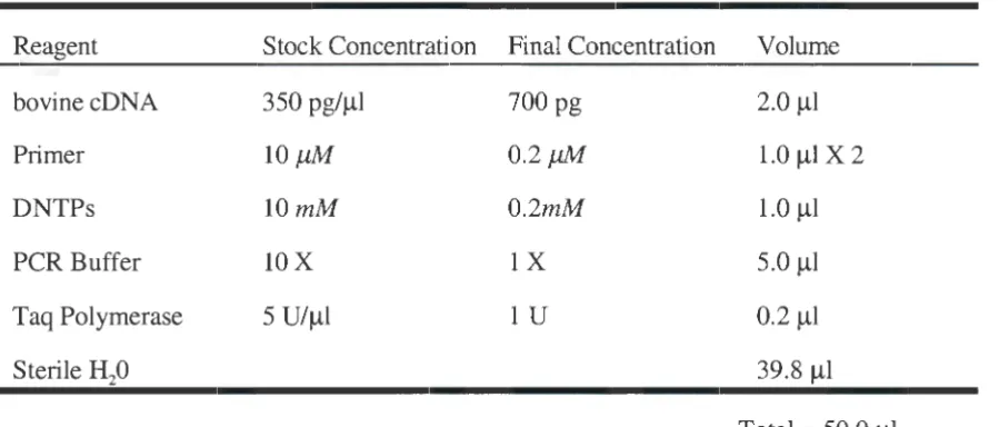
Each PCR reaction was carried out in a total volume of 50 rnicroliters (µl). 700 pico
grams (pg) (350 pg/µl) of bovine cDNA from a plasmid encoding the complete bovine ICAM-1
pairs was added to the reaction mixture from a 10 micromolar (µM) stock to a final
concentration of 0.2 µM for each primer (1 µ1/primer). dNTPs (Boehringer Mannheim,
Indianapolis, IN) from a stock of 10 milliMolar (mM) were added to a final concentration of
0.2 mM (1 µl total dNTPs). Five microliters of 10 X PCR Buffer (100 mMTris-HCl, 15 mM
MgC12, 500 mM KCl, pH 8.3; Boehringer Mannheim, Indianapolis, IN) was added for a final
dilution of 1 X along with 1 Unit (U) Taq Polymerase (5U/µl) (Boehringer Mannheim,
Indianapolis, IN). Sterile distilled water (39.8 µl) was added to bring the reaction volume to
50 µl (Table 2).
Table 2: PCR Reactions
Reagent Stock Concentration Final Concentration Volume
bovinecDNA 350 pg/µl 700 pg 2.0 µl
Primer lOµM 0.2µM 1.0 µIX 2
DNTPs lOmM 0.2mM 1.0 µl
PCR Buffer lOX lX 5.0 µl
Taq Polymerase 5 U/µ1 lU 0.2 µl
Sterile 8i0 39.8 µl
Total = 50.0 µl
The PCR parameters were as follows: 30 seconds at 94°C for denaturing, 45 seconds at
58°C for annealing, and 1 minute at 72°C for extension. A total of 30 cycles was completed
with an additional extension time of 10 minutes after the last cycle. All PCR reactions were
done in triplicate and the respective samples pooled. After pooling of the samples, the
respective cDNAs for the two constructs were cleaned using the Wizard PCR Clean-Up Kit
(Promega, Madison, WI). A double restriction enzyme digest was performed with 2.4 µg of
[image:30.568.50.500.319.511.2]incubated at 37° C for 90 minutes. After digestion the individual constructs were cleaned with
the Wizard PCR Clean-Up Kit.
The Expression Vector
The pTrxFus plasmid (Invitrogen, Carlsbad, CA) (based upon pUC18 design) contains:
a co!El origin of replication, a ~-lactarnase gene for ampicillin resistance as a selection marker,
a polylinker sequence that includes the restriction sites Barn HI and Kpn I, and a gene sequence
encoding the enterokinase recognition site for cleavage of the bacterial portion of the
recombinant peptide [106]. The plasmid uses the PL promoter from bacteriophage A to drive
expression. Regulation of the promoter for transcription of the recombinant protein is under
the control of the bacteriophage A cl repressor.
Expression of the cl repressor is also regulated. The E. coli strain used for
transformations contains the cl repressor gene under control of the trp promoter (see below).
The trp repressor and attentuator regulate expression of the cl repressor [106]. When
transformed cells are grown in tryptophan-free medium, the cl repressor gene is transcribed,
and the cl repressor protein binds the PL promoter preventing transcription [106, 107].
Expression is induced by adding tryptophan to the medium thereby stopping cl repressor
synthesis [106, 107] and allowing transcription from the PL promoter.
Ligations
The pTrxFus expression vector was digested in a 40 µ1 double reaction. Five micro
grams of the expression vector (lµg/µl) was digested in 20 U each of Barn HI and Kpn I
(lOU/µl) along with 4.0 µl 10 X REact® 4 (200 rnM Tris-HCI, 50 rnM MgC12, 500 rnM KCl,
pH 7.4; GibcoBRL), and the addition of 27 µl sterile deionized distilled water brought the
reaction volume to 40 µl. The reaction was incubated at 37° C for 90 minutes. The Wizard
Digestion of the expression vector with two different restriction enzymes resulted in a
linearized vector yielding non-compatible ends. This alleviated the problem of vector self
ligation or treating the vector with alkaline phosphatase. Both ICAM truncations contained
restriction enzyme sites at each end to aid in site directed cloning and maximize the probability
of obtaining a clone with the proper orientation. The goal was to ligate each construct
separately into the pTrxFus expression vector down stream and in frame with the sequence
coding for the expression tag, thioredoxin.
To produce a construct encoding the first two domains of bovine ICAM-1, the PCR
product generated from primers 1 and 2 (Bl) was ligated with linearized pTrxFus in the
presence of 1 U T4 DNA ligase (GibcoBRL, Rockville, MD). The insert to vector molar ratio
was 2:1. The PCR product derived from primers 3 and 4 (A6) encoding all five extracellular
domains of bovine ICAM-1 was ligated into pTrxFus in the same manner as above. All
ligations were incubated at 15.8°C for 12 hours.
Transformations
Escherichia coli strain 01724 (Invitrogen) was streaked out on a Luria-Bertani (LB)
agar plate and placed in a 37° C incubator overnight. A single colony from this plate was used
to inoculate 50 ml of LB Medium and grown for 16 hours in a 37° C shaking incubator. One
liter of LB Medium was inoculated with the 50 ml overnight culture and grown at 37° C until the
optical density (OD) at 550 nm was 0.7. The 1 L culture was split into two cold 500 ml
centrifuge bottles and incubated on ice for 30 minutes. Cultures were centrifuged at 2000 X g
for 15 minutes at 4°C and the supernatant decanted. Each pellet was resuspended in 250 ml
cold sterile distilled water and centrifuged at 2000 X g for 15 minutes at 4°C. Supernatant was
decanted and 20 ml cold sterile 10% glycerol (diluted in water) was added to each pellet and the
pellets were transferred to separate cold 50 ml centrifuge tubes. Cells were centrifuged at 4000
cold sterile 10% glycerol and pooled into one 50 ml tube and placed on ice. The cell
suspension was pipetted into 1.5 ml microcentrifuge tubes in 45 µ1 aliquots, quick frozen in a
dry ice-ethanol bath, and stored at -70°C until ready for use.
The electrocompetent cells were transformed with 1 µl of each ligation reaction in
separate tubes. The mixture was added to a Gene Pulser® IE. coli Pulser'M Cuvette (BIO-RAD,
Hercules, CA). Electroporation was carried out using the BIO-RAD Gene Pulser™ with the
following parameters: volts= 2.5, resistance (ohms)= 200, and capacitance (micro Faradays) =
25. After electroporation 800 µ1 of SOC Medium (Invitrogen, Carlsbad, CA) was added to each
cuvette and the mixtures were quickly transferred to separate 14 ml sterile polystyrene Falcon®
tubes (Benton Dickinson, Franklin Lakes, NJ). The cultures were placed in a 30° C shaking
incubator for 1 hour.
Fifty microliters of transformed cells were plated on RMG-Agar (Invitrogen, Carlsbad,
CA) plates containing 100 µg/ml ampicillin and incubated at 30° C for 24 hours. Three
milliliters RM Medium (Invitrogen, Carlsbad, CA) containing 100 µg/ml ampicillin were
inoculated with a colony containing either construct and grown at 30°C in a shaking incubator
for 24 hours. DNA plasmid minipreps (Promega, Madison, WI) were done on all cultures
containing either construct for DNA sequence analysis (Iowa State University DNA
Sequencing Facility). Cultures containing either truncated ICAM construct were frozen in 20%
glycerol (in water) at -70°C until further use. As a control for recombinant expression the
pTrxFus vector without any insert was transformed into the competent cells.
Small Scale Expression of Soluble Recombinant Bovine ICAM-1
One ml of RM medium containing 100 µg/ml ampicillin was inoculated with a
transformed colony containing either construct (B 1 or A6) or the pTrxFus expression vector
(no insert) and grown overnight in a 30° C shaking incubator. Ten milliliters of Induction
culture from either construct or control, and grown until the OD550 was 0.7. One ml was
removed and labeled as time zero. Tryptophan was added to the remaining cultures to a final
concentration of 100 µg/ml from a 10 mg/ml stock to induce expression. Cultures were placed
in a 37° C shaking incubator and expression was allowed to proceed for 4 hours. One ml from
each culture was removed and placed in a microcentrifuge tube on ice at the 1, 2, 3, and 4 hour
time points respectively.
Cells from the various time points were pelleted in a microcentrifuge at 20,000 X g for 3
minutes. Cell pellets were resuspended in 500 µl cold Osmotic Shock Solution #2 (lnvitrogen,
Carlsbad, CA) and placed on ice. Cells were sonicated using a sonicator equipped with a mico
tip one at a time with three 10 second bursts then flash frozen in a dry ice-ethanol bath and
thawed in a 37° C water bath. Three sonication freeze/thaw cycles were completed for each
sample. After the last freeze/thaw, the cells were pelleted at 20,000 X g for 5 minutes in a
microcentrifuge. Supematants were transferred to clean microcentrifuge tubes and pellets ( cell
debris and insoluble matter) were resuspended in 500 µl cold Osmotic Shock Solution #2.
Large Scale Expression and Affinity Purification
Fifty milliliters RM-Media containing 100 µg/ml ampicillin were inoculated with a
loopful of bacteria containing either ICAM construct. Cultures were grown over night in a
shaking incubator at 30° C. One liter of Induction Medium containing 100 µg/ml ampicillin
was inoculated with 50 ml of RM-Media containing either construct. The cultures were grown
in a 30°C shaking incubator until an OD550 of 0.6 and then stimulated with 100 µg/ml
tryptophan for 4 hours at 37°C to induce expression.
Cells were harvested by centrifugation at 4000 X g for 15 minutes at 4° C and the pellets
were resuspended in 20 ml running buffer (100 mM HEPES pH 7.0, 1 mM EDTA, lmM
13-mercaptoethanol). Cells were disrupted by passing three times through a cold French Pressure
10,000 X g for 15 minutes at 4° C to pellet cell debris. The supernatant was removed and
placed in a clean tube.
The protein in the soluble lysate (supernatant) was quantitated using the Kalckar
formula [108](amino acid composition at 260 nm and 280 nm). Forty milligrams of soluble
lysate (2.5 mg/ml) was added to 2 ml equilibrated ThioBond™ Resin (lnvitrogen, Carlsbad, CA)
for affinity chromatography. The ThioBond Resinr" was equilibrated according to the
manufacturer's instructions. The resin was washed with 8 ml running buffer and the
recombinant protein was eluted off the column using running buffer containing increasing
concentrations of ~-mercaptoethanol (125 mM to 350 mM).
Inclusion Body Production, Solubilization, and Purification Using Ion Exchange
Chromatography
Fifty milliliters RM-Media containing 100 µg/ml ampicillin was inoculated with a
loopful of bacteria containing either ICAM construct. Cultures were grown over night in a
shaking incubator at 30° C. One liter oflnduction Medium containing 100 µg/ml ampicillin
was inoculated with 50 ml of RM-Media containing either construct. The cultures were grown
in a 30°C shaking incubator until an OD550 of 0.6 was reached and then stimulated with 100
µg/ml tryptophan for 4 hours at 37° C.
Cells were harvested by centrifugation at 4,000 X g for 15 minutes and resuspended in
20 ml HEPES buffer (50 mM HEPES-NaOH pH 7.5, 0.5 M NaCl, 1 mM PMSF, 5 mM DTT
(dithiothreitol) containing 0.35 mg/ml lysozyme) per liter of cell culture and placed at 20°C for
30 minutes. Triton X-100 was added to a concentration of 1 % (vol/vol) and the bacteria were
sonicated, then treated with 200 µl DNase I (10 mg/ml) and placed at 37°C for 1 hour and
finally, the extract was centrifuged at 30,000 X g for 30 minutes at 4°C.
The recombinant ICAM-1 was contained in the pelleted inclusion bodies as determined
NaCl) containing 1 % Triton X-100 at 30,000 X g for 30 minutes at 4°C and solubilized in 2 ml
50 mM HEPES-NaOH pH 7.5, 6 M guanidine-HCl, 25 mM DTT and left for 1 hour at 4°C.
Insoluble material was removed by centrifuging the extract at 100,000 X g for 10 minutes and
the supernatant was adjusted to 1 mg/ml using 50 mM HEPES-NaOH pH 7.5, 6 M
guanidine-HCl, 25 mM DTT. The solubilized proteins were immediately diluted 1: 10 in cold folding
buffer (50 mM HEPES pH 7.5, 0.2 M NaCl, 1 mM DTT, IM NDSB256
(dimethylbenzlammonium propane sulfonate, Cal Biochem, San Diego, CA) vortexed for 30
seconds, and kept at 4°C for 1 hour. Protein concentration was determined by using the
BIO-RAD (Hercules, CA) Protein Standard Assay, based on the Bradford dye-binding procedure.
To remove guanidine-HCl the protein solution was dialyzed using snake skin dialysis
tubing (molecular weight cutoff 10,000) (Pierce, Rockford, IL) against low salt ion exchange
buffer (20 mM Tris-HCI, 25 mM NaCl, 10 mM ~-mercaptoethanol; pH 8.0) with frequent
buffer changes.
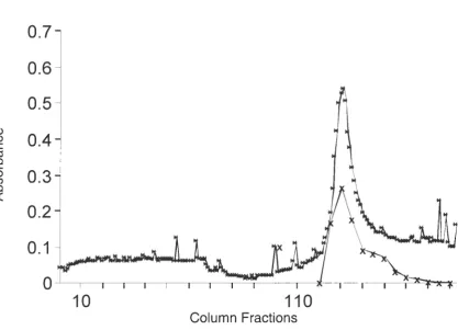
Ion Exchange Chromatography
Partially purified recombinant protein containing bovine ICAM-1 domains 1 through 5
was obtained by ion exchange chromatography using DEAE-Sephacel (Sigma, St. Louis, MO)
anion-exchange. Sixty milliliters of preswollen DEAE-Sephacel anion exchanger were poured
into a 250 ml beaker. One hundred milliliters of filtered, degassed low salt ion exchange buffer
were added to the beaker. The resin was allowed to settle and the buffer and fines were
aspirated off. This step was repeated three times. The resin was resuspended in a small volume
of degassed column buffer and packed into a 25 X 10 cm glass column yielding 35 ml of anion
exchanger. The column was equilibrated with 10 column volumes (350 ml at 1 ml/min) of
degassed low salt ion exchange buffer at 4° C. The pre-equilibrated sample (400 ml,
220 µg/ml) (see above) was loaded onto the column at a flow rate of 1 ml/min. The column was
of protein were detected using UV spectrometry at 280 nm. Bound proteins were eluted using a
salt gradient. The gradient was generated by mixing the low salt ion exchange buffer with a
degassed high salt buffer (20 mM Tris-HCl, 500 mM NaCl, 10 mM B-mercaptoethanol; pH
8.0). Four hundred milliliters of each buffer were used for the gradient. Fractions were
collected in 5 ml aliquots.
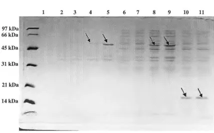
Electrophoresis
Electrophoresis of all protein samples was run on precast 12% and 14% SDS-PAGE
(sodium dodecyl sulfate-polyacrylamide gel electrophoresis) Tris-Glycine mini gels (Novex,
San Diego California) based on the Laemmli system [109]. Samples were prepared by adding
equal volumes of sample and 2 X NuPAGE SDS Sample Buffer pH 8.5 (Novex) supplemented
with 5 mM B-mercaptoethanol and heated at 100° C for 4 minutes. The XCell II™ Blot Module
(Novex) slab gel apparatus was used with a BIO-RAD Power Pac 200 power supply. Samples
were electrophoresed for 90 minutes at 125 volts and either silver or Commassie stained, or
prepared for western blot transfer.
Coomassie Blue Stain
A coomassie blue (Coomassie Brilliant Blue R-250, Boehringer Mannheim) working
solution (12.5% wt/vol Coomassie Blue Powder, 25% reagent grade methanol, 10% reagent
grade acetic acid; Mallinckrodt, Paris, Kentucky) was filtered through a 0.45 µm membrane
(CORNING® Costar, Coming, NY). Destaining solutions of 50% methanol, 10% acetic acid
(Destain I) and 5% methanol, 7% acetic acid (Destain II) were used to remove unbound
Coomassie. Briefly, after samples were electrophoresed gels were stained in Commassie
Working solution for 2 hours, placed in Destain I for 1 hour, and finally placed in Destain II
Silver Stain
Following electrophoresis of samples (see above) gels were stained with the Silver Stain
Plus Kit (BIO-RAD) to detect proteins. Briefly, gels were fixed for 20 minutes in Fixative
Enhancer Solution (200 ml reagent grade methanol, 40 ml reagent grade acetic acid, 40 ml
Fixative Enhancer Concentrate (BIO-RAD), 120 ml sterile deionized distilled water), washed
twice for 10 minutes with deionized distilled water, and stained for 15 minutes in Staining and
Developing Solution (5 ml Silver Complex, 5 ml Reduction Moderator Solution, 5 ml Image
Development Reagent, 50 ml Development Accelerator Solution; BIO-RAD). The staining
procedure was stopped after 15 minutes by placing the gels in 5 % acetic acid in water.
Western Blotting
Following electrophoresis of samples on 12% SDS-PAGE gels, proteins were
transferred to 0.2 µm nitrocellulose (Novex, San Diego, CA). Protein transfer was carried out
using the XCell II™ Blot Module powered by the BIO-RAD Power Pac 200. Eight hundred
milliliters cold transfer buffer (700 mM Glycine, 25 mM Tris; pH 7.4) were added to the
module and elution of the proteins from the gel was allowed to proceed for 75 minutes at 20
volts.
Western blotting to detect recombinant protein was carried out using SuperSignal®
West Pico Chemiluminescent Substrate (Pierce, Rockford, IL). After transfer, the nitrocellulose
membranes were washed in 15 ml PBS (0.8 % NaCl) for 5 minutes, incubated in 50 ml
azide-hydrogen peroxide (5 ml 30%
Ri0
2 , 0.05 g NaN3, 45 ml PBS) for 5 minutes and rewashed for5 minutes in 15 ml PBS. To block non-specific sites, membranes were incubated overnight at
4°C in 25 ml SuperBlock® blocking buffer (Pierce, Rockford, IL) supplemented with 0.05%
(vol/vol) Tween-20 (Sigma, St. Louis, MO). Blocked membranes were incubated at room
temperature for 1 hour with an Anti-Thioredoxin"' IgG monoclonal antibody (lnvitrogen,
0.05 % Tween-20. Membranes were washed 5 times (10 minutes/wash) with 15 ml wash
buffer (PBS supplemented with 0.05 %Tween-20). After the last wash, membranes were
incubated for 1 hour in 12 ml goat-anti mouse IgG horseradish peroxidase (HRP)-conjugated
secondary antibody diluted 1:50,000 in SuperBlock®blocking buffer supplemented with 0.05 %
Tween-20. Membranes were washed again 5 times for 10 minutes with 15 ml Wash Buffer and
after the last wash were incubated for 15 minutes in 24 ml SuperSignal® Substrate Working
Solution (12 ml Luminol/Enhancer Solution, 12 ml Stable Peroxide Solution) (Pierce,
Rockford, IL). Blots were removed from the Working Solution, drained, and enclosed in Saran
Wrap™. Wrapped membranes were placed in a Kodak BioMax™ Cassette (Kodak, Rochester,
NY) with intensifying screen and Kodak Bio-Max™MS Film was applied on top for forty-five
seconds. Films were developed using a Kodak X-OMAT M35 Processor (Kodak).
Antibody Production
The immuogen was prepared by pooling fractions eluted from the ThioBond Resin™
(Invitrogen) containing rbICAM-1 and contaminating proteins and dialyzed in snake skin
dialysis tubing (10,000 molecular weight cutoff; Promega) at 4° C against PBS (0.83% NaCl)
with frequent buffer changes. The dialyzed sample was concentrated using a Centricon Plus-80
5,000 molecular weight cut-off centrifugal filter device (Millipore, Bedford, MA) and diluted to
1 µg/µl.
Four 5 month old female BALB/c mice were immunized with the impure antigen. Each
mouse was immunized with 50 µg antigen (1 µg/µl), 50 µl PBS (0.83% NaCl), and 100 µl
Freund's Complete Adjuvant (FCA) intraperitoneally (IP).
Mice were boosted twice IP at two week intervals, first with 50 µg antigen (1 µg/µl),
50 µl PBS (0.83% NaCl), 100 µl Freund's Incomplete Adjuvant (FIA), and the second time