open-angle glaucoma and their effects on
trabecular meshwork cell function
Esther Lara Ashworth Briggs
BSc (Hons)
September 2018
Submitted in fulfilment of the requirements for the Degree of Doctor of Philosophy
i
This thesis contains no material which has been accepted for a degree or diploma by
the university or any other institution, except by the way of background information
and duly acknowledged in the thesis, and to the best of my knowledge and belief no
material previously published or written by another person except where due
acknowledgement is made in the text of the thesis, nor does the thesis contain any
material that infringes copyright.
16.09.2018
STATEMENT OF AUTHORITY OF ACCESS
This thesis may be made available for loan and limited copying and communication in
accordance with the Copyright Act 1968.
The publishers of the papers comprising Chapters 2 and 3 hold the copyright for that
content and access to the material should be sought from the respective journals. The
remaining non-published content of the thesis may be made available for loan and
limited copying and communication in accordance with the Copyright Act 1968.
Due to the inclusion of published material there is unavoidable repetition of material
between chapters in this thesis.
16.09.2018
STATEMENT OF ETHICAL CONDUCT
The research associated with this thesis abides by the international and Australian
codes on human and animal experimentation, the guidelines by the Australian
Government’s Office of the Gene Technology Regulator and the rulings of the Safety,
Ethics and Institutional Biosafety Committees of the University. This study was
approved by the Human Research Ethics Committee Network, Tasmania (Approval
no. H0013502 and H0013264).
iii
The following people and institutions contributed to the publication of work
undertaken as part of this thesis:
Candidate: Esther L Ashworth Briggs (ELAB), School of Health Sciences, College of Health and Medicine, University of Tasmania
Author 1: Anthony L Cook (ALC), School of Health Sciences, College of Health and Medicine, University of Tasmania and Wicking Dementia Research and Education Centre, College of Health and Medicine, University of Tasmania
Author 2: Alex W Hewitt (AWH), Menzies Institute for Medical Research, College of Health and Medicine, University of Tasmania and Centre for Eye Research Australia, University of Melbourne, Victoria
Author 3: Rajaraman Eri (RE), School of Health Sciences, College of Health and Medicine, University of Tasmania
Author 4: Tze’Yo Toh (TYT), Launceston Eye Institute and Launceston Eye Doctors, Tasmania
Author 5: Stephen Myers (SM), School of Health Sciences, College of Health and Medicine, University of Tasmania
Contribution of work by co-authors for each published manuscript:
MANUSCRIPT 1: Located in Chapter 2
Ashworth Briggs EL, Toh TY, Eri R, Hewitt AW, and Cook AL. TIMP1, TIMP2, and
TIMP4 are increased in aqueous humor from primary open angle glaucoma patients.
The candidate performed the laboratory data collection, analysis and interpretation
and drafted the manuscript. The study was conceived and designed by author 1 with
the assistance of author 2 and author 3. Author 4 recruited all study participants,
recorded clinical data, and performed sample collection during surgery. Author 1 and
author 2 provided assistance with data analysis and interpretation. Each author
assisted in the refinement and presentation of the final manuscript.
Percentage estimate of the contribution made by each author:
Candidate: 65%
Author 1: 15%
Author 2: 5%
Author 3: 5%
Author 4: 10%
MANUSCRIPT 2: Located in Chapter 3
Ashworth Briggs EL, Toh TY, Eri R, Hewitt AW, and Cook AL. Uteroglobin and FLRG
concentrations in aqueous humor are associated with age in primary open angle
glaucoma patients. BMC Ophthalmology 2018;18:57-64.
Authors’ contributions
The candidate performed the laboratory data collection, analysis and interpretation
and drafted the manuscript. The study was conceived and designed by author 1 with
the assistance of author 2 and author 3. Author 4 recruited all study participants,
v
assisted in the refinement and presentation of the final manuscript.
Percentage estimate of the contribution made by each author:
Candidate: 65%
Author 1: 15%
Author 2: 5%
Author 3: 5%
Author 4: 10%
We, the undersigned agree with the above stated contributions to the listed
manuscripts.
Signed: Date: 11.09.2018
Anthony Cook
Primary supervisor
School of Health Sciences, College of Health and Medicine, University of
Tasmania
Signed: Date: 17.09.2018
James Fell
Associate Head of Research
School of Health Sciences, College of Health and Medicine, University of
CHAPTER 4:
Molecular and functional characterisation of primary human trabecular meshwork
cells and the effect of uteroglobin on trabecular meshwork contraction in vitro.
Author contributions:
The candidate conceived and designed the study, performed all laboratory data
collection, analysis and interpretation and wrote the chapter. Author 1, author 2, and
author 5 assisted with the conception and design of the study, and gave input with
regard to analysis, interpretation and data presentation.
Percentage estimate of the contribution made by each author:
Candidate: 85%
Author 1: 5%
Author 2: 5%
vii Published manuscripts:
Ashworth Briggs EL, Toh TY, Eri R, Hewitt AW, and Cook AL. TIMP1, TIMP2, and TIMP4
are increased in aqueous humor from primary open angle glaucoma patients. Mol Vis
2015;21:1162-1172.
Ashworth Briggs EL, Toh TY, Eri R, Hewitt AW, and Cook AL. Uteroglobin and FLRG
concentrations in aqueous humor are associated with age in primary open angle
glaucoma patients. BMC Ophthalmology 2018;18:57-64.
Conference presentations:
Ashworth Briggs EL, Toh TY, Eri R, Hewitt AW, and Cook AL. Imbalance between MMPs
and TIMPs in aqueous humour from primary open angle glaucoma patients due to
increased levels of TIMP1, TIMP2 and TIMP4. Oral presentation, abstract no. 83.
Proceedings of the Australian Health and Medical Research Congress 2014, 16th– 19th November, Melbourne Convention and Exhibition Centre, Victoria, Australia.
Ashworth Briggs EL, Hewitt AW, SA Myers and Cook AL. Effect of uteroglobin on
trabecular meshwork cell contraction. Poster presentation, presentation no. 3519.
Proceedings of the Association for Research in Vision and Ophthalmology 2018
ix
I am deeply grateful to all the people who supported me during my PhD. You all
contributed to its success in your own wonderful ways. Firstly, I would like to thank
my supervisors Dr. Anthony Cook, Dr. Steve Myers, and Prof. Alex Hewitt. Tony, I am
deeply grateful that you took me on as your PhD student and made this thesis possible.
I am yet to find out about all the hoops you had to jump through to make it happen!
Thank you for all the support you have provided throughout my candidature. Steve,
thank you for your kindness, enthusiasm, and support, and for always carrying a smile
on your face. It is always a joy to see you. Alex, thank you for your support and
guidance, especially with regards to the clinical aspects of my research. I would also
like to extend my sincere gratitude to Dr. Murray Adams and Prof. Dominic Geraghty,
my graduate research coordinators and avid supporters. Your support and
encouragement have been invaluable throughout my candidature.
I would like to acknowledge the generous support of the Clifford Craig Medical
Research Trust (CCMRT, Launceston, Australia), who funded the research contained
in this thesis, and the Schulthess’sche Familienstiftung (Zürich, Switzerland), whose
support enabled me to move across the world to obtain my PhD. Without their
financial support this thesis would not have been possible. Furthermore, I would like
to extend my gratitude to the study participants, Dr. Tze’Yo Toh, and Sally Baxter,
without whom the clinical studies in chapters 2 and 3 would not have been possible.
I would like to thank all the beautiful souls who work tirelessly in the prep room and
the technical support provided throughout my PhD, and the teaching staff within the
School of Health Sciences for their general support and encouragement.
I wish to acknowledge my peers within the School of Health Sciences, Sarron
Randall-Demllo, Waheeda Basheer, Safa Al-maghrabi, Kate Herbert, Liz Witherden, Nicole
Ranson, Dana Lis, Sally McLaine, Shada Norouzi, John Adulcikas, Laura Danderian, and
Nicola McDonald. Thank you for your support, friendship and all the fun times
together! I would like to especially thank Emma Zadow for our impromptu chats
whilst my coffee was brewing (and going cold again :D ). Our conversations kept me
going when I was feeling isolated and alone in my little glaucoma world. Also, a
heartfelt thank you to Kate Edwards, thank you for your friendship and support, the
many coffees, and the lunch time runs, which helped keep me mostly sane in my final
months!
Not only did I learn and grow a lot through this experience, I also made great friends
to whom I am very grateful for their support: Chris and Natalie Polis, the Cook family,
Phil Marsh and Deb Osterhage, Rob and Miranda Gracie, Andrew Rath and Sharon
Fraser, Kate and Fergus Edwards, Zhi Quan Leong, and Alex Conway. Thank you for
cheering me along and for all the good times together, which provided a much-needed
balance to my studies.
I would like to acknowledge the fellow researchers I had the pleasure of meeting at
ARVO 2018, especially Prof. Daniel Stamer, Prof. Rudolf Fuchshofer, Dr. Mary Kelley,
xi
to make this thesis the best it can be. Furthermore, I would like to thank Simon Bakker
of Kugler Publications. Our conversations at ARVO 2018 gave me confidence in my
ideas for the future and our joint plans provided me with extra motivation to finish
my PhD.
I wish to extend a deep and heartfelt thank you to Prof. Dr. med. Christoph W. Spraul
and Dr. Christian Lingenfelder. Thank you for your friendship, encouragement, and
support in the final months of this epic journey. Your mentorship has been truly
invaluable and your enthusiasm and curiosity with regards to my research gave me
the motivation and drive required to finish my thesis; to finish it well and with
purpose. I will forever be grateful that we met at ARVO 2018. Herzlichen Dank!
Finally, I would like to thank my family for their ongoing support. Thank you, Mum,
Dad, and Chris, for your encouragement and for biting your tongue and letting me fly
far, far away from home to explore a new life and tackle this challenge. And most
importantly, my deepfelt gratitude towards my husband Alex. You never failed to
show me your belief in my abilities, supported me at all hours of the day and days of
the week (hello cell culture :D ) and helped me navigate the stormy waters. Many
people thought we were mad to be doing our PhDs simultaneously, I believed all along
that it was the best way for us. Thank you all for believing in me and for your patience,
I am truly grateful.
Yours sincerely,
xiii
DECLARATION OF ORIGINALITY ... I
STATEMENT OF AUTHORITY OF ACCESS ... I
STATEMENT REGARDING PUBLISHED WORK ... II
STATEMENT OF ETHICAL CONDUCT ... II
STATEMENT OF CO-AUTHORSHIP... III
PUBLICATIONS AND CONFERENCE PRESENTATIONS DURING PHD
CANDIDATURE ... VII
ACKNOWLEDGEMENTS ... IX
TABLE OF CONTENTS ... XIII
LIST OF FIGURES ... XVII
LIST OF TABLES ... XXIII
LIST OF APPENDICES ... XXV
LIST OF ABBREVIATIONS... XXVII
ABSTRACT ... XXIX
LITERATURE REVIEW ... 1
1.1 GLAUCOMA – A COMPLEX OPTIC NEUROPATHY ... 1
1.1.1 Definition ... 2
1.1.2 Glaucoma subtypes ... 2
1.1.3 Primary open-angle glaucoma ... 6
1.2 OCULAR ANATOMY AND AQUEOUS HUMOUR DYNAMICS ... 10
1.2.1 Ocular anatomy - an overview ... 10
1.2.2 Aqueous humour and the generation of intraocular pressure... 13
1.2.3 Altered aqueous humour composition in primary open-angle glaucoma ... 14
1.3 TRABECULAR MESHWORK DYSFUNCTION ... 18
1.4.1 Thesis objective and specific aims ... 28
1.4.2 Significance... 29
1.5 THESIS OUTLINE ... 30
TIMP1, TIMP2, AND TIMP4 ARE INCREASED IN AQUEOUS
HUMOR FROM PRIMARY OPEN ANGLE GLAUCOMA PATIENTS ... 32
2.1 ABSTRACT... 32
2.1.1 Purpose ... 32
2.1.2 Methods... 32
2.1.3 Results ... 33
2.1.4 Conclusions ... 33
2.2 INTRODUCTION ... 34
2.3 MATERIALS & METHODS... 36
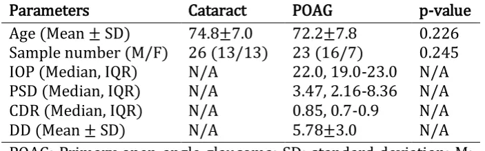
2.3.1 Patient eligibility and recruitment ... 36
2.3.2 Clinical assessment ... 36
2.3.3 Aqueous humour collection ... 37
2.3.4 Quantification of MMPs and TIMPs ... 37
2.3.5 Statistical analyses ... 38
2.4 RESULTS ... 39
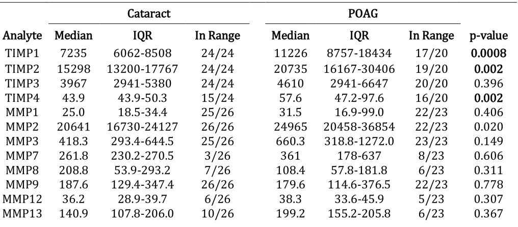
2.4.1 The levels of TIMP1, TIMP2 and TIMP4 are significantly increased in glaucomatous aqueous humour samples ... 41
2.4.2 Several MMP/TIMP molar ratios correlate with IOP and PSD in glaucomatous aqueous humour samples ... 48
2.5 DISCUSSION ... 49
2.6 ACKNOWLEDGEMENTS ... 54
UTEROGLOBIN AND FLRG CONCENTRATIONS IN AQUEOUS
HUMOUR ARE ASSOCIATED WITH AGE IN PRIMARY OPEN ANGLE
GLAUCOMA PATIENTS ... 55
3.1 ABSTRACT... 55
3.1.1 Background ... 55
3.1.2 Methods... 55
3.1.3 Results ... 56
xv
3.3.1 Patient eligibility and recruitment ... 58
3.3.2 Aqueous humour collection ... 60
3.3.3 Multiplex immunoassay ... 60
3.3.4 Normalisation to total protein concentration ... 61
3.3.5 Statistical analyses ... 62
3.4 RESULTS ... 62
3.4.1 Significant correlation of FLRG and uteroglobin with age in POAG but not cataract ... 65
3.4.2 HGF correlated significantly with POAG disease duration since commencing treatment 67 3.5 DISCUSSION ... 68
3.6 CONCLUSION ... 72
3.7 ACKNOWLEDGEMENTS ... 72
MOLECULAR AND FUNCTIONAL CHARACTERISATION OF
PRIMARY HUMAN TRABECULAR MESHWORK CELLS AND THE EFFECT OF
UTEROGLOBIN ON TRABECULAR MESHWORK CONTRACTION
IN VITRO
73
4.1 ABSTRACT ... 734.1.1 Purpose ... 73
4.1.2 Methods... 73
4.1.3 Results ... 74
4.1.4 Conclusions ... 74
4.2 INTRODUCTION ... 75
4.3 MATERIALS & METHODS ... 77
4.3.1 pHTMC culture... 77
4.3.2 HDFn and HeLa cell culture ... 78
4.3.3 RNA extraction... 78
4.3.4 cDNA synthesis ... 79
4.3.5 Quantitative PCR and analysis ... 79
4.3.6 Immunocytofluorescence ... 80
4.3.7 Dexamethasone-stimulated myocilin assay ... 80
4.3.8 MTT assay ... 81
4.3.9 Collagen gel contraction assay ... 82
4.3.10 Statistical analyses ... 83
4.5 DISCUSSION ... 100
4.6 ACKNOWLEDGEMENTS ... 106
GENERAL DISCUSSION AND CONCLUSION ... 109
5.1 PURPOSE AND AIMS OF THE RESEARCH ... 109
5.2 SUMMARY OF NOVEL FINDINGS AND IMPLICATIONS FOR THE FIELD ... 109
5.3 STUDY LIMITATIONS ... 111
5.4 FUTURE DIRECTIONS ... 112
5.4.1 Improved trabecular meshwork cell models ... 112
5.4.2 Further investigations of aqueous humour proteins ... 114
5.5 CONCLUSION... 116
REFERENCES ... 117
xvii
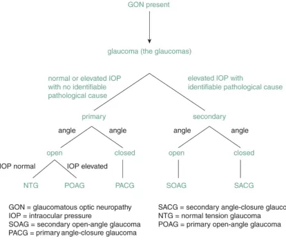
Figure 1.1 Glaucoma subcategories based on cause, anterior chamber angle
morphology, and IOP. Figure adapted from (15). ... 4
Figure 1.2 Iridocorneal angle morphology and aqueous humour outflow pathways
(yellow arrows). A) Aqueous humour flow in a healthy eye. B) Aqueous humour
outflow is reduced despite an open iridocorneal angle. C) Angle closure prevents
aqueous humour from passing between the lens and iris into the anterior chamber.
Image reproduced from (18)... 5
Figure 1.3 Anatomy of the human eye – subdivision into anterior and posterior
segment. The anterior segment is further subdivided into the anterior and posterior
chamber. Figure reproduced from (55). ... 12
Figure 1.4 Anatomy of the human eye. Reproduced with permission from Carlson
Stock Art. ... 13
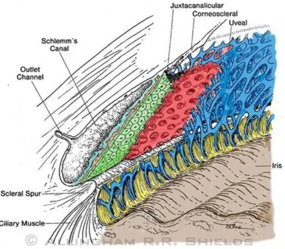
Figure 1.5 Anatomy of the trabecular meshwork. The trabecular meshwork consists
of three layers: the uveal meshwork (blue), the corneoscleral meshwork (red), and
the juxtacanalicular tissue (green). Figure reproduced from (119). ... 19
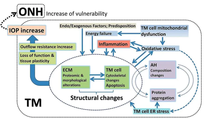
Figure 1.6 Model illustrating the wide range of factors thought to be involved in TM
dysfunction, leading to increased outflow resistance and elevated IOP. Figure
reproduced from (202). ONH: optic nerve head; IOP: intraocular pressure; TM:
trabecular meshwork; ECM: extracellular matrix; AH: aqueous humour; ER:
endoplasmic reticulum. ... 28
Figure 2.1 Distribution of TIMP1 (A), TIMP2 (B), TIMP4 (C) and MMP2 (D)
concentrations in aqueous humor from non-glaucomatous cataract (blue) versus
interquartile ranges are indicated. See Table 2.2 for complete list of analyte
concentrations measured. ... 44
Figure 2.2 Distribution of MMP/TIMP ratios in cataract versus POAG patients.
Stoichiometric ratios were calculated for individual aqueous humour samples from
non-glaucomatous cataract (blue; N=23-24) and POAG (orange; N=16-20) patients.
Median ratios and interquartile ranges are indicated. The ratios for MMP2/TIMP1 (A),
MMP2/TIMP3 (B), MMP3/TIMP3 (C) and MMP9/TIMP1 (D) were significantly
different between cataract and glaucoma (p<0.05), as determined using
Mann-Whitney U. See Table 2.3 for full set of ratios calculated. ... 47
Figure 3.1 Normalised analyte distributions in cataract and primary open-angle
glaucoma (POAG) samples. Distribution of CHI3L1 (A), FLRG (B), HGF (C), MIF (D),
p-selectin (E) and uteroglobin (F) concentrations in aqueous humour normalised to
total aqueous humour protein concentration from non-glaucomatous cataract (blue)
and POAG (orange). Median and interquartile range are indicated. ... 66
Figure 4.1 TM marker expression in pHTMCs in vitro. A) Subconfluent (S; blue) and
contact-inhibited (CI; orange) pHTMCs were assessed for expression of TM markers
relative to HPRT1 using qPCR. Statistical significance was tested by unpaired T-tests
for individual genes; no significant differences were determined. Gene expression was
measured in triplicate and the experiment was performed three times (n=3). Data
shown as mean SD. B) Subconfluent pHTMCs were stained against selected TM
markers (AQP1 and CHI3L1; red) and nuclei with Hoechst (blue). Images were taken
on a confocal microscope using a 10x objective with 3.6x zoom and an exposure time
xix
pHTMCs in vitro. Myocilin gene expression levels relative to HPRT1 in pHTMCs
treated with vehicle control (0.1% ethanol, black), 100 nM Dex (blue), or 500 nM Dex
(orange) in vitro. Gene expression was assessed by qPCR after 3 and 5 days of
treatment. Statistical significance was assessed using two-way ANOVA with Tukey
post-test, * indicates p 0.05. The experiment was performed three times (n=3). Data
are shown as mean SD. ... 87
Figure 4.3 Representative photographs of pHTMC, HDFn, and HeLa cells contracting
3D collagen gels. Images of collagen gel contraction by subconfluent (S) and
contact-inhibited (CI) pHTMCs, HDFn, and HeLa cells A) in the absence and B) presence of
Y-27632 at 0, 24, and 96 hours. ... 89
Figure 4.4 Contractibility of pHTMC, HDFn, and HeLa cells embedded in a 3D collagen
gel. Collagen gel contraction by subconfluent (S) and contact-inhibited (CI) pHTMCs,
HDFn, and HeLa cells A) in the absence and B) in the presence of 100 M Y-27632 was
determined by measuring changes in gel surface area over 96 hours, presented as %
contraction relative to 0 hours. Data shown as mean SD (n=3). ... 90
Figure 4.5 Gene expression of uteroglobin, HGF, and their respective receptors in
pHTMCs in vitro. Subconfluent (S; blue) and contact-inhibited (CI; orange) pHTMCs
were assessed for expression of A) HGF and its receptor MET and B) uteroglobin (UG)
and receptors LMBR1L and FPR2, relative to HPRT1 using qPCR. Statistical
significance was assessed using unpaired T-tests for individual genes, with p 0.05
considered significant. No significant differences were determined. Gene expression
was measured in triplicate and the experiment was performed three times (n=3).
Subconfluent pHTMCs were stained for A) LMBR1L (red) and B) MET (red).
Phalloidin (green) was used to stain actin and nuclei were stained using Hoechst
(blue). Images were taken on a confocal microscope with a 40x objective and an
exposure time of 8 s/pixel. The scale bar corresponds to 20 m. ... 94
Figure 4.7 Effect of exogenous uteroglobin on pHTMC viability in vitro as determined
by MTT assays. Subconfluent pHTMCs were treated with vehicle control (1% HBSS)
or 0.05, 0.5, 1.0, 2.0, or 5.0 g/ml uteroglobin (UG). Viability was assessed at 24, 48,
and 72 hours. Each variable was tested in triplicate and the experiment was
performed twice (n=2). Data are shown as mean SD... 96
Figure 4.8 Representative photographs of collagen contraction by pHTMCs treated
with uteroglobin. Images of collagen gel contraction by A) subconfluent (S) and B)
contact-inhibited (CI) pHTMCs treated with vehicle control (VC; 1% HBSS), 1.0, or 2.0
g/ml uteroglobin (UG) at 0, 24, and 96 hours. ... 98
Figure 4.9 Effect of exogenous uteroglobin on pHTMC contraction. Collagen gel
contraction by A) subconfluent (S) and B) contact-inhibited (CI) pHTMCs treated with
vehicle control (1% HBSS), 1.0, or 2.0 g/ml uteroglobin (UG) was determined by
measuring changes in gel surface area over 96 hours, presented as % contraction
relative to 0 hours. The experiment was performed in triplicate and repeated three
times (n=3). Data are shown as mean SD. No statistically significant differences
were found (assessed by 2-way ANOVAs with Tukey’s post-test). ... 99
Figure 4.10 Diagram illustrating how uteroglobin may influence aqueous humour
outflow through the trabecular meshwork, through interactions with tissue
xxi
uncertain are highlighted with a question mark (?). ... 106
Figure 5.1 Differentiation of pluripotent stem cells to trabecular meshwork and
xxiii
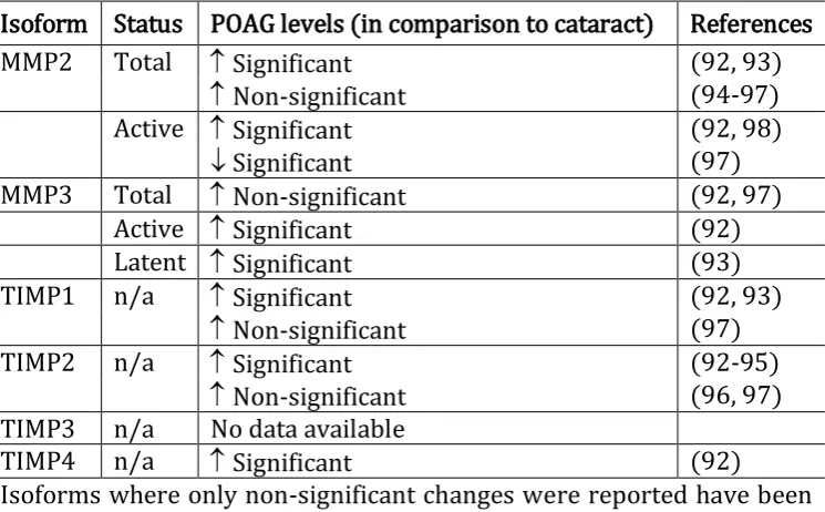
Table 1.1 Overview of published data regarding levels of MMPs and TIMPs found in
human aqueous humour from POAG compared to cataract patients. ... 17
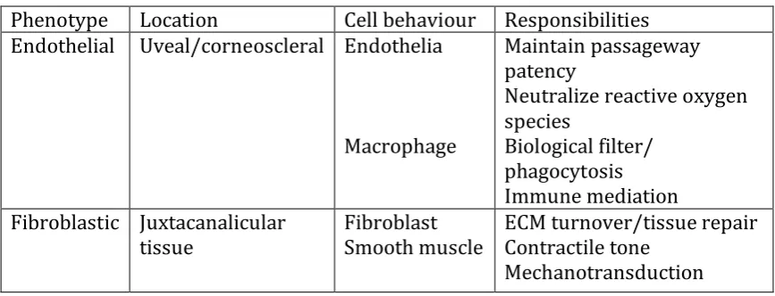
Table 1.2 The diverse biological functions of trabecular meshwork cells correspond to
different cell phenotypes and behaviour. Table reproduced from (71). ... 21
Table 2.1 Clinical data of non-glaucomatous cataract and POAG patients ... 40
Table 2.2 Aqueous humour analyte concentrations in cataract control versus POAG
... 43
Table 2.3 Stoichiometric analysis of MMP and TIMP ratios in cataract control versus
POAG samples ... 46
Table 2.4 Correlation of measured analytes and MMP/TIMP ratios to age for cataract
control and POAG samples ... 48
Table 2.5 Correlation of measured analytes and MMP/TIMP ratios to PSD, IOP and
disease duration for POAG samples ... 49
Table 3.1 Clinical data for non-glaucomatous cataract and POAG patients ... 64
Table 3.2 Aqueous humour analyte concentrations in non-glaucomatous cataract
versus POAG ... 65
Table 3.3 Correlation of measured analytes to age for non-glaucomatous cataract and
POAG samples ... 67
xxv
Appendix 1 Calculation of TIMP and MMP molecular weights for stoichiometric
analysis ... 155
Appendix 2 Correlation of measured analytes & MMP/TIMP ratios to CDR for POAG
samples ... 156
Appendix 3 Standard curve ranges of multiplex assays ... 157
Appendix 4 Correlation of measured analytes to IOP for non-glaucomatous cataract
and POAG samples ... 158
Appendix 5 Correlation of measured analytes to CDR for non-glaucomatous cataract
and POAG samples ... 158
Appendix 6 Correlation of measured analytes to MD for non-glaucomatous cataract
and POAG samples ... 159
Appendix 7 Correlation of measured analytes to PSD for non-glaucomatous cataract
and POAG samples ... 159
Appendix 8 TaqMan probes used for TM marker and housekeeper gene expression
analysis ... 160
Appendix 9 TaqMan probes used for gene expression analysis of HGF, uteroglobin, and
their respective receptors. ... 160
Appendix 10 Primary antibodies used for immunocytofluorescence ... 161
Appendix 11 Secondary Antibody used for immunocytofluorescence ... 161
Appendix 12 Western blot of myocilin in pHTMC conditioned media with
Dexamethasone (Dex) treatment. Conditioned media from pHTMCs in vitro was
harvested after 3 or 5 days of exposure to vehicle control (0.1% ethanol), 100 nM, or
gels followed by Western blotting. Naïve media was run as a control (on a separate
gel). The samples show a single band at the expected size of approx. 57 kDa,
corresponding to monomeric myocilin. Samples from three separate Dex treatments
xxvii
The following abbreviations are used in this thesis. Due to the inclusion of published
manuscripts, some abbreviations are defined more than once.
ADAM A disintegrin and metalloprotease
ADAMT ADAM metallopeptidase with thrombospondin type 1 motif ANOVA Analysis of variance
AQP1 Aquaporin 1
SMA Alpha-smooth muscle actin
bFGF Basic fibroblast growth factor BMP Bone morphogenic protein BSA Bovine serum albumin
CCL27 C-C motif chemokine ligand 27 cDNA Complementary deoxynucleic acid CDR Cup-to-disc ratio
CHI3L1 Chitinase 3 like 1
CLANs Cross-linked actin networks
CT Threshold cycle
CTGF Connective tissue growth factor CXCL9 C-X-C motif chemokine ligand 9 DcR3 Decoy receptor 3
DMEM Dulbecco’s modified Eagle’s medium
DMSO Dimethyl sulfoxide DNA Deoxyribonucleic acid ECM Extracellular matrix
EDTA Ethylenediaminetetraacetic acid EGF Epidermal growth factor
EPO Erythropoietin
ER Endoplasmic reticulum FBS Foetal bovine serum FI Fluorescence intensity FLRG Follistatin-related gene FOXC1 Forkhead Box C1
FPR2 Formyl peptide receptor 2 GON Glaucomatous optic neuropathy GWAS Genome-wide association studies HBSS Hank’s balanced salt solution
HDFn Human dermal fibroblasts, neonatal HepII Heparin-binding domain II
HGF Hepatocyte growth factor
HPRT1 Hypoxanthine phosphoribosyltransferase 1 HRP Horseradish peroxidase
IGFBP Insulin-like growth factor binding protein
IOP Intraocular pressure
iPSCs Induced pluripotent stem cells JCT Juxtacanalicular tissue
kDa Kilodalton
LIF Leukemia inhibitory factor
LMBR1L Limb development membrane protein 1 like LMX1B LIM Homeobox Transcription Factor 1 beta
MD Mean deviation
MFG-E8 Milk fat globule-EGF factor 8 MGP Matrix gla protein
MIF Macrophage migration inhibitory factor MMP Matrix metalloproteinase
MYOC Myocilin
OCT Optical coherence tomography PAX6 Paired Box 6
PBS Phosphate-buffered saline
pHTMCs Primary human trabecular meshwork cells
PITX2 Paired Like Homeodomain Transcription Factor 2 PLA2 Phospholipase A2
POAG Primary open-angle glaucoma POM Periocular mesenchyme PSD Pattern standard deviation PVDF Polyvinylidene difluoride
qPCR Quantitative polymerase chain reaction RGC Retinal ganglion cell
RNA Ribonucleic acid
ROCK Rho-associated, coiled-coil containing protein kinase
rs Spearman’s correlation coefficient
SAA Serum amyloid A
SCGB1A1 Secretoglobin 1A1 (uteroglobin)
SDS-PAGE Sodium dodecyl sulfate polyacrylamide gel TBST Tris-buffered saline Tween20
TGF- Transforming growth factor beta TGM2 Tissue transglutaminase
TIMP Tissue inhibitor of metalloproteinase
TM Trabecular meshwork
xxix Purpose
Glaucoma is the leading cause for irreversible blindness worldwide. The term
encompasses several ocular disease subtypes with the same end result, namely
damage to the optic nerve head, of which primary open-angle glaucoma (POAG) is the
most common. Despite extensive research, the aetiology and progression of POAG
remain largely elusive. The development of early diagnostic strategies and more
effective treatment approaches requires a better understanding of the cellular
mechanisms involved in POAG.
Problem
Elevated intraocular pressure (IOP) is a key risk factor for POAG and the focus of all
current treatment strategies. IOP homeostasis is regulated by the trabecular
meshwork (TM), a specialised tissue within the anterior chamber of the eye that
determines the rate of aqueous humour outflow. In POAG, a dysfunctional TM leads
to elevated IOP and consequently damage to the optic nerve head. To date, the
processes involved in TM dysfunction remain unclear.
Methodology
Two studies investigated imbalances in human aqueous humour protein
concentrations using multiplex immunoassays. Proteins measured included matrix
metalloproteinases (MMPs) and their endogenous tissue inhibitors (TIMPs), various
cytokines, and growth factors. Samples from POAG patients were compared to
deviation (PSD), and disease duration since diagnosis. Based on the results from the
second study, the anti-inflammatory cytokine uteroglobin was selected for in vitro
investigation using a human TM cell model. TM cells were first characterised using a
panel of TM marker genes and two functional assays: Dexamethasone-stimulated
myocilin upregulation and collagen gel contraction. For the latter, contractile (human
fibroblast) and non-contractile (HeLa) control cell lines were included, and ROCK
inhibitor Y-27632 was used to inhibit contraction. Subsequently, the collagen gel
contraction assay was used to investigate the effect of 1 and 2 g/ml uteroglobin on
TM cell contraction over a period of 96 hours.
Results
The first study found significantly elevated levels of TIMP1, TIMP2, TIMP4, and MMP2
concentrations in aqueous humour samples from POAG compared to cataract control.
Imbalances in MMP/TIMP molar ratios were also determined in POAG, several of
which correlated with IOP and PSD, but not with other disease descriptors. In the
second study, several cytokines and growth factors, including uteroglobin, HGF, and
FLRG, were detected in aqueous humour samples from POAG and cataract control
patients. Uteroglobin and FLRG concentrations correlated significantly with age in
POAG but not cataract samples. Furthermore, HGF concentrations resulted in a
negative correlation with disease duration. Gene expression and
immunofluorescence experiments using primary TM cells determined expression of
the uteroglobin receptor LMBR1L but not uteroglobin itself. Treatment of primary TM
cells with recombinant uteroglobin did not significantly affect cell contraction at the
xxxi
The differences identified in MMPs, TIMPs, and various cytokine concentrations in
aqueous humour from POAG patients add to our knowledge on aqueous humour
imbalances in POAG. The analysis of MMP-to-TIMP ratios showed a shift towards
increased TIMP levels, suggesting a potential increase in MMP inhibition, which may
affect extracellular matrix composition and thus contribute to elevated aqueous
humour outflow resistance. Several of the cytokines reported in the second study
have not previously been quantified in aqueous humour. Correlations of age with
uteroglobin and FLRG in POAG may indicate an increased need for anti-inflammatory
(uteroglobin) or anti-calcification (FLRG) activity in the aging glaucomatous TM.
While no effect of uteroglobin on TM cell contraction was found in the final study,
further experiments may elucidate the effects this protein has on TM cell function.
Specifically, phospholipase A2 activity, tissue transglutaminase activity, and
phagocytosis assays are required to determine the potential effect of uteroglobin on
TM cells. Determination of the effects of the aqueous humour proteins quantified in
this thesis on TM cell behaviour may lead to new insights with regards to TM
dysfunction in POAG and possibly provide new targets for pharmaceutical
1
LITERATURE REVIEW
1.1
G
LAUCOMA–
A COMPLEX OPTIC NEUROPATHYGlaucoma is the leading cause of irreversible blindness, affecting an estimated 60.5
million people worldwide in 2010, with a predicted increase to 111.8 million by 2040
(1), due to the rapid rise in the aging population. In Australia, a prevalence of 3% has
been reported in people over the age of 49 (2, 3), which equates to approximately
300,000 individuals. Typically, glaucoma affects peripheral vision first and blindness
can ensue if left untreated (4). Whilst treatment can slow down or even arrest disease
progression (5), existing visual field defects are irreversible, making early detection
critical. The slow, insidious progression of chronic glaucoma, occurring over many
years, combined with the late appearance of symptomatic visual field defects, means
that diagnosis often does not occur until the disease has reached an advanced stage,
and for the same reasons, many cases remain undiagnosed (6, 7). By these later
stages, central visual acuity and the ability to read are affected, which severely impact
on a patient’s quality of life (8). Glaucoma therefore presents a considerable burden
on health care systems (9), particularly due to the chronic nature of the disease,
requiring life-long treatment and regular clinical reviews. Specifically, a study by the
Centre for Eye Research Australia (CERA) reported an annual health care cost due to
glaucoma of AUD 342 million in 2005 (10). As a consequence, efforts have been made
to introduce population screenings for glaucoma to enable early detection and
treatment, although the lack of a single, definitive test for glaucoma presents a
considerable challenge (11, 12). Current research mainly focusses on more reliable
early diagnosis and improved treatment strategies to lessen the social burden and
1.1.1 Definition
The term glaucoma encompasses a heterogeneous group of ocular diseases with the
common clinical denominator of progressively degenerating retinal ganglion cell
(RGC) axons, which leads to a distinct cupping of the optic nerve head and
characteristic visual field damage (13-15). This common clinical characteristic is
known as glaucomatous optic neuropathy (GON). Whilst the pathogenesis of GON
remains unclear, several potential mechanisms leading to RGC degeneration and
resultant visual field damage have been described in the literature and are outlined
in section 1.1.3.3. In addition to the uncertain development of glaucomatous damage,
the numerous glaucoma subtypes present with varied aetiologies, risk factors,
demographics, symptoms, disease durations, and prognoses and require differing
treatment strategies (8). For these reasons, correct diagnosis and treatment of
glaucoma remains a challenge.
1.1.2 Glaucoma subtypes
The different forms of glaucoma are divided into subcategories, based on the
morphology of the anterior chamber angle, the age of onset, and whether the disease
results from an identifiable cause or not (4, 15). This subdivision is summarised in
Figure 1.1. Whilst intraocular pressure (IOP) is not a defining criterion for glaucoma
(13), it does play a role in categorising different forms of glaucoma. The existence of
a clear cause for IOP elevation in association with GON, such as an inflammation, eye
injury, or steroid treatment, divides the glaucomas into primary and secondary forms.
The former lacks an identifiable cause, whereas in the latter, the source of increased
IOP is recognizable. A second subdivision is made based on the status of the
3
unobstructed (Figure 1.2B), and any increase in IOP cannot be explained by visible
changes to the anterior chamber. Contrarily, a closed, physically obstructed angle
blocks aqueous humour outflow at a macroscopic level and is labelled closed-angle
glaucoma (Figure 1.2C). Although the relevance is disputed (16), a further subdivision
exists in primary open-angle glaucoma (POAG), depending on the level of IOP. A
subset of POAG patients do not present with elevated IOP, which has been termed
normal tension glaucoma (13, 15). Depending on the age of onset, glaucoma subtypes
can be further classified as congenital, juvenile, or adult onset glaucoma. Among these
different types of glaucoma, all of which are potentially progressive and can lead to
blindness (15), POAG is the most common form (14, 17) and will be the focus of this
Figure 1.1 Glaucoma subcategories based on cause, anterior chamber angle
[image:38.595.75.490.77.429.2]5
Figure 1.2 Iridocorneal angle morphology and aqueous humour outflow pathways
(yellow arrows). A) Aqueous humour flow in a healthy eye. B) Aqueous humour
[image:39.595.129.480.69.639.2]aqueous humour from passing between the lens and iris into the anterior chamber.
Image reproduced from (18).
1.1.3 Primary open-angle glaucoma
1.1.3.1 Definition and clinical assessment
POAG is a complex, multifactorial disease with a development that remains largely
elusive. The disease is defined as glaucomatous damage to the optic nerve head in the
presence of an open iridocorneal angle (Figure 1.2B), with no determinable cause for
IOP elevation (15). Early POAG diagnosis is critical but also challenging, as the
diagnosis is often not clear-cut. Due to this uncertainty, regular clinical reviews are
required to assess whether there is a progression of symptoms necessitating medical
treatment.
POAG diagnosis is based on the presence of characteristic optic disc cupping,
corresponding visual field defects, and a thinning of the retinal nerve fibre layer (19).
Optic disc cupping is assessed by means of the vertical cup-to-disc ratio (CDR). An
enlarged optic cup results in an increased CDR value and is due to the loss of RGC
axons. RGC axonal degeneration is also responsible for the thinning of the retinal
nerve fibre layer, which is measurable by ocular coherence tomography (14). The
resulting glaucomatous visual field defects can be detected by means of perimetry,
although functional loss is generally not detectable until at least 20-50% of RGCs have
been lost (20, 21). A visual field test maps specific locations of damage and provides
parameters such as mean deviation (MD) and pattern standard deviation (PSD). The
7
normative database, whereas the latter highlights focal variation within the visual
field of one patient (22). Additional methods can be used to assess visual field defects,
such as blue-on-yellow perimetry to test loss of colour-contrast sensitivity (23, 24).
To determine whether the patient is presenting with open- or closed-angle glaucoma,
the anatomy of the iridocorneal angle is assessed by means of gonioscopic
examination. As POAG can present without IOP elevation, the diagnosis is made
irrespective of the pressure measured.
1.1.3.2 Risk factors
POAG aetiology involves a variety of ocular, systemic, environmental, and genetic risk
factors. The main risk factors for POAG are advancing age and elevated IOP. A family
history of POAG and African ethnicity are also strongly associated with an increased
risk for developing the disease (25). Of these, IOP is the only modifiable risk factor
and the current focus of all treatment strategies.
It is well known that genetics play an important role in POAG. A first-degree relative
with POAG increases the risk of developing the disease by ten-fold in comparison to
the general population (26). However, POAG is a genetically complex and
heterogeneous disease and the majority of cases are adult onset and not inherited in
a Mendelian fashion. The use of family linkage studies has revealed causative
mutations for adult-onset POAG in a number of genes, including myocilin, optineurin,
and WD repeat domain 36 (reviewed in (27)), yet these only explain up to 10% of all
POAG cases (28). More recently, genome-wide association studies (GWAS) have
identified numerous loci associated with POAG. The variants detected in GWAS are
than causative mutations (29, 30). The information obtained through GWAS has
highlighted several cellular pathways that may be involved in POAG pathogenesis,
such as extracellular matrix (ECM) metabolism, TGF- signalling, and the
rhoA/rho-associated kinase pathway (reviewed in (31)).
Whilst several additional factors have been associated with an increased prevalence
of POAG, their validity as risk factors is uncertain. Association of gender with the
prevalence of POAG has been inconsistent (32), yet some meta-analyses of population
studies have found a higher prevalence in men (1, 33, 34). The validity of myopia as a
risk factor has been questioned due to the difficulty of detecting GON in myopic eyes
(35). Further factors that may be associated with a greater risk for POAG include
diabetes mellitus, arterial hypertension, body mass index, central corneal thickness,
obstructive sleep apnoea, and atherosclerosis, although these associations are
uncertain and require further research (reviewed in (8, 27, 36)).
1.1.3.3 Aetiology of primary open-angle glaucoma and the relevance of intraocular
pressure
Many mechanisms have been suggested to explain the loss of RGC axons that
underlies optic nerve head damage, some of which are directly related to elevated IOP.
These mechanisms include impaired axonal transport, which deprives RGCs of
neurotrophic factors critical to survival, glial cell activation in response to elevated
IOP, and hypoxia caused by reduced blood flow at the optic nerve head (reviewed in
9
Elevated IOP has commonly been defined as a pressure that exceeds 21 mm Hg. This
value represents a purely statistical concept and corresponds to two standard
deviations above the mean value of 16 mm Hg, assuming a Gaussian distribution (16).
Elevated IOP was long thought to be a causative factor for POAG but is now considered
merely a risk factor (15) for two reasons: First, increased IOP does not necessarily
lead to GON (39). Secondly, GON may present itself in the absence of elevated IOP,
which is often referred to as normal tension glaucoma (13, 15). Thus, elevated IOP is
not a defining feature of POAG and IOP-independent mechanisms are likely involved
in the development of GON, such as ischemia, loss of neurotrophic signalling, and
neurotoxicity (37, 38).
Although POAG can occur in people with normal IOP, ocular hypertension is a risk
factor for the development of GON, and the likelihood of developing POAG is increased
at higher IOP levels (40, 41). Several randomized clinical trials have shown that
reduction of IOP by means of hypotensive medication, laser therapy, or surgical
intervention delays the onset and progression of glaucoma (5, 42-44), and lowering
IOP not only benefits patients with elevated pressures, but also improves outcomes
for so-called normal tension glaucoma (45, 46). The fact that hypotensive treatment
benefits these patients suggests that normal tension glaucoma is also linked to IOP
(15).
Whilst there is consensus that elevated IOP is due to increased aqueous humour
outflow resistance rather than increased aqueous production (47, 48), the precise
mechanisms involved in altered outflow resistance remain unclear. Many theories
outflow resistance forms a key area of active research in glaucoma. The precise link
between elevated IOP and optic nerve damage is also yet to be determined. Several
animal studies have demonstrated that elevation of IOP leads to RGC degeneration
and optic nerve head damage (49-51), and it is thought that the pressure increase
causes mechanical changes at the lamina cribrosa, leading to RGC axonal injury and
death (52).
1.1.3.4 Current treatment strategies
To date, reduction of IOP remains the only proven treatment strategy for glaucoma
(8), which is achieved by a range of topical or oral medications, laser trabeculoplasty,
or incisional surgery, such as trabeculectomy or the placement of a drainage implant
(reviewed in (53)). The topical treatments available alter IOP by either increasing
uveoscleral outflow or reducing aqueous humour production. A new class of drugs
called rho kinase inhibitors, which increase trans-trabecular outflow, have recently
been approved for clinical use (54). The aqueous humour outflow pathways
mentioned above are further discussed in section 1.2.2.
1.2
O
CULAR ANATOMY AND AQUEOUS HUMOUR DYNAMICS1.2.1 Ocular anatomy - an overview
The human eye is divided into two main compartments: the anterior segment and the
posterior segment (Figure 1.3). The former consists of the lens and all the structures
in front of it, whereas the latter comprises all structures that lie behind the lens. The
anterior segment of the eye is further subdivided into the anterior and posterior
chamber (Figure 1.3), both of which are filled with aqueous humour. The posterior
11
chamber by the iris. The cornea, the anterior face of the iris, the trabecular meshwork
(TM), and Schlemm’s canal jointly form the anterior chamber (55, 56).
In the posterior segment, the inner surface of the eye, called the fundus, consists of
the retina, the optic disc, the macula, and the fovea (Figure 1.4). RGCs and their axons
form the retinal nerve fibre layer, which makes up the innermost layer of the fundus.
The visual impulses generated by the photoreceptors in the retina are transmitted via
bipolar cells to the RGCs. The signal is then relayed to the lateral geniculate nucleus
in the brain via the RGC axons, which leave the eye at the optic disc to form the optic
nerve. The optic disc is therefore also known as the optic nerve head (57). The base
of the optic nerve head is formed by a mesh-like, collagenous structure called the
lamina cribrosa, through which the optic nerve fibres pass as they exit the eye. The
lamina cribrosa is thought to be the principal site of RGC axonal injury, leading to the
Figure 1.3 Anatomy of the human eye – subdivision into anterior and posterior
segment. The anterior segment is further subdivided into the anterior and posterior
[image:46.595.104.473.89.341.2]13
Figure 1.4 Anatomy of the human eye. Reproduced with permission from Carlson Stock
Art.
1.2.2 Aqueous humour and the generation of intraocular pressure
Aqueous humour is the clear fluid secreted by the ciliary body (see Figure 1.4) that
circulates through the anterior chamber to supply the avascular iris, lens, TM, and
cornea with nutrients, as well as removing waste from these tissues (Figure 1.2). It
consists of a complex mixture of organic solutes, ions, and proteins (59-62). In
addition, the volume of aqueous humour determines IOP, which is critical for eye
health, maintenance of correct ocular shape, and clear vision. IOP is closely regulated
and drainage through the trabecular and uveoscleral pathways (63-66). The majority
of aqueous humour drains through the trabecular pathway, which involves the TM
and Schlemm’s canal (67). As aqueous humour secretion is relatively constant and
pressure-independent up to high levels of pressure, IOP is predominantly determined
by outflow resistance through the trabecular pathway, which is modulated in order
to enable IOP homeostasis (47, 68, 69). Whilst it is unclear exactly where and how
outflow resistance is generated and regulated (47, 48, 70), there is consensus that the
TM plays a key role in the regulation of IOP (71), which will be discussed in section
1.3.1.2.
1.2.3 Altered aqueous humour composition in primary open-angle glaucoma
Aqueous humour contains a vast range of proteins, including many growth factors
and cytokines, and several studies have shown that the composition of aqueous
humour is altered in POAG, as detailed below. In most cases, the comparison is made
to aqueous humour from patients with cataract, as it can be sampled with ease during
routine cataract surgery. Ideally, aqueous humour from healthy eyes would serve as
the control, but such sample collection would be unethical. Whilst many studies of
aqueous humour composition in animal models exist, this review focusses exclusively
on human aqueous humour.
Due to the low protein concentration in aqueous humour and the small sample
volumes available for testing, laboratory techniques with high sensitivity are
preferential for composition analysis, in order to maximise the data generated (72,
73). Two main approaches have been used to date: mass spectrometry and multiplex
15
provided insight into the composition of aqueous humour (74-76) and highlighted
several proteins with altered abundance in POAG when compared to cataract control
samples. The proteins identified include ones involved in oxidative stress, apoptosis,
mitochondrial function, inflammation, metabolism, and proteolysis (77-82),
highlighting these processes as potentially relevant to POAG pathogenesis. Whilst
mass spectrometry has allowed researchers to take a global, unbiased view of the
aqueous humour proteome, the technique’s sensitivity limitations make the detection
of low abundance proteins (ng/ml and lower), such as cytokines, growth factors, and
soluble receptors challenging (72). Multiplex immunoassays on the other hand allow
the simultaneous quantification of dozens of proteins per sample. These assays allow
the determination of specific protein concentrations, yet the proteins to be tested
need to be pre-selected, thus resulting in a biased view of the aqueous humour
proteome. Despite the increased sensitivity compared to mass spectrometry
techniques, many analytes still escape detection in multiplex immunoassay screens
(72).
Of the many proteins present in aqueous humour, only a small number have been
studied in some detail with regards to POAG. Several studies have shown significantly
increased levels of TGF-2 in POAG aqueous humour compared to non-glaucomatous
cataract samples (summarised in (83)). The effects of TGF-2 on the TM have been
investigated extensively and studies have demonstrated a multitude of consequences
on TM physiology, including increased ECM production, inhibition of ECM
in 1.3.2.4). Several ex vivo and in vivo studies have also indicated that elevated levels
of TGF-2 cause reduced aqueous humour outflow (86-88).
Two further protein families that have been investigated in detail are the matrix
metalloproteinases (MMPs) and their endogenous tissue inhibitors (TIMPs). MMPs
are proteolytic enzymes that degrade a variety of ECM molecules. Due to their
destructive nature, MMP activity requires tight regulation. MMPs are secreted in a
latent form and require extracellular cleavage of the pro-domain for activation (89).
In addition, TIMPs reversibly bind to MMPs in a 1:1 stoichiometry to obstruct the
MMP proteolytic site (90), thus inhibiting their activity. Several members of the MMP
family and all four known TIMPs are expressed by TM cells and numerous MMPs and
most TIMPs have been measured in human aqueous humour samples (reviewed
extensively in (91)). Table 1.1 provides an overview of the reported changes in MMP
17
Table 1.1 Overview of published data regarding levels of MMPs and TIMPs found in human aqueous humour from POAG compared to cataract patients.
Isoform Status POAG levels (in comparison to cataract) References
MMP2 Total Significant (92, 93)
Non-significant (94-97)
Active Significant (92, 98)
Significant (97)
MMP3 Total Non-significant (92, 97)
Active Significant (92)
Latent Significant (93)
TIMP1 n/a Significant (92, 93)
Non-significant (97)
TIMP2 n/a Significant (92-95)
Non-significant (96, 97)
TIMP3 n/a No data available
TIMP4 n/a Significant (92)
Isoforms where only non-significant changes were reported have been excluded from the list. n/a: not applicable.
Whilst some previous publications include the calculation of direct MMP-to-TIMP
ratios (ratio of concentrations measured) (92, 93, 97, 99, 100), none have considered
the sizeable differences in molecular weight between MMPs and TIMPs. Given that
MMPs and TIMPs interact at a 1:1 ratio, a stochiometric analysis that includes the
molecular sizes would provide a more accurate indication of MMP-to-TIMP
imbalances. Such imbalances are of interest due to the known role of MMPs in outflow
facility (101) and the reported changes in TM ECM composition. Specifically, it has
been suggested that the excessive accumulation of ECM material within the TM of
POAG eyes may be due to imbalanced MMP-to-TIMP ratios (97).
The aqueous humour concentrations of numerous other growth factors and cytokines
are also altered in POAG, although the effects of these changes on TM function remain
CCL2, and CXCL9 have been reported (102-106), although not all studies were able to
detect these factors and only some adjusted their p-values to correct for multiple
testing. On the other hand, IL-6 concentrations are reduced in POAG (103, 107).
Collectively, these observations have led some researchers to suggest the
involvement of inflammatory processes in TM dysfunction and POAG (104, 108, 109).
Other specific proteins present in the aqueous humour that have been investigated in
relation to POAG include CD44S (110), EPO (111, 112), fibronectin (113), HGF (114),
myocilin (115), and VEGF (116).
For the most part, it is unclear how these changes in aqueous humour composition
relate to clinical descriptors of POAG, as few studies have investigated correlations
extensively. Studies that have assessed correlations have predominantly focussed on
IOP, medication, and age (95, 96, 103, 104, 107, 117). Changes in protein abundance
may still be relevant to TM dysfunction, even if they do not differ significantly from
the concentrations found in cataract control samples. Thus, correlation with clinical
descriptors offers an additional approach to analysing data gained from aqueous
humour studies and may provide new insights into relevant disease mechanisms.
1.3
T
RABECULAR MESHWORK DYSFUNCTION1.3.1 Structure and function of the healthy trabecular meshwork
1.3.1.1 Trabecular meshwork architecture - an overview
The TM is a complex, connective tissue located in the iridocorneal angle, between the
cornea, the sclera, and the iris (Figure 1.2). The anatomy of the TM has been reviewed
in detail (71, 118). Briefly, the tissue consists of three different structural regions
19
passes through the uveal meshwork, the corneoscleral meshwork, and finally the
juxtacanalicular tissue (JCT), before entering Schlemm’s canal. Subsequently, the
aqueous humour drains into the episcleral veins. This fluid movement occurs as bulk
[image:53.595.107.510.210.561.2]flow driven by the pressure gradient rather than active transport (118).
Figure 1.5 Anatomy of the trabecular meshwork. The trabecular meshwork consists of
three layers: the uveal meshwork (blue), the corneoscleral meshwork (red), and the
juxtacanalicular tissue (green). Figure reproduced from (119).
The uveal meshwork is comprised of a network of trabecular beams covered in a thin,
and elastic fibres and the TM cells are situated such that they bridge the
intertrabecular spaces and connect adjacent beams. The corneoscleral meshwork,
which forms the thickest layer of the TM, is made of perforated collagen and elastin
sheets covered in TM cells. Both the uveal and the corneoscleral meshwork are highly
porous and thus do not contribute significantly to outflow resistance (reviewed in
(118, 120)). Instead, they function as a self-cleansing biological filter, responsible for
clearing cellular debris and reactive oxygen species that could prevent the JCT from
functioning (71). Unlike the uveal and corneoscleral meshwork, the JCT, which lies
adjacent to the inner wall of Schlemm’s canal, lacks trabecular beams and instead
consists of TM cells loosely arranged in a fibrillar ECM forming an irregular network
(120). The JCT, in combination with the inner wall endothelium of Schlemm’s canal,
is thought to be the site where outflow resistance is generated (47, 48).
1.3.1.2 Biological properties of trabecular meshwork cells
The TM has two primary roles: filtration of the aqueous humour and generation of
outflow resistance. In order to fulfil these responsibilities, TM cells execute a diverse
range of biological functions (summarized in Table 1.2). Due to their morphology, TM
cells have often been referred to as endothelial-like cells (121-123). Whilst they do
exhibit endothelial cell behaviours (see Table 1.2), this description fails to recognise
many critical aspects of TM cells. Recently, Stamer and Clark published a
comprehensive review of the TM cell’s diverse biological properties (71), which
highlighted that TM cells assume behaviours typical of macrophages, fibroblasts,
endothelia and smooth muscle cells, in order to maintain outflow resistance and
successfully respond to the wide-ranging stressors experienced by the TM, which
21
Table 1.2 The diverse biological functions of trabecular meshwork cells correspond to different cell phenotypes and behaviour. Table reproduced from (71).
Phenotype Location Cell behaviour Responsibilities Endothelial Uveal/corneoscleral Endothelia
Macrophage
Maintain passageway patency
Neutralize reactive oxygen species
Biological filter/ phagocytosis Immune mediation Fibroblastic Juxtacanalicular
tissue Fibroblast Smooth muscle ECM turnover/tissue repair Contractile tone Mechanotransduction
1.3.1.2.1 Resistance generation
The TM’s central role is the generation and regulation of aqueous humour outflow
resistance, in order to maintain IOP within an acceptable physiological range.
Resistance generation is thought to occur in the JCT, in conjunction with the inner wall
endothelium of Schlemm’s canal and its basement membrane (47, 48, 124, 125).
Specifically, the ECM of the JCT has been suggested to play a major role in determining
aqueous humour outflow resistance (68, 120, 126-128), although other mechanisms
have been proposed and resistance generation is yet to be fully understood (48, 129).
The TM ECM and its components, which have been reviewed in detail (68, 118), are
not static, as the ECM undergoes continuous remodelling to maintain open outflow
pathways (120). In addition, elevated IOP is sensed in the JCT through mechanical
stretch, triggering a cascade of events to alter ECM composition and thus outflow
resistance. These processes, referred to as IOP homeostasis, involve the secretion of
ECM proteins and their corresponding degradation enzymes (reviewed in (69, 130)).
One major group of proteinases involved in ECM degradation are MMPs. Several
treatment with specific cytokines can lead to an increase in MMP expression levels
(91, 130). The importance of MMPs, and thus ECM turnover, in controlling aqueous
humour outflow was initially demonstrated by treating perfused anterior segments
with MMPs or agents that stimulate MMP expression, which led to increased aqueous
outflow. On the contrary, inhibition of MMP activity through the addition of TIMPs
reduced outflow facility (101).
In addition to ECM composition, cellular contraction is also involved in regulating
aqueous humour egress and the two are linked through integrin signalling. Thus,
changes to the ECM are transmitted to the intracellular space and can affect cell
contraction (131, 132). Both the TM and the JCT contain contractile cells, allowing
them to alter the geometry of the outflow pathways (129). Specifically, contraction of
TM cells leads to increased outflow resistance, whereas relaxation enhances aqueous
humour egress (reviewed in (133, 134)). The contractile state of the ciliary muscle
and the scleral spur cells (see Figure 1.5) further influence the geometry of the
trabecular outflow pathways (reviewed in (129, 135)).
1.3.1.2.2 Aqueous humour filtration
Filtration of aqueous humour is thought to be critical for TM homeostasis, as the
presence of debris in the JCT could obstruct aqueous humour outflow and prevent
proper resistance generation (136-138). The TM cells in the uveal and corneoscleral
meshwork are highly phagocytic (139-142), which allows them to clear cellular
debris, reactive oxygen species, pigment granules, and pseudoexfoliative material
23
shown to affect the expression of ECM remodelling enzymes (143), suggesting it may
also play a role in TM ECM homeostasis.
1.3.2 Trabecular meshwork dysfunction in primary open-angle glaucoma
Aqueous humour outflow resistance is elevated in glaucomatous eyes when
compared to age-matched controls (144). The cause of increased outflow resistance
in the TM is still unknown and many theories have been postulated. Rather than
offering an exhaustive summary of each hypothesis presented in the literature, this
section provides an overview of mechanisms that have been studied in considerable
detail and that are relevant to the investigations contained in this thesis. The diverse
range of processes studied in the field suggests that impaired regulation of outflow
facility is likely due to a complex combination of changes, both at the cellular and
tissue level, that lead to TM dysfunction.
Much of the current knowledge on TM cell dysfunction in POAG has come from studies
using cultured TM cells. To date, no unique genetic marker or feature has been
determined for TM cells, making identification of TM cells reliant on a combination of
criteria. Due to a lack of consensus, different laboratories have used varying criteria
to characterise TM cell cultures in the past, including morphology, the expression of
various marker genes, corticosteroid induction of cross-linked actin networks
(CLANs), phagocytic capacity, and Dexamethasone-stimulated myocilin upregulation
(145-148). Further known cellular responses, as well as a list of positive and negative
selection markers, are summarised in a recent review by Stamer and Clark (71). The
lack of consensus for TM cell characterisation in vitro was recently addressed in a
in combination with functional assays, in particular myocilin induction by
Dexamethasone, which is considered the most reliable assay for functional TM
characterisation.
1.3.2.1 Reduced trabecular meshwork cellularity
A gradual reduction in TM cell numbers is part of the aging process in healthy eyes
(150, 151). Continuous TM cell loss occurs at a similar rate in POAG, but overall
cellularity is lower than in age-matched controls (152). In addition, a greater number
of senescent cells has been determined in the glaucomatous TM (153). The loss of TM
cells leads to areas where trabecular beams are no longer covered, which has been
suggested to cause the fused trabecular beams observed in eyes with advanced POAG
(152, 154, 155). It has been hypothesised that reduced cell numbers affect the TM’s
ability to respond to increased pressure, which TM cells sense through mechanical
distortion (120).
The mechanisms involved in TM cell death remain unclear. In a recent review, Green
et al. (156) examined five cell death pathways (apoptosis, autophagy, necroptosis,
pyroptosis, and immunogenic apoptosis), all of which may contribute to reduced TM
cellularity, and suggested that immunogenic apoptosis is the most likely, due to the
involvement of oxidative and ER stress in TM dysfunction. Based on animal studies, it
has been suggested that phagocytic TM cells may leave the meshwork upon engulfing
foreign particles, which could also contribute to reduced cellularity (140, 142, 157,
158). However, subsequent studies using human perfused anterior segments saw
little to no cell loss in response to phagocytic challenge in single exposure