promoting access to White Rose research papers
Universities of Leeds, Sheffield and York
http://eprints.whiterose.ac.uk/
This is an author produced version of a paper, subsequently published in the
Journal of Alzheimer’s Disease. (This paper has been peer-reviewed but does not include final publisher proof-corrections or journal pagination.)
White Rose Research Online URL for this paper: http://eprints.whiterose.ac.uk/3661
Published paper
Vardy, ERLC, Hooper, NM, Rice PJ et al (2007) Increased circulating insulin-like
growth factor-1 in late-onset Alzheimer's disease Journal of Alzheimer’s
Increased circulating insulin-like growth factor-1 in late-onset Alzheimer’s disease
Emma R. L. C. Vardy1, Penny J. Rice1, Peter C. W. Bowie2, John D. Holmes3, Peter J.
Grant1 and Nigel M. Hooper4
1Academic Unit of Molecular Vascular Medicine, Leeds Institute of Genetics, Health and
Therapeutics, University of Leeds, Leeds, LS2 9JT, UK
2Sheffield Care Trust, Grenoside Grange Hospital, Salt Box Lane, Sheffield, S35 8QS, UK
3Academic Unit of Psychiatry and Behavioural Sciences, University of Leeds, Leeds, LS2
9JT, UK
4Institute of Molecular and Cellular Biology, Faculty of Biological Sciences, and Leeds
Institute of Genetics, Health and Therapeutics, University of Leeds, Leeds, LS2 9JT, UK
Corresponding author: Nigel M. Hooper, Institute of Molecular and Cellular Biology, Faculty
of Biological Sciences, LIGHT Laboratories, Clarendon Way, University of Leeds, Leeds,
LS2 9JT, UK. e-mail: [email protected]; tel. +44 113 343 3163; fax. +44 113 343
6603
Short title: Circulating IGF-1 in Alzheimer’s
Abstract
Background: Insulin-like growth factor (IGF)-1 has been implicated in the pathogenesis of
Alzheimer’s disease (AD).
Methods: We compared the level of circulating total and bioavailable IGF-1, by simultaneous
measurements of IGF-1 and IGF binding protein (IGFBP)-3, between 87 patients diagnosed
with AD and 126 age and sex matched control subjects without cognitive impairment. Blood
samples were collected and IGF-1 and IGFBP-3 measured by ELISA. Subjects were also
genotyped for apolipoprotein E.
Results: Total circulating IGF-1 levels were significantly raised in the AD group as compared
to the control group (p=0.022). There was no significant difference in the circulating level of
IGFBP-3 between the two groups. When the IGF-1 levels were ratioed against IGFBP-3
levels as an indicator of unbound, bioavailable circulating IGF-1, there was a significant
increase in the molar IGF-1:IGFBP-3 ratio in the AD subjects (0.181 ± 0.006) as compared to
the controls (0.156 ± 0.004) (p<0.001). Logistic regression analysis revealed that an increase
in the IGF-1:IGFBP-3 molar ratio increased the risk of AD significantly.
Conclusion: The results of increased total and free circulating IGF-1 support the hypothesis
that in its early stages late-onset AD reflects a state of resistance to IGF-1.
Key words: Alzheimer’s, insulin-like growth factor-1, insulin-like growth factor binding
Introduction
Alzheimer’s disease (AD) is the most prevalent form of dementia and understanding the
factors which influence the pathophysiology of the disease is essential for impacting on
incidence and defining new therapeutic strategies. Insulin-like growth factor (IGF)-1
modulates growth and metabolic function in the brain, enhances neuronal survival and exerts
neurotrophic activities in the hippocampus, which is involved in learning and memory [10,
11]. At a cellular level, disruption of IGF-1 input lowers neuronal resistance to amyloid β
(Aβ) peptide toxicity, increases cellular susceptibility to cell death signals, enhances
phosphorylation of tau and leads ultimately to accumulation of Aβ in the brain [5]. Aβ, which
is proteolytically cleaved from the larger amyloid β-protein precursor (AβPP) [30], is the key
constituent of the amyloid plaque and hyperphosphorylation of tau is integral to the formation
of neurofibrillary tangles, both of which are diagnostic of AD pathologically.
Although IGF-1, its receptor and binding proteins are present and locally produced in the
brain [27], IGF-1 is actively transported across the blood-brain barrier, and therefore changes
in circulating IGF-1 can lead to changes in IGF-1 input to the brain [4]. The bioavailability
and bioactivity of IGF-1 is regulated by six IGF binding proteins (IGFBPs) and several
IGFBP-proteases [7, 8]. Quantitatively the most important binding protein in the circulation is
IGFBP-3 which binds more than 80% of the circulating IGF-1 [15]. Thus measurement of
circulating IGFBP-3 levels, in addition to IGF-1 levels, allows the amount of bioavailable
IGF-1 to be determined [16, 23]. Although other studies have measured circulating IGF-1
levels in AD subjects as compared to control subjects, the results have been conflicting with
one showing an increase [29] and two a decrease [22, 32] in IGF-1 levels in AD. However,
none of these studies measured the circulating levels of IGFBP-3 which would reflect the
amount of bioavailable IGF-1. This lack of measurement of bioavailable IGF-1 was identified
compare the level of circulating total and bioavailable IGF-1, by simultaneous measurements
of IGF-1 and IGFBP-3, between patients with late-onset, sporadic AD and age-matched
controls without dementia.
Materials and Methods
Subjects
Two hundred and thirteen elderly subjects were enrolled for the purposes of this study.
These subjects consisted of 87 patients diagnosed with AD and 126 age and sex matched
control subjects without cognitive impairment. AD patients were recruited through memory
clinics in Leeds and Dewsbury (West Yorkshire, England). Control subjects were recruited
through the Leeds Family Health Services Authority, day hospitals and elderly medicine
outpatient clinics within the Leeds area. All participants were of European Caucasian
extraction and gave written informed consent (assent from relatives of the AD patients was
given where appropriate) according to a protocol approved by the Leeds (East) Research
Ethics Committee. Diagnosis of probable AD was made in accordance with international
diagnostic criteria (National Institute of Neurological and Communicative Disorders and
Stroke-Alzheimer’s Disease and Related Disorders Association Work Group;
NINCDS-ADRDA) [18]. All patients and control subjects underwent a standardized clinical evaluation
which included medical history, anthropometric measurements and cognitive function
assessment (Mini-Mental State Examination; MMSE and Modified Mini-Mental State
examination; 3MS) [12, 28]. Demographic and clinical profiles of the subjects participating in
Assays
Blood samples were collected between 8 am and noon after an overnight fast in lithium
heparin on ice, centrifuged at 4°C at 2997g for 30 min and the plasma samples stored at -40°C
until analysis. Enzyme-linked immunosorbent assay (ELISA) kits were used to measure
insulin (Biosource, Nivelles, Belgium), total IGF-1 and IGFBP-3 (R&D Systems, Oxford,
UK). According to the manufacturer’s data sheets, the minimum detection limit for the
IGF-1 assay was 0.026 ng/ml (3.42 pM) and for the IGFBP-3 assay was 0.05 ng/ml (IGF-1.72 pM), and
the IGF-1 assay does not detect IGF-2, insulin, IGFBP-2, -3 or -4, and the IGFBP-3 assay
does not detect IGF-1 or -2, IGFBP-1, -2, -4, -5 or -6. The apolipoprotein E (ApoE) genotype
was determined by polymerase chain reaction as described previously [6].
Statistical Analysis
All statistical analyses were performed using SPSS for windows version 12.0. Distribution
of variables was tested for skewness using the Kolmogrov-Smirnov test. Normal data are
presented as mean and standard error of the mean; non-parametric data are presented as
median and range. The unpaired Students t-test was used to compare mean values of normally
distributed data; the Mann-Whitney U-test was used to compare non-parametric data.
Differences in categorical data (expressed as percentages) groups were assessed using the
chi-squared test. Logistic regression analysis was used to investigate factors contributing to the
risk of AD.
Results
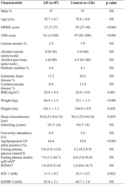
There were no significant differences between the AD group and the control group in
terms of age, gender, alcohol intake (past or present), education (years of schooling or
cardiovascular disease, fasting blood sugar, fasting plasma insulin or insulin resistance as
defined by the homeostasis model assessment (HOMA) [31] (Table 1). HOMA is a validated
standard method of assessing insulin resistance using glucose and insulin concentrations,
calculated by dividing the product of fasting plasma insulin concentration and fasting plasma
glucose concentration by 22.5 [31]. The low prevalence of diabetes mellitus in the AD
subjects and control group (Table 1) is very similar to general population figures quoting
prevalence to be 3.45% in England [19]. AD subjects were significantly shorter in stature
than the control group (p=0.038), had significantly smaller head circumference (p=0.039) and
were significantly lighter in weight (p<0.001) than control subjects (Table 1). Body mass
index (BMI) was significantly lower in the AD subjects as compared to the control group
(p=0.001) (Table 1). There was a significantly greater proportion of subjects with at least one
copy of the ApoE4 allele in the AD group (p<0.001) (Table 1).
Comparison of total plasma IGF-1 between AD subjects and control subjects revealed it to
be significantly raised in the AD group as compared to the control group (p=0.022) (Table 1).
There was no significant difference in the plasma level of IGFBP-3 between the two groups.
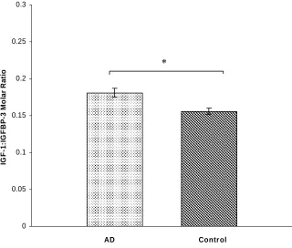
When the IGF-1 was ratioed against IGFBP-3 as an indicator of unbound, bioavailable
circulating IGF-1 [16], there was a significant increase in the IGF-1:IGFBP-3 molar ratio in
the AD subjects (0.181 ± 0.006) as compared to the controls (0.156 ± 0.004) indicating a
greater amount of circulating bioavailable IGF-1 in the AD subjects (p<0.001) (Fig 1).
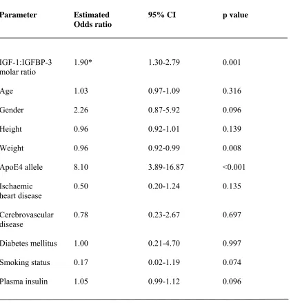
Logistic regression analysis was performed to look for independent associations between
particular parameters and the risk of AD (Table 2). Of the factors in the model, the presence
of an ApoE4 allele and also an increase in the IGF-1:IGFBP-3 molar ratio each increased the
odds of AD significantly. In particular, for an increase of one standard deviation of
IGF-1:IGFBP-3 molar ratio, the estimated odds ratio of being a patient with AD was 1.90
detected and there was no significant difference in IGF-1:IGFBP-3 molar ratio when the
subjects were grouped based on ApoE4 status. Although weight was also significantly
associated, ischaemic heart disease, cerebrovascular disease, diabetes, smoking status and
plasma insulin were not associated with the risk of AD (Table 2). There was no correlation
between MMSE and IGF-1, IGFBP-3 or IGF-1:IGFBP-3 molar ratio in the AD group.
Discussion
In this study, the primary findings were that circulating total IGF-1 was raised in
late-onset, sporadic AD subjects as compared to controls. Furthermore, the IGF-1:IGFBP-3 ratio,
indicative of bioavailable IGF-1, was raised significantly in the AD subjects as compared to
control subjects. Secondary findings included significant associations between stature, head
circumference, weight, BMI, ApoE4 allele and AD consistent with previous studies [1, 2, 21,
26]. There was no significant difference between the groups in terms of plasma insulin
measurements and insulin resistance as measured by HOMA [31]. Although other studies
have shown plasma insulin to be raised in AD [3, 9, 20], these studies included smaller
numbers of subjects (12-25 AD patients; 14-66 controls) and also excluded diabetic subjects.
A larger study found cognitive impairment with subcortical features to be associated with
biochemical and clinical features of the insulin resistance syndrome [13], however, this work
was not limited to AD.
Our data are consistent with a previous small study with 10 Caucasian sporadic AD
subjects (age range 63-74 years with probable AD according to the NINCDS-ADRDA criteria
and mean MMSE ± S.D. 15 ± 9) and 10 controls (age range 65-83 years) which found
circulating total IGF-1 to be raised in the AD group [29]. Increased circulating IGF-1, both
total and bioavailable, is consistent with the hypothesis that late-onset AD may reflect a state
sensitivity to IGF-1 action [5], possibly due to reduced receptor expression or function in the
AD brain [25]. In an attempt to overcome this resistance to 1 signalling, circulating
IGF-1 increases. Although it has been suggested that the IGF-IGF-1 resistance in late-onset, sporadic
AD may either originate from prior insulin resistance or lead to the development of insulin
resistance [5, 14], there was no difference in measures of peripheral insulin resistance
between the AD subjects and controls in our study.
In contrast to the increase in IGF-1 observed in our study and that of Tham et al. [29],
reduced circulating total IGF-1 was observed in a study involving 106 Japanese sporadic AD
subjects (mean age 79 years with probable AD according to NINCDS-ADRDA criteria and
mean MMSE ± S.D. 11 ± 7) compared with 227 control subjects (mean age 76 years) [32].
One possible explanation for the observed difference between our study and that of Watanabe
et al. [32] is that in the earlier stages of AD IGF-1 is raised, as seen in our study where the
mean MMSE score was 21, whereas in the later stages of the disease IGF-1 decreases, as seen
in the study by Watanabe et al. [32] where the mean MMSE score was 11. Although it should
be noted that we did not observe any correlation between either total or free circulating IGF-1
and MMSE score.
Circulating total IGF-1 was reported to be reduced in 5 familial AD subjects (mean age
60.8 years with probable AD according to NINCDS-ADRDA criteria) compared with 10
controls (mean age 55.8 years) [22]. However, all the patients in this study carried the
Swedish AβPP 670/671 mutation that causes early onset AD. It has been suggested that
alterations in IGF-1 in this case may be due to a greater demand for IGF-1 neuroprotection
due to the primary pathogenic event (the mutation in AβPP) in this case causing an increase in
the production of the neurotoxic Aβ peptide [5].
In conclusion, the increased circulating IGF-1 observed in the present study provides
IGF-1 signalling. At what stage in the dementia process resistance to IGF-1 develops and
whether this state predates the onset of cognitive impairment have yet to be determined.
However, it should be emphasised that the results presented here refer to plasma IGF-1 levels
and, as cerebrospinal fluid samples were not taken, we cannot be certain that these levels of
IGF-1 would be replicated in the brain and thus whether blood vessels, brain parenchymal
cells or both are IGF-1 resistant. Also as higher total IGF-1 has also been associated with
better cognitive performance and lower risk of cognitive decline in older individuals [17, 23,
24], the measurement of circulating IGF-1 is unlikely to be a useful biomarker to screen for
the onset of AD. If in the early stages late-onset, sporadic AD is indeed a state of resistance to
1 signalling, this may explain why MK-0677, a compound that induces secretion of
IGF-1 and increased circulating IGF-IGF-1 by 60%, failed to alter cognitive function in a double-blind
trial in 563 AD patients with baseline MMSE scores between 14 and 26
(http://www.alzforum.org/new/detail.asp?id=1458). Rather, it may be that therapeutics that
increase the sensitivity of cells to IGF-1 would hold greater potential in the pharmacological
treatment of patients in the early stages of AD.
Acknowledgments
This work was supported by grants from the Health Foundation and the Alzheimer’s
Research Trust. NMH, PCWB and JDH are members of the Yorkshire Centre in the
Alzheimer’s Research Trust’s Alzheimer’s Disease Network. We thank the consultant
psychiatrists in Leeds (Dr Burn, Dr Laybourn, Dr Nightingale, Dr Dearden, Dr Branton, Dr
Brindle) and Dewsbury (Dr Shukla, Dr Brodie, Dr Booya) for assistance with recruitment of
AD subjects. We also thank all the patients, their carers and control subjects without whom
References
[1] M. S. Beeri, M. Davidson, J. M. Silverman, S. Noy, J. Schmeidler and U. Goldbourt,
Relationship between body height and dementia, Am. J. Geriatr. Psychiatry 13 (2005)
116-123.
[2] A. Borenstein Graves, J. A. Mortimer, J. D. Bowen, W. C. McCormick, S. M. McCurry,
G. D. Schellenberg and E. B. Larson, Head circumference and incident Alzheimer's disease:
modification by apolipoprotein E, Neurology 57 (2001) 1453-1460.
[3] M. Carantoni, G. Zuliani, M. R. Munari, K. D'Elia, E. Palmieri and R. Fellin, Alzheimer
disease and vascular dementia: relationships with fasting glucose and insulin levels, Dement.
Geriatr. Cogn. Disord. 11 (2000) 176-180.
[4] E. Carro, A. Nunez, S. Busiguina and I. Torres-Aleman, Circulating insulin-like growth
factor I mediates effects of exercise on the brain, J. Neurosci. 20 (2000) 2926-2933.
[5] E. Carro and I. Torres-Aleman, Insulin-like growth factor I and Alzheimer's disease:
therapeutic prospects?, Expert Rev. Neurother. 4 (2004) 79-86.
[6] A. J. Catto, L. J. McCormack, M. W. Mansfield, A. M. Carter, J. M. Bamford, P.
Robinson and P. J. Grant, Apolipoprotein E polymorphism in cerebrovascular disease, Acta
Neurol. Scand. 101 (2000) 399-404.
[7] D. R. Clemmons, Insulin-like growth factor binding proteins and their role in controlling
IGF actions, Cytokine Growth Factor Rev. 8 (1997) 45-62.
[8] P. F. Collett-Solberg and P. Cohen, The role of the insulin-like growth factor binding
proteins and the IGFBP proteases in modulating IGF action, Endocrinol. Metab. Clin. North
[9] S. Craft, E. Peskind, M. W. Schwartz, G. D. Schellenberg, M. Raskind and D. Porte, Jr.,
Cerebrospinal fluid and plasma insulin levels in Alzheimer's disease: relationship to severity
of dementia and apolipoprotein E genotype, Neurology 50 (1998) 164-168.
[10] S. M. de la Monte and J. R. Wands, Review of insulin and insulin-like growth factor
expression, signaling, and malfunction in the central nervous system: relevance to
Alzheimer's disease, J. Alzheimers Dis. 7 (2005) 45-61.
[11] S. Dore, S. Kar and R. Quirion, Insulin-like growth factor I protects and rescues
hippocampal neurons against beta-amyloid- and human amylin-induced toxicity, Proc. Natl.
Acad. Sci. USA 94 (1997) 4772-4777.
[12] M. F. Folstein, S. E. Folstein and P. R. McHugh, "Mini-mental state". A practical
method for grading the cognitive state of patients for the clinician, J. Psychiatr. Res. 12
(1975) 189-198.
[13] C. Geroldi, G. B. Frisoni, G. Paolisso, S. Bandinelli, M. Lamponi, A. M. Abbatecola, O.
Zanetti, J. M. Guralnik and L. Ferrucci, Insulin resistance in cognitive impairment: the
InCHIANTI study, Arch. Neurol. 62 (2005) 1067-1072.
[14] S. Jain, D. W. Golde, R. Bailey and M. E. Geffner, Insulin-like growth factor-I
resistance, Endocr. Rev. 19 (1998) 625-646.
[15] J. I. Jones and D. R. Clemmons, Insulin-like growth factors and their binding proteins:
biological actions, Endocr. Rev. 16 (1995) 3-34.
[16] A. Juul, P. Dalgaard, W. F. Blum, P. Bang, K. Hall, K. F. Michaelsen, J. Muller and N.
E. Skakkebaek, Serum levels of insulin-like growth factor (IGF)-binding protein-3 (IGFBP-3)
in healthy infants, children, and adolescents: the relation to IGF-I, IGF-II, 1,
IGFBP-2, age, sex, body mass index, and pubertal maturation, J. Clin. Endocrinol. Metab. 80 (1995)
[17] S. Kalmijn, J. A. Janssen, H. A. Pols, S. W. Lamberts and M. M. Breteler, A prospective
study on circulating insulin-like growth factor I (IGF-I), IGF-binding proteins, and cognitive
function in the elderly, J. Clin. Endocrinol. Metab. 85 (2000) 4551-4555.
[18] G. McKhann, D. Drachman, M. Folstein, R. Katzman, D. Price and E. M. Stadlan,
Clinical diagnosis of Alzheimer's disease: report of the NINCDS-ADRDA Work Group under
the auspices of Department of Health and Human Services Task Force on Alzheimer's
Disease, Neurology 34 (1984) 939-944.
[19] G. McLean, B. Guthrie and M. Sutton, Differences in the quality of primary medical
care for CVD and diabetes across the NHS: evidence from the quality and outcomes
framework, BMC Health Serv. Res. 7 (2007) 74.
[20] G. S. Meneilly and A. Hill, Alterations in glucose metabolism in patients with
Alzheimer's disease, J. Am. Geriatr. Soc. 41 (1993) 710-714.
[21] J. A. Mortimer, D. A. Snowdon and W. R. Markesbery, Head circumference, education
and risk of dementia: findings from the Nun Study, J. Clin. Exp. Neuropsychol. 25 (2003)
671-679.
[22] A. Mustafa, L. Lannfelt, L. Lilius, A. Islam, B. Winblad and A. Adem, Decreased
plasma insulin-like growth factor-I level in familial Alzheimer's disease patients carrying the
Swedish APP 670/671 mutation, Dement. Geriatr. Cogn. Disord. 10 (1999) 446-451.
[23] O. I. Okereke, J. H. Kang, J. Ma, J. M. Gaziano and F. Grodstein, Midlife plasma
insulin-like growth factor I and cognitive function in older men, J. Clin. Endocrinol. Metab.
91 (2006) 4306-4312.
[24] G. Paolisso, S. Ammendola, A. Del Buono, A. Gambardella, M. Riondino, M. R.
Tagliamonte, M. R. Rizzo, C. Carella and M. Varricchio, Serum levels of insulin-like growth
leptin and lipid concentrations, insulin action, and cognitive function, J. Clin. Endocrinol.
Metab. 82 (1997) 2204-2209.
[25] E. J. Rivera, A. Goldin, N. Fulmer, R. Tavares, J. R. Wands and S. M. de la Monte,
Insulin and insulin-like growth factor expression and function deteriorate with progression of
Alzheimer's disease: link to brain reductions in acetylcholine, J. Alzheimers Dis. 8 (2005)
247-268.
[26] P. W. Schofield, G. Logroscino, H. F. Andrews, S. Albert and Y. Stern, An association
between head circumference and Alzheimer's disease in a population-based study of aging
and dementia, Neurology 49 (1997) 30-37.
[27] R. J. Schulingkamp, T. C. Pagano, D. Hung and R. B. Raffa, Insulin receptors and
insulin action in the brain: review and clinical implications, Neurosci. Biobehav. Rev. 24
(2000) 855-872.
[28] E. L. Teng and H. C. Chui, The Modified Mini-Mental State (3MS) examination, J.
Clin. Psychiatry 48 (1987) 314-318.
[29] A. Tham, A. Nordberg, F. E. Grissom, C. Carlsson-Skwirut, M. Viitanen and V. R. Sara,
Insulin-like growth factors and insulin-like growth factor binding proteins in cerebrospinal
fluid and serum of patients with dementia of the Alzheimer type, J. Neural. Transm. Park.
Dis. Dement. Sect. 5 (1993) 165-176.
[30] E. R. L. C. Vardy, A. J. Catto and N. M. Hooper, Proteolytic mechanisms in
amyloid-beta metabolism: therapeutic implications for Alzheimer's disease, Trends Mol. Med. 11
(2005) 464-472.
[31] T. M. Wallace, J. C. Levy and D. R. Matthews, Use and abuse of HOMA modeling,
[32] T. Watanabe, A. Miyazaki, T. Katagiri, H. Yamamoto, T. Idei and T. Iguchi,
Relationship between serum insulin-like growth factor-1 levels and Alzheimer's disease and
Table 1. Clinical and biochemical characteristics of study subjects
Characteristic AD (n=87) Control (n=126) p value
Male % 47 47 NS
Age (yrs) 78.7 ± 0.7 78.8 ± 0.6 NS
MMSE score 21 (7-27) 29 (27-30) <0.001
3MS score 63 (12-88) 97 (82-100) <0.001
Current smoker % 2.3 7.9 NS
Alcohol current (units/week)
0 (0-28) 2 (0-60) NS
Alcohol past (max.
units/week) 2 (0-80) 4.5 (0-140) NS
Diabetes mellitus % 4.6 4.1 NS
Ischaemic heart disease %
17.2 26.2 NS
Cerebrovascular
disease % 8.0 11.9 NS
BMI (kg/m2) 24.9 ± 0.4 26.9 ± 0.4 0.001
Weight (kg) 66.4 ± 1.5 74.1 ± 1.3 <0.001
Height (cm) 163.1 ± 1.1 166.0 ± 0.9 0.038
Head circumference (cm)
56.0 (51.0-61.0) 56.5 (52.0-62.0) 0.039
Schooling (years) 10 (7-14) 10 (7-14) NS
University attendance (%)
6.9 5.6 NS
Apolipoprotein E4
allele positive (%) 64.4 19.8 <0.001
Fasting plasma glucose (mmol/l)
5.0 (3.8-12.8) 5.2 (4.2-8.9) NS
Fasting plasma insulin (µIU/ml)*
7.9 (3.3-44.7) 8.0 (3.0-56.0) NS
HOMA* 1.8 (0.9-21.0) 1.8 (0.6-16.7) NS
IGF-1 (nM) 11.3 ± 0.3 10.3 ± 0.3 0.022
IGFBP-3 (nM) 65.8 ± 2.1 68.7 ± 1.6 NS
Table 2. Association of IGF-1:IGFBP-3 ratio and other parameters with AD
Parameter Estimated 95% CI p value
Odds ratio
IGF-1:IGFBP-3 1.90* 1.30-2.79 0.001
molar ratio
Age 1.03 0.97-1.09 0.316
Gender 2.26 0.87-5.92 0.096
Height 0.96 0.92-1.01 0.139
Weight 0.96 0.92-0.99 0.008
ApoE4 allele 8.10 3.89-16.87 <0.001
Ischaemic 0.50 0.20-1.24 0.135
heart disease
Cerebrovascular 0.78 0.23-2.67 0.697
disease
Diabetes mellitus 1.00 0.21-4.70 0.997
Smoking status 0.17 0.02-1.19 0.074
Plasma insulin 1.05 0.99-1.12 0.096
Figure 1. Plasma IGF-1:IGFBP-3 molar ratio in controls and AD subjects.
Bars represent mean ± SEM. * = p<0.001.
0 0.05 0.1 0.15 0.2 0.25 0.3
AD Contr ol
IG
F
-1
:IG
F
B
P
-3
M
o
la
r R
a
ti