THE ROLE OF NET1 PHOSPHORYLATION IN REGULATING CDC14 RELEASE DURING MITOTIC EXIT
Thesis by Ramzi Issam Azzam
In Partial Fullfillment of the Requirements for the Degree of
Doctor of Philosophy
California Institute of Technology Pasadena, California
2004
ACKNOWLEDGEMENTS
I would like to thank many people who have contributed so much to my development as a scientist and as a person. I owe my advisor, Ray Deshaies, so much for helping me mature for the last five years both personally and scientifically. His support has always been there even when things were not working and his enthusiasm for my work has never faltered. His door was always open to me whenever I needed to talk to him about my work or life in general. Most importantly, he has shown me that science with all its ups and downs is still and always will be a passion that is worth pursing. I will always cherish those conversations.
I would also like to thank my committee members Paul Sternberg, Bruce Hay, and Bill Dunphy. You all have inspired and challenged me to think critically and creatively about my work and that of others. Your comments and support have helped me throughout my time here and will always be appreciated. I especially want to thank Bruce Hay for his advice on the multitude of carrier options, his insight, and willingness to share his experience as a scientist.
Wenying Shou has been an inspiration to me and I want to thank her for “setting the bar” and teaching me not just about yeast genetics, but also about what one can do, and aspire to be, if they are dedicated and passionate about their work. I will always remember that lesson Wenying.
ABSTRACT
Exit from mitosis is as an important phase in the cell cycle. The molecular event that triggers the cell cycle transition from anaphase into the G1 state involves the
inactivation of the cyclin-dependent kinase complex (Cdk) through multiple mechanisms that lead to both destruction of the cyclin subunit co-activator and direct Cdk kinase inhibition. These multiple mechanisms indicate the importance of regulating the
inactivation of Cdk to ensure proper cell cycle progression and cytokinesis. We set out to examine the regulation of the protein phosphatase Cdc14. Cdc14 is thought to act through reversal of phosphorylation on key Cdk substrates that promote mitotic exit by stimulating the destruction and inactivation of Cdk. In Saccharomyces cerevisiae, activation of Cdc14 is achieved via release from its nucleolar inhibitor Net1/Cfi1. This activation is correlated with multi-site phosphorylation of Net1 in cells where Cdc14 appears to be released from the nucleolus. We set out to identify new components of the nucleolar complex known as RENT (Regulator of Nucleolar Silencing and Teleophase) which holds Cdc14 in an
TABLE OF CONTENTS
Acknowledgements... iii
Abstract ...v
Table of Contents ... vii
Chapter I: The End of the Cell Cycle...1
Overview:The motor of the cell cycle; Cyclin-dependent kinase (Cdk). ...1
Exit from mitosis in Saccharomyces cerevisiae ...2
The Mitotic Exit Network (MEN) ...2
Fourteen Early Anaphase Release (FEAR) Network ...4
Roles of FEAR...6
FEAR and Mitotic Exit Networks in Meiosis and Mitosis...8
Thesis Overview ...9
References ...10
Chapter II: Net1 Phosphorylation by Clb1,2–Cdk Regulates Cdc14 Release from the Nucleolus during Exit from Mitosis ...22
Summary ...22
Introduction ...23
Results ...26
Net1 N-terminus mediates regulated localization of Cdc14 and is highly phosphorylated in cdc14-1 cells...26
Net1 phosphorylation is required for transient release of Cdc14 in early anaphase and proper meiosis...27
Mitotic cyclin-Cdk phosphorylates sites on Net1 required for FEAR...29
Over-expression of non-degradable Clb2 is sufficient to drive Cdc14 out of the nucleolus in metaphase-arrested cells...31
Slk19, Spo12, and Cdc5 modulate phosphoryltion of Net1 on Thr212 32 Discussion ...33
FEAR network promotes phosphorylation of Net1 by Clb1,2-Cdk ...34
Is Net1 phosphorylation by Clb-cdk sufficient for FEAR? ...36
On the roles of Cdc5 and Clb-Cdk protein Kinase activities in FEAR .37 Acknowledgements...39
Experimental Procedures...39
References ...44
Figure II-1 ...54
Figure II-2 ...56
Figure II-3 ...59
Figure II-4 ...62
Figure II-5 ...68
Figure II-6 ...71
Figure II-7 ...74
Figure II-8 ...77
Figure II-9 ...79
Figure II-10 ...81
Figure II-11 ...83
Table II-1...85
Table II-2...87
Chapter III: New Components of the RENT Complex...89
Introduction ...89
RNA Polymerase I (Pol I) ...90
Casein Kinase II (CKII)...91
Results ...92
RNA Polymerase I and Casein Kinase II Interact with Net1...92
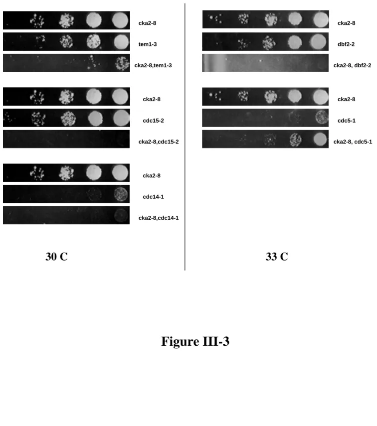
Casein Kinase II Mutants Display Synthetic Interactions with MEN Mutants ...93
Conclusions ...95
Acknowledgements...96
Experimental Procedures...96
References ...98
Figure III-1...104
Figure III-2...106
Figure III-3...108
Figure III-4...110
Figure III-5...112
Chapter IV: Future Directions...114
Summary ...114
C h a p t e r I - T h e E n d o f t h e C e l l C y c l e
Overview: The motor of the cell cycle; Cyclin-dependent kinase (Cdk)
The ability of cells to coordinate important events such as spindle disassembly (Li and Cai, 1997), chromosomal condensation (Loidl, 2003), and DNA replication (Piatti, 1997) with cellular division rely on the activity of the cyclin-dependent kinase complex (Amon et al., 1994; Holloway et al., 1993; Surana et al., 1993). The Cdk complex consists of at least two components in Saccharomyces cerevisiae. The first is a cyclin subunit that both activates and is thought to impart substrate specificity to the second component, the kinase subunit. The cyclin subunit appears to be interchangeable during various phases of the cell cycle where G1 cyclins (Cln1,2,3) (Levine et al., 1995); S-phase cyclins (Clb5,6) (Kuntzel et al., 1996; Toone et al., 1997); and G2 cyclins (Clb1,2,3,4) (Fitch et al., 1992) activate the kinase subunit to effect phosphorylation on cell cycle-specific substrates. Direct regulation of the kinase subunit in cis involves the post-translationalphosphorylation of key residues that lead to modification of enzymatic activity
(Mendenhall and Hodge, 1998). Regulation of the Cdk complex in trans also plays a key role in S. cerevisiae. This is achieved through the action of Cdk inhibitors (Sic1) (Donovan et al., 1994) and (Cdc6) (Calzada et al., 2001) which directly bind to and inhibit Cdk complexes.
break, in effect, by regulating the activation of the molecular trigger that leads to the ultimate extinction of Cdk activity, thus paving the road for the return back to the G1 state.
Exit from mitosis in Saccharomyces cerevisiae
The Mitotic Exit Network (MEN)To achieve the goal of Cdk inactivation by the various mechanisms listed
previously, a signal transduction network has been identified to be involved in mitotic exit. This group of proteins consists of at least 10 genetically interacting proteins collectively known as the mitotic exit network (MEN) (Jaspersen et al., 1998; McCollum and Gould, 2001). They include 4 protein kinases (Cdc15, Cdc5, Dbf2, and Dbf20) (Johnston et al., 1990; Kitada et al., 1993; Schweitzer and Philippsen, 1991; Toyn et al., 1991), a spindle pole body (SPB) protein (Nud1) (Adams and Kilmartin, 1999), a protein phosphatase (Cdc14) (Wan et al., 1992), a GTPase (Tem1) (Shirayama et al., 1994), a GTP/GDP exchange protein (Lte1) (Shirayama et al., 1994), a negative regulator of Cdc15 (Amn1) (Wang et al., 2003), and a protein of unknown function (Mob1) (Luca and Winey, 1998). All of the MEN proteins, with the exception of Lte1, are essential in budding yeast and many are found to be conserved between yeast and higher eukaryotic organisms (Li et al., 1997; Li et al., 2000).
cyclin proteolysis by removing inhibitory phosphorylation from Hct1/Cdh1, an activator of APC in late anaphase (Jaspersen et al., 1999; Visintin et al., 1998; Zachariae et al., 1998). Cdc14 is also thought to be responsible for promoting Sic1 accumulation and stability by reversal of Cdk phosphorylation events on Swi5 (a Sic1 transcriptional activator) and Sic1, respectively (Moll et al., 1991; Toyn et al., 1991; Toyn et al., 1997; Verma et al., 1997; Visintin et al., 1998).
Evidence suggests that the MEN acts as part of the Bub2-dependent spindle positioning checkpoint that monitors spindle pole body (SPB) position with respect to the bud neck (Bardin et al., 2000; Bloecher et al., 2000; Pereira et al., 2000). Upon spindle pole body duplication, the old spindle pole body is thought to contain the Tem1 GTPase which migrates to the bud. Activation of Tem1 occurs as the SPB interacts with the Bud cortex containing the GTP/GDP exchange protein Lte1 (Bardin et al., 2000; Bloecher et al., 2000; Pereira et al., 2000). Tem1 then in turn is thought to activate Cdc15 kinase
(Asakawa et al., 2001; Bardin et al., 2003) which in turn leads to the activation of the Dbf2/Dbf20 kinase complex bound to the cyclin-like subunit Mob1 (Mah et al., 2001; Visintin and Amon, 2001). It remains unclear how the Dbf2/Dbf20/Mob1 complex then controls the activation of Cdc14 to promote mitotic exit. One possible role for the action of MEN involves the control of Cdc14’s ability to transition between the nucleus and
allow precocious release of Cdc14 independently of MEN function. Indeed, Net1
mutations such as net1-1 have a significant impact on general nucleolar structure as judged by localization of multiple nucleolar antigens and regulation of rDNA morphology (Shou et al., 2001), which helps to explain this allele’s ability to bypass cdc15∆ cells (Shou et al., 1999). It is clear though that the cell cycle function of Net1 can be uncoupled from its other nucleolar functions as demonstrated by the dominant mutation in CDC14 (TAB6) (Shou et al., 1999).
Fourteen Early Anaphase Release (FEAR) Network
Recently, a new network of proteins has been identified in regulating the release of Cdc14 from the nucleolus in early anaphase (Stegmeier et al., 2002). This network consists of a separase (Esp1), the polo-like kinase (Cdc5), Spo12, and Slk19 which have unknown functions.
The first component of this network is a protease known as separase (Esp1)
allows metaphase-arrested cells to complete mitosis and cycle to the next G1 phase but is dependent on Spo12, Slk19, and to a lesser extent Cdc5 (Sullivan and Uhlmann, 2003).
The second component is (Cdc5), the only known homolog to the polo kinase in Saccharomyces cerevisiae. Cdc5 is interesting because it acts as both a component of the MEN as well as the FEAR networks. Cdc5 is thought to act in the MEN pathway through inhibitory phosphorylations of the Bub2/Bfa1 complex which leads to the activation of Tem1 (Hu et al., 2001). Cdc5 is also thought to act on the RENT complex to promote Cdc14 release from Net1, although this is thought to be primarily through an indirect mechanism (Shou et al., 2002). The function of Cdc5 in the FEAR pathway is less clear, whereas over-expression of Cdc5 can promote Cdc14 release in metaphase-arrested cells, it appears to be independent of the other FEAR network genes (Sullivan and Uhlmann, 2003; Visintin et al., 2003).
The third component of the FEAR is Spo12, a protein of unknown function. Spo12 was originally identified as playing an important role in the proper progression of meiosis (Klapholz and Esposito, 1980a; Klapholz and Esposito, 1980b). Interestingly, Spo12 levels appear to be cell-cycle regulated (Shah et al., 2001) and functions as a multi-copy
The fourth component of this network is Slk19, another protein of unknown function. Slk19 was originally identified and later confirmed to be required for the proper maintenance of mitotic anaphase spindles (Sullivan et al., 2001; Zeng et al., 1999). Slk19 localizes to kinetochores and the spindle mid-zone during anaphase and is a substrate of Esp1, yet interestingly cleavage of Slk19 by Esp1 does not seem to be necessary for Cdc14 release from the nucleolus (Stegmeier et al., 2002). The function of Slk19 in the FEAR pathway could also be complicated by the fact that Slk19 appears to be required upstream to promote Cdc14 release through the FEAR pathway yet also required for Cdc14 function downstream to promote anaphase spindle integrity through Cdc14 mediated localization of the INCENP-Aurora B complex to the spindle midzone (Pereira and Schiebel, 2003).
Roles of FEAR
In a relatively short amount of time, the FEAR network has been implicated to play a key role in organizing multiple events during early anaphase. FEAR was initially
identified as a group of proteins required for the timely progression through mitosis by promoting the release of Cdc14 in early anaphase (Stegmeier et al., 2002). Cdc14 is then able to positively feed back and activate the kinase Cdc15 as part of the MEN pathway as has been proposed previously (Jaspersen and Morgan, 2000). FEAR mutants, in the context of mitosis, displayed a delay in proper progression through mitosis, and synthetic phenotypes when combined with MEN mutants (Stegmeier et al., 2002).
segregation of rDNA (Buonomo et al., 2003). Anaphase I spindle disassembly is delayed in cdc14-1, spo12∆, and slk19∆ mutants to the point where anaphase II equational division occurs on meiosis I spindles (Marston et al., 2003). Remarkably, meiosis II events still take place and deletion of SPO11, preventing recombination, rescues the nuclear division defect of these mutants (Marston et al., 2003). Both Spo12 and Slk19 appear to be critical for executing meiosis than mitosis as judged by the sporulation and meiotic defects of spo12∆ and slk19∆ mutant cells (Buonomo et al., 2003; Grether and Herskowitz, 1999; Klapholz and Esposito, 1980b; Zeng et al., 1999). Mutant spo12∆ asci contain only two spores and half of slk19∆ contain two and half three or four spores (unpublished
observations) (Marston et al., 2003). More so, Spo12 and Slk19 are required for the division of the nucleolus (specifically, the segregation of newly replicated rDNA) and release of Cdc14 during Anaphase I of meiosis implying that Cdc14 release and activation is important for nucleolar segregation as has been suggested previously for cells exiting mitosis (Granot and Snyder, 1991). The polo kinase Cdc5 has also been linked to
controlling chromosomal segregation during meiosis I (Lee and Amon, 2003). Removal of meiotic cohesin from chromosomes and sister-kinetochore co-orientation during meiosis I are coupled through their dependence on Cdc5 (Lee and Amon, 2003).
phosphatase activity in vitro (Pereira and Schiebel, 2003) which corresponds with Cdc14 being a proline-directed phosphatase (Gray et al., 2003). Sli15 dephosphorylation is
sufficient to trigger microtubule binding of the INCEP-Aurora complex as demonstrated by the Sli15-6A mutant which remarkably is able to rescue the spindle defect of cdc14-2 mutant cells. Mutant Sli15-6A cells also displayed a 1000-fold increase in chromosome loss rate due to constitutive localization of Ipl1 and consequent stabilization of microtubules (Pereira and Schiebel, 2003) again arguing that proper activation of Cdc14 in early anaphase is important for timely progression through the cell cycle towards mitotic exit.
FEAR and Mitotic Exit Networks in Meiosis and Mitosis
Clb-Cdk activity must be completely extinguished to enable an irreversible return to an interphase state that can support DNA replication. This requirement in turn may
necessitate a more extended release of Cdc14 from the nucleolus, which requires the MEN. Positive feedback loops built into the MEN (Jaspersen and Morgan, 2000) may act to ensure that once the MEN is switched on, Clb-Cdk activity will inevitably be extinguished completely to allow the cell to divide and enter a subsequent S phase. Owing to these positive feedback loops, even a very modest initial stimulus could snowball into a complete mobilization of Cdc14, which could explain why the FEAR is not essential in mitotic cycles.
Conversely, the budding lifestyle of Saccharomyces cerevisiae offers a simple explanation for why the FEAR may not be sufficient to drive exit from mitosis (in contrast to the situation at the completion of meiosis I). Accurate partitioning of sister
chromosomes to mother and daughter yeast cells is supported by a post-anaphase
checkpoint that monitors the position of the spindle relative to the bud neck (Bardin et al., 2000; Pereira et al., 2000). If the FEAR by itself could drive exit from mitosis, then the cell would be irreversibly committed to exit mitosis upon activation of separase at the metaphase-anaphase transition, regardless of whether the two sets of sister chromatids were properly distributed to the two daughter cells.
Thesis Overview
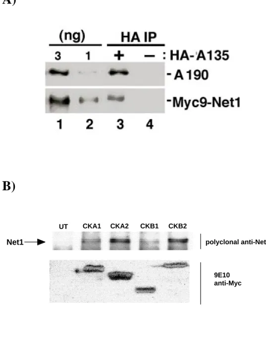
II describes how phosphorylation of Net1 by Clb-Cdk underlies disruption of the RENT complex during anaphase and illustrates a fascinating aspect of the switch that governs the return of mitotic cells to an interphase state. Although Clb-Cdk initiates feedback loops that help sustain a mitotic state with high Clb-Cdk activity (Deshaies, 1997), there must be mechanisms for subverting the reign of Clb-Cdk to allow growing cells to continue to cycle. Fittingly, at least two of these mechanisms – activation of Cdc20 binding to APC (Rudner et al., 2000; Rudner and Murray, 2000) and disruption of the Cdc14/Net1 complex – are initiated directly by the very enzyme whose activity helps to specify the mitotic state in the first place. Chapter III describes the identification of new interactors with the RENT complex such as Casein kinase II (CKII) and RNA polymerase I in mitotic cells. Given that a CKII site was identified and mapped from in vivo samples of Net1, and CKII was found co-associated with RENT complexes by mass spectrometry and this association was confirmed by co-immunoprecipitation; CKII could be playing an important role in
regulating Cdc14 release and possibly rDNA segregation. Also, given the location of the RENT complex within the nucleolus, interactions of Net1 with RNA Pol I help explain the multi-functionality displayed by net1-1 mutants. Chapter IV will outline a potential line of experiments that address how regulation of the FEAR and MEN pathways could be
investigated through the use of Clb2–Cdk phosphorylation assays on Net1 and how these phosphorylations lead to the activation of Cdc14.
References
Amon, A., S. Irniger, and K. Nasmyth. 1994. Closing the cell cycle circle in yeast: G2 cyclin proteolysis initiated at mitosis persists until the activation of G1 cyclins in the next cycle. Cell. 77:1037-50.
Asakawa, K., and A. Toh-e. 2002. A defect of Kap104 alleviates the requirement of mitotic exit network gene functions in Saccharomyces cerevisiae. Genetics. 162:1545-56.
Asakawa, K., S. Yoshida, F. Otake, and A. Toh-e. 2001. A novel functional domain of Cdc15 kinase is required for its interaction with Tem1 GTPase in Saccharomyces cerevisiae. Genetics. 157:1437-50.
Bardin, A.J., M.G. Boselli, and A. Amon. 2003. Mitotic exit regulation through distinct domains within the protein kinase Cdc15. Mol Cell Biol. 23:5018-30.
Bardin, A.J., R. Visintin, and A. Amon. 2000. A mechanism for coupling exit from mitosis to partitioning of the nucleus. Cell. 102:21-31.
Bloecher, A., G.M. Venturi, and K. Tatchell. 2000. Anaphase spindle position is monitored by the BUB2 checkpoint. Nat Cell Biol. 2:556-8.
CDC14 during anaphase of meiosis I depends on separase, SPO12, and SLK19. Dev Cell. 4:727-39.
Calzada, A., M. Sacristan, E. Sanchez, and A. Bueno. 2001. Cdc6 cooperates with Sic1 and Hct1 to inactivate mitotic cyclin-dependent kinases. Nature. 412:355-8.
Ciosk, R., W. Zachariae, C. Michaelis, A. Shevchenko, M. Mann, and K. Nasmyth. 1998. An ESP1/PDS1 complex regulates loss of sister chromatid cohesion at the metaphase to anaphase transition in yeast. Cell. 93:1067-76.
Cohen-Fix, O., and D. Koshland. 1999. Pds1p of budding yeast has dual roles: inhibition of anaphase initiation and regulation of mitotic exit. Genes Dev. 13:1950-9.
Deshaies, R.J. 1997. Phosphorylation and proteolysis: partners in the regulation of cell division in budding yeast. Curr Opin Genet Dev. 7:7-16.
Donovan, J.D., J.H. Toyn, A.L. Johnson, and L.H. Johnston. 1994. P40SDB25, a putative CDK inhibitor, has a role in the M/G1 transition in Saccharomyces cerevisiae. Genes Dev. 8:1640-53.
Granot, D., and M. Snyder. 1991. Segregation of the nucleolus during mitosis in budding and fission yeast. Cell Motil Cytoskeleton. 20:47-54.
Gray, C.H., V.M. Good, N.K. Tonks, and D. Barford. 2003. The structure of the cell cycle protein Cdc14 reveals a proline-directed protein phosphatase. Embo J. 22:3524-35.
Grether, M.E., and I. Herskowitz. 1999. Genetic and biochemical characterization of the yeast spo12 protein. Mol Biol Cell. 10:3689-703.
Holloway, S.L., M. Glotzer, R.W. King, and A.W. Murray. 1993. Anaphase is initiated by proteolysis rather than by the inactivation of maturation-promoting factor. Cell. 73:1393-402.
Hu, F., Y. Wang, D. Liu, Y. Li, J. Qin, and S.J. Elledge. 2001. Regulation of the Bub2/Bfa1 GAP complex by Cdc5 and cell cycle checkpoints. Cell. 107:655-65.
Iwabuchi, M., K. Ohsumi, T.M. Yamamoto, W. Sawada, and T. Kishimoto. 2000. Residual Cdc2 activity remaining at meiosis I exit is essential for meiotic M-M transition in
Xenopus oocyte extracts. Embo J. 19:4513-23.
Jaspersen, S.L., J.F. Charles, R.L. Tinker-Kulberg, and D.O. Morgan. 1998. A late
mitotic regulatory network controlling cyclin destruction in Saccharomyces cerevisiae. Mol Biol Cell. 9:2803-17.
Jaspersen, S.L., and D.O. Morgan. 2000. Cdc14 activates cdc15 to promote mitotic exit in budding yeast. Curr Biol. 10:615-8.
Johnston, L.H., S.L. Eberly, J.W. Chapman, H. Araki, and A. Sugino. 1990. The product of the Saccharomyces cerevisiae cell cycle gene DBF2 has homology with protein kinases and is periodically expressed in the cell cycle. Mol Cell Biol. 10:1358-66.
Kitada, K., A.L. Johnson, L.H. Johnston, and A. Sugino. 1993. A multicopy suppressor gene of the Saccharomyces cerevisiae G1 cell cycle mutant gene dbf4 encodes a protein kinase and is identified as CDC5. Mol Cell Biol. 13:4445-57.
Klapholz, S., and R.E. Esposito. 1980a. Isolation of SPO12-1 and SPO13-1 from a natural variant of yeast that undergoes a single meiotic division. Genetics. 96:567-88.
Kuntzel, H., A. Schulz, and I.M. Ehbrecht. 1996. Cell cycle control and initiation of DNA replication in Saccharomyces cerevisiae. Biol Chem. 377:481-7.
Lee, B.H., and A. Amon. 2003. Role of Polo-like kinase CDC5 in programming meiosis I chromosome segregation. Science. 300:482-6.
Levine, K., A.H. Tinkelenberg, and F. Cross. 1995. The CLN gene family: central regulators of cell cycle Start in budding yeast. Prog Cell Cycle Res. 1:101-14.
Li, L., B.R. Ernsting, M.J. Wishart, D.L. Lohse, and J.E. Dixon. 1997. A family of putative tumor suppressors is structurally and functionally conserved in humans and yeast. J Biol Chem. 272:29403-6.
Li, L., M. Ljungman, and J.E. Dixon. 2000. The human Cdc14 phosphatases interact with and dephosphorylate the tumor suppressor protein p53. J Biol Chem. 275:2410-4.
Li, X., and M. Cai. 1997. Inactivation of the cyclin-dependent kinase Cdc28 abrogates cell cycle arrest induced by DNA damage and disassembly of mitotic spindles in
Saccharomyces cerevisiae. Mol Cell Biol. 17:2723-34.
Luca, F.C., and M. Winey. 1998. MOB1, an essential yeast gene required for completion of mitosis and maintenance of ploidy. Mol Biol Cell. 9:29-46.
Mah, A.S., J. Jang, and R.J. Deshaies. 2001. Protein kinase Cdc15 activates the Dbf2-Mob1 kinase complex. Proc Natl Acad Sci U S A. 98:7325-30.
Marston, A.L., B.H. Lee, and A. Amon. 2003. The Cdc14 phosphatase and the FEAR network control meiotic spindle disassembly and chromosome segregation. Dev Cell. 4:711-26.
McCollum, D., and K.L. Gould. 2001. Timing is everything: regulation of mitotic exit and cytokinesis by the MEN and SIN. Trends Cell Biol. 11:89-95.
Mendenhall, M.D., and A.E. Hodge. 1998. Regulation of Cdc28 cyclin-dependent protein kinase activity during the cell cycle of the yeast Saccharomyces cerevisiae. Microbiol Mol Biol Rev. 62:1191-243.
Moll, T., G. Tebb, U. Surana, H. Robitsch, and K. Nasmyth. 1991. The role of
Noton, E., and J.F. Diffley. 2000. CDK inactivation is the only essential function of the APC/C and the mitotic exit network proteins for origin resetting during mitosis. Mol Cell. 5:85-95.
Pereira, G., T. Hofken, J. Grindlay, C. Manson, and E. Schiebel. 2000. The Bub2p spindle checkpoint links nuclear migration with mitotic exit. Mol Cell. 6:1-10.
Pereira, G., and E. Schiebel. 2003. Separase regulates INCENP-Aurora B anaphase spindle function through Cdc14. Science. 302:2120-4.
Piatti, S. 1997. Cell cycle regulation of S phase entry in Saccharomyces cerevisiae. Prog Cell Cycle Res. 3:143-56.
Rudner, A.D., K.G. Hardwick, and A.W. Murray. 2000. Cdc28 activates exit from mitosis in budding yeast. J Cell Biol. 149:1361-76.
Rudner, A.D., and A.W. Murray. 2000. Phosphorylation by Cdc28 activates the Cdc20-dependent activity of the anaphase-promoting complex. J Cell Biol. 149:1377-90.
Shah, R., S. Jensen, L.M. Frenz, A.L. Johnson, and L.H. Johnston. 2001. The Spo12 protein of Saccharomyces cerevisiae: a regulator of mitotic exit whose cell cycle-dependent degradation is mediated by the anaphase-promoting complex. Genetics. 159:965-80.
Shirayama, M., Y. Matsui, K. Tanaka, and A. Toh-e. 1994. Isolation of a CDC25 family gene, MSI2/LTE1, as a multicopy suppressor of ira1. Yeast. 10:451-61.
Shou, W., R. Azzam, S.L. Chen, M.J. Huddleston, C. Baskerville, H. Charbonneau, R.S. Annan, S.A. Carr, and R.J. Deshaies. 2002. Cdc5 influences phosphorylation of Net1 and disassembly of the RENT complex. BMC Mol Biol. 3:3.
Shou, W., and R.J. Deshaies. 2002. Multiple telophase arrest bypassed (tab) mutants alleviate the essential requirement for Cdc15 in exit from mitosis in S. cerevisiae. BMC Genet. 3:4.
Shou, W., K.M. Sakamoto, J. Keener, K.W. Morimoto, E.E. Traverso, R. Azzam, G.J. Hoppe, R.M. Feldman, J. DeModena, D. Moazed, H. Charbonneau, M. Nomura, and R.J. Deshaies. 2001. Net1 stimulates RNA polymerase I transcription and regulates nucleolar structure independently of controlling mitotic exit. Mol Cell. 8:45-55.
Stegmeier, F., J. Huang, R. Rahal, J. Zmolik, D. Moazed, and A. Amon. 2004. The
replication fork block protein Fob1 functions as a negative regulator of the FEAR network. Curr Biol. 14:467-80.
Stegmeier, F., R. Visintin, and A. Amon. 2002. Separase, polo kinase, the kinetochore protein Slk19, and Spo12 function in a network that controls Cdc14 localization during early anaphase. Cell. 108:207-20.
Sullivan, M., C. Lehane, and F. Uhlmann. 2001. Orchestrating anaphase and mitotic exit: separase cleavage and localization of Slk19. Nat Cell Biol. 3:771-7.
Sullivan, M., and F. Uhlmann. 2003. A non-proteolytic function of separase links the onset of anaphase to mitotic exit. Nat Cell Biol. 5:249-54.
Surana, U., A. Amon, C. Dowzer, J. McGrew, B. Byers, and K. Nasmyth. 1993. Destruction of the CDC28/CLB mitotic kinase is not required for the metaphase to anaphase transition in budding yeast. Embo J. 12:1969-78.
Toyn, J.H., H. Araki, A. Sugino, and L.H. Johnston. 1991. The cell-cycle-regulated budding yeast gene DBF2, encoding a putative protein kinase, has a homologue that is not under cell-cycle control. Gene. 104:63-70.
Toyn, J.H., A.L. Johnson, J.D. Donovan, W.M. Toone, and L.H. Johnston. 1997. The Swi5 transcription factor of Saccharomyces cerevisiae has a role in exit from mitosis through induction of the cdk-inhibitor Sic1 in telophase. Genetics. 145:85-96.
Uhlmann, F., D. Wernic, M.A. Poupart, E.V. Koonin, and K. Nasmyth. 2000. Cleavage of cohesin by the CD clan protease separin triggers anaphase in yeast. Cell. 103:375-86.
Verma, R., R.M. Feldman, and R.J. Deshaies. 1997. SIC1 is ubiquitinated in vitro by a pathway that requires CDC4, CDC34, and cyclin/CDK activities. Mol Biol Cell. 8:1427-37.
Visintin, R., and A. Amon. 2001. Regulation of the mitotic exit protein kinases Cdc15 and Dbf2. Mol Biol Cell. 12:2961-74.
Visintin, R., K. Craig, E.S. Hwang, S. Prinz, M. Tyers, and A. Amon. 1998. The phosphatase Cdc14 triggers mitotic exit by reversal of Cdk-dependent phosphorylation. Mol Cell. 2:709-18.
Visintin, R., F. Stegmeier, and A. Amon. 2003. The role of the polo kinase cdc5 in controlling cdc14 localization. Mol Biol Cell. 14:4486-98.
Wan, J., H. Xu, and M. Grunstein. 1992. CDC14 of Saccharomyces cerevisiae. Cloning, sequence analysis, and transcription during the cell cycle. J Biol Chem. 267:11274-80.
Wang, Y., T. Shirogane, D. Liu, J.W. Harper, and S.J. Elledge. 2003. Exit from exit: resetting the cell cycle through Amn1 inhibition of G protein signaling. Cell. 112:697-709.
Yamamoto, A., V. Guacci, and D. Koshland. 1996. Pds1p is required for faithful execution of anaphase in the yeast, Saccharomyces cerevisiae. J Cell Biol. 133:85-97.
Zachariae, W., M. Schwab, K. Nasmyth, and W. Seufert. 1998. Control of cyclin ubiquitination by CDK-regulated binding of Hct1 to the anaphase promoting complex. Science. 282:1721-4.
C h a p t e r I I - N e t 1 P h o s p h o r y l a t i o n b y
C l b 1 , 2 – C d k R e g u l a t e s C d c 1 4 R e l e a s e f r o m t h e
N u c l e o l u s d u r i n g E x i t f r o m M i t o s i s
Ramzi Azzam, Susan L. Chen, Wenying Shou, Angie S. Mah, Gabriela Alexandru, Kim Nasmyth, Roland S. Annan, Steven A. Carr, Raymond J. Deshaies
Submitted
Summary
The Cdc14 Early Anaphase Release (FEAR) network promotes a transition in the M-phase state that demarcates meiosis I and II, and together with the mitotic exit network enables timely exit from mitosis. These functions derive from FEAR’s ability to promote release of active Cdc14 from the nucleolar anchor Net1/Cfi1. We report here a molecular basis for the release of Cdc14 during early anaphase. Mitotic cyclin Clb2–Cdk
complexes phosphorylate Net1 in vitro and in vivo. Underscoring the relevance of these phosphorylations, net1 phosphosite and clb1∆clb2∆ mutants are deficient in FEAR, and net1 phosphosite mutants fail to execute proper meiosis. Over-expression of
Introduction
Exit from mitosis is an essential step in the progression of cells through the cell cycle. In late mitosis, inactivation of mitotic cyclin–Cdk complexes causes mitotic spindle disassembly (Li and Cai, 1997), chromosome decondensation, and return of cells to G1 phase (Amon et al., 1994; Holloway et al., 1993; Surana et al., 1993). In budding yeast, this inactivation is achieved by accumulation of the Cdk inhibitor Sic1 (Donovan et al., 1994; Schwab et al., 1997; Visintin et al., 1998), and destruction of the mitotic cyclins by the Anaphase Promoting Complex (APC), a multi-subunit ubiquitin protein ligase that catalyzes the ubiquitination of substrates containing a destruction-box sequence (Glotzer et al., 1991; Irniger et al., 1995; King et al., 1995; Morgan, 1999). Both of these
processes require a network of genetically interacting proteins collectively known as the mitotic exit network (MEN) (Jaspersen et al., 1998; McCollum and Gould, 2001). The most upstream component is the spindle pole body (SPB) protein Nud1 (Adams and Kilmartin, 1999), which tethers MEN components to the SPB. Among those components is the GTPase Tem1 (Shirayama et al., 1994b), which is activated by the guanine
et al., 2001; Pereira et al., 2002; Visintin and Amon, 2001; Yoshida and Toh-e, 2001). Activation of Cdc14, a protein phosphatase (Wan et al., 1992) that serves as a key
mediator of mitotic exit, appears to be the eventual target of the MEN, yet the mechanism by which this activation occurs remains unknown.
During interphase, Cdc14 is retained in an inactive form within the nucleolus as a component of the RENT complex, which comprises Cdc14 and Sir2 tethered to the nucleolar protein Net1/Cfi1 (Shou et al., 1999; Straight et al., 1999; Visintin et al., 1999). During anaphase, active Cdc14 is released from Net1, and diffuses throughout the cell. Cdc14 is thought to activate cyclin proteolysis by removing inhibitory phosphate groups from Hct1/Cdh1, an activator of APC in late anaphase (Jaspersen et al., 1999; Visintin et al., 1998; Zachariae et al., 1998). Cdc14 also promotes Sic1 accumulation and stability by removal of Cdk phosphorylations on Swi5 (a Sic1 transcriptional activator) and Sic1, respectively (Moll et al., 1991; Toyn et al., 1997; Verma et al., 1997; Visintin et al., 1998).
Recently, Cdc5, Esp1, Spo12, and Slk19 have been proposed to comprise a
Although the FEAR network plays a critical coordinating role in the M-phases of mitosis and meiosis, the molecular mechanisms underpinning FEAR’s ability to promote Cdc14 activation are not fully understood. Investigations on the role of separase in this process demonstrated that ectopic disassembly of RENT induced by over-expression of separase is not affected by a point mutation that disrupts separase’s endoproteolytic
activity, but is blocked completely by deletion of SPO12 or SLK19, and partially in cdc5-1 temperature sensitive (ts) mutants (Sullivan and Uhlmann, 2003). These observations suggest that a novel activity of Esp1 stimulates Spo12, Slk19, and Cdc5 to promote release of Cdc14 from Net1. In contrast, over-expressed Cdc5 can promote ectopic release of Cdc14 from the nucleolus in spo12∆, slk19∆, and esp1-1 mutants, and over-expressed Spo12 accelerates the same event in slk19∆, esp1-1, and Cdc5-depleted cells. These observations suggest that Spo12 and Cdc5 collaborate on parallel pathways to dislodge Cdc14 from Net1 (Visintin et al., 2003). Interestingly, separase- and Cdc5-induced disassembly of RENT correlates with hyper-phosphorylation of Net1 (Shou et al., 2002a; Sullivan and Uhlmann, 2003; Visintin et al., 2003). It has long been thought that
phosphorylation of Net1, Cdc14, or both underlies disassembly of the RENT complex during early and late anaphase (Shou et al., 2002a; Shou et al., 1999; Sullivan and
the appealing hypothesis that phosphorylation underlies the release of Cdc14 from Net1 during anaphase. We propose that mitotic Clb-Cdk phosphorylates Net1 to bring about Cdc14 release and activation and that this phsophorylation is regulated by components of the FEAR network.
Results
Net1 N-terminus mediates regulated localization of Cdc14 and is highly phosphorylated in cdc14-1 cells.
To map determinants that sustain regulated disassembly of the RENT complex, we examined a Net1 (1-621) truncation mutant along with a set of strains that contain
transposon insertions in NET1 (Burns et al., 1994; Ross-Macdonald et al., 1999).
Net1 phosphorylation is required for the transient release of Cdc14 in early anaphase and proper meiosis.
Endogenous NET1 was substituted by a mutant (net1-13m) allele in which thirteen, N-terminal, in vivo phosphorylation sites were converted to Alanine (Table1). The mutant protein, designated Net1-13m, localized to the nucleolus (data not shown) and directed the nucleolar localization of Cdc14 (Figure 7A). However, net1-13m cells were deficient in the transient release of Cdc14 from the nucleolus that occurs during early anaphase (i.e., FEAR; compare Figure 7B to Figure 2B). To determine which phosphorylation sites were responsible for the phenotype of net1-13m, we constructed mutants lacking subsets of sites. This effort yielded a mutant lacking three CDK
consensus sites (hereafter referred to as net1-3Cdk) that almost completely recapitulated the phenotypes of net1-13m (Figures 2A, 2B), whereas another mutant (net1-3Ax) lacking the 3 non-Cdk consensus sites in the first 341 aa of Net1 displayed a very minor
reduction in FEAR (Figure 7C). Upon further analysis of the phosphosite mapping data (see Experimental Procedures), we constructed an additional mutant lacking all six Cdk consensus sites in the first 341 aa of Net1 (hereafter referred to as net1-6Cdk).
in Cdc14 release from the nucleolus in net1-6Cdk mutants similar to that seen for
spo12∆ cells (Figure 8 and (Visintin et al., 2003). A third phenotype exhibited by FEAR mutants is that they exhibit synthetic genetic phenotypes when combined with MEN mutants (Stegmeier et al., 2002). Likewise, the net1-3Cdk and net1-6Cdk mutants showed synthetic-lethal interactions with the MEN mutants dbf2-2 (Figure 2C) and cdc15-2 (Figure 2D), respectively. For example, whereas cdc15-2 and net1-6Cdk mutants were viable at 30.7°C, cdc15-2 net1-6Cdk double mutants were not. Taken together, these observations implicate net1-6Cdk as a FEAR mutant.
Recent work has implicated the FEAR network in proper segregation of rDNA and cell cycle progression during meiosis (Buonomo et al., 2003; Lee and Amon, 2003; Marston et al., 2003). Given that net1-6Cdk mutants exhibited phenotypes reminiscent of FEAR network mutants, we evaluated their ability to segregate rDNA and form meiotic spores. In parallel with the 10’ delay in Cdc14 release from the nucleolus, spo12∆ and net1-6Cdk mutants exhibited a 10’ delay in rDNA segregation during mitosis (Figure 8). Likewise, net1-6Cdk mutants exhibited a meiotic defect: whereas greater than 90% of nitrogen-starved wild-type cells produced tri- or tetranucleate asci, net1-6Cdk mutants produced 43% binucleate and 57% tri- or tetranucleate asci with the majority of those being tri-nucleate (data not shown). The meiotic defect of net1-6Cdk mutants is less severe than seen for spo12∆, but closely resembles that previously reported for slk19∆
mutants undergoing meiosis (Buonomo et al., 2003).
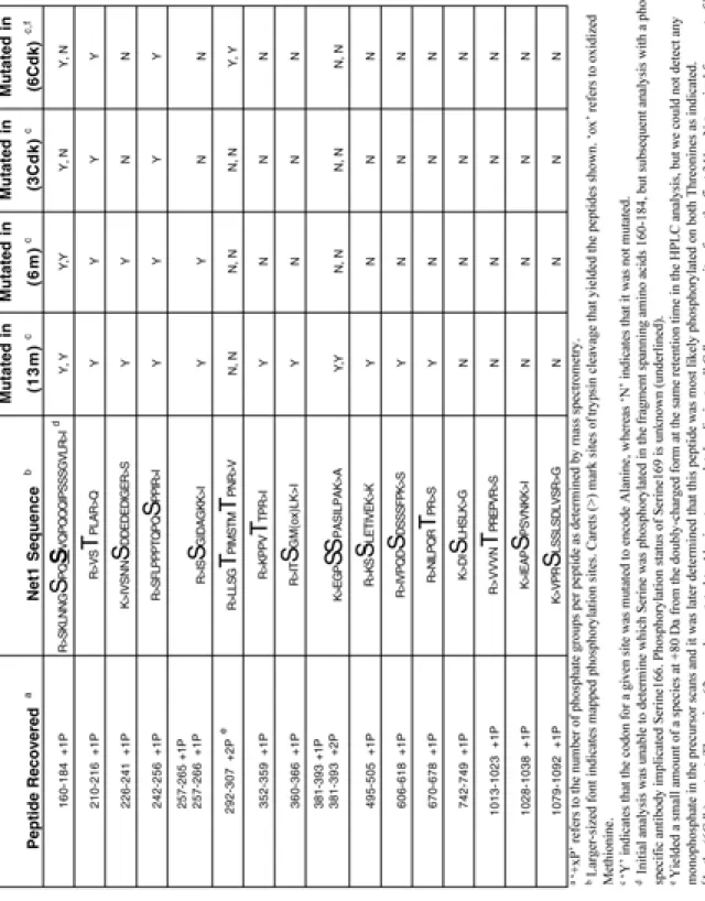
Given the striking phenotypes of net1-3Cdk and net1-6Cdk cells, we prepared phospho-specific antibodies using phosphorylated peptides corresponding to the three in vivo phosphorylation sites that were mutated in net1-3Cdk (designated PP-A, PP-B, and PP-C). All three antibodies reacted with Net1 isolated from arrested cdc14-1 cells (when Net1 is maximally phosphorylated) but not with Net1-3Cdk (Figure 3A). Moreover, the antibodies failed to detect Net1 from α factor-arrested cells (when Net1 is not detectably phospho-shifted) (Figure 3B). In many of the subsequent experiments we chose to focus on the PP-B epitope (which is formed by phosphorylation on Threonine 212) because it yielded a better signal in western blot analyses.
In vitro protein kinase assays provided the first concrete evidence that Net1 is a specific Cdk substrate. Recombinant Clb2–Cdk both generated the PP-B and PP-C epitopes on (Figure 3C), and incorporated radiolabel from γ-32P-ATP into recombinant Net1 (Figure 3D). Remarkably, phosphorylation of Net1 by Clb2–Cdk was exquisitely specific, in that an equivalent amount of Clb5–Cdk histone H1 kinase activity did not phosphorylate Net1 (Figure 3D).
glucose-repressible GAL1p-CLB2 construct. Whereas the timing and level of PP-B epitope formation were normal in clb1∆ cells (data not shown), clb1∆clb2∆ GAL1p-CLB2 cells depleted of the majority of Clb2 by growth on glucose after centrifugal elutriation, proceeded from G1 phase through anaphase but arrested in late anaphase/telophase without undergoing either detectable phosphorylation on Threonine 212 (Figure 4B) or release of Cdc14 from the nucleolus (Figures 4B, 4C, 4D). This observation was further corroborated by using an analog-sensitive version of the yeast Cdk (cdc28-as1) (Bishop et al., 2000; Ubersax et al., 2003) to demonstrate that phosphorylation of Net1 on Threonine 212 was dependent on Cdk activity as cells exited mitosis (Figure 9).
Moreover, CLB2 fulfilled the criteria established for FEAR network genes in that clb2∆
cdc15-2 cells failed to exhibit FEAR (Figures 4C and 4D), and clb2∆ enhanced the ts phenotype of cdc15-2 (Figure 4F) and dbf2-2 mutants (data not shown). Interestingly, Cdc14 localization in both clb1∆clb2∆ and clb2∆ cdc15-2 cells appeared to remain nucleolar despite arresting in late anaphase (Figure 4C, 2nd
panel). Moreover, upon meiotic segregation we could not obtain viable colonies containing clb2∆ in combination with cdc14-1, cdc5-1, or msd2-1 (data not shown) (Yuste-Rojas and Cross, 2000).
Importantly, the synthetic phenotype of clb2∆ MEN ts double mutants arose from an exacerbation of the MEN defect because the terminal phenotype of cdc15-2 clb2∆
Over-expression of non-degradable Clb2 is sufficient to drive Cdc14 out of the nucleolus in metaphase-arrested cells.
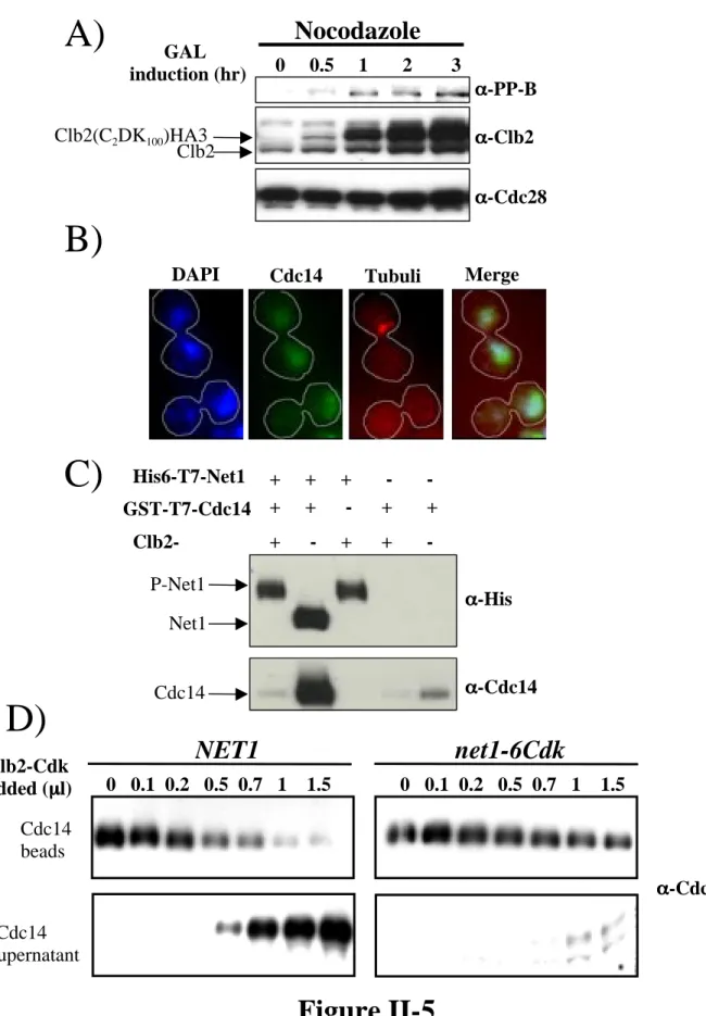
Our results with cis mutations in sites of Net1 in vivo phosphorylation coupled with trans mutations in CLB1 and CLB2 provide strong evidence that phosphorylation of Net1 on a set of N-terminal CDK sites by Clb1/Clb2-Cdk is necessary for FEAR. Over-expression of a stable form of Clb2 (GAL1p-CLB2∆db) has been reported to arrest cells in late anaphase with Cdc14 released from the nucleolus at the onset of anaphase (Stegmeier et al., 2002; Surana et al., 1993). To determine if regulation of Clb2-Cdk activity might normally govern the timing of FEAR, we tested whether elevated levels of Clb2 are sufficient to bring about ectopic release of Cdc14 from the nucleolus in cells arrested in metaphase. Whereas endogenous levels of Clb2 did not sustain Threonine 212 phosphorylation in metaphase-arrested cells (Figure 5A, Lane 1), over-expression of a hyper-stable form of Clb2 (CLB2C2DK100) (Hendrickson et al., 2001) was sufficient to drive Net1 phosphorylation and release of Cdc14 into the nucleus (Figures 5A, 5B). By contrast, induction of stable Clb2 in cells arrested in G1 phase with alpha factor did not bring about formation of the PP-B epitope on Net1 or disrupt association of Cdc14 with Net1 (data not shown).
strains expressing wild-type Net1 were dismantled by Clb2–Cdk in a dose-dependent manner, whereas complexes isolated from net1-6Cdk cells were not (Figure 5D). Taken together, the in vivo and in vitro data indicate that Cdk phosphorylation sites on Net1 can drive RENT disassembly.
Slk19, Spo12, and Cdc5 modulate phosphorylation of Net1 on Threonine 212. Analysis of Threonine 212 phosphorylation in synchronized cells revealed an unexpected result: appearance of Clb2 antigen preceded appearance of PP-B epitope by 10-20 min (Figure 3B). Because Clb2-associated protein kinase activity rises in parallel with Clb2 antigen (Stegmeier et al., 2002), we concluded that the ability of Clb2–Cdk to promote accumulation of phosphorylated Net1 must somehow be regulated. Moreover, because FEAR requires both the FEAR network genes and phosphorylation of Net1 by Clb1 or Clb2–Cdk complexes, and because ectopic induction of FEAR with GAL1p-ESP1 induces Net1 hyperphosphorylation (Sullivan and Uhlmann, 2003), we
(Figure 6B) and FEAR (Figure 6C) were greatly diminished in the double mutant cells compared to wild-type and cdc15-2 cells.
We have previously reported that Cdc5 influences the phosphorylation state of Net1 and promotes the release of Cdc14 in vivo (Shou et al., 2002a). This regulation appears to operate at least in part through Clb-Cdk, because phosphorylation of
Threonine 212 was absent in cdc14-1 cdc5-1 double mutant cells (Figure 6D) and over-expression of stable Cdc5 promoted phosphorylation of Threonine 212 in nocodazole-arrested cells (Figure 6E). Thus, Cdc5, like Spo12 and Slk19, promotes the
phosphorylation of Net1, at least in part, through Clb-Cdk.
Discussion
Phosphorylation of Net1 by Clb-Cdk underlies FEAR
Protein phosphatase Cdc14 plays a critical role in promoting exit from the M-phase state during both mitotic and meiotic cell cycles (Buonomo et al., 2003; Jaspersen et al., 1998; Marston et al., 2003). Cdc14 is kept under negative control during most of the cell cycle by binding to its nucleolar partner Net1, but is released from Net1 during anaphase through the actions of the FEAR and mitotic exit (MEN) networks (Shou et al., 1999; Stegmeier et al., 2002; Visintin et al., 1999). Because the transient release of Cdc14 from Net1 during anaphase is a signature event that drives exit from M-phase, we sought to investigate how this association is regulated in the context of the more well defined pathway for mitotic exit.
vivo phosphorylation sites on Net1 (isolated from cdc14ts cells) revealed that a set of Cdk sites in the N-terminal portion of Net1 were required for the proper release of Cdc14 in early anaphase (FEAR). Phosphorylation of these Cdk sites in Net1 was catalyzed by Cdk complexes formed by the late mitotic cyclins Clb1 and Clb2, in that they were completely absent in clb1∆ clb2∆ and cdc28-as1 mutants and that Clb2–Cdk was
sufficient to bring about phosphorylation of Net1 and disassembly of the RENT complex both in vivo and in vitro. Interestingly, a proteomic screen by Morgan and colleagues revealed that Net1 is one of the best substrates for Clb2-Cdk in vitro (Ubersax et al., 2003). In accordance with the observation that the FEAR network is important for proper progression through meiosis, the FEAR-deficient net1-6Cdk mutant also displayed a sporulation defect reminiscent of the slk19∆ FEAR mutant. Net1 phosphorylation by Clb-Cdk is likely to be a recurrent theme of M-phase progression in budding yeasts, because the majority of the N-terminal Cdk phosphorylation sites in Net1 are highly conserved among orthologs found in other fungal species (Figure 10). A similar phosphorylation-based mechanism may likewise control the transient association of human Cdc14A with the nucleolus (Mailand et al., 2002).
FEAR network promotes phosphorylation of Net1 by Clb1,2-Cdk
Release of Cdc14 from Net1 during early anaphase requires the FEAR network components Spo12, Slk19, Cdc5, and Esp1 in addition to phosphorylation of Net1 by Clb1,2-Cdk. Since the phosphorylation of Net1 by Clb-Cdk lagged behind the
was only moderately delayed in both spo12∆ and slk19∆ mutants, it was severely impaired in cdc5-1 mutants. We propose that either the FEAR network or the MEN can dissociate Cdc14 from Net1, and they act by independent mechanisms. FEAR network brings about RENT complex disassembly by promoting the phosphorylation of Net1 by Clb-Cdk. In contrast, the mild phenotype of the net1-6Cdk mutant indicates that the MEN can bring about release of Cdc14 from Net1 even in the absence of these same phosphorylations. Nevertheless, the MEN promotes phosphorylation of Net1 by Clb-Cdk, but possibly as an indirect consequence of exposing phosphorylation sites on Net1 that are normally shielded by bound Cdc14. Thus, whereas Net1 phosphorylation by Clb-Cdk is required for FEAR, we propose the same phosphorylations can occur as a passive consequence of Cdc14 release that is triggered by the MEN via a distinct mechanism. Consistent with this interpretation, in the absence of both the FEAR and mitotic exit networks (spo12∆ cdc15-2, slk19∆ cdc15-2, and cdc5-1 mutants), Net1 phosphorylation on Cdk sites was drastically reduced. It remains unknown how the FEAR network promotes phosphorylation of Net1 by Clb-Cdk. The putative ‘effector’ components of the FEAR network, Cdc5 and Spo12 (Sullivan and Uhlmann, 2003; Visintin et al., 2003), may modulate Net1, Clb-Cdk, or a Net1 phosphatase to promote accumulation of phosphate groups on Net1.
Is Net1 phosphorylation by Clb–Cdk sufficient for FEAR?
phosphorylation the only function of the FEAR, and is phosphorylation of Net1 on Cdk sites sufficient to dislodge Cdc14? Mutant spo12∆ cells have a sporulation defect that is more severe than that of net1-6Cdk mutants, suggestive of additional roles for Spo12 that go beyond phosphorylation of Net1. Moreover, the early anaphase release of Cdc14 promoted by the FEAR network in a cdc15-2 mutant is transient (Stegmeier et al., 2002), whereas the phosphorylation of Net1 on Threonine 212 is not (Figure 6). Thus, although the FEAR network promotes phosphorylation of Net1 and dissociation of Cdc14 from Net1, the former does not appear to be sufficient for the latter. On the other hand, Clb2–Cdk was sufficient to dislodge Cdc14 from Net1 in vitro, and over-expression of Clb2 brought about release of Cdc14 from Net1 in nocodazole-arrested cells (in which the FEAR network is inactive due to inhibition of Esp1 by securin). One possible explanation is that the FEAR network mobilizes Clb–Cdk to promote stable
phosphorylation of Net1 and labile phosphorylation of a second target (possibly Cdc14?), and that both of these phosphorylations are needed to sustain release of Cdc14 from Net1 in vivo. Attempts to resolve this issue through expression of NET1 mutants bearing phosphomimetic substitutions have been thwarted by the apparent hypomorphic nature of these alleles (data not shown).
Regardless of whether phosphorylation of Net1 by Clb2–Cdk is sufficient to dismantle RENT in vivo, there clearly is yet another mysterious dimension to this
issues, it is clear that phosphorylation of Net1 by Clb–Cdk is a key mechanism by which the FEAR network instigates disassembly of the RENT complex during mitosis.
On the roles of Cdc5 and Clb-Cdk protein kinase activities in FEAR
In previous work, we reported that Cdc5 influences the phosphorylation of Net1 and disassembly of the RENT complex (Shou et al., 2002a). Our current observations that Cdc5 is necessary and sufficient for phosphorylation of Net1 by Clb–Cdk lead us to propose that Cdc5 controls Net1 phosphorylation primarily through Clb–Cdk. However, to evaluate the relative role of Cdc5 in more detail requires careful consideration of our prior findings. To address the role of Cdc5 in RENT disassembly in vivo, we originally constructed a mutant Net1 (net1-7m) lacking the 7 sites of in vitro phosphorylation that contributed most prominently to the Cdc5-induced dissociation of recombinant
phosphorylation of Net1. However, our cis mutant analysis of Net1 clearly indicates that the former role is critical, and the latter role is, at best, minor. We wish to note that the putative Cdc5 phosphorylation sites (S231 and S259) do not lie in the recently reported consensus for Cdc5/Polo kinases (Nakajima et al, 2003), and thus it remains possible that these sites may normally be phosphorylated by an unknown protein kinase in vivo, and Cdc5 may act solely through Clb–Cdk.
Acknowledgements
We thank J. Huangh (Moazed lab) for the (Net1 1-621) construct, I. Lesur (Campbell lab) for help with elutriation, Doug Kellogg for Anti-Clb2, Kevan Shokat for NaPP1 analog, and the Nasmyth, Morgan, Amon, and Holloway labs for various strains and GAL1p-CLB2 mutant constructs. We also thank Mike Olson for the Clb2-MBP bacterial construct, Rati Verma for preparing the Baculo (GST-Cdc28-HA) virus, Matthew Petroski for purified Clb5–Cdk, Jennifer Sanders for mutagenesis efforts, and members of the Deshaies lab for critical comments during the course of this work. This work was supported by an NIH grant (GM59940) to RJD. RJD is an Assistant
Investigator of the Howard Hughes Medical Institute. RA was supported by fellowships from the Norris and Baxter foundations.
Experimental Procedures
Strain construction, materials, and Net1 mutagenesis
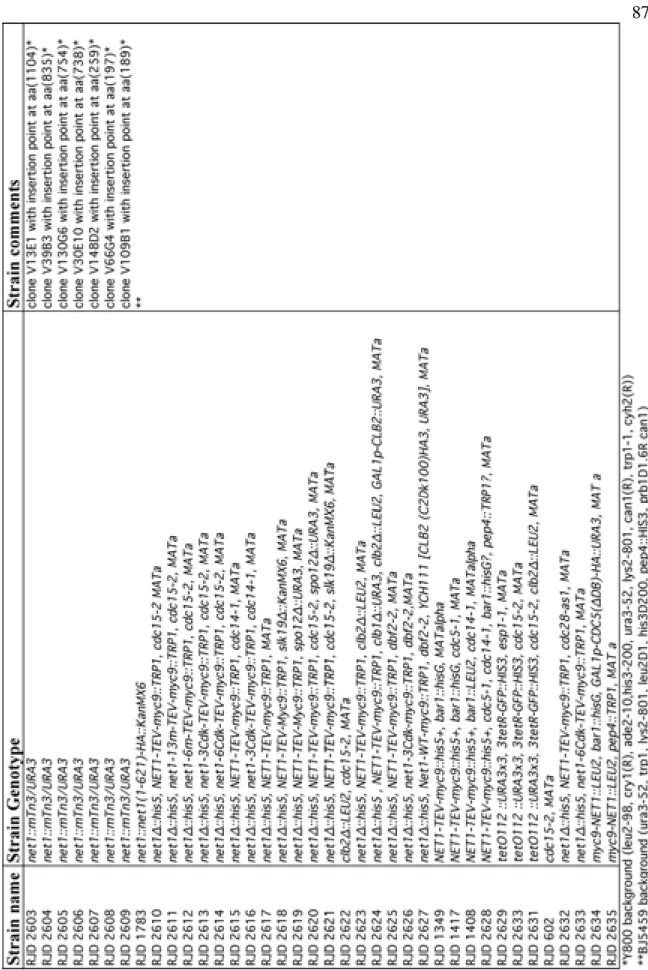
All strains used are in the W303 background (can1-100, leu2-3, his3-11, trp1-1, ura3-1, ade2-1) except where noted in the strain table (Supplementary Table 1). A strain expressing a stable form of Clb2 lacking both the KEN and destruction boxes
(Clb2C2DK100)HA3 was used in over-expression experiments with Clb2 (Hendrickson et al., 2001).
using QuikChange Site-Directed mutagenesis kit from Stratagene (La Jolla, CA). The indicated Serine/Threonine were mutated to Alanine in Net1-13m
(166,169,212,231,252,259,356,362,384,385,497,611,676), Net1-6m
(166,169,212,231,252,259), Net1-3-Cdk (166,212,252), and Net1-6Cdk (62*,
166,212,252,297,304) where * indicates that residue 62 was mutated to ensure complete elimination of all Cdk consensus sites even though it was not determined to be
phosphorylated in vivo. Mutagenesis was confirmed using restriction digests followed by DNA sequencing. All constructs were targeted by linearization with BstXI to the trp1 locus in a NET1/net1::his5+ heterozygous diploid. The strains were sporulated and tetrads were dissected to obtain a haploid isolate of the integrant over net1::his5+
. Copy number of integrants was estimated by normalizing extract protein from transformed and wild-type cells and blotting for Net1 levels. Proper localization of all net1 phosphosite mutants was confirmed by indirect immuno-fluorescence against the myc epitope (data not shown).
Antibodies specifically reactive against the phosphopeptides were positively selected on a resin derivatized with the phosphopeptide immunogen and negatively selected by passage through a resin derivatized with the unphosphorylated version of the peptide. Anti-phosphopeptide B (α-PP-B) was used in all experiments described since it generated the strongest signal against Phospho-Net1 (Figure 3A).
Cell Growth and Synchronization Procedures
Cells were grown in yeast extract-peptone (YP) or in yeast minimal (YM) media containing 2% glucose (YPD,YMD), 2% raffinose (YPR,YMR) or 2% galactose
Cell Extract Preparation and Western Blotting
Cells were grown to an O.D.600 of 1.0, and for every time point 2 ml of culture was collected and TCA added to a final concentration of 20%. Cells were collected by centrifugation and washed with 2 ml of Tris-HCl (pH 7.5). SDS loading buffer [70 µl of 100mM Tris-HCl (pH 7.5), 20% glycerol, 4% SDS, 2M Urea, 200mM DTT] was added, tubes were boiled for 3-minutes, and 100 µl of acid-washed glass beads (500 µm) were added to each tube followed by boiling for an additional 2-minutes. Tubes were vortexed for 45 sec using a Bio 101 multi-bead vortexer at setting 5.5. Tubes were boiled again for 2-minutes and 5 µl of sample was fractionated on a 10% SDS-PAGE gel followed by transfer to a nitrocellulose membrane. Western blot analysis was performed with the following primary antibodies at the indicated dilutions: All anti-phospho Net1 antibodies (α-PP-A, α-PP-B, α-PP-C) at 2 µg/ml; Clb2 (1:3000), Cdc28 (1:5000), Clb3 (1:2000), myc (9E10) (1:5000), His (1:250), HA (1:5000), and anti-Cdc14 (1:1000).
Immunoprecipitation and Clb2–Cdk release/kinase assay.
acid-washed glass beads (500 µm). Samples were vortexed using a Bio 101 multi-beads vortexer at setting 5.5 (speed) and 45sec (time). Tubes were then centrifuged for 5-minutes at 14,000 rpm and the supernatant was collected. Clarified extract (400 µl) was incubated with 60 µl of 9E10-coupled protein A beads for 1 hour on a rotator at 4°C. Beads were collected and washed ten times in wash buffer [25 mM HEPES/KOH
(pH7.5), 150 mM NaCl, 1 mM DTT, 0.2% Triton], and divided to approximately 15 µl beads per reaction condition. For protein kinase assays, 3 µl of either Clb2–Cdk or Clb5–Cdk was used with either 1 µg of myc9-Net1 (purified from insect cells infected with a recombinant baculovirus) (Shou et al., 2001), or 5 µg of Histone H1. For assays that monitored release of Cdc14 from bead-bound Net1-myc9, varying concentrations of in vitro assembled Clb2–Cdk in 30µl kinase buffer [25 mM Tris-HCl (pH 7.5), 10 mM MgCl2, 1 mM ATP, 1 mM DTT, 0.1 mg/ml BSA, 50 mM NaCl] were mixed with 15 µl 9E10 beads coated with RENT complex. Reactions were allowed to proceed for 30-minutes on a rotator at 25°C. Supernatant and beads were processed for Western blot analysis as previously described (Shou et al., 2002a). For in vitro assays with bacterially expressed constructs, approximately 116 ng of Net1 per 20 µl of Ni+2
-NTAbeads, 10 ng Cdc14, and 5 µl of roughly 30 ng/µl stock of Clb2–Cdk was used per reaction.
Immuno-fluorescence and Cdc14 release quantification
indicated dilutions. Images of synchronized cells at 70 to 110 minutes following
release from α factor were collected on a Zeiss Axioskop or Axiovert 200M microscope using a Hamamatsu CCD digital camera. Spindle length measurements were performed using Zeiss Axiovision software.
References
Adams, I.R., and J.V. Kilmartin. 1999. Localization of core spindle pole body (SPB) components during SPB duplication in Saccharomyces cerevisiae. J Cell Biol. 145:809-23.
Amon, A. 2002. Synchronization procedures. Methods Enzymol. 351:457-67.
Amon, A., S. Irniger, and K. Nasmyth. 1994. Closing the cell cycle circle in yeast: G2 cyclin proteolysis initiated at mitosis persists until the activation of G1 cyclins in the next cycle. Cell. 77:1037-50.
Bardin, A.J., R. Visintin, and A. Amon. 2000. A mechanism for coupling exit from mitosis to partitioning of the nucleus. Cell. 102:21-31.
Shokat. 2000. A chemical switch for inhibitor-sensitive alleles of any protein kinase. Nature. 407:395-401.
Buonomo, S.B., K.P. Rabitsch, J. Fuchs, S. Gruber, M. Sullivan, F. Uhlmann, M. Petronczki, A. Toth, and K. Nasmyth. 2003. Division of the nucleolus and its release of CDC14 during anaphase of meiosis I depends on separase, SPO12, and SLK19. Dev Cell. 4:727-39.
Burns, N., B. Grimwade, P.B. Ross-Macdonald, E.Y. Choi, K. Finberg, G.S. Roeder, and M. Snyder. 1994. Large-scale analysis of gene expression, protein localization, and gene disruption in Saccharomyces cerevisiae. Genes Dev. 8:1087-105.
Deshaies, R.J. 1997. Phosphorylation and proteolysis: partners in the regulation of cell division in budding yeast. Curr Opin Genet Dev. 7:7-16.
Donovan, J.D., J.H. Toyn, A.L. Johnson, and L.H. Johnston. 1994. P40SDB25, a putative CDK inhibitor, has a role in the M/G1 transition in Saccharomyces cerevisiae. Genes Dev. 8:1640-53.
Hendrickson, C., M.A. Meyn, 3rd, L. Morabito, and S.L. Holloway. 2001. The KEN box regulates Clb2 proteolysis in G1 and at the metaphase-to-anaphase transition. Curr Biol. 11:1781-7.
Holloway, S.L., M. Glotzer, R.W. King, and A.W. Murray. 1993. Anaphase is initiated by proteolysis rather than by the inactivation of maturation-promoting factor. Cell. 73:1393-402.
Irniger, S., S. Piatti, C. Michaelis, and K. Nasmyth. 1995. Genes involved in sister chromatid separation are needed for B-type cyclin proteolysis in budding yeast. Cell. 81:269-78.
Jaspersen, S.L., J.F. Charles, and D.O. Morgan. 1999. Inhibitory phosphorylation of the APC regulator Hct1 is controlled by the kinase Cdc28 and the phosphatase Cdc14. Curr Biol. 9:227-36.
Jaspersen, S.L., J.F. Charles, R.L. Tinker-Kulberg, and D.O. Morgan. 1998. A late mitotic regulatory network controlling cyclin destruction in Saccharomyces cerevisiae. Mol Biol Cell. 9:2803-17.
Johnston, L.H., and A.L. Johnson. 1997. Elutriation of budding yeast. Methods Enzymol. 283:342-50.
King, R.W., J.M. Peters, S. Tugendreich, M. Rolfe, P. Hieter, and M.W. Kirschner. 1995. A 20S complex containing CDC27 and CDC16 catalyzes the mitosis-specific conjugation of ubiquitin to cyclin B. Cell. 81:279-88.
Lee, B.H., and A. Amon. 2003. Role of Polo-like kinase CDC5 in programming meiosis I chromosome segregation. Science. 300:482-6.
Li, X., and M. Cai. 1997. Inactivation of the cyclin-dependent kinase Cdc28 abrogates cell cycle arrest induced by DNA damage and disassembly of mitotic spindles in Saccharomyces cerevisiae. Mol Cell Biol. 17:2723-34.
Loughrey Chen, S., M.J. Huddleston, W. Shou, R.J. Deshaies, R.S. Annan, and S.A. Carr. 2002. Mass spectrometry-based methods for phosphorylation site mapping of
hyperphosphorylated proteins applied to Net1, a regulator of exit from mitosis in yeast. Mol Cell Proteomics. 1:186-96.
Luca, F.C., and M. Winey. 1998. MOB1, an essential yeast gene required for completion of mitosis and maintenance of ploidy. Mol Biol Cell. 9:29-46.
Mah, A.S., J. Jang, and R.J. Deshaies. 2001. Protein kinase Cdc15 activates the Dbf2-Mob1 kinase complex. Proc Natl Acad Sci U S A. 98:7325-30.
Mailand, N., C. Lukas, B.K. Kaiser, P.K. Jackson, J. Bartek, and J. Lukas. 2002. Deregulated human Cdc14A phosphatase disrupts centrosome separation and chromosome segregation. Nat Cell Biol. 4:317-22.
Marston, A.L., B.H. Lee, and A. Amon. 2003. The Cdc14 phosphatase and the FEAR network control meiotic spindle disassembly and chromosome segregation. Dev Cell. 4:711-26.
McCollum, D., and K.L. Gould. 2001. Timing is everything: regulation of mitotic exit and cytokinesis by the MEN and SIN. Trends Cell Biol. 11:89-95.
Moll, T., G. Tebb, U. Surana, H. Robitsch, and K. Nasmyth. 1991. The role of
phosphorylation and the CDC28 protein kinase in cell cycle- regulated nuclear import of the S. cerevisiae transcription factor SWI5. Cell. 66:743-58.
Pereira, G., T. Hofken, J. Grindlay, C. Manson, and E. Schiebel. 2000. The Bub2p spindle checkpoint links nuclear migration with mitotic exit. Mol Cell. 6:1-10.
Pereira, G., C. Manson, J. Grindlay, and E. Schiebel. 2002. Regulation of the Bfa1p-Bub2p complex at spindle pole bodies by the cell cycle phosphatase Cdc14p. J Cell Biol. 157:367-79.
Ross, K.E., and O. Cohen-Fix. 2003. Multitasking at mitotic exit. Nat Cell Biol. 5:188-90.
Ross-Macdonald, P., A. Sheehan, C. Friddle, G.S. Roeder, and M. Snyder. 1999.
Transposon mutagenesis for the analysis of protein production, function, and localization. Methods Enzymol. 303:512-32.
Rudner, A.D., K.G. Hardwick, and A.W. Murray. 2000. Cdc28 activates exit from mitosis in budding yeast. J Cell Biol. 149:1361-76.
Rudner, A.D., and A.W. Murray. 2000. Phosphorylation by Cdc28 activates the Cdc20-dependent activity of the anaphase-promoting complex. J Cell Biol. 149:1377-90.
Schweitzer, B., and P. Philippsen. 1991. CDC15, an essential cell cycle gene in Saccharomyces cerevisiae, encodes a protein kinase domain. Yeast. 7:265-73.
Shirayama, M., Y. Matsui, K. Tanaka, and A. Toh-e. 1994a. Isolation of a CDC25 family gene, MSI2/LTE1, as a multicopy suppressor of ira1. Yeast. 10:451-61.
Shirayama, M., Y. Matsui, and E.A. Toh. 1994b. The yeast TEM1 gene, which encodes a GTP-binding protein, is involved in termination of M phase. Mol Cell Biol. 14:7476-82.
Shou, W., R. Azzam, S.L. Chen, M.J. Huddleston, C. Baskerville, H. Charbonneau, R.S. Annan, S.A. Carr, and R.J. Deshaies. 2002a. Cdc5 influences phosphorylation of Net1 and disassembly of the RENT complex. BMC Mol Biol. 3:3.
Shou, W., K.M. Sakamoto, J. Keener, K.W. Morimoto, E.E. Traverso, R. Azzam, G.J. Hoppe, R.M. Feldman, J. DeModena, D. Moazed, H. Charbonneau, M. Nomura, and R.J. Deshaies. 2001. Net1 stimulates RNA polymerase I transcription and regulates nucleolar structure independently of controlling mitotic exit. Mol Cell. 8:45-55.
Shou, W., R. Verma, R.S. Annan, M.J. Huddleston, S.L. Chen, S.A. Carr, and R.J. Deshaies. 2002b. Mapping phosphorylation sites in proteins by mass spectrometry. Methods Enzymol. 351:279-96.
Stegmeier, F., R. Visintin, and A. Amon. 2002. Separase, polo kinase, the kinetochore protein Slk19, and Spo12 function in a network that controls Cdc14 localization during early anaphase. Cell. 108:207-20.
Straight, A.F., W. Shou, G.J. Dowd, C.W. Turck, R.J. Deshaies, A.D. Johnson, and D. Moazed. 1999. Net1, a Sir2-associated nucleolar protein required for rDNA silencing and nucleolar integrity. Cell. 97:245-56.
Sullivan, M., and F. Uhlmann. 2003. A non-proteolytic function of separase links the onset of anaphase to mitotic exit. Nat Cell Biol. 5:249-54.
Surana, U., A. Amon, C. Dowzer, J. McGrew, B. Byers, and K. Nasmyth. 1993. Destruction of the CDC28/CLB mitotic kinase is not required for the metaphase to anaphase transition in budding yeast. Embo J. 12:1969-78.
Toyn, J.H., A.L. Johnson, J.D. Donovan, W.M. Toone, and L.H. Johnston. 1997. The Swi5 transcription factor of Saccharomyces cerevisiae has a role in exit from mitosis through induction of the cdk-inhibitor Sic1 in telophase. Genetics. 145:85-96.
Ubersax, J.A., E.L. Woodbury, P.N. Quang, M. Paraz, J.D. Blethrow, K. Shah, K.M. Shokat, and D.O. Morgan. 2003. Targets of the cyclin-dependent kinase Cdk1. Nature. 425:859-64.
Verma, R., R.S. Annan, M.J. Huddleston, S.A. Carr, G. Reynard, and R.J. Deshaies. 1997. Phosphorylation of Sic1p by G1 Cdk required for its degradation and entry into S phase. Science. 278:455-60.
Visintin, R., and A. Amon. 2001. Regulation of the mitotic exit protein kinases Cdc15 and Dbf2. Mol Biol Cell. 12:2961-74.
Visintin, R., K. Craig, E.S. Hwang, S. Prinz, M. Tyers, and A. Amon. 1998. The
phosphatase Cdc14 triggers mitotic exit by reversal of Cdk- dependent phosphorylation. Mol Cell. 2:709-18.
Visintin, R., E.S. Hwang, and A. Amon. 1999. Cfi1 prevents premature exit from mitosis by anchoring Cdc14 phosphatase in the nucleolus. Nature. 398:818-23.
Walker, G.M. 1999. Synchronization of yeast cell populations. Methods Cell Sci. 21:87-93.
Wan, J., H. Xu, and M. Grunstein. 1992. CDC14 of Saccharomyces cerevisiae. Cloning, sequence analysis, and transcription during the cell cycle. J Biol Chem. 267:11274-80.
Yoshida, S., and A. Toh-e. 2001. Regulation of the localization of Dbf2 and mob1 during cell division of Saccharomyces cerevisiae. Genes Genet Syst. 76:141-7.
Yoshida, S., and A. Toh-e. 2002. Budding yeast Cdc5 phosphorylates Net1 and assists Cdc14 release from the nucleolus. Biochem Biophys Res Commun. 294:687-91.
Yuste-Rojas, M., and F.R. Cross. 2000. Mutations in CDC14 result in high sensitivity to cyclin gene dosage in Saccharomyces cerevisiae. Mol Gen Genet. 263:60-72.
Figure II-1: Cell cycle-regulated binding site for Cdc14 resides in the
N-terminal half of Net1
DAPI
Cdc14
Tubulin
Merge
WT
3Cdk
6Cdk
A)
B)
0 20 40 60 80 1000-2 2-4 4-6 6-8 8-10 >10
0 20 40 60 80 100
0-2 2-4 4-6 6-8 8-10 >10
NET1 (WT)
net1-3Cdk
0 20 40 60 80 1000-2 2-4 4-6 6-8 8-10 >10 Spindle length (µµµµm)
net1-6Cdk
% Cdc14 released
dbf2-2
3Cdk
dbf2-2
25
°°°°
C
33.5
°°°°
C
C)
cdc15-2
6Cdk
cdc15-2
25
°°°°
C
30.7
°°°°
C
D)
Spindle length (µµµµm)
% Cdc14 released
Spindle length (µµµµm)
% Cdc14 released
Full Release Partial Release
Figure II-2: Net1 phosphorylation site mutants display Cdc14 release
defects and synthetic interactions with MEN mutants.
(A) Net1 mutants are defective in Cdc14 release in early anaphase. Mutant cdc15-2 cells carrying either NET1 (WT) (RJD2617), net1-3Cdk (3Cdk) (RJD2613), or net1-6Cdk (6Cdk) (RJD2614) alleles were synchronized with α factor at 25°C and released in YP 2% glucose at 37°C. Cells were collected at 10 to15 minute time intervals for analysis by indirect immuno-fluorescence. Staining was performed with DAPI, Cdc14 and anti-tubulin antibodies for determination of nuclear position, Cdc14 localization, and
microtubule spindle length, respectively. Cell outlines are indicated for comparison.
(B) Quantitation of Fourteen Early Anaphase Release (FEAR) defect of Net1
phosphorylation site mutants. Synchronized cells collected at 70 to110 minutes after release from α factor were double-labeled with anti-Cdc14 and anti-tubulin antibodies. Spindle length was measured and localization of Cdc14 was determined to be either: 1, full release (black boxes; complete release of Cdc14 from the nucleolus into the nucleus); or 2, partial release (white boxes; Cdc14 was nuclear in one of the DAPI masses and nucleolar in the other DAPI mass in the same cell, or Cdc14 was not completely restricted to the nucleolus in either DAPI mass). Over 350 cells were counted for each panel.
incubated at the indicated temperature for 2-3 days before the picture was taken. Two independent isolates of each strain were used. The first two isolates in the 33.5°C panel were compiled from different sections of the same plate.