Experimental human pneumococcal carriage
Thesis submitted in accordance with the requirements of the
University of Liverpool for the degree of Doctor in Philosophy
by
Jenna Faye Gritzfeld
i
This thesis is the result of my own work and effort. In some instances, work was
done in collaboration with other colleagues and institutions. Table I details in full
the attribution of work and responsibility related to the project.
The research in this thesis was carried out at the Liverpool School of Tropical
Medicine.
ii
Activity Responsibility
Recruitment, sample taking,
inoculation, symptoms data collection
Angela D. Wright, Andrea M. Collins, Carole Hancock, David Shaw
(Liverpool School of Tropical Medicine)
Microarray Jason Hinds
(St. George's, University of London) Pneumococcal phase morphology Aoife M. Roche
(University of Pennsylvania) Complement deposition flow cytometry
analysis
Shaun Pennington
(Liverpool School of Tropical Medicine) Whole genome sequence comparison
of serotypes 6B and 23F
Jen E. Cornick
(University of Malawi) Genetic comparison of two 23F strains
and detection of amiC frameshift mutation
Ankur B. Dalia
(Tufts University School of Medicine, Boston) Mouse model of colonization Ankur B. Dalia
Pneumococcal DNA extraction Amelieke J. H. Cremers
(Radboud University Medical Center)
LytA qPCR Amelieke J. H. Cremers
Detection of URT viruses Mark J. Hopkins
(Royal Liverpool and Broadgreen University Hospital)
Factor H ELISAs from the Dose-Ranging study
Nicholas Coombes
(Liverpool School of Tropical Medicine)
Anti-PspC IgG ELISA Michael Garner-Jones
(Liverpool School of Tropical Medicine) Depletion and purification of antibodies
from nasal wash and sera samples
Angela D. Wright and Michael Garner-Jones Factor H binding and antibody binding
assays and flow cytometry analysis
Nicholas Coombes, Michael Garner-Jones; Adriana T. Moreno (Instituto Butantan) Epithelial pIgR and PAFr expression by
flow cytometry
Sarah Glennie (University of Bristol)
Anti-PspC antibody epitope mapping Cintia F. Vadesilho (Instituto Butantan) Generalized estimating equation (GEE)
model
Duolao Wang
iii Pneumococcal disease is preceded by nasopharyngeal colonization, which is also the source of transmission. Current pneumococcal conjugate vaccines protect against invasive disease and reduce carriage in children, but are less effective against mucosal disease and have limited serotype coverage. There is an urgent need for new vaccines and colonization has been suggested as an alternative endpoint in vaccine licensure. Experimental human pneumococcal carriage, although potentially risky, offers a way to examine colonization in the context of vaccination. Experimental carriage also allows the investigation of the impact of a pathogen on the immunological complexity and normal microbiota of humans, both of which cannot be done using animal models. We developed a safe and reproducible method of experimental human pneumococcal carriage, described bacteriological and immune factors associated with carriage, and examined the density and duration of experimental carriage.
iv
Gritzfeld JF, Dalia AB, Ferreira DM, Roche AM, Glennie S, Pennington SH, Bangert M, Cornick JE, Wright AD, Collins AM, Camilli A, Weiser JN, Everett DB, Kadioglu A, Gordon SB. The pneumococcal permease protein amiC has a key role in experimental human pneumococcal carriage. Manuscript in preparation
Gritzfeld JF*, Glennie S*, Pennington SH, Garner-Jones M, Coombes N, Hopkins MJ, Vadesilho CF, Miyaji EN, Wang D, Wright AD, Collins AM, Gordon SB, Ferreira DM (2015). Modulation of nasopharyngeal innate defences by viral co-infection predisposes individuals to experimental pneumococcal carriage. Mucosal Immunol, In press
Gritzfeld JF*, Cremers AJH*, Ferwerda G, Ferreira DM, Kadioglu A, et al. (2014). Density and duration of experimental human pneumococcal carriage. Clin Microbiol Infec 20: O1145-O1151
Gritzfeld JF, Cremers AK, Gordon SB (2013). Detection limits in pneumococcal carriage. Pediatr Infect Dis J 32: 425-426
Gritzfeld JF, Wright AD, Collins AM, Pennington SH, Wright AKA, et al. (2013). Experimental human pneumococcal carriage. J Vis Exp 72, e50115
Book Chapter
:Gritzfeld JF, Gordon SB (2013). Pneumococcal Vaccines: Experimental Human Pneumococcal Carriage as a Model for Vaccine Development. Vaccines: Benefits and Risks. iConcept Press. ISBN: 978-1477554-95-1
Papers arising during the course of this PhD
Collins AM, Wright AD, Mitsi E, Gritzfeld JF, Hancock CA, Pennington SH, Wang D, Morton B, Ferreira DM, Gordon SB. Pneumococcal conjugate vaccine reduces the rate, density, and duration of experimental human pneumococcal colonisation. Manuscript submitted for publication
v Cremers JH, Zomer AL, Gritzfeld JF, Ferwerda G, van Hijum SAFT, et al. (2014). The adult nasopharyngeal microbiome as a determinant of pneumococcal acquisition. Microbiome 2: 44
Wall EC, Gritzfeld JF, Scarborough M, Ajdukiewicz KMB, Mukaka M, et al. (2014). Genomic pneumococcal load and CSF cytokines are not related to outcome in Malawian adults with meningitis. J Infection 69: 440-446
Shak JR, Cremers AJH, Gritzfeld JF, de Jonge MI, Hermans PWM, et al. (2014). Impact of experimental human pneumococcal carriage on nasopharyngeal bacterial densities in healthy adults. PLoS One 9: e98829
Neill DR, Coward WR, Gritzfeld JF, Richards L, Garcia-Garcia FJ, et al. (2014). Density and duration of pneumococcal carriage is maintained by transforming growth factor β1and T regulatory cells. Am J Respir Crit Care Med 189: 1250-1259
Ferreira DM, Neill DR, Bangert M, Gritzfeld JF, Green N, et al. (2013). Controlled human infection and re-challenge with Streptococcus pneumoniae reveals the protective efficacy of carriage in healthy adults. Am J Respir Crit Care Med 187: 855-864
Wright AK, Bangert M, Gritzfeld JF, Ferreira DM, Jambo KC, Wright AD, Collins AM, Gordon SB (2013). Experimental human pneumococcal carriage augments IL-17A-dependent T-cell defence of the lung. PLoS Pathog 9(3), e1003274
vi I would first like to thank my supervisors, Professor Stephen Gordon and Professor Aras Kadioglu. Their support and guidance has been unwavering. I am very grateful to Stephen for putting his trust in me, not only in building this model, but also for being the first volunteer and allowing me to put pneumococci up his nose.
The work presented in this thesis was funded by the Gates Foundation and would not have been possible without the assistance, support, and advice of numerous colleagues and collaborators. These include, but are not limited to: Daniela, Shaun, Elena, Angie, Andrea, Jessica, Sarah, Carole, Adam, Mathieu, Jane and Tracy. My thanks also go to Amelieke for the productive collaboration and for hosting me in Nijmegen. And to Andrew and Katy for the necessary venting sessions whilst writing up. A big thank you also goes to all the volunteers without whom this work would not have been possible.
Thank you to my parents, Brenda and Greg, who never tired of my numerous “whys”. I love you to the moon and back. Thank you to my sister Tessa and my brother Colton for their advice and humour. And a special thanks to Dave for his patience and support. I think we are both happy you no longer have to ask "have you finished the PhD yet?”.
vii
Declaration ... i
Table I: People that have contributed to the work presented in this thesis ... ii
Abstract ... iii
Papers arising from work presented in this thesis ... iv
Papers arising during the course of this PhD ... iv
Acknowledgements ... vi
Abbreviations ... xviii
List of Figures ... xix
List of Tables ... xxi
Chapter 1 ... 1
Introduction ... 1
1.1 Streptococcus pneumoniae ... 1
1.1.1 Epidemiology of pneumococcal carriage ... 2
1.1.1.1 Risk factors associated with carriage ... 3
1.1.1.1.2 Crowding ... 3
1.1.1.1.2 Environment... 3
1.1.1.1.3 Socioeconomics... 3
1.1.2 Epidemiology of pneumococcal disease ... 4
1.1.3 The association of pneumococcal carriage with disease ... 5
1.1.3.1 Association between carriage and disease in animal models ... 5
1.1.3.2 Association between carriage and disease in humans ... 6
1.1.3.3 Association of serotype with the incidence of carriage and disease ... 6
1.2 Mechanisms of S. pneumoniae carriage ... 6
1.2.1 Expression of capsule during colonization ... 6
1.2.2 Pneumococcal surface proteins involved in carriage ... 7
1.2.2.1 Choline-binding proteins mediate adherence ... 7
1.2.2.2 Lipoproteins mediate adherence to cell-surface carbohydrates ... 8
1.2.2.3 Protease production increases attachment ... 8
1.2.2.4 Adhesins assist in binding to the extracellular matrix ... 8
1.2.2.5 Neuraminidase facilitates persistence in the nasopharynx ... 8
1.3 Immunity to S. pneumoniae ... 9
viii
1.3.1.3 Complement system action against pneumococcus ... 10
1.3.1.3.1 Complement pathways involved in protection against pneumococcus ... 10
1.3.1.3.2 The role of complement in the progression from carriage to disease ... 11
1.3.1.3.3 Pneumococcal mechanisms used to resist complement ... 11
1.3.1.4 Toll-like receptors involved in protection against pneumococcus ... 13
1.3.1.4.1 TLR2 ... 13
1.3.1.4.2 TLR4 ... 14
1.3.1.4.3 TLR9 ... 14
1.3.2 Adaptive immunity to the pneumococcus ... 14
1.3.2.1 Humoral immunity against pneumococcus ... 14
1.3.2.1.1 Agglutination of pneumococcus ... 15
1.3.2.2 Cellular immunity against pneumococcus ... 15
1.3.2.2.1 Macrophages ... 15
1.3.2.2.2 Neutrophils ... 16
1.3.2.2.3 CD4+ T cells ... 16
1.3.2.2.4 Th17 cells ... 16
1.3.3 The effect of carriage on defence against future carriage and disease ... 17
1.4 Vaccination against S. pneumoniae ... 17
1.4.1 Early attempts at pneumococcal vaccination ... 18
1.4.2 Pneumococcal polysaccharide vaccines ... 18
1.4.2.1 Pneumococcal polysaccharide vaccine efficacy ... 19
1.4.5 Pneumococcal conjugate vaccines... 20
1.4.5.1 Pneumococcal conjugate vaccine efficacy ... 20
1.4.5.1.1 Immune correlates of protection ... 20
1.4.5.1.2 Protection against carriage ... 21
1.4.5.1.3 Protection against invasive disease ... 21
1.4.5.1.4 Protection against mucosal disease ... 21
1.4.5.1.5 Indirect effects of pneumococcal conjugate vaccination ... 21
1.4.5.2 Drawbacks of pneumococcal conjugate vaccines ... 22
1.4.6 Novel pneumococcal vaccine development ... 23
1.4.6.1 Live attenuated vaccines ... 23
ix
1.5.5.2 Co-colonization of pneumococcus and respiratory viruses ... 25
1.5.5.3 Co-colonization of pneumococcus and Staphylococcus aureus ... 25
1.5.5.4 Co-colonization of pneumococcus and Gram negatives... 25
1.5.5.5 The nasopharyngeal microbiome ... 26
1.6 Detection of S. pneumoniae carriage ... 26
1.6.1 Nasopharyngeal sample collection ... 26
1.6.1.1 Sampling methods in children and infants ... 26
1.6.1.2 Sampling methods in adults ... 27
1.6.2 Methods used to detect pneumococcal carriage ... 27
1.6.2.1 Microbiological culture ... 27
1.6.2.2 Culture enrichment ... 28
1.6.2.3 Non-culture based detection methods ... 28
1.6.2.4 Serotyping ... 29
1.6.3 Determining pneumococcal carriage density ... 29
1.6.3.1 Methods to determine density ... 29
1.6.3.2 Role of density in disease and transmission ... 29
1.7 Models of S. pneumoniae carriage ... 30
1.7.1 Mouse model of carriage ... 30
1.7.2 Infant rat and chinchilla models of carriage ... 31
1.7.3 Nonhuman primate model of carriage ... 31
1.8 Experimental human challenge studies ... 31
1.8.1 Ethical framework behind human challenge models ... 32
1.8.1.1 Regulation of human challenge models ... 32
1.8.2 Human challenge models with potential respiratory pathogens ... 32
1.8.3 Experimental human pneumococcal carriage ... 33
1.8.3.1 Experimental human pneumococcal carriage in the United States ... 33
1.8.3.2 Experimental human pneumococcal carriage in England ... 34
1.8.3.3 Applications of the experimental human pneumococcal carriage model ... 34
1.8.3.4 Usefulness of an experimental human pneumococcal carriage model in novel vaccine testing ... 35
x
1.9 Project aims... 37
Chapter 2 ... 38
Materials and methods ... 38
2.1 Initial model development ... 38
2.2 Clinical procedures ... 41
2.2.1 Recruitment and ethics ... 41
2.2.2 Study schedules ... 41
2.2.2.1 Dose-Ranging study schedule ... 42
2.2.2.2 Reproducibility study schedule ... 44
2.2.2.3 Re-challenge study schedules ... 44
2.2.3 Safety monitoring ... 46
2.2.3.1 Data monitoring and safety committee ... 46
2.2.4 Symptom reporting ... 47
2.2.4.1 Passive reporting of symptoms ... 47
2.2.4.2 Active reporting of symptoms ... 47
2.3 Laboratory procedures... 49
2.3.1 Bacterial strains and growth conditions ... 49
2.3.2 Experimental pneumococcal challenge ... 49
2.3.2.1 Inoculum stock preparation ... 49
2.3.2.1.1 Confirmation of inoculum serotype and sensitivity ... 52
2.3.2.2 Quantification of S. pneumoniae – Miles and Misra method ... 52
2.3.2.2.1 How to calculate CFU per ml ... 53
2.3.2.4 Determination of inoculum dose ... 53
2.3.2.4.1 Quantification of inoculum dose ... 53
2.3.2.5 Preparation of inoculum on day of challenge ... 54
2.3.2.6 Nasopharyngeal inoculation ... 55
2.3.2.7 Nasal wash sampling method ... 56
2.3.2.7.1 Adaptations for the nasal wash procedure ... 56
2.3.2.8 Nasal wash sample processing ... 57
2.3.2.8.1 Removal of microbial flora from nasal wash plates ... 57
2.3.3 Detection of pneumococcal carriage ... 58
xi
2.3.3.4 Quantification of pneumococcal DNA by qPCR ... 59
2.3.4 In vitro assays ... 60
2.3.4.1 Microarray... 60
2.3.4.2 Determination of phase morphology ... 60
2.3.4.3 Complement deposition assay ... 61
2.3.4.4 Measurement of anti-pneumococcal polysaccharide antibodies in intravenous immunoglobulin by ELISA ... 61
2.3.4.5 Opsonophagocytic killing assay ... 62
2.3.4.5.1 Isolation of neutrophils from peripheral blood ... 62
2.3.4.5.2 Neutrophil opsonophagocytic killing assay... 63
2.3.4.6 Sequencing ... 63
2.3.4.6.1 Genetic comparison of the 6B and 23F strains ... 63
2.3.4.6.2 Genetic comparison of 23F strains P833 and P1123 ... 64
2.3.4.7 Determination of a mutation in amiC ... 64
2.3.4.7.1 Confirmation of amiC mutation by sequencing ... 64
2.3.4.7.2 Phenotypic confirmation of a mutation in the ami locus ... 64
2.3.4.8 Pneumococcal adherence and internalization assays ... 64
2.3.4.8.1 Epithelial cell inflammation ... 65
2.3.4.8.2 Epithelial pIgR and rPAF expression by flow cytometry ... 65
2.3.4.9 Detection and identification of URT viruses ... 65
2.3.4.10 Levels of FH, lactoferrin, SLPI, and beta defensin 2 in nasal wash and anti-PspC in serum ... 66
2.3.4.11 Depletion and purification of antibodies from nasal wash and sera samples . 67 2.3.4.12 FH binding and antibody binding assays ... 68
2.3.4.13 Anti-PspC antibody epitope mapping ... 68
2.3.5 Mouse model of colonization ... 69
Chapter 3 ... 70
Dose-dependency and reproducibility of an experimental human pneumococcal carriage model ... 70
3.1 Introduction ... 70
3.2 Materials and methods ... 72
3.2.1 Recruitment ... 72
xii
3.2.3 Nasopharyngeal inoculation ... 72
3.2.4 Detection of carriage rate and density of carriage ... 72
3.2.5 Safety and symptoms ... 72
3.2.6 Microarray ... 73
3.2.7 Statistical analysis ... 73
3.3 Results ... 74
3.3.1 Dose-Ranging study ... 74
3.3.1.1 Inoculation doses were within the targeted range ... 74
3.3.1.2 Serotype 6B was more successful at establishing carriage ... 75
3.3.1.3 Carriage density was not a function of inoculation dose ... 76
3.3.2 Reproducibility study ... 78
3.3.2.1 The model was reproducible above a dose of 4x10⁴ CFU/naris ... 78
3.3.2.2 Carriage density was stable up to one month post-challenge ... 79
3.3.2.3 Half of all carriers had cleared carriage one month after challenge ... 80
3.3.2.4 Detection of potential co-colonization ... 81
3.3.3 Experimental carriage was not symptomatic ... 81
3.3.3.1 Passive symptom detection ... 81
3.3.3.2 Active symptom detection ... 84
3.3.3.2.1 Non-nasal symptoms ... 84
3.3.3.2.1 Nasal symptoms ... 85
3.4 Discussion ... 87
3.4.1 Dose-dependent establishment of carriage ... 87
3.4.2 Experimental carriage was reproducible ... 87
3.4.2.1 Detecting potential pneumococcal co-colonization ... 88
3.4.3 Experimental carriage was not symptomatic ... 88
Chapter 4 ... 90
The protective effect of a carriage episode on subsequent experimental carriage ... 90
4.1 Introduction ... 90
4.2 Materials and methods ... 91
4.2.1 Recruitment ... 91
4.2.1.1 Homologous re-challenge ... 91
xiii
4.2.4 Detection of carriage ... 91
4.2.5 Statistical analysis ... 91
4.3 Results ... 93
4.3.1 Re-challenge with the homologous serotype 6B was protective against reacquisition of carriage ... 93
4.3.2 Challenge with a heterologous serotype was not protective against reacquisition of carriage ... 94
4.3.3 Carriage density following experimental challenge was similar to density following a recent natural carriage episode ... 95
4.4 Discussion ... 97
4.4.1 Carriage was protective against reacquisition of the same serotype ... 97
4.4.2 Carriage was not protective against acquisition of a different serotype ... 97
4.4.3 Previous carriage was not associated with reduced density of subsequent carriage ... 98
Chapter 5 ... 99
Bacteriological and genetic factors influence experimental human pneumococcal carriage ... 99
5.1 Introduction ... 99
5.2 Materials and methods ... 101
5.2.1 Recruitment and ethics ... 101
5.2.2 Bacterial strains and growth conditions ... 101
5.2.3 Inoculation and sampling ... 101
5.2.4 Determination of phase morphology ... 101
5.2.5 Complement deposition assay ... 101
5.2.6 Measurement of anti-pneumococcal polysaccharide antibodies in intravenous immunoglobulin by ELISA ... 101
5.2.7 Opsonophagocytic killing assay ... 101
5.2.8 Sequencing ... 102
5.2.8.1 Genetic comparison of the 6B and 23F strains ... 102
5.2.8.2 Genetic comparison of 23F strains P833 and P1123 ... 102
5.2.9 Determination of a mutation in amiC ... 102
5.2.9.1 Confirmation of amiC mutation by sequencing ... 102
xiv
5.2.12 Statistical analysis ... 102
5.3 Results ... 103
5.3.1 Transparent colonies were the dominant phenotype in the 6B inoculum stock ... 103
5.3.2 The 23F inoculum strain was more susceptible to complement deposition as compared to the 6B strain ... 104
5.3.3 Opsonophagocytic killing did not differ between the 6B and 23F strains ... 105
5.3.4 Two genetic mutations in the 23F strain are relevant for adherence ... 107
5.3.4.1 The 23F inoculum also contained a frameshift mutation in amiC ... 107
5.3.4.2 There were six genetic differences between the serotype 23F inoculum and a derivative strain ... 109
5.3.5 The 23F strain had decreased adherence to Detroit 562 epithelial cells as compared to the 6B strain ... 109
5.3.6 An amiC mutant did not establish sustained carriage in a mouse model of colonization ... 111
5.4 Discussion ... 112
5.4.1 Serotype 23F was more susceptible to complement deposition but this did not translate to increased killing ... 112
5.4.2 Two genes shown to have roles in adherence were attenuated in serotype 23F .. 112
5.4.2.1 Serotype 23F had decreased adherence in vitro and decreased carriage in a mouse model ... 113
Chapter 6 ... 114
Density and duration of experimental human pneumococcal carriage ... 114
6.1 Introduction ... 114
6.2 Materials and methods ... 116
6.2.1 Recruitment and ethics ... 116
6.2.2 Inoculation ... 116
6.2.3 Quantification of pneumococci by culture ... 116
6.2.4 Bacterial DNA extraction ... 116
6.2.5 Quantification of pneumococcal DNA by qPCR ... 116
6.2.6 Statistical analysis ... 116
6.3 Results ... 117
6.3.1 Comparison of culture and qPCR in the detection of pneumococci ... 117
xv 6.3.3 Fluctuations in carriage density are accurately detected when both culture and
qPCR are used for detection ... 120
6.4 Discussion ... 122
6.4.1 A higher pneumococcal carriage rate was detected using qPCR, as compared with culture ... 122
6.4.2 Culture and qPCR were complementary in determining pneumococcal carriage density, as well as the number and duration of carriage episodes ... 123
Chapter 7 ... 125
Modulation of nasopharyngeal innate defences by viral co-infection predisposes individuals to experimental pneumococcal carriage ... 125
7.1 Introduction ... 125
7.2 Materials and methods ... 127
7.2.1 Recruitment and ethics ... 127
7.2.2 Inoculation ... 127
7.2.3 Quantification of pneumococci by culture ... 127
7.2.4 Detection and identification of URT viruses ... 127
7.2.5 Levels of FH, lactoferrin, SLPI, and beta defensin 2 in nasal wash and anti-PspC in serum ... 127
7.2.6 Depletion and purification of antibodies from nasal wash and sera samples ... 127
7.2.7 Bacterial strains and growth conditions ... 128
7.2.8 FH binding and antibody binding assays ... 128
7.2.9 Epithelial cell assays ... 128
7.2.9.1 Epithelial cell growth and inflammation of epithelium ... 128
7.2.9.2 Epithelial pIgR and rPAF expression by flow cytometry ... 128
7.2.9.3 Pneumococcal adherence and internalization assays ... 128
7.2.10 Anti-PspC antibody epitope mapping ... 128
7.2.11 Statistical analysis ... 128
7.3 Results ... 130
7.3.1 Asymptomatic URT viral infections increased susceptibility to pneumococcal colonization ... 130
7.3.2 Levels of mucosal FH were increased in individuals co-infected with virus and pneumococci and associated with increased colonization density ... 132
xvi 7.3.5 Anti-PspC epitope mapping revealed lack of human antibodies recognizing the
binding site for FH after intranasal exposure to pneumococcus ... 142
7.4 Discussion ... 145
7.4.1 Virus was associated with increased likelihood of colonization and levels of FH .. 145
7.4.2 Epithelial inflammation and FH binding resulted in increased pneumococcal adherence to the epithelium ... 146
7.4.3 PspC epitope mapping revealed individuals lacked antibodies against the FH binding region ... 147
Chapter 8 ... 149
General discussion ... 149
8.1 Introduction ... 149
8.2 Summary and discussion of findings ... 149
8.2.1 Development of an experimental human pneumococcal carriage model ... 149
8.2.1.1 Experimental carriage was reproducible ... 150
8.2.1.2 Experimental carriage was not symptomatic ... 151
8.2.1.3 Experimental carriage was protective against reacquisition of the same serotype ... 151
8.2.1.3.1 Previous carriage was not associated with reduced density of subsequent carriage ... 152
8.2.2 Factors involved in establishment of experimental carriage ... 153
8.2.2.1 Bacteriological differences between the 6B and 23F inoculum strains ... 153
8.2.2.2 Asymptomatic URT virus was associated with increased likelihood of colonization ... 156
8.2.3 Density and duration of experimental carriage ... 156
8.3 Implications ... 157
8.3.1 Implications for testing novel vaccines and vaccine development ... 158
8.3.2 Implications for detection of pneumococcal carriage ... 158
8.4 Future work ... 159
8.4.1 Novel vaccine testing ... 159
8.4.2 Pneumococcal biology ... 159
8.4.3 Host susceptibility to experimental carriage ... 159
8.5 Conclusion ... 160
References ... 161
xviii
Abbreviations are explained at their first use. The following list may also be useful.
BAL bronchoalveolar lavage
CAP community-acquired bacterial pneumonia
CbpA choline-binding protein
CFU colony forming units
ChoP phosphorylcholine
EHPC experimental human pneumococcal carriage ELISA enzyme-linked immunosorbent assay EMEM eagles minimal essential media
FH factor H
IPD invasive pneumococcal disease
IVIG intravenous immunoglobulin
lytA autolysin
M&M miles and misra quantification
MFI mean fluorescence intensity
OD optical density
OPA opsonophagocytic killing assay
PBS phosphate buffered saline
PcpA pneumococcal choline-binding protein A
PCV pneumococcal conjugate vaccine
PNPP p-nitrophenyl phosphate
PspA pneumococcal surface protein A
PspC pneumococcal surface protein C
qPCR quantitative real-time polymerase chain reaction rPAF receptor for platelet-activating factor
RSV respiratory syncytial virus
SLPI secretory leukocyte protease inhibitor STGG skim milk, tryptone, glucose, glycerol media THY todd-hewitt broth with 0.5% yeast extract
xix
Figure 1.1 Photomicrograph of S. pneumoniae grown from blood culture ... 2
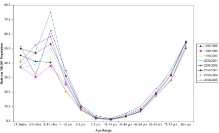
Figure 1.2 Invasive pneumococcal disease (IPD) incidence rate per 100,000 in England and Wales ... 4
Figure 1.3 Pathogenesis of pneumococcal disease ... 5
Figure 1.4 Pneumococcal mechanisms used to resist complement ... 13
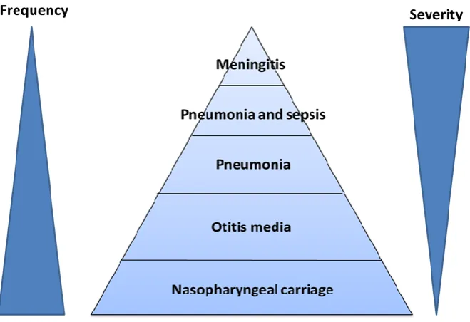
Figure 1.5 Burden of pneumococcal disease demonstrating the inverse relationship between frequency and severity ... 18
Figure 1.6 Overnight growth of S. pneumoniae on a blood agar plate ... 28
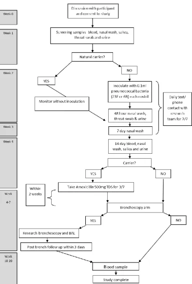
Figure 2.1 Flow chart of Dose-Ranging study appointments ... 43
Figure 2.2 Flow chart of Reproducibility study appointments. ... 45
Figure 2.3 Daily symptom log ... 48
Figure 2.4 Flow chart diagram of inoculum stock preparation ... 51
Figure 2.5 Miles and Misra bacterial quantification ... 52
Figure 2.6 Quantification of inoculum on blood agar plate ... 54
Figure 2.7 Inoculation of the nasal mucosa with 100 µl of S. pneumoniae ... 55
Figure 2.8 Nasal wash ... 56
Figure 2.9 Blood agar plate inoculated with nasal wash, without and with gentamicin added to the media ... 58
Figure 3.1 Experimental human pneumococcal carriage Dose-Ranging curve ... 75
Figure 3.2 Density of carriage is not a function of inoculated dose and shows stability over time ... 77
Figure 3.3 Density of carriage, when present, is stable up to one month post-challenge ... 79
Figure 3.4 Kaplan-Meier survival curve for time to carriage clearance ... 80
Figure 3.5 Percentage of volunteers with actively reported symptoms ... 86
Figure 4.1 Carriage density is similar between natural carriage and experimental carriage ... ... 95
Figure 4.2 Carriage density is similar between an initial experimental carriage episode and an experimental carriage episode following recent natural carriage ... 96
Figure 5.1 Transparent colonies are the dominant phenotype in the 6B inoculum stock ... 103
xx
Figure 5.4 The percentage of opsonophagocytic killing does not differ between the 6B and 23F strains ... 106
Figure 5.5 Genes unique to the 6B strain include those important for colonization ... 108
Figure 5.6 A 23F strain of pneumococci is less adherent to Detroit 562 epithelial cells in vitro than a 6B strain and a derivative 23F strain ... 110
Figure 5.7 AmiC is critical for colonization in a mouse model of colonization ... 111
Figure 6.1 Proportion of carriage positive volunteers detected by bacterial culture or qPCR over time ... 118
Figure 6.2 Correlation between bacterial culture and qPCR in quantifying S. pneumoniae in nasal wash samples ... 119
Figure 6.3 Culture and qPCR are complementary for following a carriage episode ... 121
Figure 7.1 Asymptomatic URT viral infections are associated with susceptibility to pneumococcal colonization but not increased density ... 131
Figure 7.2 Levels of mucosal FH are increased in individuals co-infected with virus and pneumococci and correlate with colonization density ... 133
Figure 7.3 Confirmation of epithelium inflammation ... 135
Figure 7.4 Pneumococcal epithelial adherence and internalization are increased in the presence of human nasal wash or FH ... 137
Figure 7.5 Binding of human FH to pneumococcus is mediated by PspC ... 139
Figure 7.6 Purified human anti-PspC IgG binds to pneumococcus and partially inhibits FH binding and adherence to human pharyngeal epithelial cells ... 141
Figure 7.7 PspC epitope mapping using sera of healthy adults exposed to pneumococcus reveals the lack of antibodies to the FH binding site ... 143
Figure 7.8 PspC epitope mapping using sera from mice immunized with recombinant PspC3 reveals the presence of antibodies to the FH binding site ... 144
xxi
Table 1.1 Summary of EHPC model applications ...34
Table 2.1 Summary of EHPC pilot studies ...40
Table 3.1 Average inoculation dose per group for both serotype 6B and 23F ...74
Table 3.2 Determination of serotype in a natural carrier challenged with serotype 6B ...81
Table 3.3 Passive symptom detection following challenge with the first five 6B doses and the first four 23F doses in the Dose-Ranging study ...83
Table 4.1 Details of volunteers re-challenged with serotype 6B ...93
Table 4.2 Reacquisition of carriage following heterologous experimental challenge ...94
Table 5.1 Five genetic lesions between P833 and P1123 resulting in an amino acid change .... ...109
Table 6.1 Comparison of bacterial culture and qPCR in detection of S. pneumoniae ...117
Table 6.2 Detection of pneumococci in nasal wash by bacterial culture and qPCR, categorized according to qPCR density ...120
Table 7.1 Association of virus presence and mucosal FH levels with carriage presence and carriage density ...134
1
Chapter 1
Introduction
1.1 Streptococcus pneumoniae
Streptococcus pneumoniae (pneumococcus) is an encapsulated Gram positive diplococcus with more than 90 structurally and serologically distinct pneumococcal serotypes (Figure 1.1). The serotype is determined by the polysaccharide capsule which is the outermost layer of the cell and is made of repeating units of simple sugars. Of the more than 90 described polysaccharide capsular types, all but four are negatively charged due to acidic sugars, phosphate, or pyruvate, with the remainder being neutral [1].
2
Figure 1.1:Photomicrograph of S. pneumoniae grown from blood culture (taken from CDC Public Health Image Library, image ID# 2896).
1.1.1 Epidemiology of pneumococcal carriage
Pneumococcal carriage rates are dependent on an individual’s age and vary by geographic area. In children, carriage rates range from 27% in developed countries to 85% in developing countries [3]. In developing countries, children can become colonized within days of birth. In The Gambia >80% of children have acquired pneumococcus within 33 days of birth [4]. In contrast, less than 50% of children from developed nations are likely to become colonized within the first year of life [5]. After the first year, carriage incidence rises steadily until around 4 years of age and then decreases to around 10% in adults [6].
Not only does carriage incidence decrease with age, but so also does the duration of carriage. Large variations in the duration of carriage have been observed in different cohorts. In a study done in neonates from Alabama in the 1970s, duration of carriage ranged from one to 17 months and, on average, the younger the infant was at the time of acquisition, the longer a strain was likely to be carried [7]. This trend was also observed in Sweden where the mean carriage duration of penicillin-resistant pneumococci in children under five was 43 days as compared to 25 days in those over the age of five [8].
3
1.1.1.1 Risk factors associated with carriage
Risk factors for pneumococcal carriage and disease are multi-factorial and can include crowding, the environment, and socio-economic factors [11-13].
1.1.1.1.2 Crowding
Crowding is very important in the spread of pneumococcus and particularly so in young children who are both in close contact with other young children and have a poorly developed immune system. In The Netherlands, children attending day care centres have been found to be at a 1.6- to 3.4-fold increased risk of nasopharyngeal colonization than those not attending [14].
1.1.1.1.2 Environment
Exposure to smoke increases the risk of pneumococcal carriage. Smoking, both passive and active, increases carriage in the smoking parent and the child exposed to the smoke [15-17]. In Australian Aboriginal adults, carriage is also associated with the frequency of sitting at an outside fire [18].
1.1.1.1.3 Socioeconomics
Socioeconomic factors can influence transmission. Overcrowded housing combined with inferior hygiene facilitates transmission, allowing pneumococcus to sustain in the community [18,19]. Aerosol transmission has been demonstrated in a ferret model where ferrets carrying S. pneumoniae were able to transmit the bacteria to other ferrets in the same cage or up to 1m away [20]. This could be comparable to the conditions people face in overcrowded, unsanitary housing.
In rural Alaskan communities, a lack of in-home running water has been associated with increased carriage in children under 10 [21]. It is suggested that conserving water for cooking and dish washing decreases handwashing, especially in children who need assistance. Hand contamination in Australian Indigenous children also correlates with carriage; Aboriginal children living in remote areas were 2 times more likely to be carrying pneumococcus and 8 times more likely to have hand contamination than children attending urban child-care centers [22].
4
1.1.2 Epidemiology of pneumococcal disease
[image:26.595.114.546.185.448.2]Similar to the pattern observed with carriage, IPD rates also correlate with age (Figure 1.2) and disease rates and mortality vary geographically, with the majority of deaths occurring in Asia and Africa.
Figure 1.2: Invasive pneumococcal disease (IPD) incidence rate per 100,000 in England and Wales. Reproduced with permission from Public Health England, July 2012.
Prior to the introduction of the conjugate vaccine, the incidence of IPD in children less than 2 years of age was 44.4/100,000 (range 11.3-104.4) per year in Europe and 167/100,000 in the United States [3,24]. In comparison, the incidence in the same population in Mozambique was 797/100,000. However, following the introduction of the conjugate vaccine IPD incidence dropped drastically in some countries. In the United States the overall incidence of IPD in children under 5 dropped to 23.6/100,000 by 2007 [25]. And in South Africa, the overall incidence of IPD in children under 2 declined from 54.8 cases per 100,000 to 17.0 cases in 2012 [26].
5 600,000 hospitalizations and 59,000 deaths [28]. As more and more of the general population reaches the age of 65, the burden of disease in this age group will only increase.
1.1.3 The association of pneumococcal carriage with disease
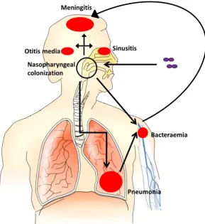
[image:27.595.172.462.256.572.2]The spectrum of disease caused by S. pneumoniae is wide and varied. Following colonization, the pneumococcus can spread and cause non-invasive illness, such as otitis media or sinusitis, or it can spread to the lungs, blood or meninges, causing pneumonia, bacteraemia, or meningitis. Disease does not occur without an initial colonization step (Figure 1.3).
Figure 1.3: Pathogenesis of pneumococcal disease.
1.1.3.1 Association between carriage and disease in animal models
6
1.1.3.2 Association between carriage and disease in humans
The first study in humans to show a link between recent acquisition of pneumococci and pneumococcal disease was done by Gray et al. who followed 82 infants from birth up to 2 years of age [7]. In these infants, serotypes causing disease were similar to commonly carried serotypes but disease was mainly associated with a newly acquired serotype; 74% of infections were caused by serotypes carried less than a month before disease. In a larger study of 329 children followed from 2 months to 2 years of age, pneumococcal acute otitis media was also associated with the newly acquired disease-causing serotype [34].
1.1.3.3 Association of serotype with the incidence of carriage and disease
The difference in disease virulence between serotypes is best illustrated by serotype 1 as it is a major cause of IPD and has been associated with outbreaks yet it is rarely found in healthy carriers [35]. This is in comparison to serotypes 6B and 23F which are frequently carried and have low invasive potential [36]. Sleeman et al. demonstrated that attack rate varies by capsule and an inverse relationship exists between attack rate and carriage duration [9].
One explanation for the differences in attack rate between serotypes is the capsular structure. Serotypes that are more heavily encapsulated are more resistant to neutrophil-mediated killing and persist for longer in the nasopharynx [37]. Pneumococcal surface charge has been shown to be one of the mechanisms behind this, with a more negative surface charge correlated with increased resistance to nonopsonic killing and increased carriage prevalence [38].
1.2 Mechanisms of S. pneumoniae carriage
For S. pneumoniae to access epithelial surfaces and establish carriage it must first reduce entrapment in the nasal cavity mucus secretions. This is achieved through expression of the polysaccharide capsule [39,40].
1.2.1 Expression of capsule during colonization
7 While sufficient capsule is needed to evade clearance mechanisms, excessive capsule has been shown to inhibit adherence to the epithelium [43,44]. To achieve a balance, the pneumococcus spontaneously switches its expression between two phenotypes - opaque and transparent - which are distinguished by differences in colony opacity [45].
Differences in colony opacity are due to the production of capsular polysaccharide; in the opaque form there is a 1.2- to 5.6-fold greater amount of capsular polysaccharide as compared to the transparent form [46]. The thinner capsule of the transparent phenotype provides better access to the epithelium and, compared to the opaque variant, enhances adherence to buccal epithelial cells [47]. This difference in colonization capability was demonstrated in infant rats where rats that received the transparent phenotype had higher rates of carriage than those that received the opaque phenotype [45]. In the rats that received the opaque inoculum, all colonies were transparent by 7 days post-inoculation. Hammerschmidt et al. used electron microscopy to visualize pneumococci during contact with epithelial cells and found that pneumococci in contact with, and starting to enter, cells lacked capsule [48]. Once pneumococcus has accessed the epithelial surface it uses surface proteins to bind to host cell-surface carbohydrates and proteins and initiate colonization.
1.2.2 Pneumococcal surface proteins involved in carriage
Underneath the capsule lies the cell wall which acts as an anchor for cell-wall-associated surface proteins responsible for assisting with pneumococcal adherence to cell-surface carbohydrates [49-51]. Adherence to the nasal epithelium is initiated when the pneumococci bind to cell-surface carbohydrates such as N-acetylglucosamine-β-(1,4)-galactose [52,53]. This initial binding is mediated by a number of bacterial structures, including phosphorylcholine (ChoP), choline-binding protein A (CbpA), and adhesins.
1.2.2.1 Choline-binding proteins mediate adherence
ChoP, a component of both cell-wall associated and lipotechoic acids, mediates adherence to the receptor for platelet-activating factor (rPAF) and is the anchor for a number of choline-binding proteins [49]. ChoP and these choline-binding proteins, including CbpA, are found in larger amounts on transparent pneumococci due to the increased amount of techoic acid in this phenotype [46,54]. This may account for the increased adherence and better colonization of the transparent phenotype.
8 mucosal barrier [55]. A PspC knockout mutant has shown reduced epithelial cell binding and decreased nasopharyngeal colonization when compared to wild-type [54]. In addition, PspC binds factor H (FH), a regulator of the alternative complement pathway, enabling the pneumococci to evade the host immune response and aiding adherence to epithelial cells [56-58].
Another choline-binding protein, pneumococcal choline-binding protein A (PcpA), is also able to facilitate pneumococcal adherence to nasopharyngeal and lung epithelial cells [59].
1.2.2.2 Lipoproteins mediate adherence to cell-surface carbohydrates
Pneumococcal lipoproteins are essential for both substrate transport and pneumococcal adherence. Pneumococcal surface adhesin A (PsaA) binds to E-cadherin, a transmembrane glycoprotein, and antibodies to PsaA have been shown to reduce pneumococcal adherence to nasopharyngeal epithelial cells [50,60]. Another group of lipoproteins, the Ami-AliA/AliB oligopeptide permease, was necessary for nasopharyngeal colonization in a mouse model [61]. Mutations in the ami locus have been suggested to decrease pneumococcal adherence to pulmonary epithelial cells as a result of reduced binding to a glycoconjugate receptor on lung epithelial or vascular endothelial cells [62].
1.2.2.3 Protease production increases attachment
Pneumococcal IgA1 protease has been shown to increase bacterial attachment to epithelial cells in vivo by cleaving human IgA1, causing a change in surface charge and resulting in ChoP interacting with rPAF [63].
1.2.2.4 Adhesins assist in binding to the extracellular matrix
Two adhesins important for the binding of pneumococci to the extracellular matrix are pneumococcal adhesion and virulence factor (PavA) and enolase. PavA, found on the outer cell surface, binds fibronectin and has been shown to be necessary for long-term carriage of S. pneumoniae D39 in a murine carriage model [64,65]. Enolase, an anchorless surface protein, binds plasminogen [66]. A third component of the extracellular matrix, vitronectin, was recently shown to interact with PspC, allowing pneumococci to use vitronectin as a molecular bridge and facilitating adherence [67,68].
1.2.2.5 Neuraminidase facilitates persistence in the nasopharynx
9 for pneumococcus [69,70]. The synergism that exists between pneumococci and respiratory viruses, such as influenza, in causing illness may be related to the action of neuraminidase and the increased adherence of pneumococci [71,72].
1.3 Immunity to S. pneumoniae
Innate immunity is the body’s first line of defence against a foreign pathogen. It is immediate, non-specific and evolutionarily conserved [73]. However, if the pathogen is able to evade or overcome the innate defences, the adaptive immune system is activated. Natural immunity against the pneumococcus is a complex interaction between the innate and adaptive immune systems.
1.3.1 Innate immunity to the pneumococcus
When the body first encounters a pathogen the aim is simply to prevent its entry. The epithelial surfaces of the body provide a physical barrier to stop the pathogen and the mucosal lining of the epithelium traps pathogens in mucus. However, the mucosal epithelium also secretes a number of chemicals that can kill pathogens or inhibit their growth.
1.3.1.1 Antimicrobial peptides against pneumococcus
10 cathelicidin LL-37 also acts by disrupting the bacterial membrane [83]. Pneumococci have been shown to induce hBDs in human pulmonary epithelial cells [84] and the pneumococcal toxin pneumolysin (PLY) has been shown to induce the release of LL-37 from human lung mast cells, which reduced pneumococcal viability [85].
In the lung, the pulmonary surfactant proteins A (SP-A) and D (SP-D), members of the collectin family, are found in the fluid that bathes the epithelial surfaces of the lung. SP-A and SP-D can bind and agglutinate a number of pathogens as well as act as opsonins [86]. In mice that were SP-D deficient, pneumococcal colonization and infection in the upper and lower respiratory tract increased [87]. The collectins, including SP-A and SP-D, are also known as pattern recognition receptors (PRRs) and they, along with numerous other PRRs, play a crucial role in host defence.
1.3.1.2 The role of pattern recognition receptors in defence against pneumococcus
When pathogens manage to breach the epithelium, the immune system must be able to distinguish self from non-self. To do this, it uses a variety of PRRs which can be expressed on the host cell surface, intracellularly, or secreted and which recognize highly conserved microbial structures known as pathogen-associated molecular patterns (PAMPs) [88]. PRRs include soluble receptors, like a number of components of the complement system, and membrane-bound receptors, including members of the Toll-like receptor (TLR) family, as well as the NOD-like receptors and cytosolic DNA sensors.
1.3.1.3 Complement system action against pneumococcus
The complement system consists of more than 30 circulating and membrane-bound proteins that, upon activation, initiate an enzyme cascade that eventually results in pathogen death. There are three overlapping pathways through which complement is activated: the classical pathway, the alternative pathway, and the lectin pathway. All three pathways converge on the activation of C3 to lead to the same effector molecules [89]. Complement activation results in cleavage of C3 to C3b which is deposited on the surface of the pathogen and processed to iC3b, both of which are important opsonins [90]. Pathogens are then eliminated either through opsonophagocytosis, membrane attack complex-mediated lysis, and/or the induction of inflammation.
1.3.1.3.1 Complement pathways involved in protection against pneumococcus
11 most important complement pathway for innate immunity to pneumococcus in mice and it is partially activated by the binding of natural IgM to bacteria [91]. However, there are other mechanisms that can activate the classical pathway during pneumococcal infection. These include the acute phase PRRs C-reactive protein (CRP) and serum amyloid protein (SAP), which bind ChoP [92-94], and SIGN-R1, a cell surface lectin, which binds capsule [95]. The alternative pathway is spontaneously activated at low levels but will only amplify when attached to a foreign surface. During pneumococcal infection, the alternative pathway is responsible for amplification of complement activation, leading to increased C3 deposition [91]. The pneumococcal cell wall can activate the alternative pathway, inducing the inflammatory reaction and cytokine production [96-98].
The lectin pathway is initiated by binding of the mannose-binding lectin (MBL), a soluble receptor, to PAMPs, mainly carbohydrates, on the microbial surface. There are two other recognition molecules, ficolin (1, 2 and 3) and collectin kidney 1 that, along with MBL, can form a complex with three MBL-associated serine proteases (MASPs) [99,100]. Until recently, the role of the lectin pathway in defence against pneumococcus appeared negligible. However, it has now been shown in mice that MASP-2, ficolin, and collectin are key PRRs that can activate complement via the lectin pathway and protect against pneumococcal disease [101,102].
1.3.1.3.2 The role of complement in the progression from carriage to disease
Humans with complement deficiencies are at an increased risk of IPD [103] but until recently it was not known if complement could prevent colonization from leading to sepsis. Using a murine model, Bogaert et al. showed that complement-depletion did not impact colonization density but mice were more likely to progress from carriage to disease than control or neutrophil-depleted mice [104]. This demonstrates a critical role for complement in stopping colonization from progressing to disease.
1.3.1.3.3 Pneumococcal mechanisms used to resist complement
Given the importance of complement in innate immunity, it is not surprising that the pneumococcus has developed a number of mechanisms to resist it (Figure 1.4).
1.3.1.3.3.1 Polysaccharide capsule
12 activity. C3b/iC3b opsonization by the alternative pathway and the conversion of C3b to iC3b on the bacterial surface were also impaired, as was phagocytosis mediated by complement, IgG, and nonopsonic phagocytic receptors [105].
1.3.1.3.3.2 Serotype
Different serotypes vary in their susceptibility to complement deposition and there exists an association between serotype prevalence in carriage and resistance to nonopsonic neutrophil-mediated killing [37,106,107]. Opsonophagocytosis can also vary between serotypes and has been linked with serotype-specific mortality [43,106,108]. However, serotype alone does not determine the effect of complement-mediated immunity, rather it is a combination of serotype and genetic background [106,109].
1.3.1.3.3.3 Proteins
A number of pneumococcal proteins also inhibit complement-mediated immunity. It has long been known that PspA inhibits complement activation but only recently was it discovered that it does so by blocking CRP binding to ChoP, thereby decreasing complement activation [110-112].
PspC mediates immune evasion by binding human FH and C3 [113,114]. The interaction between FH and PspC protects the pneumococcus from complement-mediated clearance [115]. However, FH binding varies by serotype and is negatively correlated with C3b/iC3b deposition, with serotype invasiveness associated with high levels of FH binding and resistance to complement [116].
13
Figure 1.4: Pneumococcal mechanisms used to resist complement.
1.3.1.3.3.4 Biofilm
Biofilms are increasingly being studied as they are known to be important in resistance to host-defence and antimicrobials. For S. pneumoniae, in vitro growth as a biofilm has been shown to impair C3b deposition and target both the classical and alternative pathways through impaired activation of CRP and C1q and increased recruitment of FH [120].
1.3.1.4 Toll-like receptors involved in protection against pneumococcus
As PRRs, the role of TLRs is to detect pathogens and initiate the immune response by leading to the production of mediators such as cytokines and chemokines. There are 10 known TLRs in humans, of which three have been described in protection against the pneumococcus.
1.3.1.4.1 TLR2
14 carriage and disease [125,126]. In TLR2-deficient mice, clearance of colonization is impaired and susceptibility to pneumococcal meningitis is increased, with intensified disease severity and bacterial numbers [127-129]. However, the importance of TLR2 does not extend to protection against pneumococcal pneumonia [130].
1.3.1.4.2 TLR4
TLR4 is also found on the cell surface and has been shown to interact with PLY [131]. However, its role in carriage clearance is unclear; Malley et al. [132] found TLR4-deficient mice had increased colonization density and risk of disease however, van Rossum et al. [127] saw no difference between wild-type and TLR4-deficient mice. In vivo disease studies have found TLR4-deficient mice have less upper respiratory tract (URT) cell apoptosis and higher bacterial loads in their lungs [131,133]. Recently, McNeela et al. [134] found that PLY directly activates innate immune cells, independently of TLR4, and there is no interaction of TLR4 with PLY.
1.3.1.4.3 TLR9
TLR9 is expressed within endosomal compartments and plays a protective role in the lung; TLR9-deficient mice are more susceptible to pneumococcal pneumonia and have an increased bacterial load in the lung [135].
TLRs are a crucial link between innate and adaptive immunity. TLR recognition of a PAMP induces production of inflammatory molecules through the activation of an intracellular pathway including nuclear factor-κB, mitogen-activated protein kinases and interferon-regulatory factors which initiate the activation of the adaptive immune response [136].
1.3.2 Adaptive immunity to the pneumococcus
Although slow to start, the adaptive immune system is highly specific and has memory, and is comprised of a humoral response and a cell-mediated response.
1.3.2.1 Humoral immunity against pneumococcus
15 In the mucosa, the dominant antibody is IgA and it is thought to mediate protection by blocking adherence to the host tissues. There are two forms of IgA, IgA1 and IgA2, with IgA1 making up approximately 90% of the total IgA in the URT [142]. A number of common respiratory pathogens have an IgA1 protease which, in the case of S. pneumoniae, cleaves human IgA1, increasing adherence to respiratory epithelial cells in vitro and preventing phagocytic killing [63,143] .
1.3.2.1.1 Agglutination of pneumococcus
Another important role for antibodies in the mucosa is agglutination of bacteria into a cluster that can then be cleared by the host. This was recently demonstrated in mice where, following passive immunization with IgG and intra-nasal challenge with pneumococcus, colonization was prevented, as was transmission between littermates [144]. When mice were immunized with human IgA1, colonization was only prevented following challenge with an protease-deficient mutant; challenge with an IgA1-protease-producing strain did not result in agglutination, again highlighting the role of pneumococcal virulence factors in evasion of the host immune response.
1.3.2.2 Cellular immunity against pneumococcus
If a pathogen crosses the epithelial barrier, it will often be immediately recognized by a phagocyte. Macrophages are a phagocytic cell found throughout the body, including the lung, and are derived from monocytes. Upon activation they recruit another phagocyte, the neutrophil, which is a short-lived cell found in the blood. Macrophages and neutrophils display a number of cell-surface receptors which recognize the surface molecules of pathogens, leading to phagocytosis.
1.3.2.2.1 Macrophages
In mice, recruitment of monocytes/macrophages to the upper airway cleared pneumococcal colonization by phagocytosis [145]. The retention of macrophages in the nasopharynx was the result of an innate cytokine, macrophage migration inhibitory factor (MIF). In MIF-deficient mice, carriage was prolonged and was correlated with reduced numbers of macrophages [146]. MIF activity has also been shown to inhibit alveolar macrophage migration in a rabbit model of pneumococcal pneumonia [147].
16 critical for bacterial clearance [148]. At this point the role of the macrophage changes and it begins to regulate the immune response through the production of cytokines.
1.3.2.2.2 Neutrophils
Neutrophils are crucial for phagocytic clearance of pneumococci. During pneumococcal colonization in mice, there is an influx of neutrophils into the nasal space [127]. When these neutrophils interact with pneumococcal PLY, the resulting neutrophil lysis enhances the delivery of pneumococcal antigen to the nasal-associated lymphoid tissue [149]. This neutrophil lysis combined with pneumococcal degradation by neutrophils alone results in rapid clearance of the pneumococcus from the nasopharynx.
The importance of neutrophils in carriage clearance also extends to disease. In a mouse model of colonization, neutrophil depletion resulted in a 50% death rate with a normally asymptomatic carriage strain [149]. In models of disease, depletion of neutrophils in mice increased susceptibility to pneumococcal infection [150,151].
1.3.2.2.3 CD4+ T cells
Trzciński et al. [141] and McCool et al. [140] have found that protection against colonization and clearance of carriage using murine models, respectively, were independent of antibody. Other studies have shown that in mice lacking immunoglobulin, intranasal immunization with a killed, nonencapsulated whole cell vaccine, a cell wall polysaccharide, or a selection of pneumococcal proteins, protected against colonization [152-154]. Further examination demonstrated that both protection from, and clearance of, carriage required CD4+ T cells in these models [141,153,155]. The CD4+ T cell subset responsible for protection against colonization in mice was shown to be Th17 [156].
1.3.2.2.4 Th17 cells
Interleukin-17A (IL-17A), a cytokine that induces neutrophil recruitment and activation, is secreted by the Th17 subset of CD4+ T helper cells. In mice, expression of IL-17A was correlated with increased clearance of a secondary carriage episode and levels in the blood of mice immunized with killed pneumococcal whole cell antigen prior to challenge were inversely correlated with carriage density [156]. Protection against colonization was neutrophildependent and recombinant human IL17A enhanced antibodydependent and -independent pneumococcal killing in vitro [156].
17 was dependent on the production of IL-17A-expressing CD4+ T cells via TLR2 signalling. In mice that had previously been colonized, clearance was enhanced by an influx of neutrophils.
1.3.3 The effect of carriage on defence against future carriage and
disease
Although colonization can generate an immune response that, in the majority of the population, prevents disease and leads to clearance of carriage, it is still not clear how this impacts on subsequent carriage and disease. In a mouse model of carriage, mice that had cleared carriage and were re-challenged with the same strain had similar colonization density to mice that were not initially colonized, even though they had generated a whole immunoglobulin response to the strain [140]. In contrast, a separate study found that when mice were challenged following carriage, mucosal protection and protection from sepsis required both CD4+ T cells and antibody [158].
Immunological protection against subsequent disease following carriage has also been demonstrated in mice; carriage of a PLY negative mutant strain significantly increased survival following invasive pneumococcal challenge and also resulted in cross-serotype protection [159]. In a pneumonia challenge model, prior pneumococcal colonization prevented bacteraemia by antibody-mediated phagocytosis which led to protection from lethal pneumonia [160].
In humans, carriage is known to be an immunizing event [161]. In the absence of infection, carriage has been shown to induce immunoglobulins against pneumococcal capsular polysaccharide and proteins in the serum [137,162]. In Gambian infants that were followed longitudinally, the rate of reacquisition and the duration of carriage, even with the same serotype, did not differ [4]. However, in this natural setting where sampling is intermittent, it is possible that carriage episodes can be misclassified or missed completely.
1.4 Vaccination against S. pneumoniae
18
Figure 1.5: Burden of pneumococcal disease demonstrating the inverse relationship between frequency and severity.
1.4.1 Early attempts at pneumococcal vaccination
The first vaccine against the pneumococcus consisted of heat killed, locally isolated, uncharacterized pneumococcal strains that had been suspended in saline and were given subcutaneously. The first clinical trials were conducted in 1911 in South Africa among mine workers where pneumococcal pneumonia had a case fatality rate of 20 to 40% [168]. These first whole cell vaccine trials had vaccine efficacies ranging from 25% to 82% and were generally considered to have been positive [169,170]. This was an important period in pneumococcal vaccine development as vaccine preparation, administration and dosage methods were developed, as were methods for detecting and measuring protective immunity.
1.4.2 Pneumococcal polysaccharide vaccines
19 When the widespread availability and overuse of penicillin led to a dramatic increase in pneumococcal antibiotic resistance, interest in developing new pneumococcal vaccines was renewed. Based on the study in 1945 that found links between immunization with pneumococcal polysaccharide capsule and protection from pneumonia [171], work was done to develop a new vaccine against the pneumococcus.
In 1977 a 14-valent polysaccharide vaccine was licensed for use in adults in the United States. This was expanded to a 23-valent pneumococcal polysaccharide vaccine (PPV23) in 1983 which is currently licensed for use in adults and in children over the age of 2 with underlying medical conditions. A 0.5 ml dose of the vaccine contains 25 μg of purified capsular polysaccharide from each of 23 serotypes (1, 2, 3, 4, 5, 6B, 7F, 8, 9N, 9V, 10A, 11A, 12F, 14, 15B, 17F, 18C, 19F, 19A, 20, 22F, 23F, 33F) and includes the most common drug-resistant serotypes [172].
1.4.2.1 Pneumococcal polysaccharide vaccine efficacy
Despite numerous trials over many years, the effectiveness of PPV23 in protection against pneumonia and IPD is controversial and inconclusive. It is difficult to measure the efficacy and effectiveness because of the low frequency of invasive infection, the inaccuracy of the diagnostic criteria for pneumococcal pneumonia and the variation with age and underlying illness.
A recent meta-analysis that looked at the efficacy of PPV23 in adults found the relative risk for presumptive pneumococcal pneumonia to be 0.64 [95% Confidence Interval 0.43-0.96] but with a significant heterogeneity between trials (P<0.001) due to variable trial quality [173]. In trials of higher quality there was little evidence of vaccine protection among the elderly and at-risk groups (1.04 [0.78-1.38]). These findings suggest that pneumococcal vaccination is not effective in preventing pneumonia, even among the groups for whom the vaccine is recommended.
20
1.4.5 Pneumococcal conjugate vaccines
Conjugation of purified pneumococcal polysaccharide to a nontoxic carrier protein was based on the success of the Haemophilus influenzae type B vaccine where use of a conjugate vaccine lead to a rapid reduction in disease [174]. The first pneumococcal conjugate vaccine on the market was the 7-valent pneumococcal conjugate vaccine (PCV7) (Prevnar) which offered protection against serotypes 4, 6B, 9V, 14, 18C, 19F, and 23F [175]. In 2009, conjugate vaccines containing 10 (Synflorix, PCV10) or 13 (Prevnar, PCV13) capsular polysaccharides were introduced and have led to the gradual replacement of PCV7. Serotypes 1, 5, and 7F have been added to both PCV10 and PCV13; PCV13 also includes 3, 6A, and 19A.
PCV10 and PCV13 cover >70% of serotypes that cause IPD in children from all geographic regions [3]. Both have been licensed to protect against IPD, pneumonia and acute otitis media in infants and children from 6 weeks of age to 5 years. PCV13 has also been licensed for the prevention of pneumococcal disease in adults over 50 years of age [176].
1.4.5.1 Pneumococcal conjugate vaccine efficacy
Pneumococcal conjugate vaccines are protective against invasive pneumococcal diseases caused by vaccine serotypes [25,177]. Mucosal and systemic anti-capsular antibody responses are induced by the vaccine [178] leading to protection from invasive disease, however, the vaccine is less effective against mucosal diseases such as otitis media and pneumonia.
1.4.5.1.1 Immune correlates of protection
21
1.4.5.1.2 Protection against carriage
The impact of conjugate vaccines on carriage has been shown in a number of populations. In American Indian infants, a colonization study nested within a phase 3 efficacy trial showed a decrease in vaccine-type carriage after the primary vaccination at 7 and 12 months [181]. In another nested carriage study, Gambian children were also protected against vaccine-type carriage following vaccination [182].
In both of these populations there was also a decrease in carriage density following PCV7 vaccination, with the decrease in rural Gambia seen across different age groups [181,183].
1.4.5.1.3 Protection against invasive disease
The real benefit of conjugate vaccines has been in reduction of invasive disease. In the first PCV7 trial in Northern California, invasive disease caused by vaccine serotypes was reduced by 87.3% in children under 1, by 58.1% in children under 2 and by 62.4% in children less than 5 years of age [177]. Similar decreases have been seen all over the world including South Africa where the vaccine efficacy in infants was 83% [184].
1.4.5.1.4 Protection against mucosal disease
Conjugate vaccines are less effective against mucosal disease. This may be a result of difficulty in diagnosing pneumonia or it could be due to differences between mucosal and systemic defence.
In a study of Finnish infants, those vaccinated with PCV7 had a 6% reduction in the number of acute otitis media episodes as compared to those not vaccinated [185]. The actual incidence of acute otitis media due to vaccine serotypes was reduced by 57%. This is similar to the 64% reduction in vaccine serotype otitis media in American Indian infants following vaccination with PCV7 [186].
All-cause pneumonia admissions were reduced in children under the age of 2 in both the United States and parts of Europe following vaccination with PCV7 [187,188]. In Gambian children aged 6-51 weeks, the efficacy of PCV9 was 37% against radiologically confirmed pneumonia and 16% against mortality [189]. In Filipino children under 2, PCV11 reduced community-acquired radiologically confirmed pneumonia by 23% [190].
1.4.5.1.5 Indirect effects of pneumococcal conjugate vaccination