Open Access
Methodology
A novel assay for analysis of the regulation of the function of human
osteoclasts
Barrie Kirstein
1, Urszula Grabowska
2, Bertil Samuelsson
2, Masahiro Shiroo
2,
Timothy J Chambers*
1and Karen Fuller
1Address: 1Department of Cellular Pathology, St George's, University of London, London SW17 0RE, UK and 2Medivir UK, Little Chesterford, Essex
CB10 1XL, UK
Email: Barrie Kirstein - [email protected]; Urszula Grabowska - [email protected]; Bertil Samuelsson - [email protected]; Masahiro Shiroo - [email protected]; Timothy J Chambers* - [email protected]; Karen Fuller - [email protected]
* Corresponding author
Abstract
Background: Very little is known of the regulation of the function of human osteoclasts, largely due to the virtual impossibility of obtaining human osteoclasts ex vivo. It has recently become possible to generate human osteoclasts in vitro, by incubation of peripheral blood mononuclear cells (PBMCs) in macrophage colony-stimulating factor (M-CSF) and receptor activator of nuclear factor-κB ligand (RANKL). However, the assays at present available do not distinguish clearly between the distinct effects of agents on differentiation and function.
Materials and methods: We developed a novel assay for resorptive function of human osteoclasts that minimizes inter-assay variability by using each culture as its own baseline, and that minimizes the confounding effects of agents on differentiation by assessing resorptive function over a short test period. In this assay, the development of resorptive activity is monitored in sample cultures. When resorption is underway, bone resorption (measured as the release of the C-terminal telopeptide degradation product of type I collagen (CTX-I) into the supernatant) is compared before vs after incubation for 1–24 h in test agent.
Results: Using this assay, we found that changes in bone resorption could be detected using substantially fewer cultures per variable. Moreover, we could detect effects of agents on resorption within 1 h of addition, a time sufficiently short that a change in release is likely to reflect an effect on function rather than on differentiation.
Conclusion: The assay makes it possible to distinguish the effects of agents on osteoclastic function, independent of their effects on differentiation.
Background
The maintenance of skeletal integrity depends on contin-ual resorption of bone by osteoclasts and its replacement by osteoblasts. Recently, there have been considerable
advances in our understanding of the mechanisms through which osteoclast formation is regulated [1-3]. In contrast, little is known of the mechanisms that modulate their activity once formed, even although this is a major
Published: 07 November 2006
Journal of Translational Medicine 2006, 4:45 doi:10.1186/1479-5876-4-45
Received: 21 September 2006 Accepted: 07 November 2006
This article is available from: http://www.translational-medicine.com/content/4/1/45
© 2006 Kirstein et al; licensee BioMed Central Ltd.
component of the regulation of bone resorption. Thus, after systemic administration of hormones such as PTH or CT, osteoclasts show morphological evidence of an increase or decrease in activity, with a corresponding change in plasma calcium concentration, within 30 min, while a change in osteoclast number is not detectable until 24 h later [4]. This shows that bone resorption is reg-ulated not only through modulation of the number of osteoclasts but also by modulation of the resorptive activ-ity of existing osteoclasts. It seems likely that agents exert differential actions on these distinct processes.
It is virtually impossible to obtain human osteoclasts ex vivo with which to address this question. In their absence, human osteoclastic cells can be generated in vitro by incu-bation of peripheral blood mononuclear cells (PBMCs) in macrophage colony-stimulating factor (M-CSF) and receptor activator of nuclear factor-kB ligand (RANKL) on plastic culture surfaces or on bone/dentine slices [5,6]. However, although such culture systems provide powerful insights into the regulation of osteoclastic differentiation, they do not clearly distinguish between the effects of agents on differentiation and function. For example, resorption of bone slices in such culture systems is typi-cally observed after 14–21 days of incubation [7-10]. If a putative resorption modulator is added to such cultures for a brief period, effects on resorption, classically meas-ured as the area of bone surface excavated, will be observed against a baseline of prior resorption; and if the modulator is added over a longer period, it will be diffi-cult to distinguish effects on function from those on dif-ferentiation.
Recently, it has become possible to measure bone resorp-tion as the release into culture supernatants of products of bone solubilization, the concentration of which has been shown to correlate with bone resorption [11]. This approach has the advantage that the amount released reflects the amount of bone resorbed since the last change of culture medium. This avoids the results being masked by the baseline of prior resorption. However, even in such assays, bone resorption is measured over a period of 3 days [12,13], so that an unknown and potentially sub-stantial component of an observed change in resorptive activity might have been due to an effect of the test agent on differentiation rather than function.
The difficulties in the interpretation of resorption data mentioned above are compounded by the length of time it normally takes for osteoclastic differentiation to occur: the long incubation times magnify small initial differ-ences between cultures, and in our experience can lead to substantial inter-culture variability. This variability increases the number of cultures required per variable, and because relatively small numbers of monocytes are
available from a given donor, the number of variables that can be studied in each experiment is in practice severely limited.
We therefore developed a novel assay that minimizes the confounding effects of agents on differentiation by meas-uring resorption over a shorter period; and that minimizes inter-culture variability by using each culture as its own baseline. In this assay, sample cultures are inspected to monitor the development of actively resorbing osteo-clasts. When resorption is underway, release of products of bone resorption is compared before vs after incubation for 1–24 h with test agent. Using this approach, we found that changes in bone resorption could be detected using substantially fewer cultures per variable; and that effects of agents on resorption could be detected within 1 h of addition, a time sufficiently short that a change in release is likely to reflect an effect on function rather than on dif-ferentiation.
Materials and methods
Media and reagents
Cells were incubated in MEM with Earle's salts (Invitro-gen, Paisley, UK), supplemented with 10% fetal calf serum (FCS), 2 mM glutamine, 100 IU/ml
benzylpenicil-lin, and 100 μg streptomycin (all Sigma, Poole, Dorset,
UK) (MEM/FCS). Recombinant human M-CSF and solu-ble recombinant murine RANKL were from Insight Bio-technology (Wembley, Middlesex, UK). The specific cathepsin K inhibitors MV061194, MV061748, MV061940, MV061645 and MSX-081 were provided by Medivir UK (Cambridge, UK). Cortical bovine bone slices (4 × 4 × 0.1 mm) were prepared as previously described [14]. Salmon CT, E64 and all remaining reagents were from Sigma unless otherwise stated.
Osteoclast generation
Heparinized blood was obtained from healthy human male or female volunteers (aged 22 – 57) with the consent of St George's Ethical Committee. The blood was layered over Histopaque-1077 and centrifuged for 30 min at 400 g. The opaque interface containing mononuclear cells (PBMCs) was collected with a pasteur pipette, washed in
PBS, then resuspended in MEM/FCS. 5 × 10⁵ cells were
added per well to a 96-well plate, each well of which con-tained a bone slice. The cultures were incubated in a total
volume of 200 μl MEM/FCS containing M-CSF (50 ng/ml)
and RANKL (30 ng/ml) for 24 h at 37°C in 5% CO2 in
To monitor cultures for the development of resorptive function, sample bone slices were removed at intervals, fixed in 10% formalin and stained using toluidine blue (0.1% for 1 min). Bone slices were then inspected by light microscopy for the presence of osteoclasts and excava-tions (see Fig. 1). When excavation was deemed sufficient, the remaining cultures were subjected to the osteoclast resorption assay.
Osteoclast Resorption Assay
After generation of resorptive osteoclasts (as detected above), bone slices were removed from the 24-well plates, washed in PBS, placed in the wells of a 96-well plate, and
incubated for 2 or 24 h in 100 μl of fresh MEM/FCS
con-taining M-CSF and RANKL. All the culture medium was removed after this period of incubation and stored frozen. Bone slices were washed again in PBS and transferred to new 96-well plates, the wells of which contained 100 μl of fresh MEM/FCS containing M-CSF and RANKL, together with test agent or vehicle control unless otherwise stated. Supernatants were collected and stored frozen after a fur-ther 1–24 h of incubation.
The resorption activity of the cultures was determined by quantifying the C-terminal telopeptide degradation
prod-uct of type I collagen (CTX-I/CrossLaps®) (Nordic
Bio-science Diagnostics A/S, Herlev, Denmark) in the culture supernatants. Enzyme-linked immunosorbent assay
(ELISA) was performed according to the manufacturer's instructions.
In some experiments, when the resorption assay had been completed, bone slices were further analyzed for visuali-zation of cells and excavations using toluidine blue, tar-trate-resistant acid phosphatase (TRAP) cytochemistry and reflected light or scanning electron microscopy. In other experiments, the total quantity of TRAP present in the cells at the end of the culture was assessed by enzyme assay of TRAP in cell lysates.
Reflected Light and Scanning Electron Microscopy
After incubation, bone slices were immersed in 10% (v/v) sodium hypochlorite for 10 min to remove cells. The bone slices were then washed in water, air-dried, mounted and sputter-coated with gold. Bone slices on stubs were inspected in a scanning electron microscope (S90: Cam-bridge Instruments, CamCam-bridge, UK). The extent of bone resorption on bone slices mounted onto microscope slides was quantified by counting the number of grid intersections in an eyepiece graticule that overlay an area of bone resorption using reflected light microscopy.
Enzyme Assay for TRAP
After removal of supernatant, bone slices were washed in PBS, transferred to fresh wells of a 96-well plate and
incu-bated in 100 μl 0.1% Triton in water (v/v) for 10 min.
[image:3.612.65.552.441.626.2]Osteoclast formation from human PBMCs Figure 1
TRAP enzyme activity was measured by the conversion of
p-nitrophenyl phosphate to p-nitrophenol in the presence of sodium tartrate. 80 μl of each lysate, diluted appropri-ately, was added to 96-well plate wells containing 80 μl 0.09 M citrate buffer (pH 4.8) with 20 mM phosphatase substrate and 80 mM tartaric acid and incubated at room temperature for 40 min. The reaction was stopped by
addition of 40 μl of 0.5 M sodium hydroxide. Optical
absorbance was measured at 405 nm on an Opsys MR plate reader (Thermo Electron Corporation, Basingstoke,
Hampshire, UK) against a standard curve of p
-nitrophe-nol.
Statistical Analysis
All data are expressed as mean ± standard error of mean, of four replicate cultures, unless otherwise stated. The sig-nificance of differences between control and experimental groups was assessed by Student's t test or ANOVA (Dun-nett's Multiple Comparison Test). Differences were con-sidered significant if p < 0.05
Results
Optimization of osteoclast formation and bone resorption
Previous assays have used an incubation time of three days to measure CTX-I release [12,13]. To distinguish between actions of agents on the differentiation and func-tion of osteoclasts requires as short a sampling period as possible, and this in turn depends upon optimizing the rate of bone resorption in the cultures. The optimal den-sity of cells for osteoclast generation from human PBMCs has been established [6,8]. It is also essential to optimize choice of donor, because there is considerable variation in the ability of PBMCs from different donors to generate osteoclasts. We also found that efficient osteoclastic differ-entiation was supported by only a minority of serum batches. To optimize the assay, our strategy was to test the ability of 4–5 serum batches to support osteoclast forma-tion from the blood of 4–5 donors. This approach identi-fied a single combination of donor and serum that efficiently generated osteoclasts. We found that the batch of serum so identified as optimal was able to similarly support efficient osteoclast formation in approximately 20% of donors; and that donors showed a reproducible ability for the efficient generation of osteoclasts. Thus, in our experience, some donors seemed able to generate only small or very small numbers of osteoclasts whatever serum batch was used; while some sera did not support osteoclast formation from any of the donors. Optimal combinations of donor and serum were thus identified that generated cultures in which virtually all cells were multinuclear, and almost the entire bone surface was excavated within 7 days (see Fig. 1)
Optimization of resorption assay
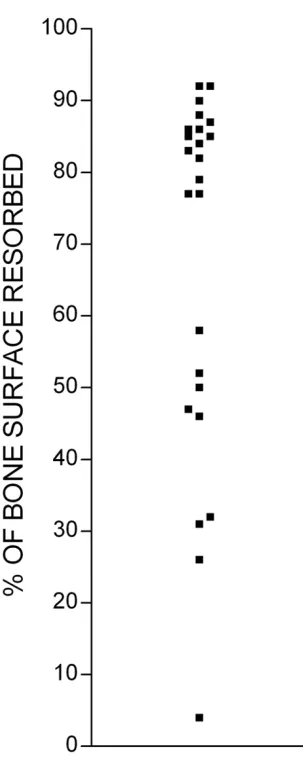
We noted that there was frequently considerable variation in the extent of bone resorption on different bone slices incubated under identical conditions in the same experi-ment (Fig. 2). This variability might reflect variation in the properties of individual bone slices. An alternative expla-nation is that variability might be due to the smallness of the numbers of osteoclast precursors in our cultures: these are known to represent a very small proportion (<1%) of PBMCs [15], and if the number of precursors in each cul-ture is very small, random variation in this number could lead to appreciable inter-culture differences in osteoclast formation. A further possibility is that osteoclast forma-tion in such cultures might reflect a cell-density depend-ent compondepend-ent in the differdepend-entiation of PBMCs to osteoclasts, which magnifies small starting differences in the number or distribution of PBMCs. Whatever the explanation for the variability, it represents an obstacle to the practical application of assays of osteoclast differenti-ation and function.
We hypothesized that the effect of inter-culture variability could be minimized by a longitudinal rather than cross-sectional assay design. These alternative approaches were compared in an assay using an inhibitor of cathepsin K, an enzyme crucial to the degradation of collagen during bone resorption by osteoclasts (Fig. 3). The inter-culture variability in CTX-I levels in samples taken after the initial 24 h incubation period lead to considerable variation in the means for each group (Fig. 3A). This inter-culture var-iability confounds the dose-response analysis derived from the CTX-I concentrations obtained after incubation in the inhibitor (Fig. 3B). However, the effect of inter-cul-ture variability is reduced when the initial 24 h period is taken as a baseline for the second sample from the same culture (Fig. 3C). Further examples of the application of this approach to the assessment of the potency of inhibi-tors of bone resorption are shown in Fig. 4.
To assess the relative statistical power of the longitudinal
vs the cross-sectional approach, we used data from the
control groups of assays similar to those shown in Fig. 3. We compared the power of CTX-I data derived from the longitudinal measurements with the power of data derived from the second 24 h incubation alone. We found, in data from 6 consecutive assays, that to have a 95% chance of detecting a change in the mean of 50% required 25 (± 20) cultures if a single CTX-I measurement is used for each culture, but only 3.4 (± 2.1) cultures if cul-tures are sampled longitudinally.
reflects the resorptive capacity of cultures. We therefore tested the relationship between the first of the pair of CTX-I readings, and the TRAP in the lysate of the corresponding culture after incubation in either control medium or the
cysteine protease inhibitor E64 (10-7 M – 10-5 M). We
found a strong correlation between initial CTX-I and TRAP in cultures incubated in control medium (r2 = 0.96)
(n = 24), but the correlation was less clear for cultures
incubated in E64 (r2 = 0.52) (n = 24). This conforms to
our previous experience, that the TRAP content of cells is influenced by agents that modulate bone resorption [16]: TRAP is released during bone resorption, and perturba-tion of resorpperturba-tion increases or decreases the amount that remains in the cell. Thus, measurement of the TRAP con-tent of lysates is an unreliable substitute for the initial CTX-I measurement.
We tested the ability of the assay to detect changes in bone resorption over shorter time periods (Fig. 5). In these experiments, bone slices were washed after osteoclasts had formed, transferred to new 96-well plates, and incu-bated in fresh medium containing M-CSF and RANKL. After 2 h of incubation this supernatant was removed for CTX-I assay, and replaced with medium containing con-trol or test reagents, and incubated for a further 1 h. This supernatant was then itself removed for assay, and the process repeated with further incubation periods of 1 and 2 h. We found that suppression of bone resorption could be detected within 1 h of incubation of osteoclasts in
MV061194 (3 × 10⁻⁷ M), a cathepsin K inhibitor, and
within 2 h of incubation in salmon CT (10 ng/ml). This result additionally suggests that inhibition of bone resorp-tion by the cathepsin K inhibitor has a more rapid onset than inhibition by CT. This might be because intracellular degradation of endocytosed matrix fragments continues to completion despite cell-inhibition, while the effects of the (membrane-permeant) enzyme inhibitor are essen-tially immediate.
Discussion
Very shortly after administration of hormones such as CT
or PTH, osteoclasts show morphological evidence in vivo
of changes in functional activity that correspond to changes in plasma calcium concentration [4]. Only much later do osteoclast numbers change. Thus, modulation of the activity of existing osteoclasts is a major component of the regulation of bone resorption. Recently, significant advances have been made in elucidating the mechanisms that govern osteoclastogenesis [1,3,17]. In contrast, much less is known about how the resorptive activity of osteo-clasts is regulated.
This is because human osteoclasts are essentially unavail-able ex vivo. In their absence, osteoclasts can be generated
in vitro, but osteoclast formation from human PBMCs typ-ically takes 14–21 days [7-10]. Thus, if a putative resorp-tion modulator is added to such cultures for a brief period, effects on resorption, measured as the area of bone surface excavated, will be observed against a high baseline of prior resorption; and if the modulator is added over a longer period, any change in resorption might be second-ary to effects on differentiation.
[image:5.612.86.238.114.494.2]Quantification of bone resorption on individual bone slices in the same experiment
Figure 2
This problem is partly circumvented in assays that meas-ure bone resorption as the release into cultmeas-ure superna-tants of products of bone solubilization [12,13,18]. This approach has the advantage that the quantity of solubi-lized product reflects only the quantity of bone resorbed since the last change of culture medium. This avoids the results being obscured by a baseline of prior resorption. However, even in such assays, bone resorption is meas-ured over a period of 3 days, so that an unknown and potentially substantial component of any observed change in resorptive activity might have been due to an effect of the test agent on differentiation rather than func-tion.
A second difficulty is the substantial inter-culture variabil-ity in osteoclast formation, which might be accentuated by the prolonged incubation required to form osteoclasts from PBMCs: long incubation times might magnify small initial differences between cultures, especially since cells capable of forming osteoclasts represent only a very small proportion (<1%) of PBMCs [15], so that random varia-tion in this number could lead to appreciable inter-culture differences in osteoclast formation. High inter-culture var-iability requires more cultures per experimental group, and this restricts the number of groups that can be studied in each experiment.
We therefore developed a novel assay that, by addressing these difficulties, facilitates the measurement of osteoclast function independent of differentiation. First, we opti-mized bone resorption, since if there is more bone resorp-tion it can be measured over a shorter period. Short
measurement periods minimize the confounding effects of agents on differentiation. We optimized bone resorp-tion by optimizing combinaresorp-tions of serum batches and PBMC-donors. This led to substantially greater and earlier release of CTX-I antigen, to levels an order of magnitude greater than previously reported [10,12,13]. This robust osteoclast formation enabled us to detect and measure CTX-I released in 1 h of incubation, a time sufficiently short that any change in resorption is likely to reflect an effect on osteoclastic function rather than on differentia-tion. Second, we minimized inter-culture variability by using each culture as its own baseline. Using this longitu-dinal approach, we found that changes in bone resorption could be detected using substantially fewer cultures per variable.
In view of the significant expense of longitudinal meas-urements of CTX-I, we tested whether measurement of the quantity of TRAP in the cell lysates at the termination of the experiment could substitute for the initial CTX-I assay. We found that, while TRAP correlated well with the initial CTX-I reading in control cultures, the correlation in exper-imental groups in which bone resorption was modulated was unreliable. We have previously noted that agents that modulate resorption also modulate lysate TRAP levels [16]. However, since TRAP release by resorbing osteoclasts is proportional to the duration of bone resorption, meas-urement of TRAP might more accurately reflect the osteo-clast content of the culture in experiments in which resorption is measured over shorter time intervals.
[image:6.612.71.529.96.273.2]CTX-I release in the 24 h period before ('basal') (A) and after (B) addition of an inhibitor of cathepsin K, and the same data expressed (C) as CTX-I released after, as a percentage of that released in the same culture before, incubation in inhibitor Figure 3
Application of assay to assessment of potency of cathepsin K inhibitors Figure 4
Conclusion
The short incubation times and the longitudinal sampling makes the assay we have described a powerful tool with which to detect and quantify the effects of agents that acti-vate or modulate the resorptive activity of osteoclasts, while minimizing the potentially confounding effects of the same agent on osteoclastic differentiation. The assay design also reduces the inter-culture variability of resorp-tion data, compared to previous approaches.
Competing interests
UG and MS are employed by Medivir UK; BS is employed by Medivir AB. TJC receives consultancy fees from Medivir UK
Authors' contributions
BK and KF performed all the experimental studies. KF pre-pared the figures. UG, BS, MS and TJC conceived the assay approach. UG, BS and MS created the novel inhibitors of cathepsin K. TJC supervised the experimental work and wrote the manuscript. All authors read and approved the final manuscript.
Acknowledgements
BK was supported by the Wellcome Trust, KF by the Arthritis and Research Council, and TJC by HEFC. We are grateful to Philip Jackson, Laia Crespo and Mark Liley (all of Medivir UK) for preparation of the cathepsin K inhibitors.
[image:8.612.82.518.100.386.2]Short-term effects of resorption-inhibitors on release of CTX-I by human osteoclasts Figure 5
Short-term effects of resorption-inhibitors on release of CTX-I by human osteoclasts. When bone resorption was underway, as judged from inspection of sample bone slices, bone slices were washed and transferred to new wells containing fresh medium containing M-CSF/RANKL. After 2 h of incubation this supernatant was removed for CTX-I assay, and replaced with medium containing M-CSF/RANKL, together with test reagents or vehicle. Cultures were incubated for a further 1 h. This supernatant was then itself removed for assay, and the process repeated with further incubation periods of 1 and 2 h. CTX-I release was calculated as nM released per hour, and expressed as a percentage of that released per hour before the test peri-ods. CTX-I release before test period (nM): controls: 22 ± 2.5; salmon CT (10 ng/ml): 35 ± 12; MV061194 3 × 10⁻⁷ M): 34 ± 13.
Publish with BioMed Central and every scientist can read your work free of charge "BioMed Central will be the most significant development for disseminating the results of biomedical researc h in our lifetime."
Sir Paul Nurse, Cancer Research UK
Your research papers will be:
available free of charge to the entire biomedical community
peer reviewed and published immediately upon acceptance
cited in PubMed and archived on PubMed Central
yours — you keep the copyright
Submit your manuscript here:
http://www.biomedcentral.com/info/publishing_adv.asp
BioMedcentral
References
1. Chambers TJ: Regulation of the differentiation and function of osteoclasts. J Pathol 2000, 192(1):4-13.
2. Suda T, Takahashi N, Udagawa N, Jimi E, Gillespie MT, Martin TJ: Modulation of osteoclast differentiation and function by the new members of the tumor necrosis factor receptor and lig-and families. Endocr Rev 1999, 20(3):345-357.
3. Takayanagi H: Mechanistic insight into osteoclast differentia-tion in osteoimmunology. J Mol Med 2005, 83(3):170-179. 4. Miller SC: Rapid activation of the medullary bone osteoclast
cell surface by parathyroid hormone. J Cell Biol 1978, 76(3):615-618.
5. Matsuzaki K, Udagawa N, Takahashi N, Yamaguchi K, Yasuda H, Shima N, Morinaga T, Toyama Y, Yabe Y, Higashio K, Suda T: Oste-oclast differentiation factor (ODF) induces osteOste-oclast-like cell formation in human peripheral blood mononuclear cell cultures. Biochem Biophys Res Commun 1998, 246(1):199-204. 6. Sabokbar A, Athanasou NS: Generating human osteoclasts from
peripheral blood. Methods Mol Med 2003, 80:101-111.
7. Fujikawa Y, Sabokbar A, Neale SD, Itonaga I, Torisu T, Athanasou NA: The effect of macrophage-colony stimulating factor and other humoral factors (interleukin-1, -3, -6, and -11, tumor necrosis factor-alpha, and granulocyte macrophage-colony stimulating factor) on human osteoclast formation from cir-culating cells. Bone 2001, 28(3):261-267.
8. Shalhoub V, Faust J, Boyle WJ, Dunstan CR, Kelley M, Kaufman S, Scully S, Van G, Lacey DL: Osteoprotegerin and osteoprote-gerin ligand effects on osteoclast formation from human peripheral blood mononuclear cell precursors. J Cell Biochem
1999, 72(2):251-261.
9. Nicholson GC, Malakellis M, Collier FM, Cameron PU, Holloway WR, Gough TJ, Gregorio-King C, Kirkland MA, Myers DE: Induction of osteoclasts from CD14-positive human peripheral blood mononuclear cells by receptor activator of nuclear factor kappaB ligand (RANKL). Clin Sci (Lond) 2000, 99(2):133-140. 10. Karsdal MA, Henriksen K, Sorensen MG, Gram J, Schaller S, Dziegiel
MH, Heegaard AM, Christophersen P, Martin TJ, Christiansen C, Bollerslev J: Acidification of the osteoclastic resorption com-partment provides insight into the coupling of bone forma-tion to bone resorpforma-tion. Am J Pathol 2005, 166(2):467-476. 11. Foged NT, Delaisse JM, Hou P, Lou H, Sato T, Winding B, Bonde M:
Quantification of the collagenolytic activity of isolated oste-oclasts by enzyme-linked immunosorbent assay. J Bone Miner Res 1996, 11(2):226-237.
12. Husheem M, Nyman JK, Vaaraniemi J, Vaananen HK, Hentunen TA: Characterization of circulating human osteoclast progeni-tors: development of in vitro resorption assay. Calcif Tissue Int
2005, 76(3):222-230.
13. Susa M, Luong-Nguyen NH, Cappellen D, Zamurovic N, Gamse R: Human primary osteoclasts: in vitro generation and applica-tions as pharmacological and clinical assay. J Transl Med 2004, 2(1):6.
14. Chambers TJ, McSheehy PM, Thomson BM, Fuller K: The effect of calcium-regulating hormones and prostaglandins on bone resorption by osteoclasts disaggregated from neonatal rab-bit bones. Endocrinology 1985, 116(1):234-239.
15. Shalhoub V, Elliott G, Chiu L, Manoukian R, Kelley M, Hawkins N, Davy E, Shimamoto G, Beck J, Kaufman SA, Van G, Scully S, Qi M, Grisanti M, Dunstan C, Boyle WJ, Lacey DL: Characterization of osteoclast precursors in human blood. Br J Haematol 2000, 111(2):501-512.
16. Kirstein B, Chambers TJ, Fuller K: Secretion of tartrate-resistant acid phosphatase by osteoclasts correlates with resorptive behavior. J Cell Biochem 2006, 98(5):1085-94.
17. Teitelbaum SL: Bone resorption by osteoclasts. Science 2000, 289(5484):1504-1508.