Int. J. Electrochem. Sci., 9 (2014) 7672 - 7679
International Journal of
ELECTROCHEMICAL
SCIENCE
www.electrochemsci.orgShort Communication
PdS Nanoparticle Label Based DNA Biosensor for Rapid
Detection of Mercury (II)
LiDong Wu1, YanHua Ding2, Nian Hong2, Lin Cheng2,*, Hao Fan2,*, Yi Song,
1
Chinese Academy of Fishery Sciences, BeiJing 100141 (China).
2
Department of Pharmacy, JiangXi University of Traditional Chinese Medicine, JiangXi 330004 (China).
*
E-mail: [email protected]; [email protected]
Received: 4 September 2014 / Accepted: 11 October 2014 / Published: 28 October 2014
In the present study, we describe an electrochemical biosensor for Hg2+ detection by using the DNA probe. Two specific sequences for the probes, one has to ensure that the probe A hybridized with probe B modified on PdS nanoparticle to form stable DNA duplexes only in the presence of Hg2+ at a given operating temperature. After dissolving the PdS particles from the electrode, a mercury-film electrode is used for electrochemical detection of these Pd2+ ions which offer sensitively electrochemical signal transduction. The limit of detection of this assay in buffer is 16 nmol L-1. The detection is also specific for Hg2+ without being affected by the other metal ions, such as Cd2+, Li+, Ba2+, K+, Ca2+.
Keywords: Electrochemical biosensor, Mercury (II), DNA hybridization, PdS nanoparticles, T-Tmismatches
1. INTRODUCTION
The development of highly sensitive and selective methods of detecting the mercury (Hg2+) contaminant in aqueous media is of great interest because of the serious threat of mercury pollution to human health and environment[1-3].
Recently, the coordinate interaction between Hg2+ and bisthymine has attracted significant interest [15-16]. In detail, T-T mismatches in DNA duplexes selectively and strongly capture Hg2+ (binding constant higher than A-T), and the metal mediated T-Hg-T forms stable DNA duplexes. The specific interaction between thymine/mercury(II)/thymine (T-Hg2+-T) has been widely used for Hg2+ detection[17-19]. A doubly-labeled Hg2+ specific probe was firstly reported to detect Hg2+ based on Hg2+ induced probe folding which brought the dye (fluorescein) and the quencher (dabcyl) into close proximity to favorintramolecular energy/electron transfer.
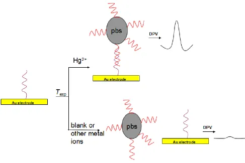
Figure 1. Schematic representation of the procedure to prepare electrochemical biosensor for the determination of mercury (II).
In the present study, we describe an electrochemical biosensor for Hg2+ detection by using the DNA probe (Figure 1). Two specific sequence for the probe [20], one has to ensure that the probe A and B form stable DNA duplexes only in the presence of Hg2+ at a given operating temperature. In the absence of Hg2+, these probes do not form DNA duplexes because of a lower melting temperature (Tm) than the operating temperature due to mismatches formed in the DNA duplexes.
[image:2.596.57.544.211.536.2]
the signal amplification effect of the labeled Au nanoparticle, resulting in high detection sensitivity. Therefore, this electrochemical biosensor is expected to have wide applications in environment monitoring.
2. EXPERIMENTAL
2.1 Reagents
The 1-ethyl-3-(3-dimethylaminopropyl) carbodiimide (EDC) were purchased from Shen energy Biocolor Biological Science and Technology Inc. (Shanghai, China). Bovine serum albumin (BSA) and thrombin were purchased from Dingguo Biotechnology Inc. (Shanghai, China). The nitric acid, cadmium chloride, sodium hydroxide, PBS (0.1 mol L-1,PH=7.3), sodium acetate buffer (1 mol L-1, pH=5.3) and other reagents were commercially available and of analytical reagent grade.
The DNA was obtained from Sangon Biotechology Inc. (Shanghai, China) with the following sequences:
Probe A: 5’-HS-C6-T13
Probe B: 5’-NH2-C6-A6TA6
2.2 Apparatus
Differential pulse voltammetry (DPV) measurements were performed using a CHI 660 Electrochemical Analyzer (CHI Instrument Inc., USA). The JB-1 stirring machine (Branson, Shanghai, China) and a TDL-16B centrifuge (Anting Science Instrument Inc., Shanghai, China) were used. The three-electrode electrochemical detection system consisted of a Au working electrode with sensing area of 3.14 mm2, a Ag/AgCl reference electrode (saturated KCl) and a platinum wire counter electrode. The detection was carried out in a 5 mL electrochemical cell containing a mercury-coated glassy carbon working electrode (2 mm diameter), an Ag/AgCl reference electrode, and a platinum wire counter electrode.
2.3 Preparation of nano PdS
Pb(NO3)2 and Na2S solutions were filtered through a 22 m microporous membrane filter prior
to use. PbS nanoparticles were prepared according to the literature [21] by using mercaptoacetic acid as the stabilizer. In brief, 9.22 L mercaptoacetic acid was added to 50 mL 0.4 mmol L-1 Pb(NO3)2
solution, and then the pH was adjusted to 7 with 0.5 mol L-1 NaOH. The solution was bubbled with nitrogen for 30 min, followed by the slow addition of 1.34 mmol L-1 Na2S to the mixture solution. The
molar ratio of Na2S to Pb(NO3)2 was kept at 2.5. The reaction was carried out for 24 h under nitrogen
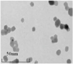
Figure 2. TEM image of the synthesized PdS nanoparticles
2.4 Preparation of DNA–PdS conjugate
200 L of 0.1 mol L-1 imidazole was added to 2 OD of 5-amido-capped detection probe B. After stirred for 30 min, 100 L of 0.1 mol L-1 EDC and 5 mL of PdS colloids were added to the mixture. The resulting mixture was stirred for 12 h at room temperature and then continued to centrifugate for at least 25 min at 14,000 rpm to remove the excessive DP. The yellow DNA-PdS precipitate was washed by 0.1 mol L-1 PBS and re-dispersed in 0.1 mol L-1 PBS. Then, the resulting solution was stored in the refrigerator for further use.
2.5 Immobilization of the probe A on Au electrode
The immobilization of probe A on Au electrode via Au-S binds was accomplished by first cleaning gold slides with a piranha solution (70% sulfuric acid, 30% H2O2) (Caution: Piranha solution
reacts violently with many organic materials and should be handled with great care), followed by treatment of the electrodes with nitric acid, then with water and drying under air. 2 L of appropriate probe A solution was dropped onto the electrode surface and the interaction was remained for 16 h. After that, the electrode was rinsed three times with phosphate buffer (0.1 mol L-1, pH = 7.3) before next program.
2.6 Hg-induced DNA hybridization reaction
electrode in the prepared solution containing appropriate Hg2+ and 600 L of 10-9 mol L-1 DNA-PdS, and then incubated at 34 oC for 35 min with stirring. During this time, probe A that initially immoblized on Au electrode preferred to form DNA duplexes structure with probe B modified on PdS nanoparticle. The amount of Hg2+ could be indicated by the signal of DNA-PdS on the electrode which came from Hg-induced DNA hybridization.
2.7 Electrochemical detection
After a thorough washing procedure, the PdS nanoparticles on the gold substrate were dissolved by adding 200 L of 0.10 mol L-1 HNO3. Then 1.8 mL acetate buffer (0.1 mol L-1, pH=5.3)
was added into 200 L of HNO3 solution (containing dissolved Pd2+). Electrochemical detection of the
dissolved Pd2+ were performed at a mercury-film electrode using a 5 min deposition at -1.0 V in an acetate buffer solution (0.1 mol L-1, pH=5.3). After the electrochemical deoxidation, DPV was immediately performed with the scan range from -0.8 to -0.2 V ( Incr E 0.004 V, amplitude 0.05 V, pulse width 0.05 s, pulse period 0.2 s), resulting in an analytical signal due to the oxidation of Pd, which relates to the amount of the PdS nanoparticles for the hybridization format. The DPV peak height at a potential of -0.50 V of the oxidation of Pd was used in all of the measurements. The mercuryfilm electrode was prepared on a polished glassy carbon electrode by applying a potential of -1.10 V for 10 min in a 0.1 mol L-1 HCl solution containing 100 mg L-1 Hg2+.
3. RESULTS AND DISCUSSION
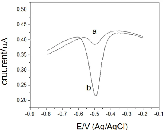
[image:5.596.163.433.505.720.2]3.1 Principle of detection of Mercury (II) based on Hg2+-induced hybirdization
The detection of mercury (II) was performed under the optimum stringency conditions determined above (0.10 mol L-1 NaNO3, 34oC). Aliquots of various concentrations of Hg2+ were
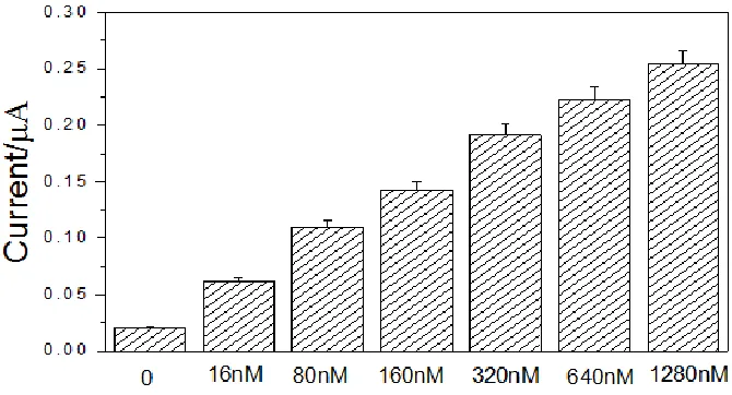
[image:6.596.131.466.225.406.2]prepared from one concentrated Hg2+ stock solution (1 mmol L-1). Firstly, the Probe A/AuE immersed into the prepared solution containing 1 nmol L-1 DNA-PbS, and then incubating it at 34oC for 30 minutes, only a relatively small DPV current signal was obtained as shown in Figure 3, curve a. Contrarily, a marked electrochemistry signal was obtained (Figure 3, curve b) after the Probe A/AuE was immersed in the solution containing the DNA-PbS and Hg2+.
Figure 4. Graph of average DPV signal intensity as a function of the Hg2+concentration in buffer.
It demonstrated that Probe A hybridization with Probe B only in present of Hg2+, and then produced a significant electrochemistry signal. Without Hg2+, there is little of adsorption of DNA-PbS onto the electrode and resulting in a small current signal. The probe A reaction with 1 nmol L-1 DNA-PdS probes in present of various concentrations of Hg2+. Significantly, signal intensity proportionally correlates with Hg2+ concentration (Figure 4) and the detection limit of Hg2+ in buffer is 16 nmol L-1.
3.2 Detecting specificity
3.3 Detection of Hg2+ in Natural Media.
Standard solutions of varying Hg2+ concentration (0, 10, 100, 200, 400, 600, 800, and 1000 nmol L-1) were prepared from a concentrated stock solution of [Hg(ClO4)2] in the collected lake. The
[image:7.596.78.510.245.473.2]DNA-PbS, the buffer, the surfactant, and the salt solutions were also prepared in the lake water to evaluate the robustness of the assay under natural conditions. The detection of mercury (II) (Hg2+) in natural media was performed in a manner similar to that used for the buffer samples and the detection limit of Hg2+ in lake water is 16 nmol L-1. In conclusion, the electrochemical biosensor could be used for detecting mercury (II) in real water, effectively.
Figure 5. DPV response of the assay in the presence of various metal ions ([Mx+]) 1 µmol L-1).
4. CONCLUSION
The present study has introduced a new mercury (II) electrochemical biosensor based on Hg2+ -induced hybirdiziation. As low as 16nmol L-1 Hg2+ has been specifically recognized, and other metal ion such as Ni2+, Cu2+, Zn2+, Cd2+, Li+ didn’t affect the target detection. Therefore, this electrochemical biosensor is expected to have wide applications in environment monitoring.
ACKNOWLEDGEMENT
References
1. Z. Torres, M. Mora, R. Taylor, D. Alvarez-Bernal, H. Buelna, A. Hyodo, Environ. Sci. Technol., 48(2014)6359.
2. J. Perrault, D. Miller, J. Garner, J. Wyneken, Sci. Total Environ., 463 (2013)61.
3. N. Zheng, Q. Wang, X. Zhang, D. Zheng, Z. Zhang, S. Zhang, Sci. Total Environ., 387(2007)96. 4. C. Chiang, C.C. Huang, C.W. Liu, H.T. Chang, Anal. Chem., 80(2008)3716.
5. L. Guo, H. Hu, R. Sun, G. Chen, Talanta, 79(2009)775.
6. Y. Wang, F. Yang, X. Yang, Biosens. Bioelectron., 25(2010)1994.
7. G. Mor-Piperberg, R. Tel-Vered, J. Elbaz, I. Willner, J. Am. Chem. Soc., 132(2010)6878. 8. B. Chen, Y. Yu, Z. Zhou, P. Zhong, Chem. Lett., 33(2004)1608.
9. X. Yan, Z. Cao, C. Lau, J. Lu, Analyst, 135(2010)2400. 10. J. Wang, B. Liu, Chem. Commun., 39(2008)4759–4761. 11. X. Zhou, D. Kong, H. Shen, Anal. Chim. Acta, 678(2010)124.
12. S. Jia, X. Liu, P. Li, D. Kong, H. Shen, Biosens. Bioelectron. 27(2011)148.
13. Liu X., Tang Y., Wang L., Zhang, J., Song S., Fan C., Wang S., Adv. Mater., 19(2007)1471. 14. D. Li, A. Wieckowska, I. Willner, Angew. Chem. Int. Ed. 120(2008)3991.
15. G. Clever, C. Kaul, T. Carell, Angew. Chem., Int. Ed., 46(2007)6226.
16. Y. Miyake, H. Togashi, M. Tashiro, H. Yamaguchi, S. Oda, M. Kudo, Y. Tanaka, Y. Kondo, R. Sawa, T. Fujimoto, T. Machinami, A. Ono, J. Am. Chem. Soc., 128(2006)2172.
17. X. Xue, F. Wang, X. Liu, J. Am. Chem. Soc., 130(2008)3244. 18. J. Liu, Y. Lu, Angew. Chem. Int. Ed., 46(2007)7587.
19. D. Li, A. Wieckowska, Angew. Chem. Int. Ed., 47(2008)3927. 20. L. Jae-Seung, A. Chad, Anal. Chem., 80(2008)6805.
21. A. Radi, L. Sanchez, E. Baldrich, C. Sullivan, Anal. Chem., 77(2005)6320. 22. T. Li, S. Dong, E. Wang, Anal. Chem., 81(2009) 2144.
23. Y. Lai, Ma Y. Y., Sun L. P., Electrochim Acta, 56(2011) 3153.
![Figure 5. DPV response of the assay in the presence of various metal ions ([Mx+]) 1 µmol L-1)](https://thumb-us.123doks.com/thumbv2/123dok_us/1902653.148427/7.596.78.510.245.473/figure-dpv-response-assay-presence-various-metal-umol.webp)
