JOURNALOFVIROLOGY, Jan. 1991, p.551-555 Vol. 65, No.1
0022-538X/91/010551-05$02.00/0
Copyright X 1991, American Society for Microbiology
Circular
DNA
of
Human
Immunodeficiency
Virus: Analysis
of Circle Junction Nucleotide Sequences
TAO HONG, KARLDRLICA, ABRAHAM PINTER, AND ELLEN MURPHY* PublicHealth Research Institute, 455 First Avenue, New York, New York10016
Received 14August 1990/Accepted 10 October 1990
During infection of cells by retroviruses, some of the nonintegratedviral DNA can be found as a circularform containing twotandem, directly repeated long terminal repeats. The nucleotide sequence at the pointwherethe long
terminal
repeats join (the circle junction) can be used to deduce the terminal nucleotides of the linear form of theviral DNA. Comparison of the termini of linear viral DNA with sequences at the junctions between the integrated provirus and the host chromosome has revealed that for most retroviruses 2 bp are removed from each end of the linear viral DNA during integration. For human immunodeficiency virus type 1 (HIV-1), however, sequence considerations involving primer-binding sites had suggested that only 1 bp is removed during integration. We obtained the nucleotide sequences at the ends of HIV-1 DNA by using the polymerase chain reaction toamplify
fragments corresponding to the HIV-1 circle junction. Of 17 clones containing amplified sequences, 10 had identical circle junctions that contained an additional 4 bp (GTAC) relative to the integrated provirus. This indicates that, as for other retroviruses, 2 bp are removed from each end of the linear HIV-1 viral DNAduringintegration. The remaining seven isolates contained insertions or deletions at the circle junction.Cells
infected with retroviruses
contain integrated viral DNA andboth
linear and circular forms of nonintegrated DNA.The
linear form is thought to be the substrate forintegration
(2, 8, 21). One of the circular species contains twotandemly
repeated long terminal repeats (LTRs); inthese molecules,
the junction between the LTRs, the circlejunction, corresponds
tothe ligated
ends of linear viralDNA.
Thus, the nucleotide
sequence across the circlejunc-tion
canbeused
todetermine
the sequences at theends of thelinear viral
DNA. For avariety
of murine and avianretroviruses, the
linear DNAcontains
2 bp at each end which are not present at the junctions between the integratedproviral
DNAand the host chromosome (4, 11, 25, 26, 31). Removalof
these 4 bparises
from a two-step reactioninvolving
the viralintegrase, the product of the 3' domain ofthe
pol
gene (5, 17, 18, 24). In thefirst
step, integrase removes twonucleotides from
the 3' ends of linearviral
DNA. In
the second
step, thetarget DNAis
cleavedand its
5'
ends
areligated
to therecessed
3' endsof
theviral
DNA(7, 8).
The2-nucleotide (nt)
protrusions
atthe 5' endsof
theviral
DNA arepresumably removed before ligation with the
3' ends
of
thehost
DNA.The
positioning of
RNAprimers used by
reversetran-scriptase
causeslinear
viral DNA toextendslightly beyond
U5
atoneend
andU3
attheother(Fig. 1).
Forinitiationof
minus-strand
DNAsynthesis,
priming involves
hybridiza-tion of the
3'end
of
atRNA toabout
18 nt within theunique
portion of
thegenomejust outside
U5. Forplus-strand
DNAsynthesis, priming involves
hybridization
of
a shortoligori-bonucleotide
toanucleotide
sequencein
theunique
region
of
the genomejust outside
U3(10, 32). With
avian and murineretroviruses, theprimer-binding
sitesfor
bothminus-and
plus-strand
DNAsynthesis
end 2 nt away from U5 andU3,
respectively.
Thus,
the 2bp removed from
each endduring
integration
areprotrusions beyond
the endsof
the LTRsthat aregenerated by
theposition of
theprimer.
For humanimmunodeficiency
virus type 1(HIV-1), however,
*Correspondingauthor.
only
1 ntseparates the tRNAprimer-binding site
andU5(30)
(the
plus-strand
primer ends
2 ntfrom
U3[10]).
Conse-quently,
it
hasbeen suggested
that HIV-1integration
in-volvesasymmetric
removalof
2bp from U3 and 1 from U5 (28, 32). Below we show that this is not the case: 2bp
are removed from eachend of
viral DNA duringintegration of
HIV-1.
We used the
polymerase chain reaction (PCR)
toamplify
afragment of
approximately
250bpcorresponding
tothecircle
junction
sequenceof
DNAextractedfrom
H9 cells infected with HIV-1(Fig.
2). Separateexperiments
werecarried
out with two strains of HIV-1. One was a viral stockof
HIVNL43
obtained by
previous transfection
withmolecular
clone
pNL4-3, obtained from
M. Martin(1).
The otherwasARV2(23),
obtained from
D. Dina ofChiron Corp.
Infected cells weregrown in RPMI 1640medium
containing 10%
fetalbovine
serum. When>90% of
thecells stained
positive for
HIV-1
by immunofluorescence
withpositive
humananti-HIV-1 serum(3 to 5
days), cells
were harvestedby
centrif-ugation
and washed and DNA wasextracted
by
amodified
Hirt
procedure (9).
PCRprimers which allowed efficient
amplification of circle
junction
sequences wereselected;
alternative
full-length
products
are notexpected
to beeffi-ciently
amplified
orcloned
because of theirlarge
sizes(Fig.
2). For eachreaction,
1 ,ugof
thetemplate,
125 nmolof
eachprimer
(described
inthelegend
toFig.
2),
and 5 Uof
Taq
polymerase
wereused.Reactionmixtures
wereincubated
at94°C
for 1min,
37°C
for
2min,
and72°C
for
3 minfor
35cycles.
Theamplified
DNA was extracted withphenol-chloroform,
precipitated,
cleaved
withHindIlI
andBamHI,
ligated
toM13mpl8,
and transformed into Escherichia coli JM109(15, 35).
Single-stranded
DNA wassequenced by
using
[35S]dATP
and T7 DNApolymerase
(Sequenase;
USBiochemicals).
Seventeen
M13mpl8
clonescontaining
HIV-1LTR-spe-cific
sequenceswereidentifiedby
sequenceanalysis
(Fig. 3).
Of the 17clones,
10(N25, N81,
N51, N70,
Al,
A5,
A3,
A8,
A46, andA47)
had circlejunction
sequencesidentical
tothose at the ends
of
theprovirus
(19),
exceptfor
theaddition
of 4 nt,
GTAC,
at the centersof
thecircle
junctions.
551
on November 10, 2019 by guest
http://jvi.asm.org/
552 NOTES
A 5. R U5 PBS gag env U3
R,
3CTAGCA GTGGCGCCC Q
.CCGcGGG-2~
~~~~~~~~~~~~~~~~5
StRNA pnmeR U ~ U3 R, 3' 5Ann
3'3 minus strand DNA
3,, USP 9 gag env 3U R
LU5.L+
U3 R U5 TCCCCGACCTCCC
it ~~~~~5'tAGGGGGG 3 '
\AGGGGGGo-opnnerplus strand DNA
translcatin
FIG. 1. Initiation of HIV-1 DNA synthesis with possible genesis of deletion and insertion mutations. The sequence of events in-volvedinproduction of double-stranded viral DNA is diagrammed insimplifiedform. For details, see reference 33. Thin lines represent RNA, and thick lines represent DNA; a few base pairs of the
sequences surrounding the U5-uniqueand U3-unique borders are shown. The two nucleotides bordering
U5
(A) or U3 (B) that are copied togenerate the 2-nt extensions on the linear DNA are shown inboldface.(A) Initiation of minus-strand DNA synthesis. Reversetranscription is initiated by binding of the
tRNA'Y'
primer on the viral RNAtemplate at the primer-binding site (PBS). Translocation to a second template occurs within R. The asterisk indicates theterminal adenosine residue of the primer tRNA which must be
removedbefore initiation of DNA synthesis to generate the predom-inant form of circle junction clone. If the tRNA primer contains
additionalbases at its 3' end or if synthesis is primed by a random
oligonucleotide
hybridizing withinU5,
deletions ofU5
will result. (B)Initiationof plus-strand DNA synthesis. An oligoribonucleotide primer19nts long, generated from the genomic RNA by RNase H,hybridizes 2 nt upstream from U3 for initiation of plus-strand
synthesis(10), which continues until the first modified base of the tRNAisencountered (plusshort-stopDNA). The tRNA is removed byRNase H. Use of an incorrect primer, hybridizing to sequences within U3, will generate U3 deletions; positioning of the primer further into the unique portion of the viral genome would generate
insertions at the circle junction containing portions of the unique region. Translocation of the plusshort-stopDNA to the 3' end of the minus strandallows extension of the minus strand through U3 and
extensionof the plus strong-stop DNA through the full length of the
genome.Failure of RNase H to remove the tRNA primer or failure ofthe strong-stop DNA to translocate could result in copying of tRNAsequences into the plus strand at the 3' end of the viral DNA;
subsequent removal of the ribonucleotides, ligation of the plus strand, and filling in of the minus strand would generate a circle
junctioncontaining an insertion of tRNA sequences.
Removal
of these 4 bp unambiguously generated these-quences
of the LTRs found in integrated proviral DNA.Thus, integration of HIV-1, as for other retroviruses,
in-volves removal of 2 bp from each end of the linear viral
DNA.
This conclusion has also been reached in two otherstudies
reported recently (14, 34).Adilemma arises from the observation that for HIV-1, the
primer-binding
site for minus-strand synthesis is only 1 ntfrom the 3' end of
U5
(Fig.1A).
The two most likelymechanisms
for generating the observed circle junctionsequences
are (i) removal of the 3' adenosine from theprimer
tRNA before reverse transcription and (ii) failure ofRNase H to remove the terminal riboadenine nucleotide of
the primer from the completed DNA. Although there are
precedents
for the use of truncated tRNA primers (6, 13, 22)or incorrectly processed tRNAs (16), Whitcomb et al. (34)
have argued that the tRNA primer used for HIV-1
replica-tionis intact and that the 5'-terminal A of the minus strand of
env
DoI
gag
A
;U3
RU51
U3R|
4-- --Ip
40--250 bp
l
l
Rjugag
pol env
y RB
R-I
bU51
4-- -P. -9kb 4- --o
c
env
pol
gag
4
3 R|U
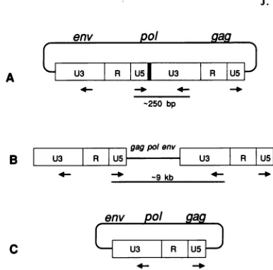
FIG. 2.
Strategy
for PCRamplification
of circle junctions. (A)An
approximately 250-bp fragment
wasamplified
across the circlejunction
(thick
blackbar)
of a two-LTR circle. Theoretically,amplification
with the sameprimers
would also yield a nearlyfull-length (approximately
9-kb)
viral DNA molecule from atwo-LTRcircle
(A),
a lineartemplate
(B),
or asingle-LTR
circle (C); no such clones were recovered. Theprimers
were5'-GCCTCAATA
AAGCTTGCCTTGAGTGC-3'
(U5)
and5'-CAggATCCAAAGGTC
AGTGGATATCTG-3'
(U3),
whichcorrespond
tonts 522 to 547 ofU5
and nts 132 to 110 of U3 of the HIVNL43 sequence (GenBankaccession number
M19921) (2a).
Nucleotides added or altered toproduce
theBamHI
restriction site in the U3 primerare shown inlowercase letters. The
HindIll
site in theU5
primer
occurs at nt 531.U5
is aribonucleotide derived from theprimer. Occasional
removal of
thisribonucleotide
by
RNase H wouldgenerate alinear
viral DNAmolecule
lacking
a T at the 3' end ofU5.
We found one clone(A20)
that could be explained in this way.The GTAC at the
circle
junction
forms
the centraltetra-nucleotide of
an Scalrestriction site
(AGTACT);
cleavage occursprecisely
at thecircle
junction.
Since
cleavage byScaI
produces
flush-ended
DNA, digestion
of
circle junctionclones
with thisenzyme generates
a linear DNA molecule that has at its ends theprecise
sequence
found in thepresumed integration
substrate,
linear viral
DNA. This DNAmay
be a useful substrate foranalyzing
biochemical
proper-ties
of
integrase,
such
as itsexpected
3'recessing
activity(2).
Thecircular form
itselfmay
be a useful substrate foranalysis
of certain interactions between DNA
andintegrase,although
it isprobably
not aproductive
intermediateforviralintegration.
Two clones contained insertions similar
inorigin
to thosethat have been
reported
for other retroviruses.
One, N72,had what
appeared
to be a342-bp
insertion at the circlejunction;
the insertioncorresponded
to
aportion
of the 5'unique region
of the
virus,
starting
at the
primer-binding
site(nt 637)
andending
at nt978 ingag.
Similar murine leukemiavirus and
copia
clones have been
interpreted
as
arisingfromintegration
of one viral
DNAintoanother
(6,
27). Theinsertin N72
contained
thesequences
located between
the 5' LTRof
thetarget
molecule and the 5'LTR
of
the integrating viralDNA;
the U3
boundary
represents
the 5' end
of theinte-grated
virus. N72 lacked the 2 nt
(AC)
at the outer
border ofU3 of the
integrating
virus,
supporting
the conclusion thatintegration
involves removal of 2
bp
from
U3. Anotherclone, A34,
contained
aremnant
of the tRNA
primer, a16-bp
insertion
precisely
at
the
circle
junction
thatcorre-J.VIROL.
on November 10, 2019 by guest
http://jvi.asm.org/
[image:2.612.340.533.58.249.2] [image:2.612.66.302.71.217.2]NOTES 553
U5
U3
9640 9650 9660 9670 9680 9690 9700 circlejunction 10 20
TGCCCGTCTGTTGTGTGACTCTGGTAACTAGAGATCCCTCAGACCCTTTTAGTCAGTGTGGAAAATCTCTAGCAGT ACTGGAAGGGCTAATTCACTCCCAAcGAAG
TGCCCGTCTGTTGTGTGACTCTGGTAACTAGAGATCCCTCAGACCCTTTTAGTCAGTGTGGAAAATCTCTAGCAGT ACTGGAAGGGCTAATTCACTCCCAAAGAAG TGCCCGTCTGTTGTGTGACTCTGGTAtCTAGAGATCCCTCAGACCCTTTTAGTCAGTGTGGAAAATCTCTAGCAGT ACTGGAAGGGCTAATTCACTCCCAAcGAAG TGCCCGTCTGTTGTGTGACTCTGGTAACTAGAGATCCCTCAGACCCTTTTAGTCAGTGTGGAAAATCTCTAGCA-ACTGGAAGGCTAATTCACTCCCAAcGAAG
TGCCCGTCTGTTGTGTGACTCcGGTAACTAGAGATCCCTCAGACCCTTTTAGTCAGTGTGGAAAATCTCTAGCAGT ---TGCCCGTCTGTTGTGTGACTCTGGTAACTAGAGATCCCTCAGACCCTTTTAGTCAGTGTGGAAAATCTCTAGCAG- ACTGGAAGGGCTAATTCACTCCCAAcGAAG TGCCCGTCTGTTGTGTGACTCTGGTAACTAGAGATCCCTCAGACCCTTTTAGTCAGTGTGGAAAATCTCTAGC
-TGCCCGTCTGTTGTGTGACTCTGGTAACTAGAGATCCC---
---gaactctggtag
A4,A47 TGCCCaTCTGTTGTGTGACTCTGGTA-CTAGAGATCCCTCAGACCCTTTTAGTCAGTGTGGAAAATCTCTAGCAGT ACTGGAAGGGCTAATTCACTCCCAAcGAAG
gaactctggtag ggcgcccgaacaggac
A34~~~~~~~~~~~~~~
AM4 TGCCCaTCTGTTGTGTGACTCTGGTA-CTAGAGATCCCTCAGACCCTTTTAGTCAGTGTGGAAAATCTCTAGCAGT'ACTGGAAGGGCTAATTCACTCCCAAcGAAG
ggcgcctgaacagggacttg - 342 nt (gag) - agataaatactgggacag
N72 TGCCCGTCTGTTGTGTGACTCTGGTAACTgGAGATCCCTCAGACCCTTTTAGTCAGTGTGGAAAATCTCTAGCAGT --TGGAAGGGCTAATTCACTCCCAAcGAAG
U3
30 40 50 60 70 80 90 100 110 120 130
10CLONES ACAAGATATCCTTGATCTGTGGATCTACCACACACAAGGCTACTTCCCTGATTGGCAGAACTACACACCAGGGCCAGGGATCAGATATCCACTGACCTTTGGAT
N51L,N70 ACAAGATATCCTTGATCTGTGGATCTACCACACACAAGGCTACTTCCCTGATTGGCAGAACTACACACCAGGGCCAGGGgTCAGATATCCACTGACCTTTGGAT
Al ACAAGATATCCTTGATCTGTGGATCTACCACACACAAGGCTACTTCCCTGATTGGCAaAACTACACACCAGGGCCAGGGATCAGATATCCACTGACCTTTGGAT
N63 ---CAGATATCCACTG-CCTTTGGAT A21 ---CTACCACACACAAGGCTACTTCCCTGATTGGCAGAACTACACACCAGGGCCAGGGATCAGATATCCACTGACCTTTGGAT
A19 ----GATATCCTTGATCTGTGGATCTACCACACACAAGGCTACTTCCCTGATTGGCAGAACTACACACCAGGGCCAGGGATCAGATATCCACTGACCTTTGGAT
A47 ACAAGATATCCTTGATCTGTtGATCTACCACACACAAGGCTACTTCCCTGATTGGCAGAACTACACACCAGGGCCAGGGATCAGATATCCACTGACCTTTGGAT
FIG. 3. Nucleotide sequencesof cloned circle junctions. Circle junctions amplified by PCRwerecloned, and the nucleotide sequences
weredetermined(seethetext). Each linerepresentsauniquesequence;clonescontaining thatsequence areindicatedattheleft. In thefirst
lineof the lowersetofsequences,the10clones with identicalsequences areA3,A8, A5,A20, A34,A46, N25, N60, N72, and N81. U3is numbered starting fromnt 1, since it is derived from the 5'LTR; U5isnumbered fromnt9636, since it is derivedfrom the 3' LTR.The additional 4 bpatthecircle junctionarenotincludedin the numbering scheme. Onlypartof theU5 portion oftheamplifiedfragmentis shown; theentire fragmentwassequenced in eachcase;andnodeviations from the publishedsequence werefound intheregionofU5notshown. Theprefix A indicates clones fromcells infected with ARV2, and the prefixNindicates clones from HIVNL43 infection. Dashed lines indicate deleted sequences. Thesequenceshown for clones N25 and N81 is identical tothatof the HIVNL43 GenBankentry(2a), exceptfor the substitutionof C for Aatnt24 in U3.This and othersubstitutions relativetothe HIVNL43sequence areshown in lowercase letters.
spondedto17 bp of the tRNAprimer-binding site(nts637to 653), less one internal nucleotide. Such clones have been postulatedtoarise from the failure ofRNase H to remove the entire tRNA primer from minus-strand DNA, which resultsin copying of the tRNA sequenceinto the 3' end of theplus-strand DNA (3,29). An alternative explanation for
theorigin of clone A34 is failure of the plusstrong-stopDNA to translocate to the 3' end of the minus strand (Fig. 1).
Clone A34also containedasecond insertion: anadenineat nt 9662 in U5 was replaced by a 12-bp imperfect tandem duplication of the adjacentsequence (nts 9652to9662). An identicalduplication wasfoundatthe samelocation intwo additional clones, A46 and A47, both ofwhich contained wild-type circle junction sequences. Thus, this duplication
probably representsa variant present in the infecting virus
population.
Nearly one-third ofour clones contained deletions that
eitherspannedthecirclejunctionorterminated preciselyat the U3-U5 boundary. Clone N63 had a 109-bp deletion
starting at thejunction andextending only into U3. Three otherclones contained deletionsat 12 (N60), 55(A21), and 73 (A19) bp that spanned the circlejunction. Similar dele-tions have been found at high frequencies in unintegrated
murineoravianretroviral DNAs(11, 12, 16, 26, 27).Inthese systems,abiastoward deletion of U3isevident;oursample size was too small to support an equivalent conclusion. Deletions spanning the circlejunction or having one end-point at the circle junction are most easily explained as arisingfromincorrectprimingorfromexonucleolytic
diges-tionofone orboth ends of the viral DNA before ligation.
Both explanations are consistent with the absence of dele-tions outside thejunction sequences, although mispriming
mightmore easilyexplainany biasindeletionfrequencyof U3overU5, sincethemechanisms ofprimingof U3 andU5 differ.
None ofourdeletions lackedonly the GT nucleotides of
U5. Suchdeletions,suggestedtoarisevia theactivityof the viral integrase, represented half of the clones found by Kulkosky et al. (14). However, deletions extending more
than2bpintoU3andU5,asdescribedabove,wereobtained atamuchhigherfrequency here than intwootherstudies. In thosestudies,therelativerecoveryoffull-lengthcloneswas increased by gel purification of the amplified DNA before
cloning(34)orby screeningofclones forfull-lengthinserts beforesequencing (14). Thus,ourresultsprobablyreflect the situation invivo moreaccurately, since therewasno
prese-lectionbasedon insert size.
To determine whether PCR provided a representative sample of the population ofcirclejunction sequences, we performed several reconstruction amplifications. Cloned DNAs from M13 clonescontaining wild-typecirclejunction (clone Al), plusDNA fromeithercloneA19(73-bpdeletion atthecirclejunction) orN72(342-bp insertion),wereadded to4 ,ug of DNA from uninfected H9 cellsto approximately
0.4to4viralmoleculespercell. The molarratio ofwild-type
to mutant clones was varied over a 10-fold range, and the mixturesweresampledafter25,30, and35cyclesof
ampli-fication. In all cases, amplified DNAs were obtained in amounts that were roughly proportional to the concentra-tions oftemplateDNA clones(datanotshown).Proportional
resultswerealso obtained when DNAsfrom all three clones weremixed inequalratios orina 10:10:1 ratio. The results were unaffected by omission of H9genomic DNA or addi-tion of 10-fold excess Scal-digested clone Al DNA to
CLONE
N25,N81,A3,AS N51,N70 Al, AS N60 N63 A20 A21 A19
VOL.65, 1991
on November 10, 2019 by guest
http://jvi.asm.org/
[image:3.612.93.521.66.300.2]554 NOTES
simulate the presence of linear viral DNA. These
experi-ments indicate that preexisting deletions are not preferen-tially amplifiedrelative to wild-typecircle junctions. Thus,it is likely that our results do reflect a high proportion of
mutant circle junction-containing DNA in infected cells. The clones we examined exhibited a small amount of
sequence variation in regions distant from the circle
junc-tion. None of our clones had a sequence identical to thatof HIVNL43; they differed by up to 3 nt (lowercase letters in Fig. 3). For the seven clones obtained after infection with molecularclone HIVNL43, the error rate was 0.4% (seven
pointmutationsin1,638 ntssequenced). This isconsiderably
higher than the misincorporation rate we haveexperienced with PCR in other systems (approximately one error per 3,000 nts sequenced;
1Sa)
and is within the range ofreportedmisincorporation rates for HIV-1 reverse transcriptase (20). Thus, these differences are likely to reflect real variation occurringduring virus replication.
There are seven positions within the amplified fragment at which the ARV2 and HIVNL43 sequences differ. All 17 clones contained the HIVNL43 sequence, even though 10 were obtained after infection with ARV2. It is likely that the ARV2 nucleotide sequence, which was obtained from a molecular clone, represents only a small fraction of the sequences present in the uncloned ARV2 virus stock. The major variant present in this stock is a virus more closely related to HIVNL43 in this region.
It is now clear that 2 bp are removed from each end of HIV-1 during integration. It is also evident that a large proportion of the circular DNA molecules in infected cells carry mutations at the circle junction. It may be thatcircular viral DNA is derived preferentially from defective linear molecules. If not, our observations could indicate that a large proportion of the linear, integrative form of HIV-1 viral DNA normally has incorrect termini, making it unable to integrate. A better understanding of mutant formation may lead to ways to increase this fraction, thereby blocking productive infection.
We thank D. Dina and M. Martin for providing strains, Jim Groarke and Joseph Hughes for stimulating discussions,andSamuel Kayman for critical comments on the manuscript.
Support for this work was provided by the Aaron Diamond Foundation, The American Foundation for AIDS Research,and the U.S. Army Medical Research and Development Command under contract DAMD17-88-C-8126. T.H. is an NIH predoctoral traineeof the Department of Microbiology, New York University School of Medicine.
REFERENCES
1. Adachi, A., H.Gendelman, S. Koenig, T. Folks, R. Willey,A. Rabson, and M. Martin. 1986. Production of acquired immuno-deficiency syndrome-associated retrovirus in human and non-human cells transfected with an infectious molecular clone. J. Virol. 59:284-291.
2. Brown, P., B. Bowerman, H. E. Varmus,and J. M.Bishop.1989. Retroviral integration: structure of the initial covalent product and its precursor, and a rolefor the viral IN protein. Proc.Natl.
Acad. Sci. USA 86:2525-2529. 2a.Buckler, C. Personal communication.
3. Colicelli, J., and S. P. Goff. 1986. Structureof acloned circular retroviral DNA containing atRNA sequence between the ter-minal repeats. J. Virol.
57:674-677.
4. Dhar, R, W. L. McClements, L. W. Enquist, andG. F. Vande Woude. 1980. Nucleotide sequences of integrated Moloney sarcoma provirus long terminal repeatsand theirhostand viral junctions. Proc. Natl. Acad. Sci. USA77:3937-3941.
5. Donehower, L. A., and H. E. Varmus. 1984. A mutant murine leukemia virus with a single missensecodon inpol isdefective
in a function affecting integration.Proc.Natl. Acad. Sci. USA 81:6461-6465.
6. Flaveli, A. J., and D. Ish-Horowicz. 1983. Theoriginof extra-chromosomal circular copia elements. Cell 34:415-419. 7. Fujiwara, T., and R. Craigie. 1989. Integration of mini-retroviral
DNA: a cell-free reaction for biochemicalanalysis of retroviral integration. Proc. Natl. Acad. Sci. USA 86:3065-3069. 8. Fujiwara, T., andK. Mizuuchi. 1988. RetroviralDNA
integra-tion: structure of an integration intermediate. Cell 54:497-504. 9. Hirt, B. 1967. Selective extraction of polyoma DNA from
infected mousecell cultures. J. Mol. Biol. 26:362-371. 10. Huber, H. E., and C. C. Richardson. 1990. Processing of the
primerfor plus strand DNA synthesis by human immunodefi-ciency virus 1reversetranscriptase.J. Biol. Chem. 265:10565-10573.
11. Ju, G., and A. M. Skalka. 1980.Nucleotide sequenceanalysisof the longterminal repeat (LTR)ofavian retroviruses: structural similarities withtransposableelements. Cell 22:379-386. 12. Katz, R. A., C. A. Omer, J. H. Weis,S. A.Mitsialis, A.J. Faras,
and R. V. Guntaka. 1982. Restriction endonuclease and nucle-otide sequence analyses of molecularly cloned unintegrated avain tumor virus DNA: structure oflarge terminal repeats in circlejunctions. J. Virol. 42:346-351.
13. Kikuchi, Y., Y. Ando, and T. Shiba. 1986. Unusual priming mechanism of RNA-directed DNA synthesis in copia retrovi-rus-like particles inDrosophila. Nature (London) 323:824-826. 14. Kulkosky, J., R. A. Katz, and A. M. Skalka. 1990. Terminal nucleotides of the preintegrative linear form of HIV-1 DNA deduced from the sequence of circular DNA junctions. J. Acquired Immune Defic. Syndr. 3:852-858.
15. Messing, J. 1983. New M13 vectors for cloning. Methods Enzymol. 101:10-89.
15a.Murphy, E. Unpublished data.
16. Olsen, J., and R. Swanstrom. 1985. A new pathway in the generationof defective retrovirus DNA. J. Virol. 56:779-789. 17. Panganiban, A. T., and H. M. Temin. 1984. Circles with two
tandem LTRs areprecursors tointegrated retrovirusDNA. Cell 36:673-679.
18. Quinn,T., and D. Grandgenett. 1988. Genetic evidence thatthe avian retrovirus DNAendonuclease domain of pol is necessary for viralintegration. J. Virol. 62:2307-2312.
19. Ratner, L., W. Haseltine, R. Patarca, K. J. Livak, B. Starcich, S. F. Josephs, E. R.Doran,J. A.Rafalski, E. A. Whitehorn,K. Baumeister,I. Ivanoff, S. R.Petteway, Jr., M. L.Pearson,J. A. Lautenberger, T. S. Papas, J. Ghrayeb, N. T. Chang, R. C. Gallo, and F. Wong-Staal. 1985. Complete nucleotide sequence of the AIDS virus, HTLV-III. Nature (London) 313:277-284. 20. Roberts, J. D., K. Bebenek, and T. A. Kunkel. 1988. The
accuracy of reverse transcriptase from HIV-1. Science 242: 1171-1173.
21. Roth, M., P.Schwartzberg, and S. Goff. 1989. Structureofthe termini of DNA intermediates in the integration of retroviral DNA: dependence onINfunction andterminalDNA sequence. Cell 58:47-54.
22. Saigo, K. 1986. A potential primer for reverse transcription of mdg3, a Drosophila copia-like element, is a leucine tRNA lacking its 3'terminal 5 bases.Nucleic AcidsRes.14:4370-4371. 23. Sanchez-Pescador, R.,M. D. Power, P. J. Barr, K. S. Steimer, M. M.Stempien, S. L.Brown-Shimer,W. W. Gee, A.Renard,A.
Randolph, J. A. Levy, D. Dina, and P. A. Luciw. 1985. Nucle-otidesequence andexpression of anAIDS-associatedretrovirus (ARV-2). Science227:484-492.
24. Schwartzberg,P., J.Coliceili,and S. P. Goff.1984. Construction and analysis of deletion mutations in the pol geneofMoloney murine leukemia virus: a new viral function required for pro-ductive infection. Cell37:1043-1052.
25. Shimotohno, K., S.Mizutani, and H. M.Temin. 1980.Sequence ofretrovirus provirus resembles thatofbacterial transposable
elements. Nature (London)285:550-554.
26. Shoemaker, C., S. Goff, E.GUlboa, M. Paskind, S. W.Mitra,and D. Baltimore. 1980. Structure of a cloned circular Moloney murine leukemia virus DNA molecule containing an inverted segment: implications for retrovirus integration. Proc. Natl. J.VIROL.
on November 10, 2019 by guest
http://jvi.asm.org/
VOL.65, 1991 NOTES 555 Acad.Sci. USA 77:3932-3936.
27. Shoemaker, C., J.Hoffmann, S. P. Goff, and D. Baltimore. 1981. Intramolecular integration within Moloney murine leukemia virus. J. Virol. 40:164-172.
28. Skalka, A. M. 1988. Integrative recombination in retroviruses, p. 701-724. In R. Kucherlapati and G. R. Smith (ed.), Genetic recombination. American Society for Microbiology, Washing-ton, D.C.
29. Sonigo, P., C. Barker, E. Hunter, and S. Wain-Hobson. 1986. Nucleotide sequence of Mason-Pfizer monkey virus: an immu-nosuppressiveD-typeretrovirus. Cell45:375-385.
30. Starcich, B., L. Ratner, S. F. Josephs, T. Okamoto, R. C.Gallo, and F. Wong-Staal. 1985. Characterization of long terminal repeatsequencesof HTLV-III. Science 227:538-540.
31. Van Beveren, C., E. Rands, S. K.Chattopadhyay, D. R. Lowy, and I. M.Verma. 1982. Longterminal repeatof murine
retro-viral DNAs: sequence analysis, host-proviral junctions, and preintegrationsite. J. Virol.41:542-556.
32. Varmus, H., and P. Brown. 1989. Retroviruses, p. 53-108.In D. E. Berg and M. M. Howe (ed.), Mobile DNA. American Society for Microbiology, Washington, D.C.
33. Varmus, H., and R. Swanstrom. 1985. Replication of retrovi-ruses, p. 75-134. In R. Weiss, N. Teich, H. Varmus, andJ. Coffin (ed.),RNA tumorviruses, vol. 2. ColdSpring Harbor Laboratory, ColdSpring Harbor,N.Y.
34. Whitcomb, J. M., R. Kumar, and S. H. Hughes.1990.Sequence of the circlejunction of humanimmunodeficiencyvirus type 1: implicationsfor reversetranscription andintegration. J. Virol. 64:4903-4906.
35. Yanisch-Perron, C., J. Vieira, and J. Messing. 1985. Improved M13 phage cloning vectors and host strains: nucleotide
se-quencesof theM13mpl8 andpUC19vectors.Gene 33:103-119.